
Section I
Functional Neuroanatomy
and Imaging
Copyright © 2005 CRC Press LLC

0-8493-1287-6/05/$0.00+$1.50
© 2005 by CRC Press LLC
1
Motor Areas in the
Frontal Lobe: The
Anatomical Substrate
for the Central Control
of Movement
Richard P. Dum and Peter L. Strick
CONTENTS
1.1 Introduction
1.2 Functional Anatomy
1.2.1.1 Organization Based on Intracortical Stimulation
1.2.1.2 Output of Single Corticomotoneuronal Cells
1.2.1.3 Peripheral Input to M1
1.2.2.1 Identification by Direct Projections to M1
1.2.2.2 Somatotopic Organization Based on Connections with
1.2.2.3 Corticospinal Output
1.2.2.4 Somatotopic Organization Based on Corticospinal
Output: Forelimb and Hindlimb Representation
1.2.2.5 Somatotopic Organization Based on Corticospinal
Output: Proximal and Distal Arm Representation
1.2.2.6 Organization Based on Intracortical Stimulation
1.2.3 Corticospinal Terminations
1.2.3.1 Primary Motor Cortex
1.2.3.2 Premotor Areas
1.3 Cortical Inputs to the Motor Areas
1.3.1.1 Frontal Cortex
1.3.1.2 Parietal Cortex
Copyright © 2005 CRC Press LLC

1.3.2.1 Interconnections among the Motor Areas
1.3.2.2 Parietal Cortex
1.3.2.3 Pre-Premotor Cortex
1.3.2.4 Prefrontal Cortex
1.3.2.5 Limbic Cortex
1.3.3 Summary of Cortical Connections
1.4 Subcortical Inputs
1.5 Summary and Conclusions
Acknowledgments
References
1.1 INTRODUCTION
The objective of this chapter is to describe the major components of the structural
framework employed by the cerebral cortex to generate and control skeletomotor
function. We will focus on motor areas in the frontal lobe that are the source of
corticospinal projections to the ventral horn of the spinal cord in primates. These
cortical areas include the primary motor cortex (M1) and the six premotor areas that
project directly to M1. We will begin by examining anatomical and physiological
evidence that demonstrates how each of these cortical areas directly accesses spinal
cord mechanisms involved in the generation and control of movement. This evidence
suggests that all these cortical areas have some direct involvement in movement
execution. Then we will examine how the pattern of cortical and subcortical inputs
could shape the functional role of each cortical area in motor control. We will show
that each of these cortical areas receives a unique pattern of cortical and subcortical
input. Taken together, these results have led to an emerging view that motor commands
can arise from multiple motor areas and that each of these motor areas makes a
specialized contribution to the planning, execution, or control of voluntary movement.
In this chapter, we will describe some of the relevant anatomical and physiological
evidence that has led to this viewpoint.
Given the breadth of the subject considered here, our review will focus on new
perspectives developed from contemporary primate studies. Even with this focus,
many topics will receive limited treatment. For instance, the physiological and
behavioral studies that provide evidence of differential involvement of each motor
area in the generation and control of movement are beyond the scope of this chapter.
For further insight into the historical development of this field and a broader coverage
of related issues, numerous reviews on this and related topics are available.
1–11
In
addition, the corticospinal system has been the subject of a recent book.
12
1.2 FUNCTIONAL ANATOMY
1.2.1 P
RIMARY
M
OTOR
C
ORTEX
The primary motor cortex (M1) owes its name to the fact that thresholds for evoking
movement with electrical stimulation are lower here than in any other cortical
region.
13–15
(For historical review, see Reference 12.) Anatomically, M1 corresponds
Copyright © 2005 CRC Press LLC
to cytoarchitectonic area 4, which is identified by the presence of giant pyramidal
cells in cortical layer V.
16–18
Based on these definitions, M1 is located in the anterior
bank of the central sulcus and on the adjacent caudal portion of the precentral gyrus
(Figure 1.1). (For more complete reviews, see References 4,5,9,12.)
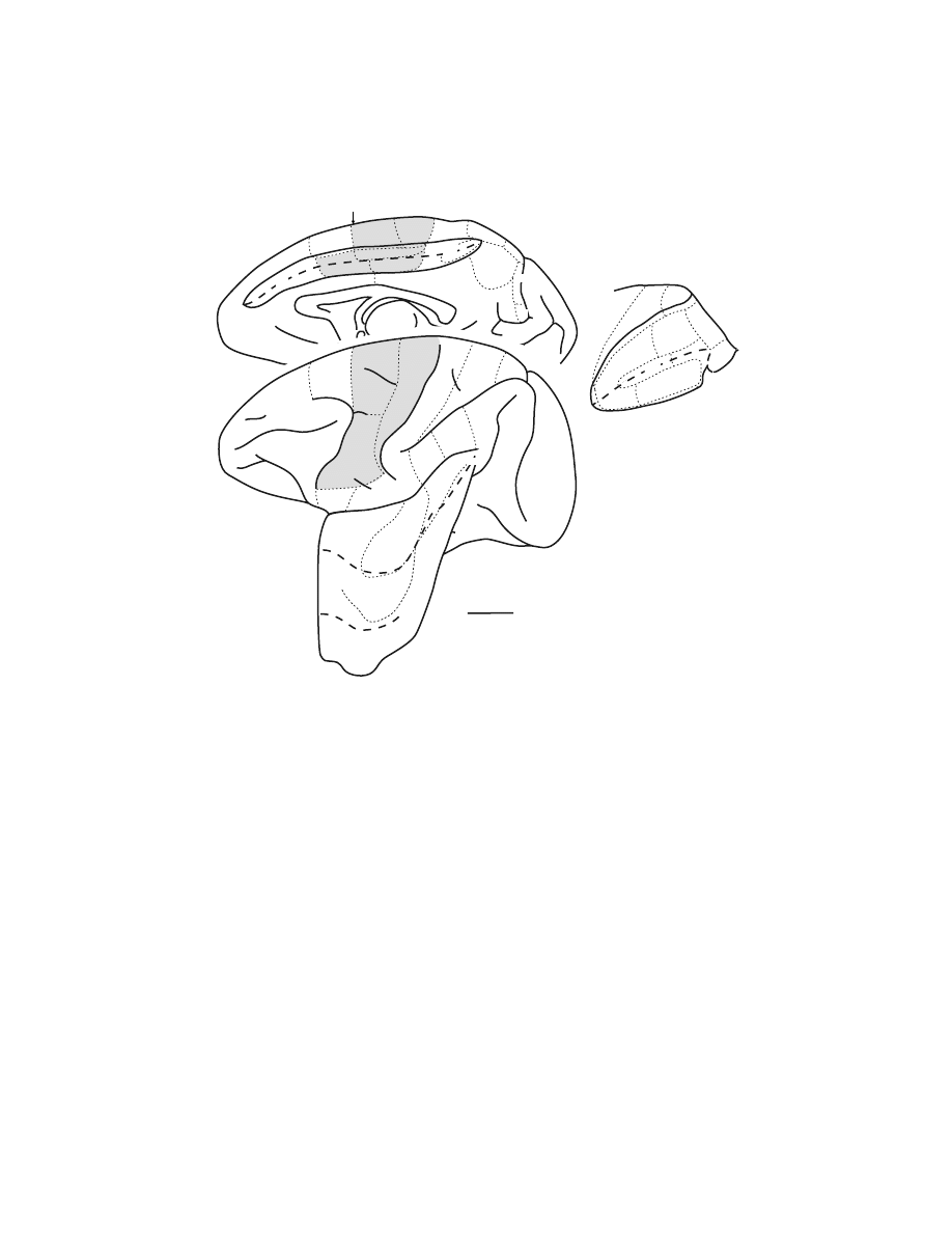
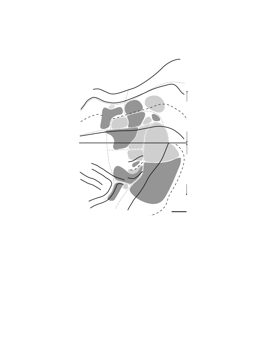
FIGURE 1.1
Identification of cortical areas in the macaque monkey. The cingulate sulcus
(CgS), lateral sulcus (LS), and intraparietal sulcus (IPS) are unfolded and each fundus is
indicated by a
dashed line.
The borders between cytoarchitectonic areas are delineated with
dotted lines.
M1 and the premotor areas are
shaded
. Abbreviations: AIP, LIP, MIP, VIP:
anterior, lateral, medial, and ventral intraparietal areas; ArS: arcuate sulcus; CGp: posterior
cingulate gyrus; CMAd, CMAv, CMAr: dorsal, ventral, and rostral cingulate motor areas;
CS: central sulcus; F1 to F7: cytoarchitectonic areas in the frontal lobe according to Matelli
et al.
77,248
; FEF: frontal eye fields; Ig: granular insular cortex; M1: primary motor cortex;
OFC: orbital frontal cortex; PMd: dorsal premotor area; PMv: ventral premotor area; PrCO:
precentral opercular cortex; prePMd: pre-premotor area, dorsal; preSMA: presupplementary
motor area; PS: principal sulcus; SEF: supplementary eye field; SI: primary somatosensory
cortex; SII: secondary somatosensory cortex; SMA: supplementary motor area; PE, PEc, PEci,
PF, PFG, PFop, PG, PGm, Pgop: parietal areas after Pandya and Selzer
249
; V6A, V6: posterior
parietal areas after Galletti et al.
177
; 9m, 9l, 46d, 46v, 12l: prefrontal areas after Walker
181
and
Barbas and Pandya.
186
V6
PEip
MIP
PEc
PE
V6A
AIP
LIP
VIP
IPS
preSMA
M1
CMAr
CMAv
CMAd
SI
PE
PEc
PGm
CGp
23a,b
24a,b
9m
V6A
PEci
1 cm
M1
PMv
SI
PE
PEc
PF
PFG
PG
PrCO
46d
46v
12l
9l
FEF
SEF
OFC
SII
PFop
Ig
PFGop
V6
LS
PS
CgS
CS
IPS
ArS
(F1)
PMd
(F2)
SMA
(F3)
(F4)
(F5)
(F6)
prePMd
(F7)
Copyright © 2005 CRC Press LLC

1.2.1.1 Organization Based on Intracortical Stimulation
Our view of the organization of M1 as based on electrical stimulation has evolved
with advances in stimulation techniques. Classically, surface stimulation suggested
that M1 contained a “motor map” that was a single, contiguous representation of
the body.
14,15
(For reviews, see References 4 and 12.) In this map, the leg, trunk,
arm, and face formed a medial to lateral procession across M1 with the distal
musculature of each limb located in the central sulcus. Electrical stimulation with
microelectrodes inserted into the cortex lowered the amount of current necessary to
evoke movement by a factor of 100.
19
Although this advance allowed a much more
detailed exploration of the cortex, intracortical stimulation confirmed the overall
somatotopy of leg, arm, and face representation described by surface stimulation.
19–32
Thus, electrical stimulation of M1 generated a somatotopic motor map with relatively
sharp boundaries between major body parts.
The organization of movements generated by intracortical stimulation within
each major body part, however, was more complex than that produced by surface
stimulation (Color
).* A consistent observation was that the same move-
ment could be evoked at multiple, spatially separate sites.
22–32
Although this obser-
vation precluded an orderly somatotopy, the general features of this map were
reproducible. Within the arm representation of macaque monkeys, distal limb move-
ments (fingers and wrist) tended to form a central core that was surrounded by a
horseshoe of proximal limb movements (elbow and shoulder) (Color Figure
1.2A).
22,33
Some intermingling of distal and proximal limb movements occurred at
the borders. This organizational structure has been confirmed with single-pulse,
stimulus-triggered averaging (Color Figure 1.2B).
34
The presence of multiple repre-
sentations of an individual movement/muscle in M1 has been proposed as an arrange-
ment that allows a muscle to engage in multiple synergies with other muscles acting
at the same or different joints. (See Reference 35.)
Other studies utilizing intracortical stimulation
20,26,28,32
reported even more com-
plex patterns of muscle activation. For example, stimulation at some sites in M1
evoked reciprocal activation of wrist antagonists, whereas at other sites it caused
their co-contraction.
26
Some stimulus locations evoked movements of several joints
at barely differing thresholds. Thus, multiple-joint movements could also be evoked
by relatively localized stimulation. These more complex relationships may allow
“automatic” coordination of postural stabilization of the proximal limb during object
manipulation by the distal limb musculature.
More recently, long trains (0.5 to 1.0 sec) of supra-threshold intracortical stim-
ulation have been reported to evoke coordinated forelimb movements in the awake
primate (Color Figure 1.2C).
36
Each stimulation site produced a stereotyped posture
in which the arm moved to the same final position regardless of its posture at the
initiation of stimulation. In the most complex example, the monkey formed a frozen
pose with the hand in a grasping position in front of the open mouth. The map of
final hand location in the workspace in front of the monkey included both M1 and
the premotor cortex (Color Figure 1.2C). In many respects, these results were a more
* Please see color insert following page 170.
Copyright © 2005 CRC Press LLC
detailed equivalent of observations made initially by Ferrier
37
who reported that in
M1 “long-continued stimulation brings the hand to the mouth, and at the same time
the angle of the mouth is retracted and elevated.” The interpretation of these complex
movements is limited by the fact that intracortical stimulation primarily activates
neurons trans-synaptically, and thereby enlarges its sphere of activation.
38,39
(See
also References 40,41.) At the extreme, long stimulus trains and high stimulus
intensities open the route for interactions at multiple levels, including local, cortical,
subcortical, and spinal. Thus, intracortical stimulation is unable to determine the
FIGURE 1.2
(see color figure) Intracortical stimulation maps of M1 in macaque monkeys.
Note that in each map, hand movements form a central core (
red
). (A) Summary map of the
movements evoked by intracortical stimulation (2–30
µ
A) in an awake macaque monkey.
(Adapted with permission from Reference 22.) (B) Summary map of muscle representation
in M1 derived from stimulus-triggered averages of rectified EMG activity (15
µ
A at 15 Hz)
in an awake monkey. Sites that influenced only proximal muscles are indicated by
light
shading
, those that influenced only distal muscles by
dark shading
, and those sites that
influenced both proximal and distal muscles by
intermediate shading
. Sites of significant
stimulus-triggered averages of rectified EMG activity for the shorthead of biceps (BIS,
blue
)
and extensor digitorum communis (EDC,
red
) are indicated with size-coded dots (3, 4, 5,
6 S.D. levels above pre-trigger level baseline activity). (Adapted with permission from Ref-
erence 34.) (C) Summary of hand and arm postures produced by long train (0.5 sec), high
intensity (25–150
µ
A) intracortical stimulation in M1, the PMd, and the PMv of an awake
monkey.
Arm
sites evoked postures involving the arm but without changes in the configuration
of the hand.
Hand + arm
indicates sites where stimulation evoked postures involving both
the hand and arm.
Hand to mouth
indicates sites that evoked grasp-like movements of the
hand which was brought to the mouth.
Bimodal/defensive
indicates sites where neurons
received visual input and stimulation moved the arm into a defensive posture. See text for
further explanation. (Adapted with permission from Reference 36.)
A
Area 3a
Area 4
Area 6
Fundus
Digits
Elbow
Elbow
Shoulder
Elbow
Wrist
Digits
Shoulder
Wrist
Wrist
Wrist
Digits + Wrist
Digits + Wrist
Shoulder
Ce
ntra
l Sulc
us, An
terior Bank
Rostral
Medial
2 mm
Shoulder
Wrist
Copyright © 2005 CRC Press LLC
output structure of M1 unambiguously or to ascertain the functional organization of
a cortical motor area.
1.2.1.2 Output of Single Corticomotoneuronal Cells
A more focused approach to examining the output structure of M1 has been to
determine the axonal branching patterns of single corticospinal neurons. Both phys-
iological and anatomical studies provide evidence that single corticospinal neurons
may have a rather widespread influence in the spinal cord. A substantial proportion
of corticospinal neurons (43%) innervates several segments of the spinal cord.
42
Reconstruction of individual corticospinal axons filled with an intracellular tracer
reveals terminal arbors located in as many as four separate motor nuclei.
43
Thus, a
single corticospinal axon can directly influence several muscles.
These anatomical observations are consistent with the results of studies employ-
ing the spike-triggered averaging technique to examine the divergence of single
corticomotoneuronal (CM) cells.
44–49
(For review see Reference 6.) In this technique,
electromyographic (EMG) activity of a sampled muscle was averaged following
each action potential of a single CM cell. Averaged muscle activity exhibiting
facilitation or suppression at a short latency after the spike was considered to indicate
a connection between the CM cell and the muscle’s motoneurons. Most CM cells
(71%) produced post-spike effects in two or more muscles (mean = 3.1, maximum
10 of 24.
49
Many of the post-spike effects were confined to distal muscles (45%)
and some were found in proximal muscles (10%). Remarkably, the remaining 45%
of CM neurons produced post-spike effects in both distal and proximal muscles.
This result strongly suggests that single CM neurons can influence muscles at both
proximal and distal joints.
FIGURE 1.2 (continued)
Fundus
Trunk
T
0
5
10
5
10
15
Face
B
Central Sulcus
C
EDC
BIS
Distal
Proximal
Distal + Proximal
CS
ArS
2 mm
Hand to Mouth
Hand + arm
Bimodal/defensive
Arm
Hindlim
Copyright © 2005 CRC Press LLC

The size of the branching patterns of individual CM cells appears to be related
to the muscles they innervate. CM cells that influence both proximal and distal
muscles have wider branching patterns than those that project to either proximal or
distal muscles.
49
In addition, half of the CM cells that facilitate intrinsic hand muscles
targeted just one of the muscles sampled.
48
These observations suggest that CM cells
have more restricted branching to distal muscles than they do to proximal muscles.
Lemon and colleagues
50–52
have emphasized, on the basis of electrophysiological
data from macaque and squirrel monkeys, that direct CM projections are important
for the control of grasp. Although Schieber
35
has argued that restricted branching is
not a requirement for producing individuated finger movements, the restricted
branching of some CM cells suggests that they may be specialized to control
individual finger muscles.
The limited branching patterns of some CM neurons as well as the observation
that small clusters of CM neurons tend to innervate the same motoneuron pool
42,46
may explain why intracortical stimulation can evoke contractions of a single muscle
at threshold.
19
This raises the possibility that a framework for muscle representation
exists at the level of small clusters of neurons. On the other hand, the highly divergent
projections of many CM neurons are consistent with some of the more complex,
multiple-joint movements observed with other variations of the intracortical stimu-
lation technique.
26,36
Thus, adjustment of the parameters of intracortical stimulation
may promote access to different structural features of the output organization of M1
as well as other portions of the motor system.
1.2.1.3 Peripheral Input to M1
Another type of map within M1 concerns the responses of its neurons to peripheral
somatosensory stimulation. In both New and Old World primates, neurons in the
caudal part of the forelimb representation of M1 were activated by peripheral input
predominantly from cutaneous afferents.
25,53–55
In contrast, neurons in the rostral part
of the M1 forelimb representation were driven by peripheral afferents originating
largely from muscles or joints. A similar segregation of peripheral input has been
observed in the hindlimb representation of M1 in the macaque.
24
Strick and Preston
54
have proposed that the segregation of peripheral inputs within M1 may represent a
functional specialization designed to solve tasks demanding high levels of sen-
sory–motor integration. For example, the portion of the hand representation in M1
that receives largely cutaneous input may be specialized to control finger coordina-
tion during object manipulation. Thus, the internal organization of M1 is quite
complicated and may include multiple, overlapping maps of sensory input and motor
output.
1.2.2 P
REMOTOR
A
REAS
The identification and characterization of the premotor cortex has been the subject
of some controversy and considerable revision over the last century.
2,9,15,56–61
The
term “premotor cortex” was originally applied to the portion of agranular cortex
(area 6) located anterior to M1 (
56,62
However, this cytoarchitectonically
Copyright © 2005 CRC Press LLC

designated premotor cortex turned out to be functionally heterogeneous. For example,
electrical stimulation of area 6 on the medial wall revealed a complete motor map
of the body in a region that has been subsequently subdivided into the supplementary
motor area (SMA) and presupplementary motor area (preSMA) (
).
15,63
(See below.) On the lateral surface, attempts to define the boundaries of the premotor
cortex using electrical stimulation or cytoarchitectonic criteria failed to produce a
consensus.
9,61
1.2.2.1 Identification by Direct Projections to M1
A more recent approach for determining the location of premotor cortex has been
based on its neuroanatomical connections. The premotor cortex in non-human pri-
mates has been operationally defined as consisting of those regions in the frontal
lobe that have direct projections to M1 (For review see References 9,59,60,64–66.)
According to this definition, the frontal lobe contains at least six spatially separate
premotor areas (Figures 1.1 and
). For example, the arm representation of M1
receives projections from two rostrally adjacent regions on the lateral surface: the
ventral premotor area (PMv) and the dorsal premotor area (PMd) (Figure 1.3A).
The PMv is located in the portion of area 6 that is lateral to the arcuate spur and
extends rostrally into the posterior bank of the inferior limb of the arcuate sulcus.
The PMd occupies the portion of area 6 that is medial to the fundus of the arcuate
spur and caudal to the genu of the arcuate sulcus. Its caudal extent typically includes
the cortex within the superior precentral sulcus (Figures 1.1, 1.3A, and
).
Four premotor areas are located on the medial wall of the hemisphere (Figures 1.1,
1.3A, and 1.4). These premotor areas include the SMA and three motor areas located
within the cingulate sulcus: the rostral, dorsal, and ventral cingulate motor areas
(CMAr, CMAd, and CMAv). The SMA is confined to the portion of area 6 on the
mesial surface of the superior frontal gyrus that lies between the arcuate genu
rostrally and the hindlimb representation in M1 caudally. The CMAr is located within
area 24c on the dorsal and ventral banks of the cingulate sulcus at levels largely
anterior to the genu of the arcuate sulcus. The CMAd occupies area 6c on the dorsal
bank of the cingulate sulcus at levels caudal to the genu of the arcuate sulcus. The
CMAv lies on the ventral bank of the cingulate sulcus in area 23c, mostly at the
same levels as the CMAd. Thus, the premotor cortex, as defined by its anatomical
connections to M1, is more complicated than previously recognized (for review see
References 2,3,8,15,57,62) and is composed of multiple, spatially separate premotor
areas (Figures 1.1, 1.3, and 1.4).
59,60,67–69
(See also References 70–76.)
The portion of area 6 (area 6aB)
17
that lies dorsal and anterior to the genu of
the arcuate sulcus can no longer be considered as part of the premotor cortex because
it lacks direct connections with M1. In fact, the connections of these rostral portions
of area 6 suggest that they are more properly considered regions of the prefrontal
cortex (see below). On the medial wall, this rostral portion of area 6 (area F6
77,78
)
has been recognized as a separate functional region and termed the preSMA (Figures
1.1 and 1.4).
65,79,80
Similarly, on the lateral surface, the rostral portion of area 6 (area
F7
77,78
) has been termed the prePMd (Figures 1.1 and 1.4). (For review see Reference
Copyright © 2005 CRC Press LLC
66.) Thus, the current definition of premotor cortex includes multiple premotor areas
located in the caudal half of area 6 as well as in additional regions within the cingulate
sulcus that were historically considered part of the limbic cortex.
9
1.2.2.2 Somatotopic Organization Based on Connections with M1
The somatotopic organization of the premotor areas has been evaluated based of
their projections to the arm, leg, and face representations of M1.
59,60,64,67–69,71–76,81,82
A number of general conclusions have come from these studies. Some premotor
FIGURE 1.3
Identification of premotor areas in the frontal lobe. (A) Premotor areas project
to M1. An unfolded map of the frontal lobe depicts the density of labeled neurons after
WGA–HRP injections into the physiologically identified digit representation of M1 in the
macaque monkey. (For details of the unfolding and the determination of cell density, see Dum
and Strick.
60
) The medial wall is unfolded and reflected upward from the midline so that it
appears upside down. The lip of each sulcus (
solid line
) and its fundus
(dashed line
) are
indicated. The labeled neurons in the PMv (
arrow
) are located in the posterior bank of the
arcuate sulcus and have been projected to the surface. This projection to the surface artificially
increases the displayed density. (B) Premotor areas project to the spinal cord. An unfolded
map of the frontal lobe shows the density of labeled corticospinal neurons after injections of
a fluorescent tracer into the C7–T1 segments of the spinal cord. Abbreviations: CC: corpus
callosum; CgSd: dorsal bank of the cingulate sulcus; CgSv: ventral bank of the cingulate
sulcus; SGm: medial superior frontal gyrus. (Reproduced with permission from Reference 64.)
5 mm
PS
ArS
LS
CS
PMv
PMd
CMAr
CMAv
SMA
CMAd
Caudal
Medial
M1
Midline
SGm
CgSd
CgSv
CgG
CC
A.
M1 Digit (OM4)
Dorsa
l
V
entra
l
11-137
8-10
5-7
2-4
1
PS
LS
CS
Midline
SGm
CgSd
CgSv
CgG
B.
C7-T1 Spinal Cord (H1)
Dorsal
V
entral
PMv
ArS
CC
5-27
4
3
2
1
Copyright © 2005 CRC Press LLC
areas lack a complete representation of the body (e.g., the PMd lacks a face area).
Indeed, complete maps of the body can only be defined for the SMA, CMAv, and
CMAr. On the other hand, the arm has the most widespread and robust representation
within each of the premotor areas. Overall, the major representations within each
premotor area originate from distinct, non-overlapping regions.
FIGURE 1.4
Somatotopy of corticospinal projections. In this map, the location of the arm
representations in M1 and the premotor areas are based on the origin of neurons that project
to upper and lower cervical segments. The location of the leg representations in each cortical
area is based on the origin of neurons that project to lower lumbosacral segments. For
conventions and abbreviations see
. ArSi: arcuate sulcus, inferior limb;
ArSs: arcuate sulcus, superior limb. (Adapted with permission from Reference 84. Also
adapted with permission from Reference 85.)
PMd
PMv
PS
ArSs
ArSi
Fundu
s
?
CS
M1
Leg
M1
Arm
Leg
Arm?
Leg?
Arm
Arm
Leg
5 mm
S M A
C M A d
C M A v
Midline
SGm
CgSd
CgSv
CgG
CC
SPcS
A r m
L e g
A r m
Leg
Arm
A r m
L e g
L e g
A r m
L e g
Leg
24a,b
23a
,b
M 1
pre-SMA
C M A r
Dorsal
V
entral
Lateral
Medial
Rostral
Copyright © 2005 CRC Press LLC

1.2.2.3 Corticospinal Output
Russell and DeMyer
83
first demonstrated that area 6 contributes about the same
number of axons to the pyramids as does area 4. However, the importance of
corticospinal projections from the premotor areas has only been appreciated recently.
With the advent of retrograde and anterograde neuronal tracing techniques, numerous
authors were able to demonstrate that each premotor area has direct access to the
spinal cord (
59,60,84,85
(See also References 86–93.) The distri-
bution of corticospinal neurons in the premotor areas that projected to cervical
segments of the spinal cord corresponded remarkably well to the distribution of
neurons in the premotor areas that projected directly to the arm representation in
M1 (Figures 1.3A and 1.3B). These results suggest that each premotor area has the
potential to influence the generation and control of movement directly at the level
of the spinal cord, as well as at the level of the primary motor cortex.
Numerically, the overall contribution of the premotor areas to the corticospinal
tract is equivalent to or greater than that of M1. This is most apparent for corticospinal
projections to the cervical segments of the spinal cord. After tracer injections con-
fined to the cervical segments (arm representation), the percentage of the total
number of corticospinal neurons in the frontal lobe that originated in the premotor
areas was always equal to or greater than the percentage of corticospinal neurons
in M1 (premotor mean = 56%, range 50–70%, n = 6).
60,84,85
For tracer injections
confined to the lumbosacral segments (leg representation), the percentage of corti-
cospinal neurons in the frontal lobe that originated in the premotor areas was less
than the percentage of corticospinal neurons in M1 (premotor mean = 43%, range
39–46%, n = 2).
85
These observations reinforce the view that the arm representation
within the premotor areas is more robustly developed than is the leg representation.
In other measures of the relative strength of corticospinal projections, M1 clearly
dominates but the premotor areas still make significant contributions. For example,
each premotor area had some localized regions in which the density of corticospinal
neurons was equivalent to that found in M1. In fact, the relative density of corti-
cospinal neurons in the SMA, CMAd, CMAv and PMd was similar to that found in
M1.
60
(See also References 84,85.) With respect to the distribution of large and small
corticospinal neurons, most large corticospinal neurons (79%) were concentrated in
M1.
60
The remaining large corticospinal neurons were located in the PMv, PMd,
SMA and CMAd.
60
Large corticospinal neurons, which comprise less than 20 percent
of the total,
60,88,94,95
are thought to be especially important for mediating corticomo-
toneuronal synapses. (See Reference 11.) Taken together, the observations on the
number, density, and size of corticospinal neurons indicate that the premotor areas
make a substantial contribution to the corticospinal system.
1.2.2.4 Somatotopic Organization Based on Corticospinal
Output: Forelimb and Hindlimb Representation
Because cervical segments of the spinal cord are known to control arm movements
and lumbosacral segments are known to control leg movements, the “arm” and the
Copyright © 2005 CRC Press LLC

“leg” representations of a cortical area also can be identified on the basis of the
origin of their projections to the cervical or lumbosacral segments of the spinal cord,
respectively. This is possible because only 0.2% of corticospinal neurons branch and
innervate both the cervical and lumbosacral levels of the spinal cord.
85
Corticospinal
projections from all of the premotor areas displayed a high degree of topographic
organization. The origin of corticospinal neurons in the premotor areas that projected
to cervical or to lumbar segments of the spinal cord corresponded remarkably well
to the origin of neurons in the premotor areas that projected directly to the M1 arm
or to the M1 leg representations, respectively (
59,60,76,84,85
Thus, the origins of corticospinal and cortico-cortical projections to M1 are in the
somatotopic register.
Five premotor areas projected to the cervical and to the lumbosacral segments
of the spinal cord (Figure 1.4). In the PMd, SMA, CMAd, and CMAv, the origin of
projections to cervical segments did not overlap with the origin of projections to the
lumbosacral segments. In the CMAr, the arm and leg representations were not as
clearly separated, whereas in the PMv, most of the corticospinal neurons projected
only to the upper cervical segments.
84
Thus, at least four premotor areas contained
arm and leg representations that appear to be as distinct as those found in M1.
1.2.2.5 Somatotopic Organization Based on Corticospinal
Output: Proximal and Distal Arm Representation
The topography of the “proximal” and “distal” arm representations has been exam-
ined by injecting different fluorescent tracers into upper cervical and lower cervical
segments of the spinal cord.
84,85
In general, lower cervical segments are primarily
involved in the control of the hand and wrist muscles, whereas upper cervical
segments are largely involved in the control of the neck, elbow, and shoulder muscles.
(He et al.
84
have discussed the topographic organization of the spinal cord motor
nuclei.) All of the premotor areas projected to upper and lower cervical segments,
but only 5% of corticospinal neurons innervated both the upper and lower cervical
segments.
85
In each premotor area, the densest concentrations of corticospinal neu-
rons that projected to upper cervical segments were separate from the densest
concentrations of neurons that projected to lower cervical segments.
84,85
This same
pattern was also evident in M1. These results suggest that some of the premotor
areas have proximal and distal representations of the arm that are as distinct as those
in M1.
One measure of the importance of each premotor area in the control of distal
versus proximal arm movements is the relative amount of cortex projecting to the
lower versus upper cervical segments.
84,85
Within M1, the region that projects to
lower cervical segments is equal in size to the region that projects to upper cervical
segments. This result suggests that the hand representation in M1 is expanded relative
to the actual physical proportion of the arm that is occupied by the hand. The
expansion of the hand representation has been viewed as a reflection of the special
role that M1 retains in the generation and control of highly skilled hand move-
ments.
12,15,96,97
Copyright © 2005 CRC Press LLC
1.2.2.6 Organization Based on Intracortical Stimulation
The anatomical framework outlined above firmly establishes that the premotor areas
are important components in the central mechanisms of skeletomotor control. Intra-
cortical stimulation with microelectrodes has been used to assess the potential of
each premotor area to generate movements and to construct a map of the body parts
represented in each area. Significantly, intracortical stimulation has evoked move-
ment in each of the premotor areas. Typically, the average threshold for evoking
movement with intracortical stimulation in a premotor area is somewhat higher than
that in M1, and the probability of evoking movement at any given site is lower in
the premotor areas than in M1.
64,76,98–101
In most respects, the body maps produced
by intracortical stimulation within the premotor areas are congruent with the topo-
graphic organization revealed by anatomical methods.
Electrical stimulation of the SMA generated a complete map of the body with
a rostral to caudal orientation of its face, arm, and leg representations (Figure
1.5).
63,80,89,98,99,102,103
Overall, this somatotopy was consistent with the body map based
on the SMA’s projections to M1 and on corticospinal projections to different segmental
FIGURE 1.5 Intracortical stimulation map of the medial wall of the hemisphere. The medial
wall is unfolded and reflected upward to display the medial wall in an “upside down”
orientation. The boundaries between cytoarchitectonic areas (dotted lines), the fundus of the
cingulate sulcus (dashed line), and the lips of the cingulate sulcus (solid lines) are indicated.
Movements were evoked by short- or long-train intracortical stimulation in a macaque monkey.
All movements were contralateral to the stimulated hemisphere. For conventions and abbre-
viations see
. ArSs: rostral limit of the superior limb of the arcuate sulcus;
Fgc: frontal granular cortex; Skc: somatic koniocortex. (Adapted from Reference 99.)
5 mm
CS
F1
F2
F3
F6
F7
ArSs
ArS Genu
Midline
SGm
CgSd
CgSv
CgG
Fgc
2 4 b
24c
24d
2 3
Skc
R
V
C
D
E y e
Face
Arm
Leg
Lower Trunk
& Tail
Upper Trunk
Neck &
No Response
Penetration Site
Arm- Long Train
Copyright © 2005 CRC Press LLC

levels (
) (see above).
67,75,76,84,85
The reported organization of body
parts within the face, arm, and leg representations has been less consistent, perhaps
due to the fact that complex movements involving multiple joints or noncontiguous
joints were evoked at some sites in the SMA. Nevertheless, sites within the arm
representation of the SMA that evoked movements of distal joints tended to be
located ventral to sites where movements of proximal joints were evoked.
80,89,98,99
Correspondingly, the origin of corticospinal neurons projecting to lower cervical
segments tended to be located ventral to the origin of corticospinal neurons projecting
to upper cervical segments.
85
Intracortical stimulation reinforced the distinction between the SMA and the
preSMA. Intracortical stimulation with parameters that were effective in the SMA
did not evoke movement in the preSMA which lies just rostral to the SMA on the
medial wall of the hemisphere (
79,80,99,104–107
Movements of the
arm and rarely the face were evoked at some sites within the preSMA when higher
currents and longer pulse trains were applied.
80,99,102,106
The movements were also
different in character from those evoked in the SMA. Movements elicited in the
preSMA were typically slow, involved multiple joints, and resembled natural postural
movements. PreSMA neurons often responded to visual but not to somatosensory
stimuli, whereas SMA neurons had the opposite characteristics, responding to somato-
sensory but not visual stimuli.
79,80
The requirement for higher currents and longer
stimulus trains is consistent with the fact that the preSMA lacks direct projections
to the spinal cord
60,85
and to M1.
59,60,74,78,79
The major features of the body maps generated with intracortical stimulation in
the cingulate motor areas are in many respects consistent with anatomically defined
somatotopy (compare
, 1.4, and 1.5). The intracortical stimulation maps,
however, are more fractured, are punctuated with nonresponsive areas, and reflect a
lower sampling frequency than in the anatomical experiments. Movements of the
arm and leg were elicited in each cingulate motor area, but face movements were
evoked only, and infrequently, in the CMAr (Figure 1.5).
98,99,101–103
In addition,
proximal and distal arm movements have been evoked within the arm representation
of each cingulate motor area.
99
The evoked movements, like those elicited in M1,
were usually limited to fast, brief contractions at a single joint.
Longer pulse trains and higher currents were required to evoke movements
within the CMAr. This observation is congruent with the relatively low density of
corticospinal neurons found in this area (Figure 1.3B). Thorough exploration of the
CMAr was limited to one animal, where arm and leg movements were found to be
somewhat intermingled.
99
Similarly, the origins of corticospinal projections to the
cervical and lumbar segments are somewhat overlapping in the CMAr.
85
In the region
corresponding to the CMAd on the dorsal bank of the cingulate sulcus, leg and trunk
movements were found rostrally, just ventral to the arm representation in the
SMA.
99,103
Thus, the orientation of the body map in the CMAd is reversed compared
to the one in the SMA, just as was predicted by the origin of corticospinal projections
to the cervical and lumbar segments (Figure 1.4).
85,108
Arm movements were consistently
Copyright © 2005 CRC Press LLC

evoked in the rostral portion of the region corresponding to the CMAv (
99
(see also Reference 109), but few penetrations have been made in the caudal portion
of the CMAv where a leg representation was reported to be located.
72,76,82,85,108
Thus,
there is a reasonable correspondence between the maps generated by intracortical
stimulation and those generated using anatomical methods.
Systematic mapping of the PMd with intracortical stimulation has been limited
to one study,
101
although numerous studies have reported the results of partial
explorations of this region.
22,36,76,110
In general, leg movements were evoked in the
region of the PMd that was medial to the superior precentral sulcus (dimple) and
arm movements were evoked in the region that was lateral to this sulcus.
101
Distal
and proximal arm movements were evoked within this region.
22,101,110
Within the
PMd, the threshold for evoking movements is highest rostrally and decreases cau-
dally.
101,111,112
These results parallel the increase in the density of corticospinal
neurons in the caudal portion of the PMd.
60,84
Eye movements were evoked by
stimulation in the prePMd which lies just rostral to the PMd.
112
Thus, here again,
the border between the prePMd and the PMd defined by intracortical stimulation
corresponds to the border defined using connections to M1 and the spinal cord.
60,64,76,84
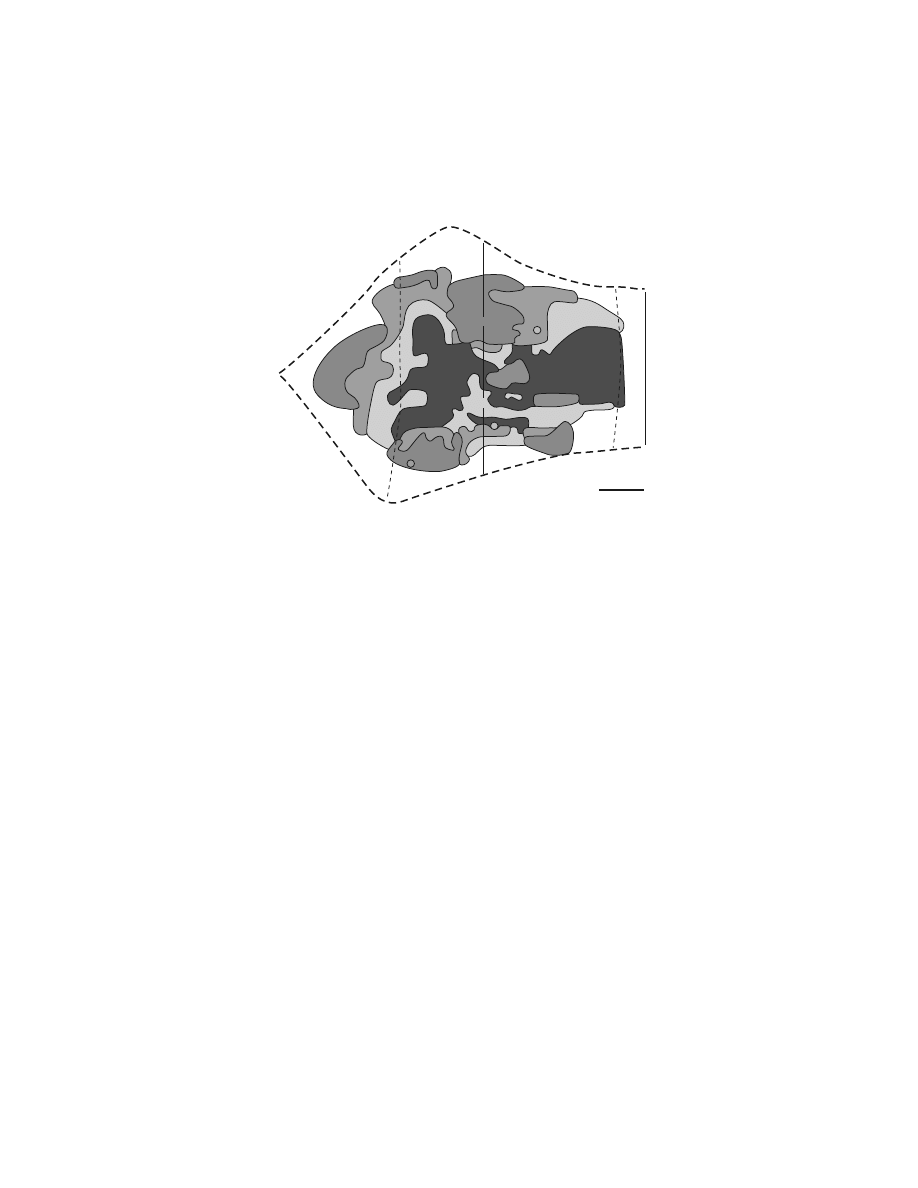
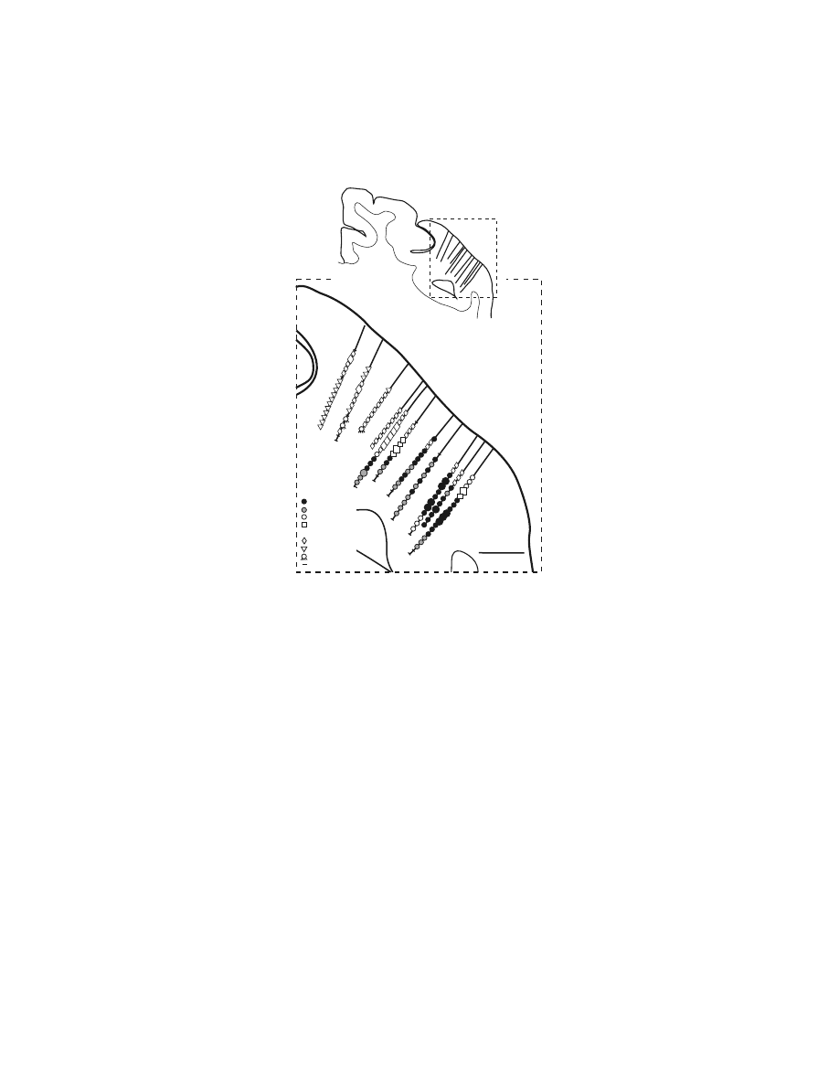
Intracortical stimulation has defined arm and face representations within the
PMv.
101
Distal arm movements dominated the portion of the arm representation that
was buried medially within the inferior limb of the posterior bank of the arcuate
sulcus (
).
64,100,101
This portion of the PMv projected almost exclusively to
upper cervical segments of the spinal cord.
60,84,113
The distal movements evoked by
stimulation at this site must therefore be mediated either by propriospinal connec-
tions from upper to lower cervical segments (for discussions see References
84,114–116) or through connections with the hand area of M1.
117,118
Proximal arm
movements tended to be evoked on the surface near the arcuate spur.
100,101
The PMv
face representation was located lateral to the arm representation both on the surface
and within the posterior bank of the arcuate sulcus (not shown in Figure 1.6).
101
Information regarding the internal organization of the face area is limited, although
laryngeal muscles appear to be represented laterally along the inferior limb of the
arcuate sulcus.
119
In summary, intracortical stimulation evokes body movements from each pre-
motor area. These stimulation effects could be mediated directly via corticospinal
efferents from each premotor area or indirectly by projections from each premotor
area to M1 and henceforth via the corticospinal efferents from M1. Examination of
this issue is limited to a short report.
120
In this study, the arm and vibrissae repre-
sentations in the SMA and M1 of the owl monkey were mapped with intracortical
stimulation. Following removal of M1, intracortical stimulation of the SMA could
still evoke movements with stimulus currents that were in the range of prelesion
values. This observation suggests that electrical activation of corticospinal efferents
in the SMA is sufficient to generate muscle contraction. This conclusion reinforces
the view that independent and parallel pathways for motor control originate in the
SMA and M1.
Copyright © 2005 CRC Press LLC
1.2.3 C
ORTICOSPINAL
T
ERMINATIONS
1.2.3.1 Primary Motor Cortex
The pattern of corticospinal terminations is one indicator of a cortical area’s potential
influence on different spinal mechanisms. (A complete discussion of this issue is
provided by Kuypers.
1
) Corticospinal efferents from M1 project to the intermediate
zone (laminae V–VIII) of the spinal cord where interneurons that innervate moto-
neurons are located as well as directly to the portions of the ventral horn where
motoneurons are located (
).
1,97,121–129
On the other hand, corticospinal
projections from somatosensory and posterior parietal cortex terminate primarily in
the dorsal horn.
1,121,122,124–126
Evidence from physiological studies suggests that these
corticospinal efferents modulate neural processing in ascending somatosensory path-
ways. (For review, see References 12,130.) Thus, both anatomical and physiological
FIGURE 1.6 Intracortical stimulation map of the arm representation of the PMv. A cross-
section from a macaque brain (Macaca nemestrina) illustrates the location of electrode
penetrations and the movements evoked at each stimulation site within the posterior bank of
the inferior limb of the arcuate sulcus. Thresholds for evoking movement are indicated by
symbol size (large symbols = #15
µA, small symbols = 16–50 µA). (Reproduced with
permission from Reference 64.)
30
32
33
34
35
36
2 mm
29
Thumb
Fingers
Wrist
Elbow,
Shoulder
Orofacial
Upper Trunk
Eye Blink
No Response
28
RP1 #239
CgS
Ar Spur
26
27
25
Copyright © 2005 CRC Press LLC
evidence suggest that the pattern of corticospinal terminations reflects the differential
involvement of these cortical areas in motor output or in somatosensory processing.
The extent of M1 terminations within the motor nuclei of the spinal cord changes
during development
129,131,132
and varies between different species.
1,133
This variation
appears to correlate with an animal’s manual dexterity.
127,132–136
For example, cebus
monkeys, which grasp small objects and manipulate tools with a modified “precision
grip,”
137–139
have abundant direct projections from M1 to spinal motor nuclei.
127
In
contrast, squirrel monkeys, which pick up small items with all fingers grasping in
concert,
139,140
have sparse monosynaptic corticospinal terminations that are located
remotely on motoneuron dendrites.
50,127
Thus, the extent of monosynaptic projections
from M1 to spinal motoneurons appears to be part of the neural substrate necessary
to make highly skilled and relatively independent movements of the fingers.
50,127,129
(For review, see References 1,12.)
1.2.3.2 Premotor Areas
The relationship between the terminations of corticospinal efferents and the motor,
sensory, and interneuronal systems of the spinal cord has been studied only for
premotor areas on the medial wall of the hemisphere.
97,128,141,142
In general, the pattern
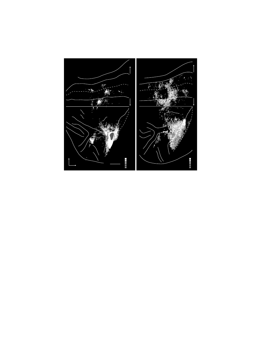
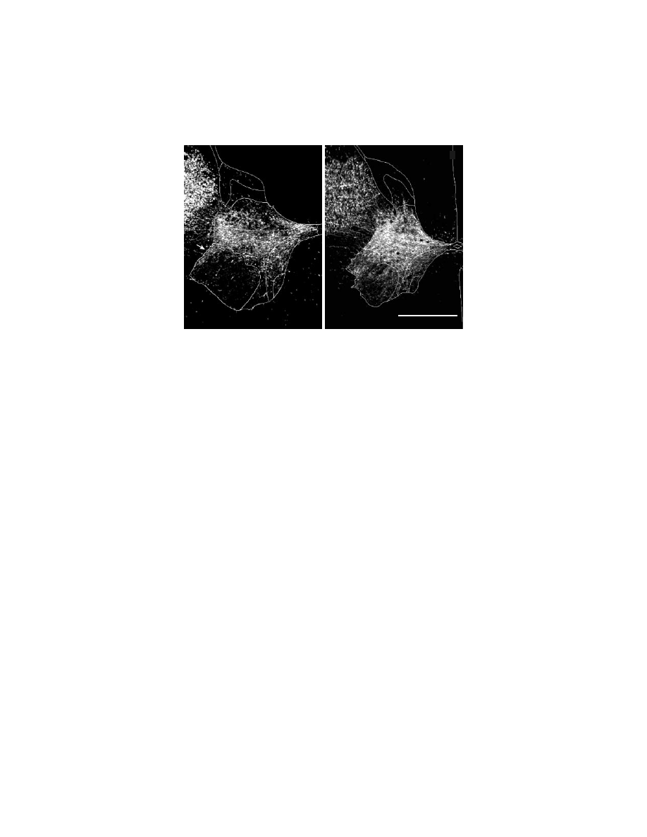
of corticospinal terminations from the SMA was quite similar to that of M1 (Figure
1.7).
97,128
The densest terminations of efferents from the SMA and M1 were located
FIGURE 1.7 Corticospinal terminations in C7 of a macaque monkey. Digital photomicro-
graphs of spinal cord sections viewed under dark-field illumination with polarized light. The
gray matter and spinal laminae are outlined. (A) SMA efferents terminate densely in inter-
mediate zone of the gray matter of the cervical spinal cord. Arrow points to terminations in
the dorsolateral part of lamina IX that contains motoneurons. (B) M1 efferents terminate in
the same regions as do SMA efferents. Compared to SMA terminations, M1 terminations are
denser and more extensive in lamina IX, and extend further into the base of the dorsal horn.
(Adapted with permission from Reference 9.)
A
A
B
B
SMA to C7
M1 to C7
1 mm
Copyright © 2005 CRC Press LLC

in the intermediate zone (laminae V–VIII) of the cervical spinal cord. Terminations
here were concentrated at three locations: (1) the dorsolateral portion of laminae
V–VII; (2) the dorsomedial portion of lamina VI at the base of the dorsal columns;
and (3) the ventromedial portion of lamina VII and adjacent lamina VIII. The
cingulate motor areas (CMAr, CMAd, CMAv) also terminated most densely within
the intermediate zone.
128,142
However, the density of their terminations was noticeably
lower than those from the SMA. In addition, terminations from the CMAr and CMAd
were concentrated in the dorsolateral portions of the intermediate zone whereas
CMAv terminations were most dense in the dorsomedial portions.
128,142
This differ-
ential pattern of terminations suggests that the CMAr, CMAd, and CMAv innervate
specific sets of spinal interneurons and thereby influence different spinal mechanisms
for controlling forelimb movements.
All of the medial wall premotor areas, like M1, had terminations that overlapped
motor nuclei in the ventral horn of the cervical segments (
).
97,128,141,142
Although the terminations from the premotor areas were less dense over lamina IX
than were those from M1, all of these studies had a consistent result: the terminations
of premotor areas that do overlap lamina IX were concentrated over the motor nuclei
innervating muscles of the fingers and wrist. Furthermore, the presence of mono-
synaptic projections onto motoneurons innervating muscles of the distal forelimb
has been confirmed electrophysiologically for the SMA.
97
This result implies that
the presence of anterogradely labeled terminations over spinal motoneurons is an
indication of direct corticomotoneuronal connections. Thus, not only the SMA but
also the CMAd, CMAv, and CMAr appear to project directly to motoneurons con-
trolling the distal forelimb. In summary, these results suggest that the premotor areas
have the anatomical substrate required to influence the generation and control of
limb movement, particularly of the hand. This influence is mediated by pathways
that are parallel to and independent of those originating in M1.
1.3 CORTICAL INPUTS TO THE MOTOR AREAS
The recognition that the premotor areas as well as M1 project to the spinal cord
suggests that motor commands may arise from multiple cortical areas. We have
proposed that each cortical area in the frontal lobe that projects to the spinal cord
could operate as a separate efferent system for the control of specific aspects of motor
behavior.
59
Obviously, identification of the cortical and subcortical inputs to these
motor areas could provide some insight into their functional contributions to motor
control.
Analysis of inputs to M1 and the premotor areas is complicated by several
factors. First, the characterization of the premotor areas is still evolving and thus,
their precise borders remain controversial.
60,77,82
(For review, see Reference 66.) For
instance, some initial examinations of the inputs to the SMA actually studied the
rostrally adjacent region that is now termed the preSMA. Second, the representations
of the face, arm, and leg within a cortical area may receive different sets of cortical
inputs.
73,75,76,78,82,143–146
For example, the arm representations of M1 and the SMA
have robust connections with the PMv whereas their leg representations do not.
75,76,78
Thus, discrepancies between studies may result from differences in the body repre-
Copyright © 2005 CRC Press LLC

sentation actually injected. Third, the boundaries and identification of the cortical
areas projecting to the motor areas are still evolving. For instance, area PE in the
parietal lobe projects to several premotor areas, but these projections tend to originate
from separate portions of this parietal area.
81,146,148,149
These results suggest that PE
may not be a single homogeneous area. Thus, precise localization and identification
of the cortical areas injected with tracers and the cortical areas containing labeled
neurons are essential for valid comparisons between different experiments.
Another aspect of comparing the inputs to each motor area is judging the relative
importance of various inputs. A small cortical region may receive input from 40 to
70 cytoarchitectonically recognized cortical areas in the ipsilateral hemisphere
alone.
82,144,150,151
However, quantitative analysis of all of the inputs to a single cortical
site has rarely been attempted.
To minimize the problems of cortical identification and strength of input, we
focused our analysis on studies that examined the inputs to the arm representation
of motor areas in macaque monkeys. We then transformed the results of these studies
onto a standarized map of the frontal and parietal lobes (
). Next, we pooled
the results from recent publications and assigned a “strength” to specific connections
based on the relative number of labeled neurons and the consistency with which a
projection was observed in all studies (
). Even with these con-
straints, we found considerable variation in the results among studies. Consequently,
our synthesis of these results reflects our consensus derived from multiple studies
and may not always fit with the data reported in an individual study.
1.3.1 P
RIMARY
M
OTOR
C
ORTEX
1.3.1.1 Frontal Cortex
Cortical input to M1 is entirely confined to cortical regions in the frontal and parietal
lobes that, like M1, are the origin of projections to the spinal cord (
, Table
1.1; see Figure 1.1 for area identification). These corticospinal tract (CST) projecting
areas include all the premotor areas in the frontal lobe (defined above) and portions
of the superior parietal lobe (SPL). M1 has no substantial connections with the
prefrontal, pre-premotor or limbic cortex.
1.3.1.2 Parietal Cortex
The densest and most extensive of projections from the parietal lobe to M1 originate
in the posterior portions of the SPL (Figure 1.8, Table 1.2; see Figure 1.1 for area
identification). This input arises in area PE on the lateral surface of the postcentral
gyrus and area PEip in the lateral portion of the dorsal bank of the intraparietal
sulcus.
59,68,71,73,76,147,152–155
M1 also receives strong inputs from the primary (SI) and
secondary (SII) somatosensory cortices. The origin of SI projections to M1 is
surprisingly widespread although their density is more modest than those from area
PE (Table 1.2). The strength of projection from the subdivisions of SI is greater for
those regions (e.g., areas 1 and 2) that are at a “later” stage in processing of the
cutaneous and proprioceptive afferent information than area 3b, which is at an
“earlier” stage of processing. Nevertheless, area 3a does have substantial input to
Copyright © 2005 CRC Press LLC

M1.
71,73,154–156
SII has heavy projections to M1 in most studies,
68,71,73,154,155
but these
projections were less substantial when injections were confined to the surface of the
precentral gyrus.
59,76
As noted earlier, neurons in M1 exhibit short latency responses to activation of
cutaneous and proprioceptive receptors. However, the route by which this soma-
tosensory input reaches M1 remains controversial. Lesion of the dorsal columns, a
major ascending pathway to the parietal cortex, extinguishes the responsiveness of
TABLE 1.1
Ipsilateral Cortical Input from the Frontal Lobe to M1, the Premotor Areas
and the Pre-Premotor Areas
Cortical
Area
M1
PMdc
PMv
SMA
CMAd
CMAv
CMAr
Pre-
PMd
Pre-
SMA
Motor
M1
Inj. Site xxx
xxx
xxx
xx
xx
x
PMd
xxx
Inj. Site xx
xxx
xx
x
x
xxx
xx
PMv
xxx
x
Inj. Site xxx
x
x
xx
x
xx
SMA
xxx
xxx
xxx
Inj. Site xxx
xxx
xx
x
x
CMAd
xx
xxx
x
xxx
Inj. Site xxx
xxx
CMAv
xx
xxx
xx
xxx
xxx
Inj. Site xxx
?
CMAr
xx
xx
xx
xxx
xxx
xxx
Inj. Site xxx
xxx
Pre-Premotor
PreSMA
xx
x
xx
?
x
xx
xxx
Inj. Site
PrePMd
xx
x
?
xx
xx
Inj. Site xxx
PrCO
xxx
?
xx
?
Prefrontal
Area 46d
?
xx
x
xxx
x
Area 46v
xx
x
x
xx
Area 9m
?
xx
xxx
Area 9l
?
?
x
FEF
x
SEF
x
Limbic
Area 24a,b
?
?
?
x
x
xx
xx
?
xxx
Area 23a,b
?
x
x
?
25, 29, 30
?
Orbital Frontal Cortex
TF, 28, 35
?
x
Prostriata
?
Note: xxx = major input, xx = moderate input, x = weak input, ? = weak input that was less than 1% of
total input and/or not observed in every case.
Copyright © 2005 CRC Press LLC

M1 neurons to peripheral input.
157
Removal of the primary and secondary somato-
sensory areas, as well as removal of the cerebellum, does not.
5,158
Some authors have
proposed that M1 receives input directly from a thalamic region innervated by a
portion of the dorsal column pathway (for references and discussion, see Reference 5),
but firm anatomical evidence for such a neural circuit remains elusive.
159
Although
the major route by which short latency somatosensory information reaches M1 has
TABLE 1.2
Ipsilateral Cortical Input from the Parietal Lobe to M1, the Premotor Areas
and the Pre-Premotor Areas
Cortical
Area
M1
PMdc
PMv
SMA
CMAd
CMAv
CMAr
Pre-PMd
Pre-SMA
Superior Parietal Lobule
Area 3a
xx
x
xx
x
?
Area 3b
x
Area 1
xx
?
x
?
Area 2
xx
?
xx
x
x
5 (PE)
xx
xx
xxx
x
xx
PEip
xxx
xxx
xx
xx
xx
xx
?
MIP
x
xxx
?
?
xxx
?
x
PEc
xxx
?
x
?
xx
PEci
xx
xx
x
x
x
xx
V6A
xxx
PGm
x
?
?
xxx
CGp
x
?
?
xxx
Inferior Parietal Lobule
AIP
x
xxx
VIP
x
?
?
LIP
?
?
7b (PF)
?
?
xxx
x
?
PFG
?
?
?
xxx
x
?
x
7a (PG)
?
?
x
xx
x
Lateral Sulcus
PFop
x
?
xx
xxx
xx
PGop
?
x
SII
xx
xxx
xx
x
xxx
Ig
xx
xx
x
xxx
x
Idg
x
xx
RI
?
Temporal
STS
?
x
Note: xxx = major input, xx = moderate input, x = weak input, ? = weak input that was less than 1%
of total input and/or not observed in every case.
Copyright © 2005 CRC Press LLC
not been determined, the interconnections between SI and M1 may be necessary for
an animal to learn a new motor skill under the guidance of somatosensory cues.
5,160
1.3.2 P
REMOTOR
A
REAS
1.3.2.1 Interconnections among the Motor Areas
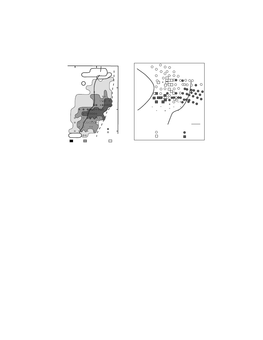
In general, the premotor areas are richly interconnected (Figure 1.8, Table 1.1).
Several features of the connections among the premotor areas stand out. First, the
SMA, of all the premotor areas, has the densest and most balanced reciprocal
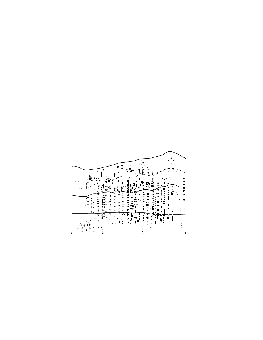
FIGURE 1.8 Major ipsilateral cortical inputs to M1, the premotor areas and the pre-premotor
areas in macaque monkeys. The premotor areas (gray shading) have reciprocal connections
with M1 and project to the spinal cord, whereas pre-premotor areas (no shading) do not. All
25 of the major cortical inputs are grouped into 8 categories, reflecting morphological location
and proposed functional similarity (see key,
and
). Sources of cortical input are
divided into cortical regions that project to the spinal cord (gray shading) and those that do
not (no shading). Note that each of the profiled cortical areas receives a unique signature of
extrinsic cortical inputs.
PMd
PEip
MIP
PEc
i
PE
c
pSM
A
pPM
d
PE
PEip
PMv
AI
P
PF
SII
Ig
46v
PrCO
SII
Ig
24a,
b
2
3a,
b
46d 46v
pSM
A
pP
Md
PrC
O
PFop
CMAr
PEi
p
3a
PFop
SII
Ig
CMAd
PE
PEip
M1
3a
2
1
SII
Pre-
PMd
PE
c
V6A
PG
m
CGp
46d 9m
pSM
A
Pre-
SMA
24a,
b
46v 9m
pPMd
PE
PEi
p
MIP
CMAv
PFG
PFop
24a,
b
46d
pPMd
2
SMA
PE
PEi
p
PEci
pS
MA
Pre-
Premotor
Prefront
al
Limbic
Medial
Posterio
r
Pariet
al
Primary
Somato-
sensory
Superior
P
ariet
al
L
obule
Inferior
Pariet
al
Lobule
Later
al
Sulcus
Target
Cortical
Area
3a
2
1
PE
PEi
p
MI
P
PEci
AI
P
PF
PF
G
P
Fop
SII
Ig
PE
c
V6
A
PG
m
CG
p
24a,
b
23a,
b
46d 46v
9m
PreSMA
PrePMd
PrCO
Project to the Spinal Cord
No Spinal Projections
Copyright © 2005 CRC Press LLC

connections with every other premotor area as well as with M1. Second, the inter-
connections among the cingulate motor areas (CMAd, CMAv, CMAr) on the medial
wall are dense and equal. Third, the PMd and the PMv on the lateral surface have
strong, reciprocal connections with M1 and the SMA. On the other hand, the con-
nections between the PMd and PMv are more limited. The PMv is connected to the
caudal portion of the PMd,
58,59,68,72,81
but only sparsely to its rostral portions.
58,161,162
The restricted connections between the PMd and the PMv may reflect differences
in the body representation in each area. The PMd was reported to project mainly to
the shoulder representation of M1, whereas the PMv projected largely to the digit
representation in M1.
73
Fourth, the projections from the lateral motor areas (PMd,
PMv, M1) to the cingulate motor areas, particularly the CMAv and the CMAr, tend
to be relatively weak.
82,163,164
These patterns of connectivity suggest that the PMd and
PMv are fundamentally distinct from each other and from the motor areas on the
medial wall. The SMA with its broad, balanced connectivity is ideally situated to
coordinate and integrate information flowing among the motor areas in the frontal lobe.
1.3.2.2 Parietal Cortex
Parietal lobe input to the premotor areas appears to follow a general trend. Most of
the parietal lobe input to the premotor areas originates from posterior portions of the
parietal lobe including the SPL (area 5, as described by Brodmann
16
), the inferior
parietal lobule (area 7, as described by Brodmann
16
) and the secondary somatosen-
sory cortex (SII) (
). These areas are thought to be concerned
with the highest levels of somatosensory processing and to participate in multimodal
sensory integration, spatial attention, or visuomotor control.
165
Every premotor area
in the frontal lobe is richly interconnected with parts of at least one of these posterior
parietal areas. On the other hand, only the SMA receives dense input from any of
the subdivisions of the primary somatosensory cortex. This input to the SMA orig-
inates in area 2, which is thought to be at an intermediate stage of somatosensory
processing (Figure 1.8, Table 1.2).
59,68,70,72,78,81,146,148,155,161–163
Portions of the superior parietal lobule project to all the premotor areas except
for the CMAr. On the postcentral gyrus, the lateral portion of area PE targets the
PMv, whereas its more medial portions project heavily to the SMA and CMAv. In
the most caudal portion of the postcentral gyrus, area PEc supplies dense input to
the PMd. Laterally within the intraparietal sulcus, area PEip has the most widespread
projections. Area PEip targets five premotor areas, including the PMd, PMv, SMA,
CMAd, and CMAv (Figure 1.8, Table 1.2). Despite this apparently broad divergence,
the origin of projections from PEip to the PMv and PMd as well as M1 tend to arise
from separate location.
59,147,149,162
Medially in the intraparietal sulcus, area MIP
projects densely to the PMd and the CMAv. In the caudal portion of the cingulate
sulcus, area PEci provides strong input to the SMA and the PMd.
59,78,146,148,149,166
Thus, although the premotor areas are broadly targeted by SPL projections, each
premotor area can be distinguished by its unique mixture of inputs from the SPL
(Figure 1.8, Table 1.2).
Projections from subdivisions of area 7 (AIP, PF, PFG, PFop) are primarily
restricted to the PMv and the three cingulate motor areas (Figure 1.8, Table 1.2).
Copyright © 2005 CRC Press LLC

Subdivisions of area 7 are characterized by more complex forms of somatosensory
processing and the presence of multimodal information that integrates visual and
somatosensory inputs.
167–169
Area PFop is the only IPL subdivision with strong links
to more than one premotor area. It has heavy projections to the CMAd, CMAv, and
CMAr. AIP in the anterior portion of the ventral bank of the intraparietal sulcus and
area PF on the adjacent IPL provide dense input to the PMv.
59,68,81,149,161,162
Area PFG,
located just caudal to area PF, projects to the CMAv.
82,163,170
Within the lateral sulcus,
SII projects densely to the PMv, CMAd, and CMAr, as well as M1.
58,59,70,149,162,163,171–174
Taken together, these observations indicate that each premotor area receives a unique
pattern of input from the various subdivisions of the parietal lobe and provides
another basis for differentiating the individual premotor areas.
The outputs from the medial posterior parietal cortex to the frontal motor areas
arise from regions that are one to two synapses removed from the primary visual
cortex. These regions include the PEc on the most caudal portion of the superior
parietal gyrus, area V6A buried on the anterior bank of the parietal occipital sulcus,
area PGm on the medial wall just rostral to the parietal occipital sulcus and the
posterior cingulate gyrus (CGp) (
). The projections from these regions are
restricted to the prePMd and PMd. The differential distribution of the projections
from this posterior parietal region to regions of the PMd and prePMd suggests that
these frontal lobe areas may require further parcellation. This is likely to be especially
evident as more physiological studies are designed to explore the visual–spatial
capabilities of these regions.
146,148,175
In general, the density of projections from these “visual areas” in posterior
parietal cortex increases as one proceeds from the caudal border of the PMd to the
prePMd.
146–149
Some have argued that these projections provide a neural substrate
for the visual guidance of reaching movements to objects in extrapersonal
space.
146–149
It is unclear, however, whether the visual information provided by these
regions is sufficient for the accurate localization of targets. For instance, area V6A
has the most direct visual input to the motor areas in the frontal lobe. V6A neurons
have large receptive fields located in the periphery of the visual field. Such fields
seem better suited to alert the motor system and shift attention to a particular quadrant
of space
176–178
than to drive the visuomotor transformation required to reach out and
grasp an object.
1.3.2.3 Pre-Premotor Cortex
Three cortical areas — the preSMA, the prePMd, and the precentral opercular cortex
(PrCO)
179
— reside at the junction between the prefrontal cortex and the premotor
areas in the frontal lobe (Figure 1.1). All three areas are located in subdivisions of
area 6. The prePMd and preSMA are part of area 6a.
17
The PrCO is part of area
6b.
17
At one time or another, each of them has been considered to be a motor area
and part of the broad term — premotor cortex. (For prePMd, see Reference 66 for
review; for preSMA, see Reference 8; for PrCO, also known as motor proisocortex
[ProM], see Reference 58). None of these areas projects directly to M1, and therefore
we do not consider any of them to be a premotor area.
9,59,64,163
Instead, the prePMd,
Copyright © 2005 CRC Press LLC

preSMA, and PrCO are one step removed from M1 and have connections with at
least one premotor area (
). For example, all of these frontal
lobe areas are interconnected with the CMAr.
58,78,82,107,144,164,180
Each area is also
connected with the adjacent premotor area — PrCO with the PMv, prePMd with
the PMd, and preSMA with the SMA.
78,107,148,161,162,180
However, the significance of
the preSMA–SMA connection and the prePMd–PMd connection is unclear. Only
immediately adjacent regions in these cortical areas appear to be interconnected and
at times these interconnections do not appear especially dense. The organization of
these connections clearly requires further investigation.
1.3.2.4 Prefrontal Cortex
Only three premotor areas — the PMv, the CMAr, and the CMAv — receive
substantial input from the dorsolateral prefrontal cortex (Walker’s area 46
181
). The
vast majority of these projections originate in the dorsal (area 46d) and ventral (area
46v) banks of the principal sulcus (
and 1.8; Table 1.1).
58,59,74,81,82,143,162
The PMv is the target of dense prefrontal projections that originate from area 46v
in a topographic manner.
59,74,81,162,182,183
The CMAv and the CMAr are the targets of
projections from both the dorsal and ventral banks of principal sulcus (areas 46d
and 46v). Additional projections to the CMAr arise from the medial and lateral
portions of area 9.
82,166
These observations indicate that specific portions of the
prefrontal cortex selectively target just three of the seven motor areas in the frontal
lobe. These projections of area 46 to the premotor areas link the prefrontal cortex
to cortical regions with direct access to the primary motor cortex and spinal mech-
anisms of motor control. Connections of the prefrontal cortex with the motor system
appear to be tightly focused and designed to provide specialized information about
particular aspects of cognitive and executive functions.
1.3.2.5 Limbic Cortex
Limbic input provides a route for emotional and affective influences over motor
behavior. Such influences may include the direction of attention toward sensory
stimuli and the expression of the motivational–affective response to noxious stimuli,
as well as the potential integration of autonomic responses.
184,185
Strong projections
from various portions of the limbic cortex are limited to the cingulate motor areas
and the PMv (Figures 1.1 and 1.8; Tables 1.1 and
). The insular cortex provides
the most widespread projections to the premotor areas. The granular insular cortex
targets the PMv, CMAd, CMAv, and CMAr
58,59,68,81,144,149,162,163,166,186
and the dysgran-
ular insular cortex targets the CMAv and CMAr.
144
From the cingulate gyrus,
areas 24a,b and 23a,b send robust projections to the CMAv and the CMAr, whereas
area 24b has additional weak projections to the SMA and CMAd.
78,82,107,163,164
The
CMAr and to a lesser degree the CMAv are also the target of weak, scattered
projections from a wide variety of cortical areas including cingulate (e.g., areas 25,
29, 30), orbitofrontal (POdg, OFdg, OFg), and temporal (e.g., TPdg, TF, areas 28
and 35) cortex.
143,144
Thus, widespread regions of the limbic cortex target the CMAr
Copyright © 2005 CRC Press LLC

and the CMAv. These connections provide the cingulate motor areas and to a lesser
degree the PMv with access to a wide spectrum of information about the state of
the entire organism as well as an integrated view of the body in space.
1.3.3 S
UMMARY
OF
C
ORTICAL
C
ONNECTIONS
We have summarized the pattern of major cortical inputs to nine cortical areas in
the frontal lobe: M1, the premotor areas and two pre-premotor areas (
Two major conclusions are evident from this analysis. First, the premotor areas are
distinguished from the two pre-premotor areas (preSMA, prePMd) on the basis of
three major characteristics. All the premotor areas (1) have reciprocal connections
with M1, (2) project directly to the spinal cord, and (3) receive input from regions
of the parietal lobe that also project directly to the spinal cord. In contrast, the pre-
SMA and pre-PMd (1) are not connected to M1, (2) do not project to the spinal
cord, and (3) do not receive dense projections from parietal areas with corticospinal
projections. These distinctions provide an anatomical basis for assigning the pre-
premotor areas to a hierarchical class that is separate from the premotor areas. The
results of imaging studies provide considerable support for this conclusion.
66
The second major conclusion from our analysis is that each of the nine cortical
areas in the frontal lobe has a unique constellation of inputs from other areas of the
cerebral cortex. Because M1 and all of the premotor areas project directly to the
spinal cord, we have proposed that each premotor area may be a “nodal point for
parallel pathways to the spinal cord.” As a consequence, each of these cortical areas
may operate as a functionally distinct system that differentially generates and/or
controls specific aspects of motor behavior.
59
1.4 SUBCORTICAL INPUTS
The basal ganglia and cerebellum are the major subcortical systems that target the
cortical motor areas in the frontal lobe. Their outputs reach the frontal lobe via
subdivisions of the ventrolateral thalamus. Efferents from the basal ganglia and
cerebellum terminate in separate sets of these thalamic nuclei.
187,188
Physiological
evidence confirms that convergence of pallidal and cerebellar input on single tha-
lamic neurons is limited (~5%).
189,190
Efferents from the globus pallidus terminate
in the rostral portions of the ventrolateral thalamus including ventralis anterior pars
parvocellularis (VApc), ventralis lateralis pars oralis (VLo) and the rostral portion
of ventralis lateralis pars caudalis (VLcr) (Figure 9) (terminology according to
Olszewski
191
).
187,188,192–195
In contrast, efferents from the deep cerebellar nuclei ter-
minate in the caudal portions of the ventrolateral thalamus including ventralis pos-
terior lateralis pars oralis (VPLo), the caudal part of ventralis lateralis pars caudalis
(VLcc), ventralis lateralis pars postrema (VLps), and area X (
).
187,188,196–204
These thalamic nuclei project to multiple regions in the frontal lobe including M1
and all the premotor areas (
). Thus, in principle, one can infer the
subcortical inputs of each cortical motor area from the origin of its thalamocortical
projections.
Copyright © 2005 CRC Press LLC
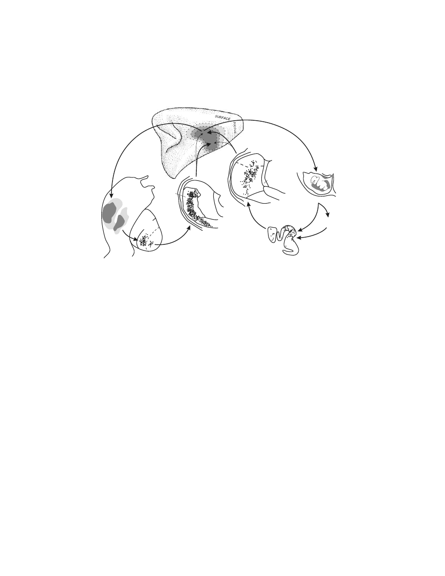
In general, multiple nuclei in the ventrolateral thalamus project to each cortical
). As a consequence, all of the cortical motor areas
except the CMAr appear to be the target of both basal ganglia and cerebellar output.
The CMAr appears to be the target largely of pallidal input (VApc and VLo).
163,164,166
In addition, the balance of pallidal and cerebellar thalamocortical projections may
vary between different portions of a single cortical area.
205–207
For example, within
the hand representation of M1, the rostral portion on the crest of the precentral gyrus
receives its densest input from VPLo, a target of cerebellar efferents (Figure 1.9).
In contrast, the caudal portion of the hand representation in the depth of the central
sulcus receives its densest input from VLo, a target of pallidal efferents. These results
suggest that pallidal and cerebellar inputs may differentially influence specific motor
areas as well as distinct regions within a single motor area. (For discussion, see
Reference 206.)
Even though each thalamic nucleus projects to more than one cortical motor
area, the neurons projecting to different cortical areas originate from largely separate
regions within the nucleus.
207–210
(See, however, References 211,212.) In fact, tha-
FIGURE 1.9 Cortical–subcortical “loops” of the basal ganglia and the cerebellum. Neurons
in the globus pallidus project via the ventrolateral thalamus mainly to the sulcal portion of
M1. Neurons in the dentate nucleus of the cerebellum project via the ventrolateral thalamus
mainly to the surface portion of M1. M1 projects to the pontine nuclei and the putamen to
complete the loops. Dark to light shading in M1 indicates hand, elbow, and shoulder repre-
sentations. The location of labeled neurons in the globus pallidus and the deep cerebellar
nuclei was determined by retrograde transneuronal transport of virus from M1. The shading
in the diagram of the pontine nuclei indicates the location of terminations from M1. The M1
terminations in the putamen are shaded according to the intensity of anterograde labeling.
(Adapted with permission from Reference 9.)
ArSi
CS
CORTEX
VPI
CL
MD
VPLo
VLcr
CEREBELLAR
NUCLEI
PONTINE NUCLEI
CEREBELLAR
CORTEX
PUTAMEN
Pcn
X
VLcr
VLo
VLm
THALAMUS
GLOBUS
PALLIDUS
ND
NI
Copyright © 2005 CRC Press LLC

lamic neurons rarely branch to innervate more than one cortical area.
207,209,210,212,213
Thus, thalamic nuclei appear to contain specific subregions, each of which projects
to a separate cortical motor area.
TABLE 1.3
Thalamocortical Connections:
Thalamic Nuclei That Receive
Pallidal and/or Nigral Afferents
Cortical Area
VApc
VLo
VLcr
VLm
M1
?
xxx
x
xx
PMd
x
xx
x
x
PMv
xx
x
?
x
SMA
x
xxx
x
xx
CMAd
x
xxx
x
CMAv
?
xx
?
CMAr
xxx
xxx
x
PreSMA
xxx
xx
xx
xx
PrePMd
xxx
x
x
x
Note: xxx = major input, xx = moderate input, x =
weak input, ? = weak input observed in a few cases.
TABLE 1.4
Thalamocortical Connections: Thalamic
Nuclei That Receive Cerebellar Afferents
Cortical Area
X
VLcc
VPLo
LP*
Pul. o*
M1
?
xx
xxx
x
x
PMd
xx
xx
?
?
PMv
xxx
xx
xx
SMA
?
xx
x
?
?
CMAd
?
xx
CMAv
xx
xx
CMAr
?
?
PreSMA
xxx
xx
?
?
PrePMd
xxx
xx
?
?
Note: xxx = major input, xx = moderate input, x = weak input,
? = weak input observed in a few cases.
* Cerebellar afferents confined to a few scattered patches in
the most rostral portions of the nuclei.
Copyright © 2005 CRC Press LLC

The presence of distinct basal ganglia and cerebellar territories in the ventrolat-
eral thalamus raises the question of how these subcortical inputs become integrated
within a cortical area. Although this issue has not been systematically investi-
gated,
214–216
both pallidal and cerebellar receiving thalamic nuclei terminate heavily
in the deeper cortical layers (layers III, V) with layer III projections being denser.
These thalamocortical projections may provide a topographically organized input
that influences corticocortical, corticothalamic, and corticostriatal circuits. Current
evidence suggests that only pallidal receiving thalamic nuclei terminate superficially
in cortical layer I.
214
These layer I terminations tend to be more broadly distributed
than are terminations in the deeper layers.
214,216
Layer I terminations may provide a
more global influence and assist in synchronizing the activity of cortical neurons
with dendrites extending into layer I.
217,218
Thus, pallidal and cerebellar circuits may
differentially access and influence some cortical laminae, but the question of whether
pallidal and cerebellar circuits converge onto the same cortical columns remains to
be addressed.
Because M1 and the premotor areas provide input to the basal ganglia via the
striatum
219,220
and to the cerebellum via the pontine nuclei,
221–224
these cortical areas
have the potential to form “closed” loops with the basal ganglia and cerebellum
(
).
219,220,225
Important aspects of the organization of these circuits remain
to be examined. For example, the extent to which related cortical areas provide
convergent input into basal ganglia and cerebellar loops with the cerebral cortex
remains to be defined. The number of distinct closed-loop circuits in each system
also remains to be determined.
To examine these and other related issues, we have employed retrograde trans-
neuronal transport of neurotropic viruses
226–231
to define the topographic organization
of the basal ganglia and cerebellar outputs to the cortical motor areas. Injections of
virus were made into different cortical motor areas. The survival time was set to
allow retrograde transport of herpes simplex virus type I (HSV1) from the injection
site to first-order neurons in the thalamus, and then retrograde transneuronal transport
from these first-order neurons to “second-order” neurons in the basal ganglia and
deep cerebellar nuclei.
Neurons labeled by retrograde transneuronal transport of virus after injections
into the arm representation of M1 were found dorsally in the dentate at mid-
rostrocaudal levels (Color
).
229,232
Injections into the leg area also
labeled neurons in a dorsal portion of the dentate, but in this case at more rostral
levels of the nucleus (Color Figure 1.10A). Likewise, virus injections into the face
area labeled neurons dorsally in the dentate, but at more caudal levels of the nucleus
(Color Figure 1.10C). This rostral to caudal arrangement of the origin of projections
to the leg, arm, and face representations in M1 corresponds well with the somatotopy
previously proposed for the dentate.
201,203,233–235
It is also clear that large portions of
the dentate nucleus were not labeled following virus injections into M1. As a
consequence, the body map generated by the projections to M1 occupied only a
portion of the dorsal third of the nucleus.
229,232
Some of the dentate regions that were not labeled after virus injections into M1
did contain labeled neurons after injections into the PMv and other premotor
Copyright © 2005 CRC Press LLC
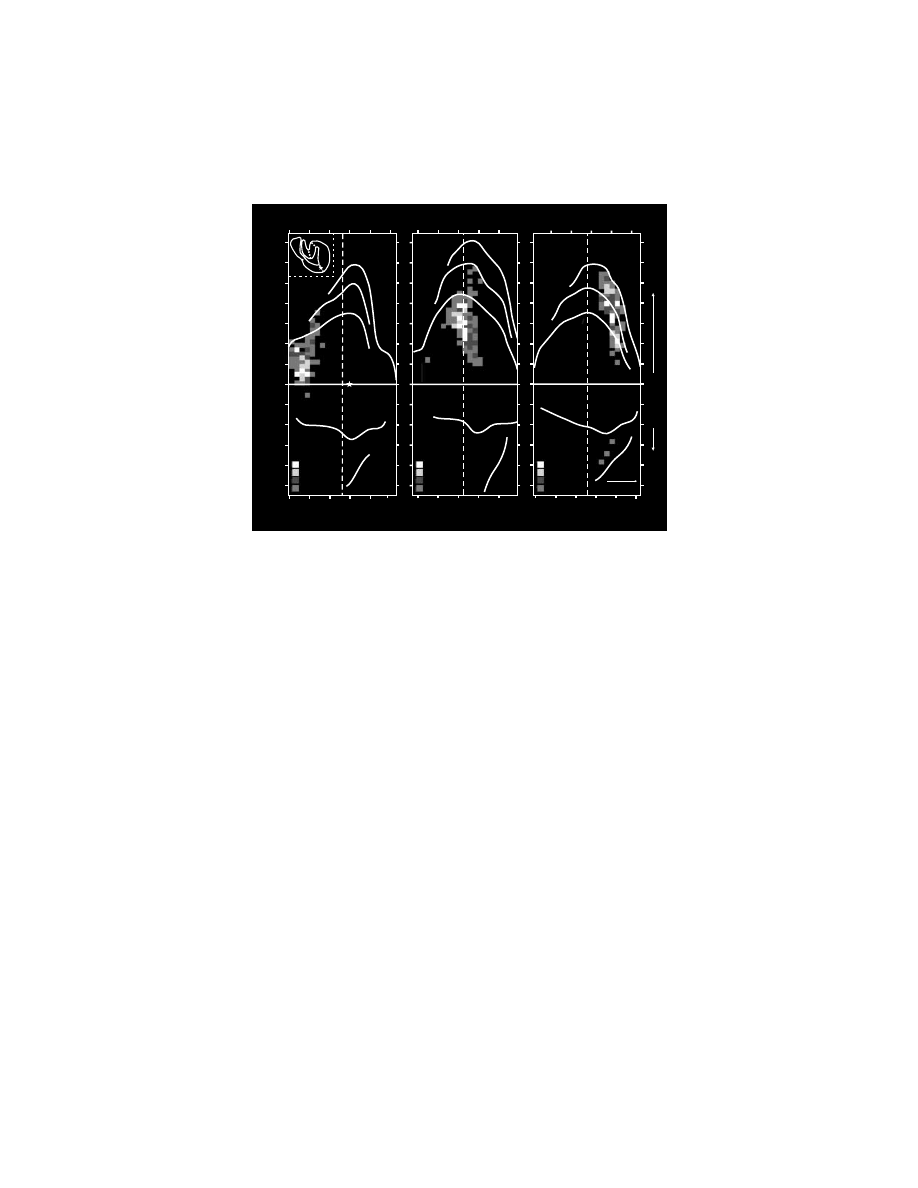
areas.
236–238
Perhaps more importantly, other regions of the dentate nucleus contained
labeled neurons after transneuronal transport of virus from prefrontal and posterior
parietal areas of the cortex.
239,240
Clearly, a substantial portion of the output from
the dentate targets nonmotor areas of cortex.
239–241
Furthermore, the dentate contains
distinct topographically organized maps of outputs to motor and nonmotor areas of
cortex (
).
232
An analogous organization is found in the globus pallidus (
).
228,229
Injection of virus into the arm representation of M1 labeled two dense clusters of
neurons in the globus pallidus — one in the inner segment and another in the outer
segment (Figure 1.12A).
229
Virus injections into the leg representation result in
similar clusters of labeled neurons that are dorsal and rostral to those projecting to
M1 arm. Virus injections into the face representation of M1 labeled neurons that are
located ventral and caudal to those projecting to M1 arm. This pattern of labeled neurons
suggests that the globus pallidus contains a rostral to caudal, dorsal to ventral body map
with respect to the leg, arm, and face representation. This somatotopic organization
is consistent with the body maps described in physiological studies.
242
As in the
dentate nucleus, large portions of the globus pallidus were not labeled following virus
FIGURE 1.10 (see color figure) Somatotopic organization of dentate output channels to M1.
Unfolded maps of the dentate illustrate the neurons labeled after HSV1 injections into the
(A) leg, (B) arm, and (C) face representations of M1. These maps of the dentate were created
by unfolding serial coronal sections through the nucleus. Inset in part A illustrates a coronal
section of the dentate where each segment in the unfolded map is identified. The dashed
vertical line indicates the rostrocaudal center of the nucleus. (Adapted with permission from
Reference 232.)
11-13
10
7-9
3-6
B
M1 Arm (Jo19)
Section Number
120
100
140 160 180
13-16
10-12
8-9
4-7
A
M1 Leg (Jo17)
Section Number
70
50
90 110 130 150
Distance (mm)
-5
-4
-3
-2
-1
0
1
2
3
4
5
6
7
Section Number
220
200
240 260 280
C
M1 Face (Jo18)
Caudal
300
10-11
9
7-8
3-6
Dorsal
Dorsal
Ventral
a
b
c
d
e
a b
c
d
e
Copyright © 2005 CRC Press LLC
injections into M1. Some of these unlabeled areas contained labeled neurons after
virus injections into premotor areas, whereas others contained labeled neurons only
after virus injections into prefrontal areas of the cortex.
243,244
Thus, the globus pallidus
contains a topographically organized map of outputs to motor and nonmotor areas
of the cortex.
We have termed the clusters of neurons in subcortical nuclei that project to a
specific cortical area an “output channel.”
245
Each of these output channels, whether
originating in the basal ganglia or cerebellum, appears to follow a simple rule:
namely, if a cortical area has dense projections to the input stage of basal ganglia
FIGURE 1.11 Topography within the cerebello-thalamocortical circuit. (A) Dentate output
channels. The origins of the peak density of dentate projections to selected cortical areas are
labeled. (B) Selected cortical targets of cerebello-thalamocortical circuits. The shading iden-
tifies cortical regions (lateral hemisphere only) that project to the cerebellum via the pons.
221–224
(C) Origin of selected cortical projections from the ventrolateral thalamus. The cortical regions
indicated receive input from regions of the ventrolateral thalamus that lie within the termi-
nation zone of cerebellar efferents. The thalamus has been turned upside down to indicate
the match between its topography and that of the dentate. Abbreviations: see
; CM/Re: nucleus centrum medianum/nucleus reuniens; IPS: intraparietal sulcus; MD:
nucleus medialis dorsalis; VLcc: caudal portion of the nucleus ventralis lateralis, pars caudalis;
VPI: nucleus ventralis posterior inferior; VPLo: nucleus ventralis posterior lateralis, pars
oralis; X: Area X. (Adapted with permission from Reference 232.)
M1
arm
M1
leg
7b
46d
9L
M1
face
1 mm
D
C
PMv
A
C
1 mm
D
M
M1
arm
M1
leg
7b
46d
PMv
9L
M1
face
MD
VLcc
VPLo
VPI
X
CM/Re
Upside Down
D
C
10 mm
M1
face
M1
leg
M1
arm
9L
46d
7b
PMv
PS
ArS
CS
B
IPS
Copyright © 2005 CRC Press LLC
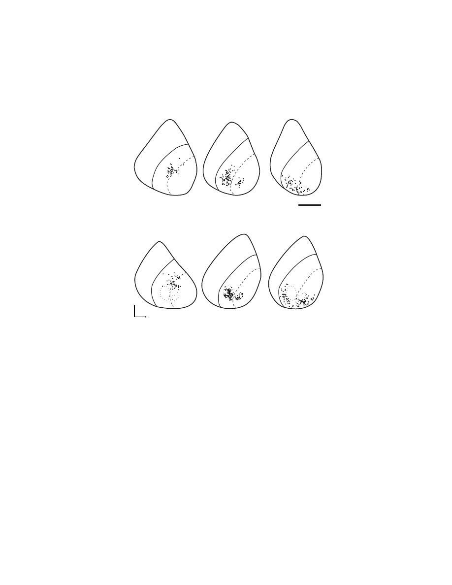
or cerebellar processing, then the cortical area is generally a target of efferents from
a distinct basal ganglia or cerebellar output channel. This rule implies that areas of
the cerebral cortex participate in multiple closed-loop circuits with the basal ganglia
(
). Not every interaction between cortex and the basal ganglia follows this
general plan. For example, regions of primary somatic sensory cortex are known to
provide input to the basal ganglia, but they do not appear to be the target of basal
FIGURE 1.12 Somatotopic organization of pallidal output channels to M1. (A) Neurons in
the internal segment of the globus pallidus (GPi) were labeled by retrograde transneuronal
transport of HSV1 following injections into the leg, arm, or face representations of M1 in
monkeys. Labeled neurons (dots) are indicated for two coronal sections separated by 0.5 mm.
(Adapted with permission from Reference 229.) (B) Neurons in the GPi were labeled follow-
ing injections of HSV1 into the arm representations of the SMA, M1, or PMv of monkeys.
Labeled neurons (dots) are indicated for two or three coronal sections near the same stereotaxic
level (A 14.0). For comparison, the dotted lines indicate the region of the GPi containing
neurons labeled from M1. The thin solid line indicates the border between the internal and
external segments of GPi. The dashed line indicates the border between the inner (i) and
outer (o) portions of the GPi. (Adapted with permission from Reference 228.)
arm
M1
leg
M1
face
M1
SMA
arm
GPe
o
i
2 mm
A 13.7
Dorsal
Medial
arm
PMv
A 14.2
arm
M1
A 14.0
A
B
Copyright © 2005 CRC Press LLC

ganglia output.
246,247
However, our evidence suggests that closed-loop circuits char-
acterize many of the interconnections between cerebral cortex and the basal ganglia
and cerebellum. Therefore, closed loops may represent a fundamental unit of basal
ganglia and cerebellar interconnections with the cerebral cortex.
231,232,243
1.5 SUMMARY AND CONCLUSIONS
A new perspective has emerged about the organization and function of the cortical
areas concerned with the control of movement. Classically, the generation of motor
commands was thought to proceed in a serial, hierarchical fashion. The output of
the premotor cortex was viewed as being funneled to M1 which served as the final
common pathway for the central control of movement.
1,4
It is now clear that the
frontal lobe contains at least six spatially separate premotor areas. Each of these
premotor areas, like M1, projects directly to the spinal cord. In fact, at least as many
corticospinal neurons originate from the premotor areas as originate from M1. Thus,
each premotor area appears to have the potential to influence the control of movement
not only at the level of the primary motor cortex, but also more directly at the level
of the spinal cord.
59,60
Why does the frontal lobe contain all of these premotor areas? Although there
is as yet no definitive answer to this question, current data from anatomical, phys-
iological, behavioral, and imaging studies suggest that each premotor area is con-
cerned with a specific aspect of movement planning, preparation, and execution.
Thus, the task of generating and controlling movement appears to be broken up into
a number of subtasks that are accomplished through parallel distributed processing
in multiple motor areas. Multiple motor areas may thereby decrease response time
and increase response flexibility. In any event, the cortical control of movement is
achieved by multiple motor areas, all of which send signals to the spinal cord.
ACKNOWLEDGMENTS
This work was supported by the Veterans Affairs Medical Research Service, and by
U.S. Public Health Service grant #24328 (PLS).
REFERENCES
1. Kuypers, H.G.J.M., Anatomy of the descending pathways, in Handbook of Physiol-
ogy, Section I: The Nervous System, Vol. II: Motor Control, Part I, Brooks, V.B., Ed.,
American Physiological Society, Bethesda, MD, 1981, 567.
2. Wise, S.P., The primate premotor cortex fifty years after Fulton, Behav. Brain Res.,
18, 79, 1985.
3. Wiesendanger, M., Recent developments in studies of the supplementary motor area
of primates, Rev. Physiol. Biochem. Pharmacol., 103, 1, 1986.
Copyright © 2005 CRC Press LLC

4. Hepp-Reymond, M.C., Functional organization of motor cortex and its participation
in voluntary movements, in Comparative Primate Biology, Vol. 4: Neurosciences,
Alan R. Liss, 1988, 501.
5. Asanuma, H., The Motor Cortex, New York, Raven Press, 1989.
6. Cheney, P.D., Fetz, E.E., and Mewes, K., Neural mechanisms underlying corticospinal
and rubrospinal control of limb movements, Prog. Brain Res., 87, 213, 1991.
7. Georgopoulos, A., Higher order motor control, Annu. Rev. Neurosci., 14, 361, 1991.
8. Tanji, J., The supplementary motor area in the cerebral cortex, Neurosci. Res., 19,
251, 1994.
9. Dum, R.P. and Strick, P.L., The corticospinal system, a structural framework for the
central control of movement, in Handbook of Physiology, Section 12: Exercise,
Regulation and Integration of Multiple Systems, Rowell, L.B. and Shepard, J.T., Eds.,
American Physiological Society, New York, 1996, 217.
10. Geyer, S. et al., Functional neuroanatomy of the primate isocortical motor system,
Anat. Embryol., 202, 443, 2000.
11. Rizzolatti, G. and Luppino, G., The cortical motor system, Neuron, 31, 889, 2001.
12. Porter, R. and Lemon, R.N., Corticospinal Function and Voluntary Movement, Oxford
University Press, Oxford, 1993.
13. Leyton, S.S.F. and Sherrington, C.S., Observations on the excitable cortex of the
chimpanzee, orangutan and gorilla, Q. J. Exp. Physiol., 11, 135, 1917.
14. Penfield, W. and Boldrey, E., Somatic motor and sensory representation in the cerebral
cortex of man as studied by electrical stimulation, Brain, 60, 389, 1937.
15. Woolsey, C.N. et al., Patterns of localization in precentral and “supplementary” motor
area and their relation to the concept of a premotor area, Assoc. Res. Nerv. Ment.
Dis., 30, 238, 1952.
16. Brodmann, K. Vergleichende Lokalisationslehre der Grosshirnrinde in ihren Prinzip-
ien dargestellt auf grund des Zellenbaues, Barth, Leipzig, 1909.
17. Vogt, C. and Vogt, O., Allgemeinere Ergebnisse unserer Hirnforschung, J. Psychol.
Neurol. (Leipzig), 25, 277, 1919.
18. Von Bonin, G. and Bailey, P., The Neocortex of Macaca Mulatta, University of Illinois
Press, Urbana, 1947.
19. Asanuma, H. and Rosen, I., Topographical organization of cortical efferent zones
projecting to distal forelimb muscles in the monkey, Exp. Brain Res., 14, 243, 1972.
20. Andersen, P., Hagan, P.J., Phillips, C.G., and Powell, T.P., Mapping by microstimu-
lation of overlapping projections from area 4 to motor units of the baboon’s hand,
Proc. R. Soc. Lond. (Biol.), 188, 31, 1975.
21. Jankowska, E., Padel, Y., and Tanaka, R., Projections of pyramidal tract cells to alpha-
motoneurones innervating hind-limb muscles in the monkey, J. Physiol. (Lond.), 249,
637, 1975.
22. Kwan, H.C., MacKay, W.A., Murphy, J.T., and Wong, Y.C., Spatial organization of
precentral cortex in awake primates. II. Motor outputs, J. Neurophysiol., 41, 1120,
1978.
23. McGuinness, E., Sivertsen, D., and Allman, J.M., Organization of the face represen-
tation in macaque motor cortex, J. Comp. Neurol., 193, 591, 1980.
24. Wise, S.P. and Tanji, J., Supplementary and precentral motor cortex, contrast in
responsiveness to peripheral input in the hindlimb area of the unanesthetized monkey,
J. Comp. Neurol., 195, 433, 1981.
25. Strick, P.L. and Preston, J.B., Two representations of the hand in area 4 of a primate.
I. Motor output organization, J. Neurophysiol., 48, 139, 1982.
Copyright © 2005 CRC Press LLC

26. Humphrey, D.R. and Reed, D.J., Separate cortical systems for control of joint move-
ment and joint stiffness, reciprocal activation and coactivation of antagonist muscles,
Adv. Neurol., 39, 347, 1983.
27. Gould, H.J., III et al., The relationship of corpus callosum connections to electrical
stimulation maps of motor, supplementary motor, and the frontal eye fields in owl
monkeys, J. Comp. Neurol., 247, 297, 1986.
28. Humphrey, D.R., Representation of movements and muscles within the primate pre-
central motor cortex, historical and current perspectives, Fed. Proc., 45, 2687, 1986.
29. Huang, C.S., Sirisko, M.A., Hiraba, H., Murray, G.M., and Sessle, B.J., Organization
of the primate face motor cortex as revealed by intracortical microstimulation and
electrophysiological identification of afferent inputs and corticobulbar projections,
J. Neurophysiol., 59, 796, 1988.
30. Sato, K.C. and Tanji, J., Digit-muscle responses evoked from multiple intracortical
foci in monkey precentral motor cortex, J. Neurophysiol., 62, 959, 1989.
31. Nudo, R.J. et al., Neurophysiological correlates of hand preference in primary motor
cortex of adult squirrel monkeys, J. Neurosci., 121, 2918, 1992.
32. Donoghue, J.P., Leibovic, S., and Sanes, J.N., Organization of the forelimb area in
squirrel monkey motor cortex, representation of digit, wrist, and elbow muscles, Exp.
Brain Res., 89, 1, 1992.
33. Murphy, J.T. et al., Spatial organization of precentral cortex in awake primates. III.
Input-output coupling, J. Neurophysiol., 41, 1132, 1978.
34. Park, M.C., Belhaj-Saif, A., Gordon, M., and Cheney, P.D., Consistent features in the
forelimb representation of primary motor cortex in rhesus macaques, J. Neurosci.,
21, 2784, 2001.
35. Schieber, M.H., Constraints on somatotopic organization in the primary motor cortex,
J. Neurophysiol., 86, 2125, 2001.
36. Graziano, M.S., Taylor, C.S., and Moore, T., Complex movements evoked by micro-
stimulation of precentral cortex, Neuron, 34, 841, 2002.
37. Ferrier, D., Experiments on the brain of monkeys, Proc. R. Soc. Lond., 23, 409, 1875.
38. Jankowska, E., Padel, Y., and Tanaka, R., The mode of activation of pyramidal tract
cells by intracortical stimuli, J. Physiol. (Lond.), 249, 617, 1975.
39. Asanuma, H., Arnold, A., and Zarzecki, P., Further study on the excitation of pyra-
midal tract cells by intracortical microstimulation, Exp. Brain Res., 26, 443, 1976.
40. Murphy, J.T. et al., Activity of primate precentral neurons during voluntary move-
ments triggered by visual signals, Brain Res., 236, 429, 1982.
41. Lemon, R.N., Muir, R.B., and Mantel, G.W., The effects upon the activity of hand
and forearm muscles of intracortical stimulation in the vicinity of corticomotor
neurons in the conscious monkey, Exp. Brain Res., 66, 621, 1987.
42. Shinoda, Y., Zarzecki, P., and Asanuma, H., Spinal branching of pyramidal tract
neurons in the monkey, Exp. Brain Res., 34, 59, 1979.
43. Shinoda, Y., Yokota, J., and Futami, T., Divergent projection of individual corticospinal
axons to motoneurons of multiple muscles in the monkey, Neurosci. Lett., 23, 7, 1981.
44. Fetz, E.E. and Cheney, P.D., Postspike facilitation of forelimb muscle activity by
primate corticomotoneuronal cells, J. Neurophysiol., 44, 751, 1980.
45. Cheney, P.D. and Fetz, E.E., Functional classes of primate corticomotoneuronal cells
and their relation to active force, J. Neurophysiol., 44, 773-791, 1980.
46. Cheney, P.D., Fetz, E.E., and Palmer, S.S., Patterns of facilitation and suppression of
antagonist forelimb muscles from motor cortex sites in the awake monkey, J. Neuro-
physiol., 53, 805, 1985.
Copyright © 2005 CRC Press LLC

47. Kasser, R.J. and Cheney, P.D., Characteristics of corticomotoneuronal postspike facil-
itation and reciprocal suppression of EMG activity in the monkey, J. Neurophysiol.,
53, 959, 1985.
48. Buys, E.J. et al., Selective facilitation of different hand muscles by single corticospinal
neurones in the conscious monkey, J. Physiol. (Lond.), 381, 529, 1986.
49. McKiernan, B.J. et al., Corticomotoneuronal postspike effects in shoulder, elbow,
wrist, digit, and intrinsic hand muscles during a reach and prehension task, J. Neuro-
physiol., 80, 1961, 1998.
50. Maier, M.A. et al., Direct and indirect corticospinal control of arm and hand moto-
neurons in the squirrel monkey (Saimiri sciureus), J. Neurophysiol., 78, 721, 1997.
51. Lemon, R.N. et al., The importance of the cortico-motoneuronal system for control
of grasp, Novartis Found. Symp., 218, 202, 1998.
52. Nakajima, K. et al., Striking differences in transmission of corticospinal excitation
to upper limb motoneurons in two primate species, J. Neurophysiol., 84, 698, 2000.
53. Lamour, Y., Jennings, V.A., and Solis, H., Functional characteristics and segregation
of cutaneous and non-cutaneous neurons in monkey precentral motor cortex (MI),
Soc. Neurosci. Abstr., 6, 158, 1980.
54. Strick, P.L. and Preston, J.B., Two representations of the hand in area 4 of a primate.
II. Somatosensory input organization, J. Neurophysiol., 48, 150, 1982.
55. Picard, N. and Smith, A.M., Primary motor cortical activity related to the weight and
texture of grasped objects in the monkey, J. Neurophysiol., 68, 1867, 1992.
56. Fulton, J.F., Definition of the “motor” and “premotor” areas, Brain, 58, 311, 1935.
57. Humphrey, D.R., On the cortical control of visually directed reaching, contribution
by nonprecentral motor areas, in Posture and Movement, Talbot, R.E. and Humphrey,
D.R., Eds., Raven Press, New York, 1979, 51.
58. Barbas, H. and Pandya, D.N., Architecture and frontal cortical connections of the
premotor cortex (area 6) in the rhesus monkey, J. Comp. Neurol., 256, 211, 1987.
59. Dum, R.P. and Strick, P.L., Premotor areas, nodal points for parallel efferent systems
involved in the central control of movement, in Motor Control, Concepts and Issues,
Humphrey, D.R. and Freund, H.-J., Eds., London, Wiley, 1991, 383.
60. Dum, R.P. and Strick, P.L., The origin of corticospinal projections from the premotor
areas in the frontal lobe, J. Neurosci., 11, 667, 1991.
61. Wise, S.P. et al., What are the specific functions of the different cortical motor areas?
in Motor Control, Concepts and Issues, Humphrey, D.R. and Freund, H-J., Eds.,
London, Wiley, 1991, 463.
62. Fulton, J.F., Physiology of the Nervous System, New York, Oxford University Press, 1949.
63. Penfield, W. and Welch, K., Supplementary motor area of the cerebral cortex, Arch.
Neurol. Psychiat., 66, 289, 1951.
64. Dum, R.P. and Strick, P.L., Motor areas in the frontal lobe of the primate, Physiol.
Behav., 77, 677, 2002.
65. Picard, N. and Strick, P.L., Motor areas of the medial wall, a review of their location
and functional activation, Cereb. Cortex, 6, 342, 1996.
66. Picard, N. and Strick, P.L., Imaging the premotor areas, Cur. Opin. Neurobiol., 11,
663, 2001.
67. Muakkassa, K.F. and Strick, P.L., Frontal lobe inputs to primate motor cortex, evidence
for four somatotopically organized ‘premotor’ areas, Brain Res., 177, 176, 1979.
68. Godschalk, M. et al., Cortical afferents and efferents of monkey postarcuate area, an
anatomical and electrophysiological study, Exp. Brain Res., 56, 410, 1984.
Copyright © 2005 CRC Press LLC

69. Strick, P.L., How do the basal ganglia and cerebellum gain access to the cortical
motor areas? Behav. Brain Res., 18, 107, 1985.
70. Künzle, H., Cortico-cortical efferents of primary motor and somatosensory regions
of the cerebral cortex, Neuroscience, 3, 25, 1978.
71. Ghosh, S., Brinkman, C., and Porter, R.A., Quantitative study of the distribution of
neurons projecting to the precentral motor cortex in the monkey (M. fascicularis),
J. Comp. Neurol., 259, 424, 1987.
72. Morecraft, R.J. and Van Hoesen, G.W., Cingulate input to the primary and supple-
mentary motor cortices in the rhesus monkey, evidence for somatotopy in areas 24c
and 23c, J. Comp. Neurol., 322, 471, 1992.
73. Tokuno, H. and Tanji, J., Input organization of distal and proximal forelimb areas in
the monkey primary motor cortex, a retrograde double labeling study, J. Comp.
Neurol., 333, 199, 1993.
74. Lu, M.-T., Preston, J.B., and Strick, P.L., Interconnections between the prefrontal
cortex and the premotor areas in the frontal lobe, J. Comp. Neurol., 341, 375, 1994.
75. Tokuno, H. and Inase, M., Direct projections from the ventral premotor cortex to the
hindlimb region of the supplementary motor area in the macaque monkey, Neurosci.
Lett., 171, 159, 1994.
76. Hatanaka, N. et al., Somatotopic arrangement and corticocortical inputs of the hind-
limb region of the primary motor cortex in the macaque monkey, Neurosci. Res., 40,
9, 2001.
77. Matelli, M., Luppino, G., and Rizzolatti, G., Architecture of superior and mesial area
6 and the adjacent cingulate cortex in the macaque monkey, J. Comp. Neurol., 311,
445, 1991.
78. Luppino, G. et al., Corticocortical connections of area F3 (SMA-Proper) and area F6
(Pre-SMA) in the macaque monkey, J. Comp. Neurol., 338, 114, 1993.
79. Matsuzaka, Y., Aizawa, H., and Tanji, J., A motor area rostral to the supplementary
motor area (presupplementary motor area) in the monkey, neuronal activity during a
learned motor task, J. Neurophysiol., 68, 653, 1992.
80. Inase, M. et al., Corticostriatal and corticosubthalamic input zones from the presup-
plementary motor area in the macaque monkey, comparison with the input zones
from the supplementary motor area, Brain Res., 833, 191, 1999.
81. Matelli, M. et al., Afferent and efferent projections of the inferior area 6 in the
macaque monkey, J. Comp. Neurol., 251, 281, 1986.
82. Morecraft, R.J. and Van Hoesen, G.W., Frontal granular cortex input to the cingulate
(M3), supplementary (M2) and primary (M1) motor cortices in the rhesus monkey,
J. Comp. Neurol., 337, 669, 1993.
83. Russell, J.R. and DeMyer, W., The quantitative cortical origin of pyramidal axons of
Macaca mulatta, Neurology (Minneapolis) 11, 96, 1961.
84. He, S.Q., Dum, R. P., and Strick, P.L., Topographic organization of corticospinal
projections from the frontal lobe, motor areas on the lateral surface of the hemisphere,
J. Neurosci., 13, 952, 1993.
85. He, S.Q., Dum, R.P., and Strick, P.L., Topographic organization of corticospinal
projections from the frontal lobe, motor areas on the medial wall of the hemisphere,
J. Neurosci., 15, 3284, 1995.
86. Catsman-Berrevoets, C.E. and Kuypers, H.G.J.M., Cells of origin of cortical projec-
tions to dorsal column nuclei, spinal cord and bulbar medial reticular formation in
the rhesus monkey, Neurosci. Lett., 3, 245, 1976.
Copyright © 2005 CRC Press LLC

87. Biber, M.P., Kneisley, L.W., and LaVail, J.H., Cortical neurons projecting to the
cervical and lumbar enlargements of the spinal cord in young and adult rhesus
monkeys, Exp. Neurol., 59, 492, 1978.
88. Murray, E.A. and Coulter, J.D., Organization of corticospinal neurons in the monkey,
J. Comp. Neurol., 195, 339, 1981.
89. Macpherson, J.M. et al., Microstimulation of the supplementary motor area (SMA)
in the awake monkey, Exp. Brain Res., 45, 410, 1982.
90. Toyoshima, K. and Sakai, H., Exact cortical extent of the origin of the corticospinal
tract (CST) and the quantitative contribution to the CST in different cytoarchitectonic
areas. A study with horseradish peroxidase in the monkey, J. Hirnforsch., 23, 257,
1982.
91. Keizer, K. and Kuypers, H.G., Distribution of corticospinal neurons with collaterals
to the lower brain stem reticular formation in monkey (Macaca fascicularis), Exp.
Brain Res., 74, 311, 1989.
92. Nudo, R.J. and Masterton, R.B., Descending pathways to the spinal cord. III. Sites
of origin of the corticospinal tract, J. Comp. Neurol., 296, 559, 1990.
93. Galea, M.P. and Darian-Smith, I., Multiple corticospinal neuron populations in the
macaque monkey are specified by their unique cortical origins, spinal terminations,
and connections, Cereb. Cortex, 4, 166, 1994.
94. Jones, E.G., and Wise, S.P., Size, laminar and columnar distribution of efferent cells
in the sensory-motor cortex of monkeys, J. Comp. Neurol., 175, 391, 1977.
95. Humphrey, D.R. and Corrie, W.S., Properties of pyramidal tract neuron system within
a functionally defined subregion of primate motor cortex, J. Neurophysiol., 41, 216,
1978.
96. Lawrence, D.G. and Kuypers, H.G.J.M., The functional organization of the motor
system in the monkey. I. The effects of bilateral pyramidal lesions, Brain, 91, 1, 1968.
97. Maier, M.A. et al., Differences in the corticospinal projection from primary motor
cortex and supplementary motor area to macaque upper limb motoneurons, an ana-
tomical and electrophysiological study, Cereb. Cortex, 12, 281, 2002.
98. Mitz, A.R. and Wise, S.P., The somatotopic organization of the supplementary motor
area, intracortical microstimulation mapping, J. Neurosci., 7, 1010, 1987.
99. Luppino, G. et al., Multiple representations of body movements in mesial area 6 and
adjacent cingulate cortex, an intracortical microstimulation study in the macaque
monkey, J. Comp. Neurol., 311, 463, 1991.
100. Hepp-Reymond, M-C. et al., Force-related neuronal activity in two regions of the
primate ventral premotor cortex, Can. J. Physiol. Pharm., 72, 571, 1994.
101. Godschalk, M. et al., Somatotopy of monkey premotor cortex examined with micro-
stimulation, Neurosci. Res., 23, 269, 1995.
102. Akazawa, T. et al., A cortical motor region that represents the cutaneous back muscles
in the macaque monkey, Neurosci. Lett., 282, 125, 2000.
103. Takada, M. et al., Organization of inputs from cingulate motor areas to basal ganglia
in macaque monkey, Eur. J. Neurosci., 14, 1633, 2001.
104. Rizzolatti, G. et al., Neurons related to reaching-grasping arm movements in the area
6 (area 6a beta), Exp. Brain Res., 82, 337, 1990.
105. Alexander, G.E. and Crutcher, M.D., Preparation for movement, neural representa-
tions of intended direction in three motor areas of the monkey, J. Neurophysiol., 64,
133, 1990.
106. Inase, M. et al., Origin of thalamocortical projections to the presupplementary motor
area (pre-SMA) in the macaque monkey, Neurosci. Res., 25, 217, 1996.
Copyright © 2005 CRC Press LLC

107. Wang, Y. et al., Spatial distribution of cingulate cells projecting to the primary,
supplementary, and pre-supplementary motor areas, a retrograde multiple labeling
study in the macaque monkey, Neurosci. Res., 39, 39, 2001.
108. Hutchins, K.D., Martino, A.M., and Strick, P.L., Corticospinal projections from the
medial wall of the hemisphere, Exp. Brain Res., 71, 667, 1988.
109. Cadoret, G. and Smith, A.M., Comparison of the neuronal activity in the SMA and
the ventral cingulate cortex during prehension in the monkey, J. Neurophysiol., 77,
153, 1997.
110. Mitz, A.R. and Humphrey, D.R., Intracortical stimulation in pyramidotomized mon-
keys, Neurosci. Lett., 64, 59, 1986.
111. Fogassi, L. et al., Visual responses in the dorsal premotor area F2 of the macaque
monkey, Exp. Brain Res., 128, 194, 1999.
112. Fujii, N., Mushiake, H., and Tanji, J., Rostrocaudal distinction of the dorsal premotor
area based on oculomotor involvement, J. Neurophysiol., 83, 1764, 2000.
113. Martino, A.M. and Strick, P.L., Corticospinal projections originate from the arcuate
premotor area, Brain Res., 404, 307, 1987.
114. Maier, M.A. et al., Does a C3-C4 propriospinal system transmit corticospinal exci-
tation in the primate? An investigation in the macaque monkey, J. Physiol. (Lond.),
511, 191, 1998.
115. Alstermark, B. et al., Disynaptic pyramidal excitation in forelimb motoneurons medi-
ated via C(3)-C(4) propriospinal neurons in the Macaca fuscata, J. Neurophysiol.,
82, 3580, 1999.
116. Kirkwood, P.A., Maier, M.A., and Lemon, R.N., Interspecies comparisons for the
C3-C4 propriospinal system, unresolved issues, Adv. Exp. Med. Biol., 508, 299, 2002.
117. Cerri, G., Shimazu, H., Maier, M.A., and Lemon, R.N., Facilitation from ventral
premotor cortex of primary motor cortex outputs to macaque hand muscles, J. Neuro-
physiol., 90, 832, 2003.
118. Shimazu, H. et al., Macaque ventral premotor cortex exerts powerful facilitation of
motor cortex outputs to upper limb motoneurons, J. Neurosci., 24, 1200, 2004.
119. Hast, M.H. et al., Cortical motor representation of the laryngeal muscles in Macaca
mulatta, Brain Res., 73, 229, 1974.
120. Hahm, J. et al., Parallel cortical pathways for the control of movement, Soc. Neurosci.
Abstr., 18, 216, 1992.
121. Kuypers, H.G.J.M., Central cortical projections to motor and somato-sensory cell
groups, Brain, 83, 161, 1960.
122. Liu, C.N. and Chambers, W.W., An experimental study of the cortico-spinal system
in the monkey (Macaca mulatta), J. Comp. Neurol., 123, 257, 1964.
123. Kuypers, H.G.J.M. and Brinkman, J., Precentral projections of different parts of the
spinal intermediate zone in the rhesus monkey, Brain Res., 24, 29, 1970.
124. Coulter, J.D. and Jones, E.G., Differential distribution of corticospinal projections
from individual cytoarchitectonic fields in the monkey, Brain Res., 129, 335, 1977.
125. Cheema, S.S., Rustioni, A., and Whitsel, B.L., Light and electron microscopic evi-
dence for a direct corticospinal projection to superficial laminae of the dorsal horn
in cats and monkeys, J. Comp. Neurol., 225, 276, 1984.
126. Ralston, D.D. and Ralston, H.J., III, The terminations of corticospinal tract axons in
the macaque monkey, J. Comp. Neurol., 242, 325, 1985.
127. Bortoff, G.A. and Strick, P.L., Corticospinal terminations in two new-world primates,
further evidence that corticomotoneuronal connections provide part of the neural
substrate for manual dexterity, J. Neurosci., 13, 5105, 1993.
Copyright © 2005 CRC Press LLC

128. Dum, R.P. and Strick, P.L., Spinal cord terminations of the medial wall motor areas
in macaque monkeys, J. Neurosci., 16, 6513, 1996.
129. Armand, J., Olivier, E., Edgley, S.A., and Lemon, R.N., Postnatal development of
corticospinal projections from motor cortex to the cervical enlargement in the
macaque monkey, J. Neurosci., 17, 251, 1997.
130. Phillips, C.G. and Porter, R., Corticospinal Neurons: Their Role in Movement, Mono-
graph of the Physiological Society, No. 34, London, Academic Press, 1977.
131. Kuypers, H.G.J.M., Corticospinal connections, postnatal development in the rhesus
monkey, Science, 138, 678, 1962.
132. Olivier, E., Edgley, S.A., Armand, J., and Lemon, R.N., An electrophysiological study
of the postnatal development of the corticospinal system in the macaque monkey,
J. Neurosci., 17, 267, 1997.
133. Heffner, R. and Masterton, R.B., Variation in form of the pyramidal tract and its
relationship to digital dexterity, Brain Behav. Evol., 12, 161, 1975.
134. Phillips, C.G., Evolution of the corticospinal tract in primates with special reference
to the hand, in Proceedings of the 3rd International Congress on Primatology, Zürich,
Vol. 2, Karger, Basel, 1971, 2.
135. Heffner, R.S. and Masterton, R.B., The role of the corticospinal tract in the evolution
of human digital dexterity, Brain Behav. Evol., 23, 165, 1983.
136. Lawrence, D.G. and Hopkins, D.A., The development of motor control in the rhesus
monkey, evidence concerning the role of corticomotoneuronal connections, Brain,
99, 235, 1976.
137. Antinucci, F. and Visalberghi, E., Tool use in Cebus apella, a case study, Int. J.
Primatol., 7, 349, 1986.
138. Westergaard, G.C. and Fragaszy, D.M., The manufacture and use of tools by capuchin
monkeys (Cebus apella), Zoo. Biol., 4, 317, 1987.
139. Costello, M.B. and Fragaszy, D.M., Prehension in Cebus and Saimiri. I. Grip type
and hand preference, Am. J. Primotol., 15, 235, 1988.
140. Fragaszy, D.M., Preliminary quantitative studies of prehension in squirrel monkeys
(Saimiri sciureus), Brain Behav. Evol., 23, 81, 1983.
141. Rouiller, E.M. et al., Evidence for direct connections between the hand region of the
supplementary motor area and cervical motoneurons in the macaque monkey, Eur. J.
Neurosci., 8, 1055, 1996.
142. Morecraft, R.J. et al., Segregated parallel inputs to the brachial spinal cord from the
cingulate motor cortex in the monkey, Neuro Report, 8, 3933, 1997
143. Bates, J.F. and Goldman-Rakic, P.S., Prefrontal connections of medial motor areas
in the rhesus monkey, J. Comp. Neurol., 336, 211, 1993.
144. Morecraft, R.J. and Van Hoesen, G.W., Convergence of limbic input to the cingulate
motor cortex in the rhesus monkey, Brain Res. Bull., 45, 209, 1998.
145. Tokuno, H. et al., Reevaluation of ipsilateral corticocortical inputs to the orofacial
region of the primary motor cortex in the macaque monkey, J. Comp. Neurol., 389,
34, 1997.
146. Matelli, M. et al., Superior area 6 afferents from the superior parietal lobule in the
macaque monkey, J. Comp. Neurol., 402, 327, 1998.
147. Johnson, P.B. et al., Cortical networks for visual reaching, physiological and anatom-
ical organization of frontal and parietal lobe arm regions, Cereb. Cortex, 6, 102, 1996.
148. Marconi, B. et al., Eye
−hand coordination during reaching. I. Anatomical relation-
ships between parietal and frontal cortex, Cereb. Cortex, 11, 513, 2001.
Copyright © 2005 CRC Press LLC

149. Tanne-Gariepy, J., Rouiller, E.M., and Boussaoud, D., Parietal inputs to dorsal versus
ventral premotor areas in the macaque monkey, evidence for largely segregated
visuomotor pathways, Exp. Brain Res., 145, 91, 2002.
150. Morecraft, R.J., Rockland, K.S., and Van Hoesen, G.W., Localization of area pros-
triata and its projection to the cingulate motor cortex in the rhesus monkey, Cereb.
Cortex, 10, 192, 2000.
151. Simonyan, K. and Jürgens, U., Cortico-cortical projections of the motorcortical larynx
area in the rhesus monkey, Brain Res., 949, 23, 2002.
152. Strick, P.L. and Kim, C.C., Input to primate motor cortex from posterior parietal
cortex (area 5). I. Demonstration by retrograde transport, Brain Res., 157, 325, 1978.
153. Vogt, B.A. and Pandya, D.N., Cortico-cortical connections of somatic sensory cortex
(areas 3, 1 and 2) in the rhesus monkey, J. Comp. Neurol., 177, 179, 1978.
154. Leichnetz, G.R., Afferent and efferent connections of the dorsolateral precentral gyrus
(area 4, hand/arm region) in the macaque monkey, with comparisons to area 8,
J. Comp. Neurol., 254, 460, 1986.
155. Darian-Smith, C. et al., Ipsilateral cortical projections to areas 3a, 3b, and 4 in the
macaque monkey, J. Comp. Neurol., 335, 200, 1993.
156. Huerta, M.F. and Pons, T.P., Primary motor cortex receives input from area 3a in
macaques, Brain Res., 537, 367, 1990.
157. Brinkman, J., Bush, B.M., and Porter, R., Deficient influence of peripheral stimuli
on precentral neurones in monkeys with dorsal column lesions, J. Physiol. (Lond.),
276, 27, 1978.
158. Asanuma, H. and Mackel, R., Direct and indirect sensory input pathways to the motor
cortex; its structure and function in relation to learning of motor skills, Jpn. J. Physiol.,
39, 1, 1989.
159. Tracey, D. J. et al., Thalamic relay to motor cortex, afferent pathways from brain
stem, cerebellum, and spinal cord in monkeys, J. Neurophysiol., 44, 532, 1980.
160. Pavlides, C., Miyashita, E., and Asanuma, H., Projection from the sensory to the
motor cortex is important in learning motor skills in the monkey, J. Neurophysiol.,
70, 733, 1993.
161. Kurata, K., Corticocortical inputs to the dorsal and ventral aspects of the premotor
cortex of macaque monkeys, Neurosci. Res., 12, 263, 1991.
162. Ghosh, S. and Gattera, R., A comparison of the ipsilateral cortical projections to the
dorsal and ventral subdivisions of the macaque premotor cortex, Somatosens. Mot.
Res., 12, 359, 1995.
163. Dum, R.P. and Strick, P.L., Cingulate motor areas, in Neurobiology of Cingulate
Cortex and Limbic Thalamus, Vogt, B.A. and Gabriel, M., Eds., Birkhauser, Boston,
1993, 415.
164. Hatanaka, N. et al., Thalamocortical and intracortical connections of monkey cingu-
late motor areas, J. Comp. Neurol., 462, 121, 2003.
165. Lynch, J.C., The functional organization of posterior parietal association cortex,
Behav. Brain Sci., 3, 485, 1980.
166. Van Hoesen, G.W., Morecraft, R.J., and Vogt, B.A., Connections of the monkey
cingulate cortex, in Neurobiology of cingulate cortex and limbic thalamus, Vogt, B.A.
and Gabriel, M., Eds., Birkhauser, Boston, 1993, 249.
167. Hyvarinen, J., Posterior parietal lobe of the primate brain, Physiol. Rev., 62, 1060, 1982.
168. Sakata, H. et al., Neural mechanisms of visual guidance of hand action in the parietal
cortex of the monkey, Cereb. Cortex, 5, 429, 1995.
Copyright © 2005 CRC Press LLC

169. Murata, A. et al., Selectivity for the shape, size, and orientation of objects for grasping
in neurons of monkey parietal area AIP, J. Neurophysiol., 83, 2580, 2000.
170. Cavada, C. and Goldman-Rakic, P., Posterior parietal cortex in rhesus monkey, II.
Evidence for segregated corticocortical networks linking sensory and limbic areas
with the frontal lobe, J. Comp. Neurol., 287, 422, 1989.
171. Baleydier, C. and Mauguiere, F., The duality of the cingulate gyrus in monkey,
neuroanatomical study and functional hypothesis, Brain, 103, 525, 1980.
172. Baleydier, C. and Mauguiere, F., Network organization of the connectivity between
parietal area 7, posterior cingulate cortex and medial pulvinar nucleus. A double
fluorescent tracer study in monkey, Exp. Brain Res., 66, 385, 1987.
173. Petrides, M. and Pandya, D.N., Projections to the frontal cortex from the posterior
parietal region in the rhesus monkey, J. Comp. Neurol., 228, 105, 1984.
174. Vogt, B.A. and Pandya, D.N., Cingulate cortex of the rhesus monkey, II. Cortical
afferents, J. Comp. Neurol., 262, 271, 1987.
175. Matelli, M. and Luppino, G., Parietofrontal circuits for action and space perception
in the macaque monkey, Neuroimage, 14, S27, 2001.
176. Shipp, S., Blanton, M., and Zeki, S., A visuo-somatomotor pathway through superior
parietal cortex in the macaque monkey, cortical connections of areas V6 and V6A,
Eur. J. Neurosci., 10, 3171, 1998.
177. Galletti, C. et al., Functional demarcation of a border between areas V6 and V6A in
the superior parietal gyrus of the macaque monkey, Eur. J. Neurosci., 8, 30, 1996.
178. Galletti, C. et al., Brain location and visual topography of cortical area V6A in the
macaque monkey, Eur. J. Neurosci., 11, 575, 1999.
179. Roberts, T.S. and Akert, K., Insular and opercular cortex and its thalamic projection
in Macaca mulatta, Schweiz. Archiv. Neurol. Neurochir. Psychiat., 92, 1, 1963.
180. Luppino, G. et al., Prefrontal and agranular cingulate projections to the dorsal pre-
motor areas F2 and F7 in the macaque monkey, Eur. J. Neurosci., 17, 559, 2003.
181. Walker, A., A cytoarchitectural study of the prefrontal area of the macaque monkey,
J. Comp. Neurol., 73, 59, 1940.
182. Barbas, H. and Mesulam, M.M., Cortical afferent input to the principalis region of
the rhesus monkey, Neuroscience, 15, 619, 1985.
183. Wang, Y. et al., Spatial distribution and density of prefrontal cortical cells projecting
to three sectors of the premotor cortex, NeuroReport 13, 1341, 2002.
184. Papez, J.W., A proposed mechanism of emotion, Arch. Neurol. Psychiat., 38, 725,
1937.
185. Vogt, B.A. and Gabriel, M., Eds., Neurobiology of Cingulate Cortex and Limbic
Thalamus, Birkhauser, Boston, 1993.
186. Barbas, H. and Pandya, D.N., Architecture and intrinsic connections of the prefrontal
cortex in the rhesus monkey, J. Comp. Neurol., 286, 353, 1989.
187. Ilinsky, I.A. and Kultas-Ilinsky, K., Sagittal cytoarchitectonic maps of the Macaca
mulatta thalamus with a revised nomenclature of the motor-related nuclei validated
by observations on their connectivity, J. Comp. Neurol., 262, 331, 1987.
188. Percheron, G., Francois, C., Talbi, B., Yelnik, J., and Fenelon, G., The primate motor
thalamus, Brain Res. Rev., 22, 93, 1996.
189. Nambu, A., Yoshida, S., and Jinnai, K., Projection on the motor cortex of thalamic
neurons with pallidal input in the monkey, Exp. Brain Res., 71, 658, 1988.
190. Nambu, A., Yoshida, S., and Jinnai, K., Movement-related activity of thalamic neurons
with input from the globus pallidus and projection to the motor cortex in the monkey,
Exp. Brain Res., 84, 279, 1991.
Copyright © 2005 CRC Press LLC

191. Olszewski, J., The Thalamus of the Macaca Mulatta: An Atlas for Use with the
Stereotaxic Instrument, S. Karger AG, Basel, 1952.
192. Nauta, W.J.H. and Mehler, W.R., Projections of the lentiform nucleus in the monkey,
Brain Res., 1, 3, 1966.
193. Kuo, J.S. and Carpenter, M.B., Organization of pallidothalamic projections in rhesus
monkey, J. Comp. Neurol., 151, 201, 1973.
194. Kim, R. et al., Projections of the globus pallidus and adjacent structures, an auto-
radiographic study in the monkey, J. Comp. Neurol., 169, 263, 1976.
195. DeVito, J.L. and Anderson, M.E., An autoradiographic study of efferent connections
of the globus pallidus in Macaca mulatta, Exp. Brain Res., 46, 107, 1982.
196. Kusama, T., Mabuchi, M., and Sumino, T., Cerebellar projections to the thalamic
nuclei in monkeys, Proc. Jpn. Acad., 47, 505, 1971.
197. Kievit, J. and Kuypers, H.G.J.M., Organization of the thalamo-cortical connections
to the frontal lobe in the rhesus monkey, Exp. Brain Res., 29, 299, 1977.
198. Batton, R.R. et al., Fastigial efferent projections in the monkey. An autoradiographic
study, J. Comp. Neurol., 174, 281, 1977.
199. Chan-Palay, V., Cerebellar Dentate Nucleus: Organization, Cytology and Transmitters,
Springer-Verlag, Berlin, 1977.
200. Percheron, G., The thalamic territory of cerebellar afferents and the lateral region of
the thalamus of the macaque in stereotaxic ventricular coordinates, J. Hirnforsch.,
18, 375, 1977.
201. Stanton, G.B., Topographical organization of ascending cerebellar projections from
the dentate and interposed nuclei in Macaca mulatta. An anterograde degeneration
study, J. Comp. Neurol., 190, 699, 1980.
202. Kalil, K., Projections of the cerebellar and dorsal column nuclei upon the thalamus
of the rhesus monkey, J. Comp. Neurol., 195, 25, 1981.
203. Asanuma, C., Thach, W.T., and Jones, E.G., Distribution of cerebellar terminations
and their relation to other afferent terminations in the ventral lateral thalamic region
of the monkey, Brain Res., 286, 237, 1983.
204. Asanuma, C., Thach, W.R., and Jones, E.G., Anatomical evidence for segregated focal
groupings of efferent cells and their terminal ramifications in the cerebellothalamic
pathway of the monkey, Brain Res., 286, 267, 1983.
205. Matelli, M. et al., Thalamic input to inferior area 6 and area 4 in the macaque monkey,
J. Comp. Neurol., 280, 468, 1989.
206. Holsapple, J.W., Preston, J.B., and Strick, P.L., The origin of thalamic inputs to the
“hand” representation in the primary motor cortex, J. Neurosci., 11, 2644, 1991.
207. Matelli, M. and Luppino, G., Thalamic input to mesial and superior area 6 in the
macaque monkey, J. Comp. Neurol., 372, 59, 1996.
208. Schell, G.R. and Strick, P.L., The origin of thalamic inputs to the arcuate premotor
and supplementary motor areas, J. Neurosci., 4, 539, 1984.
209. Darian-Smith, C., Darian-Smith, I., and Cheema, S.S., Thalamic projections to sen-
sorimotor cortex in the macaque monkey, use of multiple retrograde fluorescent
tracers, J. Comp. Neurol., 299, 17, 1990.
210. Shindo, K., Shima, K., and Tanji, J., Spatial distribution of thalamic projections to
the supplementary motor area and the primary motor cortex, a retrograde multiple
labeling study in the macaque monkey, J. Comp. Neurol., 357, 98, 1995.
211. Rouiller, E.M. et al., Cerebellothalamocortical and pallidothalamocortical projections
to the primary and supplementary motor cortical areas, a multiple tracing study in
macaque monkeys, J. Comp. Neurol., 345, 185, 1994.
Copyright © 2005 CRC Press LLC

212. Rouiller, E.M. et al., Origin of thalamic inputs to the primary, premotor, and supple-
mentary motor cortical areas and to area 46 in macaque monkeys, a multiple retro-
grade tracing study, J. Comp. Neurol., 409, 131, 1999.
213. Inase, M. and Tanji, J., Thalamic distribution of projection neurons to the primary
motor cortex relative to afferent terminal fields from the globus pallidus in the
macaque monkey, J. Comp. Neurol., 353, 415, 1995.
214. Nakano, K. et al., An autoradiographic study of cortical projections from motor
thalamic nuclei in the macaque monkey, Neurosci. Res., 13, 119, 1992.
215. Nakano, K. et al., Cortical connections of the motor thalamic nuclei in the Japanese
monkey, Macaca fuscata, Stereotact. Funct. Neurosurg., 60, 42, 1993.
216. McFarland, N.R. and Haber, S.N., Convergent inputs from thalamic motor nuclei and
frontal cortical areas to the dorsal striatum in the primate, J. Neurosci., 20, 3798, 2000.
217. Herkenhan, M., New perspectives on the organization and evolution of nonspecific
thalamocortical projections, in Cerebral Cortex, Sensory-motor Areas and Aspects of
Cortical Connectivity, Jones, E.G., and Peters. A., Eds., Plenum, New York, 1986, 403.
218. Jones, E.G., The thalamic matrix and thalamocortical synchrony, Trends Neurosci.,
24, 595, 2001.
219. Alexander, G.E., DeLong, M.R., and Strick, P.L., Parallel organization of functionally
segregated circuits linking basal ganglia and cortex, Ann. Rev. Neurosci., 9, 357, 1986.
220. Parent, A. and Hazrati, L.N., Functional anatomy of the basal ganglia. I. The cortico-
basal ganglia-thalamo-cortical loop, Brain Res. Rev., 20, 91, 1995.
221. Brodal, P., The corticopontine projection in the rhesus monkey. Origin and principles
of organization, Brain, 101, 251, 1978.
222. Glickstein, M., May, J.G., III, and Mercier, B.E., Corticopontine projection in the
macaque, the distribution of labeled cortical cells after large injections of horseradish
peroxidase in the pontine nuclei, J. Comp. Neurol., 235, 343, 1985.
223. Wiesendanger, R., Wiesendanger, M., and Ruegg, D.G., An anatomical investigation
of the corticopontaine projection in the primate (Macaca fascicularis and Saimiri
sciureus). II. The projection from frontal and parental association areas, Neuroscience,
4, 747, 1979.
224. Schmahmann, J.D. and Pandya, D.N., Anatomic organization of the basilar pontine
projections from prefrontal cortices in rhesus monkey, J. Neurosci., 17, 438, 1997.
225. Strick, P.L., Dum, R.P., and Picard, N., Macro-organization of circuits connecting the
basal ganglia with the cortical motor areas, in Models of Information Processing in
the Basal Ganglia, Houk, J., Ed., MIT Press, Boston, 1995, 117.
226. Zemanick, M.C., Strick, P.L., and Dix, R.D., Direction of transneuronal transport of
herpes simplex virus 1 in the primate motor system is strain-dependent, Proc. Nat.
Acad. Sci. U.S.A., 88, 8048, 1991.
227. Strick, P.L. and Card, J.P., Transneuronal mapping of neural circuits with alpha
herpesviruses, in Experimental Neuroanatomy, A Practical Approach, Bolam, J.P.,
Ed., Oxford University Press, Oxford, 1992, 81.
228. Hoover, J.E. and Strick, P.L., Multiple output channels in the basal ganglia, Science,
259, 819, 1993.
229. Hoover, J.E. and Strick, P.L., The organization of cerebellar and basal ganglia outputs
to primary motor cortex as revealed by retrograde transneuronal transport of herpes
simplex virus type 1, J. Neurosci., 19, 1446, 1999.
230. Kelly, R.M. and Strick, P.L., Rabies as a transneuronal tracer of circuits in the central
nervous system, J. Neurosci. Meth., 103, 63, 2000.
231. Kelly, R.M. and, Strick, P.L., Cerebellar loops with motor cortex and prefrontal cortex
of a nonhuman primate, J. Neurosci., 23, 8432, 2003.
Copyright © 2005 CRC Press LLC

232. Dum, R.P. and Strick, P.L., An unfolded map of the cerebellar dentate nucleus and
its projections to the cerebral cortex, J. Neurophysiol., 89, 634, 2003.
233. Allen, G.I., Gilbert, P.F., and Yin, T.C., Convergence of cerebral inputs onto dentate
neurons in monkey, Exp. Brain Res., 32, 151, 1978.
234. Rispal-Padel, L., Cicirata, F., and Pons, C., Cerebellar nuclear topography of simple
and synergistic movements in the alert baboon (Papio papio), Exp. Brain Res., 47,
365, 1982.
235. Thach, W.T. et al., Cerebellar nuclei, rapid alternating movement, motor somatotopy,
and a mechanism for the control of muscle synergy, Rev. Neurol. (Paris), 149, 607,
1993.
236. Akkal, D., Dum, R.P., and Strick, P.L., Cerebellar and pallidal inputs to the supple-
mentary motor area (SMA), Soc. Neurosci. Abstr., 27, 825, 2001.
237. Dum, R.P. and Strick, P.L., Pallidal and cerebellar inputs to the digit representations
of the dorsal and ventral premotor areas (PMd and PMv), Soc. Neurosci. Abstr., 25,
1925, 1999.
238. Middleton F.A. and Strick P.L., New concepts regarding the organization of basal
ganglia and cerebellar output, in Integrative and Molecular Approach to Brain Func-
tion, Ito, M., and Miyashita, Y., Eds., Elsevier Science, Amsterdam, 1996, 253.
239. Middleton, F.A. and Strick, P.L., Cerebellar “projections” to the prefrontal cortex of
the primate, J. Neurosci., 21, 700, 2001.
240. Clower, D.M., West, R.A., Lynch, J.C., and Strick, P.L., The inferior parietal lobule
is the target of output from the superior colliculus, hippocampus and cerebellum,
J. Neurosci., 21, 6283, 2001.
241. Middleton, F.A. and Strick, P.L., Anatomical evidence for cerebellar and basal ganglia
involvement in higher cognitive function, Science, 266, 458, 1994.
242. DeLong, M.R., Crutcher, M.D., and Georgopoulos, A.P., Primate globus pallidus and
subthalamic nucleus, functional organization, J. Neurophysiol., 53, 530, 1985.
243. Middleton, F.A. and Strick, P.L., Cerebellar output, motor and cognitive channels.
Trends Cognit. Sci., 2, 348, 1998.
244. Middleton, F.A. and Strick, P.L., Basal ganglia “projections” to the prefrontal cortex
of the primate, Cereb. Cortex, 12, 926, 2002.
245. Strick, P.L., Hoover, J.E., and Mushiake, H., Evidence for “output channels” in the
basal ganglia and cerebellum, in Role of the Cerebellum and Basal Ganglia in
Voluntary Movement, Mano, N., Hamada, I., and DeLong, M.R., Eds., Elsevier
Science, Amsterdam, 1993, 171.
246. Künzle, H., Projections from the primary somatosensory cortex to the basal ganglia
and thalamus in the monkey, Exp. Brain Res., 30, 481, 1977.
247. Flaherty, A. and Graybiel, A.M., Two input systems for body representations in the
primate striatal matrix, experimental evidence in the squirrel monkey, J. Neurosci.,
13, 1120, 1993.
248. Matelli, M., Luppino, G., and Rizzolatti, G., Patterns of cytochrome oxidase activity
in the frontal agranular cortex of the macaque monkey, Behav. Brain Res., 18, 125,
1985.
249. Pandya, D.N. and Selzer, B., Intrinsic connections and architectonics of posterior
parietal cortex in the rhesus monkey, J. Comp. Neurol., 204, 196, 1982.
Copyright © 2005 CRC Press LLC
Document Outline
- Motor Cortex in Voluntary Movements
- Table of Contents
- Section I: Functional Neuroanatomy and Imaging
- Chapter 1: Motor Areas in the Frontal Lobe: The Anatomical Substrate for the Central Control of Movement
- 1.1 INTRODUCTION
- 1.2 FUNCTIONAL ANATOMY
- 1.2.1 PRIMARY MOTOR CORTEX
- 1.2.2 PREMOTOR AREAS
- 1.2.2.1 Identification by Direct Projections to M1
- 1.2.2.2 Somatotopic Organization Based on Connections with M1
- 1.2.2.3 Corticospinal Output
- 1.2.2.4 Somatotopic Organization Based on Corticospinal Output: Forelimb and Hindlimb Representation
- 1.2.2.5 Somatotopic Organization Based on Corticospinal Output: Proximal and Distal Arm Representation
- 1.2.2.6 Organization Based on Intracortical Stimulation
- 1.2.3 CORTICOSPINAL TERMINATIONS
- 1.3 CORTICAL INPUTS TO THE MOTOR AREAS
- 1.4 SUBCORTICAL INPUTS
- 1.5 SUMMARY AND CONCLUSIONS
- ACKNOWLEDGMENTS
- REFERENCES
- Chapter 1: Motor Areas in the Frontal Lobe: The Anatomical Substrate for the Central Control of Movement
- Section I: Functional Neuroanatomy and Imaging
- Table of Contents
Wyszukiwarka
Podobne podstrony:
(eBook PL,matura, kompedium, nauka ) Matematyka liczby i zbiory maturalne kompedium fragmid 1287
Dz U 2008 nr 206 poz 1287 Tekst aktu
1287
CH01
ch01
1287
1287
ch01
ch01
czestosciomierz radiowy id 1287 Nieznany
Japanese for Busy People I (ch01 05)
1287
prawo geodezyjni kartograficzne Dz. U. z 2014 nr 193 poz. 1287, Geodezja
Genomes3e ppt ch01
budynas SM ch01
1287
ch01
ch01
(eBook PL,matura, kompedium, nauka ) Matematyka liczby i zbiory maturalne kompedium fragmid 1287
więcej podobnych podstron