
1
IAEA
International Atomic Energy Agency
Objective:
To familiarize students with the need for and concept of a quality
system in radiotherapy as well as with recommended quality
procedures and tests.
Chapter 12
Quality Assurance of External Beam
Radiotherapy
This set of 146 slides is based on Chapter 12 authored by
D. I. Thwaites, B. J. Mijnheer, J. A. Mills
of the IAEA publication
(ISBN 92-0-107304-6):
Radiation Oncology Physics:
A Handbook for Teachers and Students
Slide set prepared in 2006 (updated Aug2007)
by G.H. Hartmann (DKFZ, Heidelberg)
Comments to S. Vatnitsky:
dosimetry@iaea.org
IAEA
Review of Radiation Oncology Physics: A Handbook for Teachers and Students - 12.Slide 1 (2/146)
12.1 Introduction
12.2 Managing a Quality Assurance Programme
12.3 Quality Assurance Programme for Equipment
12.4 Treatment Delivery
12.5 Quality Audit
CHAPTER 12.
TABLE OF CONTENTS

2
IAEA
Review of Radiation Oncology Physics: A Handbook for Teachers and Students - 12.1.1. Slide 1 (3/146)
12.1 INTRODUCTION
12.1.1 Definitions
Commitment to
Quality Assurance (QA)
needs a sound
familiarity with some relevant terms, such as:
Quality
Assurance
Quality
Control
Quality
Standards
QA in
Radiotherapy
Quality
System
IAEA
Review of Radiation Oncology Physics: A Handbook for Teachers and Students - 12.1.1. Slide 2 (4/146)
Quality Assurance
•
Quality Assurance is all those
planned and systematic actions
necessary to provide
adequate confidence
that a product or service
will satisfy the
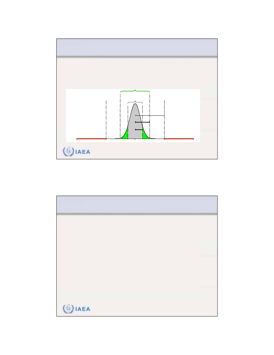
given requirements
for quality.
•
As such,
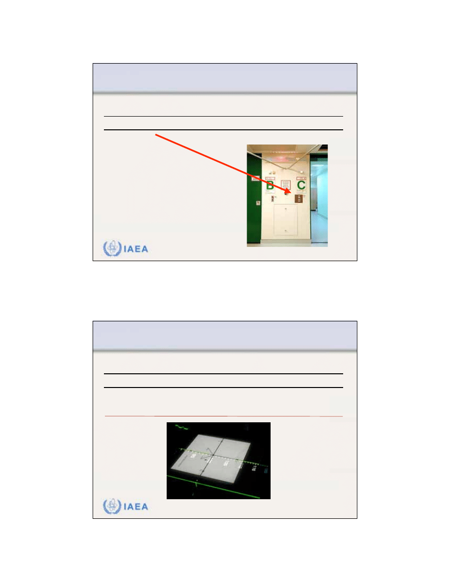
QA
is wide ranging and covering:
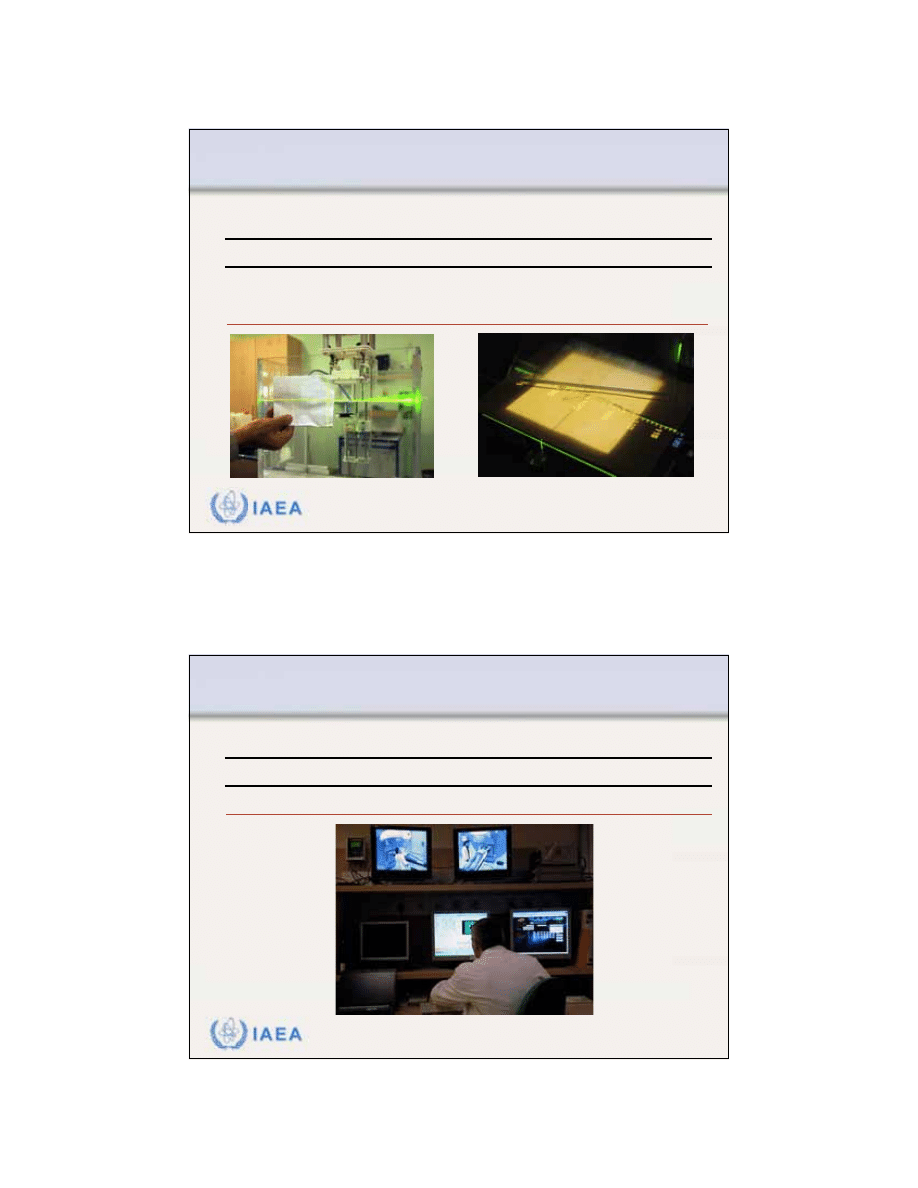
•
Procedures
•
Activities
•
Actions
•
Groups of staff.
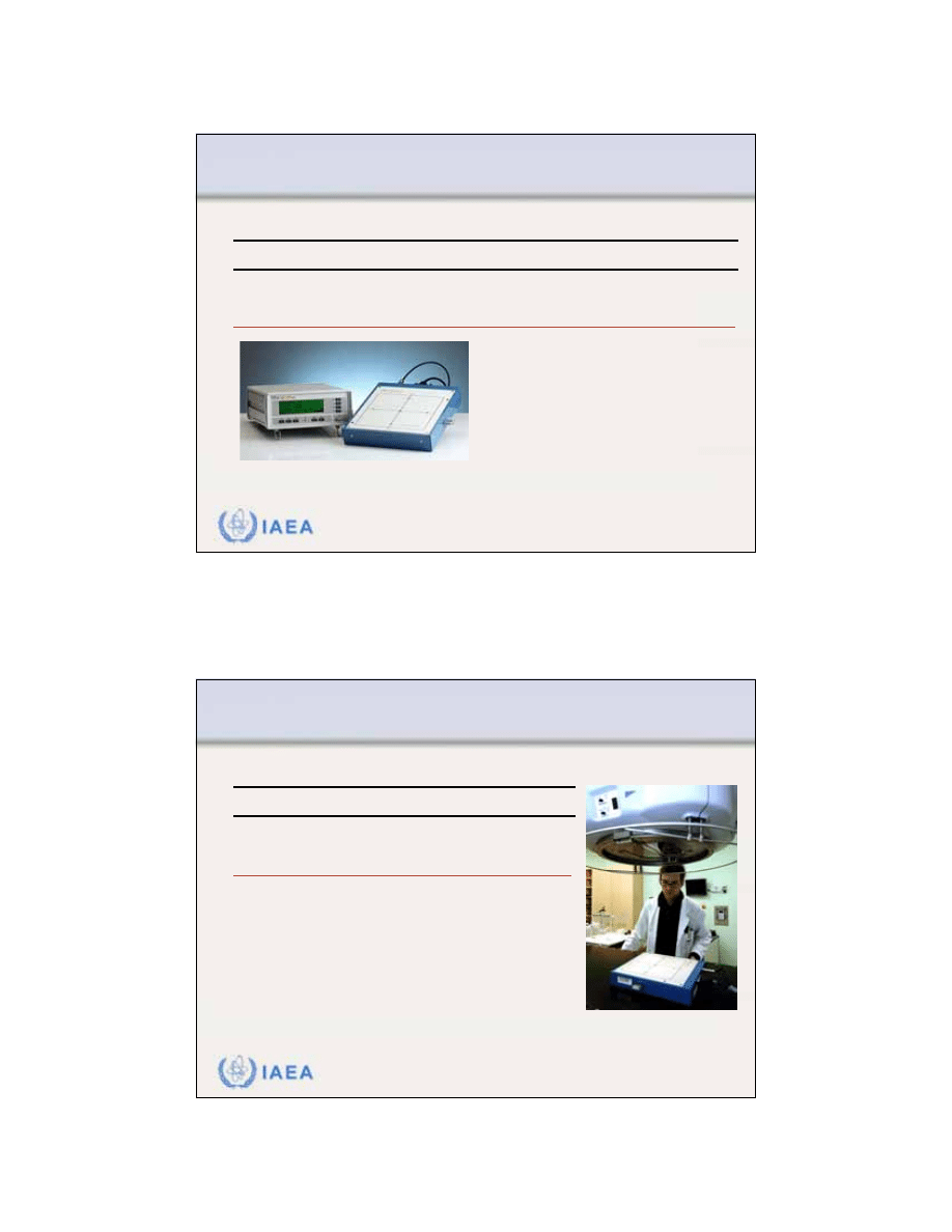
•
Management of QA program is called
Quality System Management
.
12.1 INTRODUCTION
12.1.1 Definitions

3
IAEA
Review of Radiation Oncology Physics: A Handbook for Teachers and Students - 12.1.1. Slide 3 (5/146)
Quality Control
•
Quality Control is the
regulatory process
through which the actual
quality performance is measured, compared with existing
standards, and the actions necessary to keep or regain confor-
mance with the standards.
•
Quality control forms
part of quality system management
.
•
Quality Control is concerned with operational techniques and
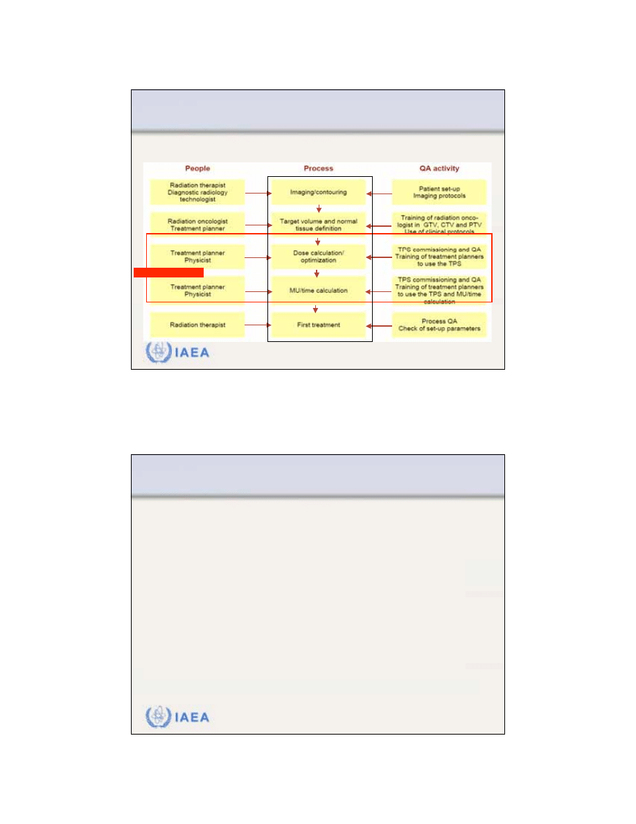
activities used:
•
To check that quality requirements are met.
•
To adjust and correct performance if requirements are found not to have
been met.
12.1 INTRODUCTION
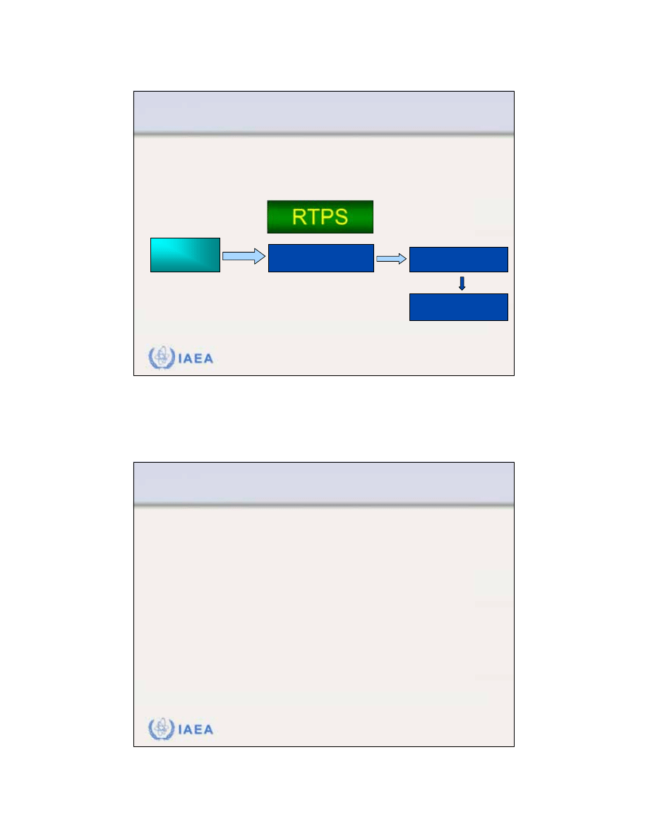
12.1.1 Definitions
IAEA
Review of Radiation Oncology Physics: A Handbook for Teachers and Students - 12.1.1. Slide 4 (6/146)
Quality Standards
•
Quality standards is the set of accepted criteria against
which the quality of the activity in question can be
assessed.
•
In other words:
Without quality standards, quality cannot
be assessed.
12.1 INTRODUCTION
12.1.1 Definitions

4
IAEA
Review of Radiation Oncology Physics: A Handbook for Teachers and Students - 12.1.1. Slide 5 (7/146)
Quality System
Quality System is a system consisting of:
•
Organizational structure
•
Responsibilities
•
Procedures
•
Processes
•
Resources
required to implement a quality assurance program.
12.1 INTRODUCTION
12.1.1 Definitions
IAEA
Review of Radiation Oncology Physics: A Handbook for Teachers and Students - 12.1.1. Slide 6 (8/146)
Quality assurance in radiotherapy
Quality Assurance in Radiotherapy
is all procedures that
ensure consistency of the medical prescription, and safe
fulfillment of that radiotherapy related prescription.
Examples of prescriptions:
•
Dose to the tumour (to the target volume).
•
Minimal dose to normal tissue.
•
Adequate patient monitoring aimed at determining the optimum
end result of the treatment.
•
Minimal exposure of personnel.
12.1 INTRODUCTION
12.1.1 Definitions

5
IAEA
Review of Radiation Oncology Physics: A Handbook for Teachers and Students - 12.1.1. Slide 7 (9/146)
Quality standards in radiotherapy
Various national or international organizations have
issued recommendations for standards in radiotherapy:
•
World Health Organization (WHO) in 1988.
•
American Association of Physicists in Medicine (AAPM) in
1994.
•
European Society for Therapeutic Radiology and Oncology
(ESTRO) in 1995.
•
Clinical Oncology Information Network (COIN) in 1999.
12.1 INTRODUCTION
12.1.1 Definitions
IAEA
Review of Radiation Oncology Physics: A Handbook for Teachers and Students - 12.1.1. Slide 8 (10/146)
Quality standards in radiotherapy
Other organizations have issued recommendations for
certain parts of the radiotherapy process:
•
International Electrotechnical Commission (IEC) in 1989.
•
Institute of Physics and Engineering in Medicine (IPEM) in
1999.
Where recommended standards are not available,
local
standards need to be developed
, based on a local
assessment of requirements.
12.1 INTRODUCTION
12.1.1 Definitions

6
IAEA
Review of Radiation Oncology Physics: A Handbook for Teachers and Students - 12.1.2. Slide 1 (11/146)
12.1 INTRODUCTION
12.1.2 The need for QA in radiotherapy
Why does a radiotherapy center need a quality system?
The following slides provide
arguments in favour
of the
need to initiate a quality project in a radiotherapy depart-
ment.
IAEA
Review of Radiation Oncology Physics: A Handbook for Teachers and Students - 12.1.2. Slide 2 (12/146)
12.1 INTRODUCTION
12.1.2 The need for QA in radiotherapy
1) You must establish a QA programme
•
This follows directly from the Basic
Safety Series (BSS) of the IAEA.
•
Appendix II.22 of the BSS states:
“Registrants and licensees, in addition to
applying the relevant requirements for
quality assurance specified elsewhere in
the Standards, shall establish a
comprehensive quality assurance program
for medical exposures
with the participation
of appropriate qualified experts in the relevant fields, such as
radiophysics or radiopharmacy, taking into account the principles
established by the WHO and the PAHO.”

7
IAEA
Review of Radiation Oncology Physics: A Handbook for Teachers and Students - 12.1.2. Slide 3 (13/146)
12.1 INTRODUCTION
12.1.2 The need for QA in radiotherapy
1) You must establish a QA programme
•
Appendix II.23 of the BSS states:
Quality assurance programs
for medical exposures shall include:
(a) Measurements of the physical
parameters of the radiation generators,
imaging devices and irradiation
installations at the time of commissioning
and periodically thereafter.
(b) Verification of the appropriate physical
and clinical factors used in patient
diagnosis or treatment …”
IAEA
Review of Radiation Oncology Physics: A Handbook for Teachers and Students - 12.1.2. Slide 4 (14/146)
12.1 INTRODUCTION
12.1.2 The need for QA in radiotherapy
2) QA programme helps to provide "the best treatment”:
•
It is a characteristic feature of the modern radiotherapy process
that this process is a multi-disciplinary process.
•
Therefore, it is extremely important that:
•
Radiation oncologist
cooperates
with specialists in the various disciplines
in
a close and effective manner.
•
Various
procedures
(related to patient and the technical aspects of radio-
therapy)
will be subjected to careful quality control
.
•
The establishment and use of a comprehensive quality system is
an adequate measure to meet these requirements.

8
IAEA
Review of Radiation Oncology Physics: A Handbook for Teachers and Students - 12.1.2. Slide 5 (15/146)
12.1 INTRODUCTION
12.1.2 The need for QA in radiotherapy
3) QA programme provides measures to achieve the following:
•
Reduction of uncertainties and errors
(in dosimetry, treatment
planning, equipment performance, treatment delivery, etc.)
•
Reduction of the likelihood of accidents and errors
occurring as
well as increase of the probability that they will be recognized and
rectified sooner
•
Providing reliable inter-comparison of results
among different
radiotherapy centers
•
Full exploitation of improved technology
and more complex treat-
ments in modern radiotherapy
IAEA
Review of Radiation Oncology Physics: A Handbook for Teachers and Students - 12.1.2. Slide 6 (16/146)
12.1 INTRODUCTION
12.1.2 The need for QA in radiotherapy
Reduction of uncertainties and errors......
Human errors in data transfer during the preparation
and delivery of radiation treatment affecting the final
result: "garbage in, garbage out"
Leunens, G; Verstraete, J; Van den Bogaert, W; Van Dam, J; Dutreix, A; van der Schueren, E
Department of Radiotherapy, University Hospital, St. Rafaël, Leuven, Belgium
Abstract
Due to the large number of steps and the number of persons involved in the preparation of a radiation
treatment, the transfer of information from one step to the next is a very critical point. Errors due to
inadequate transfer of information will be reflected in every next step and can seriously affect the final
result of the treatment. We studied the frequency and the sources of the transfer errors. A total number of
464 new treatments has been checked over a period of 9 months (January to October 1990). Erroneous data
transfer has been detected in 139/24,128 (less than 1%) of the transferred parameters; they affected 26%
(119/464) of the checked treatments. Twenty-five of these deviations could have led to large geographical
miss or important over- or underdosage (much more than 5%) of the organs in the irradiated volume, thus
increasing the complications or decreasing the tumour control probability, if not corrected. Such major
deviations only occurring in 0 1% of the transferred parameters affected 5% (25/464) of the new
Radiother. Oncol. 1992: > 50 occasions of data transfer
from one point to another for each patient.
If one of them is wrong - the overall outcome is affected.

9
IAEA
Review of Radiation Oncology Physics: A Handbook for Teachers and Students - 12.1.2. Slide 7 (17/146)
12.1 INTRODUCTION
12.1.2 The need for QA in radiotherapy
Example of improved
technology:
Use of a multileaf
collimator (MLC)
Full exploitation of improved technology.....
IAEA
Review of Radiation Oncology Physics: A Handbook for Teachers and Students - 12.1.3. Slide 1 (18/146)
12.1 INTRODUCTION
12.1.3 Requirements on accuracy in radiotherapy
Many QA procedures and tests in a QA programme for
equipment are directly related to clinical requirements on
accuracy in radiotherapy:
•
What accuracy is required on the
absolute absorbed dose
?
•
What accuracy is required on the
spatial distribution
of dose
(geometrical accuracy of treatment unit, patient positioning etc.)?

10
IAEA
Review of Radiation Oncology Physics: A Handbook for Teachers and Students - 12.1.3. Slide 2 (19/146)
12.1 INTRODUCTION
12.1.3 Requirements on accuracy in radiotherapy
Such requirements can be based on evidence from
dose
response curves
for the tumour control probability (
TCP
)
and normal tissue complication probability (
NTCP
).
TCP
and
NTCP
are usually
illustrated by plotting two
sigmoid curves, one for the
TCP (curve A)
and the other
for
NTCP (curve B).
Dose (Gy)
IAEA
Review of Radiation Oncology Physics: A Handbook for Teachers and Students - 12.1.3. Slide 3 (20/146)
12.1 INTRODUCTION
12.1.3 Requirements on accuracy in radiotherapy
The steepness of a given
TCP or NTCP curve
defines the change in
response expected for
a given change in
delivered dose.
Thus, uncertainties in delivered dose translate into
either reductions in the TCP or increases in the NTCP,
both of which worsen the clinical outcome.
Dose (Gy)

11
IAEA
Review of Radiation Oncology Physics: A Handbook for Teachers and Students - 12.1.3. Slide 4 (21/146)
12.1 INTRODUCTION
12.1.3 Requirements on accuracy in radiotherapy
The ICRU Report No. 24 (1976) concludes that:
An uncertainty of 5% is tolerable in the delivery of dose
to the target volume
The value of 5% is generally interpreted to represent a
confidence level of 1.5 - 2 times the standard deviation.
Currently, the recommended accuracy of dose delivery
is generally 5–7% at the 95% confidence level.
IAEA
Review of Radiation Oncology Physics: A Handbook for Teachers and Students - 12.1.3. Slide 5 (22/146)
12.1 INTRODUCTION
12.1.3 Requirements on accuracy in radiotherapy
Geometric uncertainty
, for example systematic errors on
the field position, block position, etc., relative to target
volumes or organs at risk,
also leads to dose problems:
•
Either
underdosing of the required volume
(decreasing the TCP).
•
Or
overdosing of nearby structures
(increasing the NTCP).
Figures of 5–10 mm (95% confidence level) are usually
given on the tolerable
geometric uncertainty
.

12
IAEA
Review of Radiation Oncology Physics: A Handbook for Teachers and Students - 12.1.4. Slide 1 (23/146)
12.1 INTRODUCTION
12.1.4 Accidents in radiotherapy
Generally speaking, treatment of a disease with radio-
therapy represents a
twofold risk for the patient
:
•
Firstly, and primarily, there is the
potential failure
to control
the
initial disease, which, when it is malignant, is eventually lethal to
the patient;
•
Secondly, there is the
risk to normal tissue
from increased
exposure to radiation.
Thus, in radiotherapy an accident or a misadministration
is
significant
if it results
in either an
underdose or an
overdose
, whereas in conventional radiation protection
only overdoses are generally of concern.
IAEA
Review of Radiation Oncology Physics: A Handbook for Teachers and Students - 12.1.4. Slide 2 (24/146)
12.1 INTRODUCTION
12.1.4 Accidents in radiotherapy
From the general aim of an accuracy approaching 5%
(95% confidence level), a
definition for an accidental
exposure
can be derived:
A generally accepted limit
is
about twice the accuracy
requirement, i.e. a 10% difference should be taken as
an accidental exposure
In addition, from clinical observations of outcome and
of normal tissue reactions, there is good evidence that
differences of 10% in dose are detectable in normal
clinical practice.

13
IAEA
Review of Radiation Oncology Physics: A Handbook for Teachers and Students - 12.1.4. Slide 3 (25/146)
12.1 INTRODUCTION
12.1.4 Accidents in radiotherapy
IAEA has analyzed a series of
accidental exposures in
radiotherapy
to draw lessons
in methods for prevention of
such occurrences.
Criteria for classifying:
•
Direct causes of mis-
administrations
•
Contributing factors
•
Preventability of
misadministration
•
Classification of potential
hazard.
IAEA
Review of Radiation Oncology Physics: A Handbook for Teachers and Students - 12.1.4. Slide 4 (26/146)
12.1 INTRODUCTION
12.1.4 Accidents in radiotherapy
1
Wrong repair followed by error
1
Accelerator software error
3
Transcription error of prescribed
dose
1
Treatment unit mechanical
failure
3
Error in calibration of cobalt-60
source
1
Malfunction of accelerator
4
Error involving lack of/or misuse of
a wedge
2
Technologist misread the
treatment time or MU
4
Error in identifying the correct
patient
2
Error in commissioning of TPS
8
Error in anatomical area to be
treated
2
Decommissioning of
teletherapy source error
9
Inadequate review of patient chart
2
Human error during simulation
15
Calculation error of time or dose
Number
Cause
Number
Cause
Examples of direct causes of misadministrations

14
IAEA
Review of Radiation Oncology Physics: A Handbook for Teachers and Students - 12.2 Slide 1 (27/146)
12.2 MANAGING A QUALITY ASSURANCE PROGRAMME
It must be understood that the
required quality system is
essentially a total management system
:
•
For the total organization
•
For the total radiation therapy process
The total radiation therapy process includes:
•
Clinical radiation oncology service
•
Supportive care services (nursing, dietetic, social, etc.)
•
All issues related to radiation treatment
•
Radiation therapists
•
Physical quality assurance (QA) by physicists
•
Engineering maintenance
•
Management
IAEA
Review of Radiation Oncology Physics: A Handbook for Teachers and Students - 12.2 Slide 2 (28/146)
A number of organizations and publications have given
background discussion and recommendations on the
structure and management of a quality assurance
programme in radiotherapy or radiotherapy physics:
•
WHO in 1988
•
AAPM in 1994
•
ESTRO in 1995 and 1998
•
IPEM in 1999
•
Van Dyk and Purdy in 1999
•
McKenzie et al. in 2000
12.2 MANAGING A QUALITY ASSURANCE PROGRAMME

15
IAEA
Review of Radiation Oncology Physics: A Handbook for Teachers and Students - 12.2.2. Slide 1 (29/146)
12.2 MANAGING A QUALITY ASSURANCE PROGRAMME
12.2.1 Multidisciplinary radiotherapy team
One of the reasons to implement a
Quality System
is that
radiotherapy is a multidisciplinary process
.
•
Responsibilities are shared between the different disciplines and
must be clearly defined.
•
Each group has an important
part in the output of the entire
process, and their overall roles
as well as their specific quality
assurance roles, are inter-
dependent requiring close
cooperation.
Radiation
Oncology
Medical
Physics
RTTs
Dosimetrists
Engineering
etc.
Radiotherapy
Process
IAEA
Review of Radiation Oncology Physics: A Handbook for Teachers and Students - 12.2.1. Slide 2 (30/146)
12.2 MANAGING A QUALITY ASSURANCE PROGRAMME
12.2.1 Multidisciplinary radiotherapy team
The
multidisciplinary radiotherapy team
consists of:
•
Radiation oncologists
•
Medical physicists
•
Radiotherapy technologists
•
Sometimes referred to as radiation therapists (RTT), therapy radiographers,
radiation therapy technologists.
•
Dosimetrists
•
In many systems there is no separate group of dosimetrists; these functions
are carried out variously by physicists, medical physics technicians or
technologists, radiation dosimetry technicians or technologists, radiotherapy
technologists, or therapy radiographers.
•
Engineering technologists
•
In some systems medical physics technicians or technologists, clinical tech-
nologists, service technicians, electronic engineers or electronic techni-
cians.
16
IAEA
Review of Radiation Oncology Physics: A Handbook for Teachers and Students - 12.2.2. Slide 1 (31/146)
12.2 MANAGING A QUALITY ASSURANCE PROGRAMME
12.2.2 Quality system/comprehensive QA programme
It is now widely appreciated that the concept of a
Quality
System in Radiotherapy
is broader than a restricted
definition of technical maintenance and quality control of
equipment and treatment delivery.
Instead it should encompass a comprehensive approach
to all activities in the radiotherapy department:
•
Starting from the moment a patient enters the department.
•
Until the moment he or she leaves the department.
•
Continuing into the follow-up period.
IAEA
Review of Radiation Oncology Physics: A Handbook for Teachers and Students - 12.2.2. Slide 2 (32/146)
12.2 MANAGING A QUALITY ASSURANCE PROGRAMME
12.2.2 Quality system/comprehensive QA programme
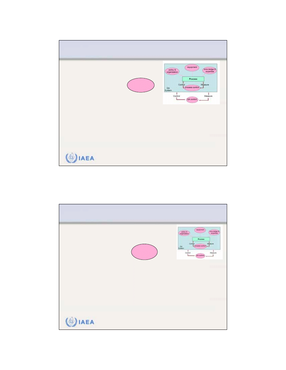
The patient enters
the process
seeking treatment.
The patient leaves
the department
after treatment.
Outcome can be considered of good quality when the handling of the qua-
lity system organizes well the five aspects shown in the illustration above.
Input
Output
Control
Measure
Control
Measure
QA control
process control
policy &
organization
equipment
knowledge &
expertise
QA
System
Process
17
IAEA
Review of Radiation Oncology Physics: A Handbook for Teachers and Students - 12.2.2. Slide 3 (33/146)
12.2 MANAGING A QUALITY ASSURANCE PROGRAMME
12.2.2 Quality system/comprehensive QA programme
A
comprehensive quality system
in radiotherapy is a management
system that:
•
Should be supported by the department
management in order to work effectively.
•
Must have a clear definition of its scope and of all the quality standards
to be met.
•
Must be regularly reviewed as to operation and improvement. To this
end a quality assurance committee is required, which should represent
all the different disciplines within radiation oncology.
•
Must be consistent in standards for different areas of the program.
Policy &
organization
IAEA
Review of Radiation Oncology Physics: A Handbook for Teachers and Students - 12.2.2. Slide 4 (34/146)
12.2 MANAGING A QUALITY ASSURANCE PROGRAMME
12.2.2 Quality system/comprehensive QA programme
A
comprehensive quality system
in radiotherapy is a management
system that:
•
Requires availability of adequate test equipment.
Equipment
18
IAEA
Review of Radiation Oncology Physics: A Handbook for Teachers and Students - 12.2.2. Slide 5 (35/146)
12.2 MANAGING A QUALITY ASSURANCE PROGRAMME
12.2.2 Quality system/comprehensive QA programme
A
comprehensive quality system
in radiotherapy is a management
system that:
•
Requires every staff member to have qualifications (education,
training and experience) appropriate to his or her role and
responsibility.
•
Requires every staff member to have access to appropriate
opportunities for continuing education and development.
Knowledge &
expertise
IAEA
Review of Radiation Oncology Physics: A Handbook for Teachers and Students - 12.2.2. Slide 6 (36/146)
12.2 MANAGING A QUALITY ASSURANCE PROGRAMME
12.2.2 Quality system/comprehensive QA programme
A
comprehensive quality system
in radiotherapy is a management
system that:
•
Requires the development of a
formal written quality assurance
programme
that details the quality assurance policies and
procedures, quality control tests, frequencies, tolerances, action
criteria, required records and personnel.
•
Must be consistent in standards for different areas of the
programme.
•
Must incorporate compliance with all the requirements of national
legislation, accreditation, etc.
process control

19
IAEA
Review of Radiation Oncology Physics: A Handbook for Teachers and Students - 12.2.2. Slide 7 (37/146)
Formal written quality assurance programme
is also
called referred to as the
Quality Manual
.
•
The quality manual has a double purpose:
•
External
•
Internal.
•
Externally
to collaborators in other departments, in manage-
ment and in other institutions, it helps to indicate that the
department is strongly concerned with quality.
•
Internally
, it provides the department with a framework for
further development of quality and for improvements of existing
or new procedures.
12.2 MANAGING A QUALITY ASSURANCE PROGRAMME
12.2.2 Quality system/comprehensive QA programme
IAEA
Review of Radiation Oncology Physics: A Handbook for Teachers and Students - 12.2.2. Slide 8 (38/146)
12.2 MANAGING A QUALITY ASSURANCE PROGRAMME
12.2.2 Quality system/comprehensive QA programme
ESTRO Booklet 4:
PRACTICAL GUIDELINES FOR THE
IMPLEMENTATION OF A QUALITY
SYSTEM IN RADIOTHERAPY
A project of the ESTRO Quality Assurance Committee sponsored by
'Europe against Cancer'
Writing party: J W H Leer, A L McKenzie, P Scalliet, D I Thwaites
Practical guidelines for writing a quality manual:

20
IAEA
Review of Radiation Oncology Physics: A Handbook for Teachers and Students - 12.2.2. Slide 9 (39/146)
12.2 MANAGING A QUALITY ASSURANCE PROGRAMME
12.2.2 Quality system/comprehensive QA programme
A
comprehensive quality system
in radiotherapy is a management
system that:
•
Requires control of the system itself, including:
•
Responsibility for quality assurance and the quality system: quality
management representatives.
•
Document control.
•
Procedures to ensure that the quality system is followed.
•
Ensuring that the status of all parts of the service is clear.
•
Reporting all non-conforming parts and taking corrective action.
•
Recording all quality activities.
•
Establishing regular review and audits of both the implementation of the quality
system (quality system audit) and its effectiveness (quality audit).
QA control
IAEA
Review of Radiation Oncology Physics: A Handbook for Teachers and Students - 12.2.2. Slide 10 (40/146)
12.2 MANAGING A QUALITY ASSURANCE PROGRAMME
12.2.2 Quality system/comprehensive QA program
When starting a quality assurance (QA) program, the
setup of a
QA team
or a
QA committee
is the most
important first step.
•
The QA team should reflect composition of the multi-
disciplinary radiotherapy team.
•
The quality assurance committee must be appointed by the
department management/head of department with the
authority to manage quality assurance.
21
IAEA
Review of Radiation Oncology Physics: A Handbook for Teachers and Students - 12.2.2. Slide 11 (41/146)
12.2 MANAGING A QUALITY ASSURANCE PROGRAMME
12.2.2 Quality system/comprehensive QA program
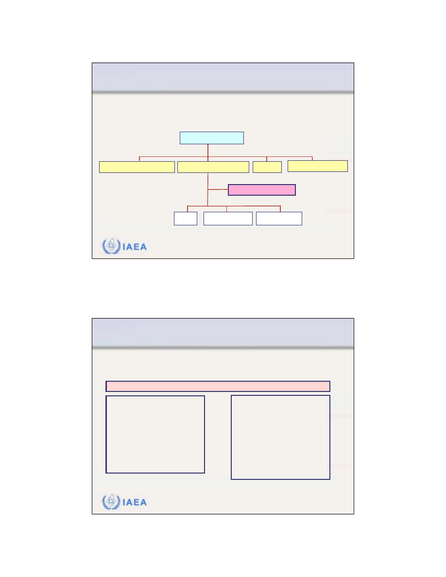
Example for the
organizational structure of a radiotherapy
department and the integration of a QA team
Systematic Treatment Program
Radiation Treatment Program
Management Services
............
QA Team (Committee)
Physics
Radiation Oncology
Radiation Therapy
Chief Executive Officer
IAEA
Review of Radiation Oncology Physics: A Handbook for Teachers and Students - 12.2.2. Slide 12 (42/146)
12.2 MANAGING A QUALITY ASSURANCE PROGRAMME
12.2.2 Quality system/comprehensive QA program
Membership and Responsibilities
of the QA team (QA Committee)
Membership:
Radiation Oncologist(s)
Medical Physicist(s)
Radiation Therapist(s)
..........
Chair:
Physicist or
Radiation Oncologist
Responsibilities:
Patient safety
Personnel safety
Dosimetry instrumentation
Teletherapy equipment
Treatment planning
Treatment delivery
Treatment outcome
Quality audit
QA Team (Committee)

22
IAEA
Review of Radiation Oncology Physics: A Handbook for Teachers and Students - 12.3. Slide 1 (43/146)
12.3 QUALITY ASSURANCE PROGRAMME
FOR EQUIPMENT
The following slides are focusing on the
equipment
related QA programme.
•
They concentrate on the
general items and systems
of a QA
program.
•
Therefore, they should be "digested" in conjunction with
Chapter 10 and other appropriate material concerned with
each of the different categories of equipment.
IAEA
Review of Radiation Oncology Physics: A Handbook for Teachers and Students - 12.3. Slide 2 (44/146)
Appropriate material:
Many documents are available:
12.3 QUALITY ASSURANCE PROGRAMME FOR EQUIPMENT

23
IAEA
Review of Radiation Oncology Physics: A Handbook for Teachers and Students - 12.3. Slide 3 (45/146)
Examples of useful published material:
•
AMERICAN ASSOCIATION OF PHYSICISTS IN MEDICINE (AAPM),
“Comprehensive QA for radiation oncology: Report of AAPM Radiation
Therapy Committee Task Group 40”, Med. Phys. 21, 581-618 (1994)
•
INTERNATIONAL ELECTROTECHNICAL COMMISSION (IEC),
“Medical
electrical equipment - Medical electron accelerators-Functional performance
characteristics”, IEC 976, IEC, Geneva, Switzerland (1989)
•
INSTITUTE OF PHYSICS AND ENGINEERING IN MEDICINE (IPEM),
“Physics aspects of quality control in radiotherapy”, IPEM Report 81, edited by
Mayles, W.P.M., Lake, R., McKenzie, A., Macaulay, E.M., Morgan, H.M.,
Jordan, T.J. and Powley, S.K, IPEM, York, United Kingdom (1999)
•
VAN DYK, J.,
(editor), “The Modern Technology for Radiation Oncology: A
Compendium for Medical Physicists and Radiation Oncologists”, Medical
Physics Publishing, Madison, Wisconsin, U.S.A. (1999)
•
WILLIAMS, J.R., and THWAITES, D.I.,
(editors), “Radiotherapy Physics in
Practice”, Oxford University Press, Oxford, United Kingdom (2000)
12.3 QUALITY ASSURANCE PROGRAMME FOR EQUIPMENT
IAEA
Review of Radiation Oncology Physics: A Handbook for Teachers and Students - 12.3.1. Slide 1 (46/146)
12.3 QUALITY ASSURANCE PROGRAMME FOR EQUIPMENT
12.3.1 The structure of an equipment QA programme
(1) Initial specification,
acceptance testing and
commissioning
for clinical use, including
calibration where applicable
(2) Quality control tests
before the equipment is put into
clinical use, quality control tests
should be established and a
formal QC program initiated
General structure of a quality assurance program for equipment
(3) Additional quality control
tests
after any significant repair,
intervention or adjustment or
when there is any indication
of a change in performance
(4) Planned preventive
maintenance programme
in accordance with the
manufacturer’s
recommendations

24
IAEA
Review of Radiation Oncology Physics: A Handbook for Teachers and Students - 12.3.1. Slide 2 (47/146)
Step 1:
Equipment specification and assessment of
clinical needs:
•
In preparation for procurement of equipment, a detailed
specification document must be prepared.
•
A multidisciplinary team from the department should be
involved in the decision process.
•
This should set out the essential aspects of the equipment
operation, facilities, performance, service, etc., as required
by the customer.
12.3 QUALITY ASSURANCE PROGRAMME FOR EQUIPMENT
12.3.1 The structure of an equipment QA programme
IAEA
Review of Radiation Oncology Physics: A Handbook for Teachers and Students - 12.3.1. Slide 3 (48/146)
Questions to be answered in assessment of clinical needs:
•
Which patients will be affected by this technology?
•
What is the likely number of patients per year?
•
Number of procedures or fractions per year?
•
Will the new procedure provide cost savings over old techniques?
•
Would it be better to refer patients to a specialist institution?
•
Is the infrastructure available to handle the technology?
•
Will the technology enhance the academic program?
•
What is the organizational risk in implementing this technology?
•
What is the cost impact?
•
What maintenance is required?
12.3 QUALITY ASSURANCE PROGRAMME FOR EQUIPMENT
12.3.1 The structure of an equipment QA programme

25
IAEA
Review of Radiation Oncology Physics: A Handbook for Teachers and Students - 12.3.1. Slide 4 (49/146)
Equipment specification and assessment of clinical needs:
•
Once this information is compiled, the purchaser is in a good
position to develop clearly his own specifications.
•
The specification can also be based on:
•
Manufacturers specification (brochures)
•
Published information
•
Discussions with other users of similar products
•
All specification data must be expressed clearly in well defined
and measurable units.
•
Decisions on procurement should again be made by a multi-
disciplinary team.
12.3 QUALITY ASSURANCE PROGRAMME FOR EQUIPMENT
12.3.1 The structure of an equipment QA programme
IAEA
Review of Radiation Oncology Physics: A Handbook for Teachers and Students - 12.3.1. Slide 5 (50/146)
Acceptance of equipment
•
Acceptance of equipment is the process in which the
supplier
demonstrates the baseline performance of the equipment to
the satisfaction of the customer.
•
After new equipment is installed, it must be tested in order to
ensure that it meets the specifications and that the
environment is free of radiation and electrical hazards to staff
and patients.
•
The essential performance required and expected from the
machine should be agreed upon
before
acceptance of the
equipment begins.
12.3 QUALITY ASSURANCE PROGRAMME FOR EQUIPMENT
12.3.1 The structure of an equipment QA programme

26
IAEA
Review of Radiation Oncology Physics: A Handbook for Teachers and Students - 12.3.1. Slide 6 (51/146)
Acceptance of equipment
•
It is a matter of professional judgment of the responsible medical
physicist to decide whether or not any aspect of the agreed
acceptance criteria is to be waived.
•
This waiver should be recorded along with an agreement from the
supplier, for example to correct the equipment should
performance deteriorate further.
•
The equipment can only be formally accepted to be transferred
from the supplier to the customer when the responsible medical
physicist either is satisfied that the performance of the machine
fulfils all specifications as listed in the contract document or
formally accepts any waivers.
12.3 QUALITY ASSURANCE PROGRAMME FOR EQUIPMENT
12.3.1 The structure of an equipment QA programme
IAEA
Review of Radiation Oncology Physics: A Handbook for Teachers and Students - 12.3.1. Slide 7 (52/146)
Commissioning of equipment
•
Commissioning is the process of preparing the equipment for
clinical service.
•
Expressed in a more quantitative way:
A full
characterization of its performance
over the whole range of
possible operation must be undertaken.
•
In this way the
baseline
standards of performance
are estab-
lished to which all future performance and quality control tests will
be referred.
•
Commissioning includes the preparation of procedures, proto-
cols, instructions, data, etc., on the clinical use of the equipment.
12.3 QUALITY ASSURANCE PROGRAMME FOR EQUIPMENT
12.3.1 The structure of an equipment QA program

27
IAEA
Review of Radiation Oncology Physics: A Handbook for Teachers and Students - 12.3.1. Slide 8 (53/146)
Quality control
•
It is essential that the
performance of treatment equip-ment
remain consistent within accepted tolerances throughout its
clinical life.
•
An ongoing quality control programme of regular perfor-mance
checks must begin immediately after commissioning to test
this.
•
If these quality control measurements identify departures from
expected performance, corrective actions are required.
12.3 QUALITY ASSURANCE PROGRAMME FOR EQUIPMENT
12.3.1 The structure of an equipment QA programme
IAEA
Review of Radiation Oncology Physics: A Handbook for Teachers and Students - 12.3.1. Slide 9 (54/146)
Quality control (continued)
•
Equipment quality control programme should specify the
following:
•
Parameters
to be tested and the
tests
to be performed.
•
Specific equipment
to be used for the tests.
•
Geometry
of the tests.
•
Frequency
of the tests.
•
Staff group
or
individual
performing the tests, as well as the individual
supervising and responsible for the standards of the tests and for
actions that may be necessary if problems are identified.
12.3 QUALITY ASSURANCE PROGRAMME FOR EQUIPMENT
12.3.1 The structure of an equipment QA program

28
IAEA
Review of Radiation Oncology Physics: A Handbook for Teachers and Students - 12.3.1. Slide 10 (55/146)
Quality control (continued)
•
An equipment quality control program should specify the
following:
•
Expected
results
.
•
Tolerance and action levels
.
•
Actions
required when the tolerance levels are exceeded.
•
The actions required must be based on a systematic analysis of
the uncertainties involved and on well defined tolerance and
action levels.
12.3 QUALITY ASSURANCE PROGRAMME FOR EQUIPMENT
12.3.1 The structure of an equipment QA programme
IAEA
Review of Radiation Oncology Physics: A Handbook for Teachers and Students - 12.3.2. Slide 1 (56/146)
If corrective actions are required:
Role of Uncertainty
•
When reporting the result of a measurement, it is obliga-tory that
some quantitative indication of the
quality of the result
be given.
Otherwise the receiver of this information cannot adequately
asses its reliability.
•
The
"Concept of Uncertainty"
is used for this purpose.
•
In 1993, the International Standards Organisation (ISO) published
a
“Guide to the expression of uncertainty in measurement”
, in
order to ensure that the method for evaluating and expressing
uncertainty is uniform all over the world.
12.3 QUALITY ASSURANCE PROGRAMME FOR EQUIPMENT
12.3.1 The structure of an equipment QA programme

29
IAEA
Review of Radiation Oncology Physics: A Handbook for Teachers and Students - 12.3.2. Slide 2 (57/146)
If corrective actions are required:
Role of Tolerance Level
•
Within the tolerance level, the performance of equipment gives
acceptable accuracy
in any situation.
•
Tolerance values should be set with the aim of achieving the
overall uncertainties desired
.
•
However, if the
measurement uncertainty
is greater than the
tolerance level set, then random variations in the measurement
will lead to unnecessary intervention.
•
Thus, it is practical to
set a tolerance level at the measurement
uncertainty at the 95% confidence level.
12.3 QUALITY ASSURANCE PROGRAMME FOR EQUIPMENT
12.3.1 The structure of an equipment QA programme
IAEA
Review of Radiation Oncology Physics: A Handbook for Teachers and Students - 12.3.2. Slide 3 (58/146)
12.3 QUALITY ASSURANCE PROGRAMME FOR EQUIPMENT
12.3.2 Uncertainties, tolerances and action levels
If corrective actions are required:
Role of Action Level
•
The performance outside the action level is
unacceptable
and
demands action
to remedy the situation.
•
It is useful to set action levels higher than tolerance levels thus
providing flexibility in monitoring and adjustment.
•
Action levels are often set at
approximately twice the tolerance
level.
•
However, some critical parameters may require tolerance and
action levels to be set much closer to each other or even at the
same value.
30
IAEA
Review of Radiation Oncology Physics: A Handbook for Teachers and Students - 12.3.2. Slide 4 (59/146)
Illustration of a possible relation between
uncertainty, tolerance level and action level
action level =
2 x tolerance level
mean
value
tolerance level
equivalent to
95% confidence interval of uncertainty
action level =
2 x tolerance level
standard
uncertainty
1 sd
2 sd
4 sd
12.3 QUALITY ASSURANCE PROGRAMME FOR EQUIPMENT
12.3.2 Uncertainties, tolerances and action levels
IAEA
Review of Radiation Oncology Physics: A Handbook for Teachers and Students - 12.3.2. Slide 5 (60/146)
The system of actions:
•
If the measurement result
is within tolerance level
, no action is
required.
•
If the measurement result
exceeds the action level
, immediate
action is necessary and the equipment must not be clinically
used until the problem is corrected.
•
If the measurement falls
between tolerance and action levels
,
this may be considered as currently acceptable.
•
Inspection and repair can be performed later, for example, after
patient irradiations.
•
If repeated measurements remain consistently between the
tolerance and action level, adjustment is required.
12.3 QUALITY ASSURANCE PROGRAMME FOR EQUIPMENT
12.3.2 Uncertainties, tolerances and action levels

31
IAEA
Review of Radiation Oncology Physics: A Handbook for Teachers and Students - 12.3.3. Slide 1 (61/146)
12.3 QUALITY ASSURANCE PROGRAMME FOR EQUIPMENT
12.3.3 QA programme for cobalt-60 teletherapy machines
A
sample quality assurance programme
(quality control
tests) for a cobalt-60 teletherapy machine with recom-
mended test procedures, test frequencies and action
levels is given in the following tables.
They are structured according to daily, weekly, monthly,
and annual test schedules.
IAEA
Review of Radiation Oncology Physics: A Handbook for Teachers and Students - 12.3.3. Slide 2 (62/146)
functional
Audiovisual monitor
2 mm
Lasers
functional
Radiation room monitor
2 mm
Distance indicator
functional
Door interlock
Action level
Procedure or item to be tested
Daily Tests
12.3 QUALITY ASSURANCE PROGRAMME FOR EQUIPMENT
12.3.3 QA programme for cobalt-60 teletherapy machines
32
IAEA
Review of Radiation Oncology Physics: A Handbook for Teachers and Students - 12.3.3. Slide 3 (63/146)
functional
Door interlock
Action level
Procedure or item to be tested
Daily Tests
12.3 QUALITY ASSURANCE PROGRAMME FOR EQUIPMENT
12.3.3 QA programme for cobalt-60 teletherapy machines
IAEA
Review of Radiation Oncology Physics: A Handbook for Teachers and Students - 12.3.3. Slide 4 (64/146)
2 mm
Optical distance indicator
2 mm
Lasers
Action level
Procedure or item to be tested
Daily Tests
12.3 QUALITY ASSURANCE PROGRAMME FOR EQUIPMENT
12.3.3 QA programme for cobalt-60 teletherapy machines

33
IAEA
Review of Radiation Oncology Physics: A Handbook for Teachers and Students - 12.3.3. Slide 5 (65/146)
12.3 QUALITY ASSURANCE PROGRAMME FOR EQUIPMENT
12.3.3 QA programme for cobalt-60 teletherapy machines
3 mm
Check of source position
Action level
Procedure or item to be tested
Weekly Tests
IAEA
Review of Radiation Oncology Physics: A Handbook for Teachers and Students - 12.3.3. Slide 6 (66/146)
12.3 QUALITY ASSURANCE PROGRAMME FOR EQUIPMENT
12.3.3 QA programme for cobalt-60 teletherapy machines
functional
Latching of wedges and trays
1º
Gantry and collimator angle indicator
1 mm
Cross-hair centering
2 mm
Field size indicator
functional
Emergency off
3 mm
Light/radiation field coincidence
functional
Wedge interlocks
2%
Output constancy
Action level
Procedure or item to be tested
Monthly Tests

34
IAEA
Review of Radiation Oncology Physics: A Handbook for Teachers and Students - 12.3.3. Slide 7 (67/146)
12.3 QUALITY ASSURANCE PROGRAMME FOR EQUIPMENT
12.3.3 QA programme for cobalt-60 teletherapy machines
1%
Timer linearity and error
2%
Transmission factor constancy for all standard
accessories
2%
Wedge transmission factor constancy
2%
Central axis dosimetry parameter constancy
2%
Output constancy versus gantry angle
2%
Field size dependence of output constancy
2%
Output constancy
Action level
Procedure or item to be tested
Annual Tests
IAEA
Review of Radiation Oncology Physics: A Handbook for Teachers and Students - 12.3.3. Slide 8 (68/146)
12.3 QUALITY ASSURANCE PROGRAMME FOR EQUIPMENT
12.3.3 QA programme for cobalt-60 teletherapy machines
2 mm diameter
Coincidence of collimator, gantry and table
axis with the isocenter
2 mm diameter
Gantry rotation isocenter
2 mm diameter
Table rotation isocenter
2 mm diameter
Collimator rotation isocenter
functional
Safety interlocks: Follow procedures of
manufacturer
3%
Beam uniformity with gantry angle
Action level
Procedure or item to be tested
Annual tests (continued)

35
IAEA
Review of Radiation Oncology Physics: A Handbook for Teachers and Students - 12.3.3. Slide 9 (69/146)
12.3 QUALITY ASSURANCE PROGRAMME FOR EQUIPMENT
12.3.3 QA programme for cobalt-60 teletherapy machines
2 mm diameter
Coincidence of the radiation and mechanical
isocenter
functional
Field light intensity
2 mm
Vertical travel of table
2 mm
Table top sag
Action level
Procedure or item to be tested
Annual Tests (continued)
IAEA
Review of Radiation Oncology Physics: A Handbook for Teachers and Students - 12.3.4. Slide 1 (70/146)
12.3 QUALITY ASSURANCE PROGRAMME FOR EQUIPMENT
12.3.4 QA programme for linear accelerators
Typical
quality assurance procedures
(quality control
tests) for a dual mode linac with frequencies and action
levels are given in the following tables.
They are again structured according to daily, weekly,
monthly, and annual tests.
36
IAEA
Review of Radiation Oncology Physics: A Handbook for Teachers and Students - 12.3.4. Slide 2 (71/146)
2 mm
Optical distance indicator
2 mm
Lasers
Action level
Procedure or item to be tested
Daily Tests
12.3 QUALITY ASSURANCE PROGRAMME FOR EQUIPMENT
12.3.4 QA programme for linear accelerators
IAEA
Review of Radiation Oncology Physics: A Handbook for Teachers and Students - 12.3.4. Slide 3 (72/146)
functional
Audiovisual monitor
Action level
Procedure or item to be tested
Daily Tests
12.3 QUALITY ASSURANCE PROGRAMME FOR EQUIPMENT
12.3.4 QA programme for linear accelerators
37
IAEA
Review of Radiation Oncology Physics: A Handbook for Teachers and Students - 12.3.4. Slide 4 (73/146)
3%
Electron output constancy
3%
X ray output constancy
Action level
Procedure or item to be tested
Daily Tests
Daily output checks and verification
of flatness and symmetry can be
done using different multi-detector
devices
.
12.3 QUALITY ASSURANCE PROGRAMME FOR EQUIPMENT
12.3.4 QA programme for linear accelerators
IAEA
Review of Radiation Oncology Physics: A Handbook for Teachers and Students - 12.3.4. Slide 5 (74/146)
3%
Electron output constancy
3%
X ray output constancy
Action level
Procedure or item to be tested
Daily Tests
12.3 QUALITY ASSURANCE PROGRAMME FOR EQUIPMENT
12.3.4 QA programme for linear accelerators

38
IAEA
Review of Radiation Oncology Physics: A Handbook for Teachers and Students - 12.3.4. Slide 6 (75/146)
2%
X-ray beam flatness constancy
2%
X ray central axis dosimetry parameter
constancy (PDD, TAR, TPR)
2 mm at thera-
peutic depth
Electron central axis dosimetry
parameter constancy (PDD)
2%
Backup monitor constancy
2%
Electron output constancy
2%
X ray output constancy
Action level
Procedure or item to be tested
Monthly Tests
12.3 QUALITY ASSURANCE PROGRAMME FOR EQUIPMENT
12.3.4 QA programme for linear accelerators
IAEA
Review of Radiation Oncology Physics: A Handbook for Teachers and Students - 12.3.4. Slide 7 (76/146)
1º
Gantry/collimator angle indicators
functional
Wedge and electron cone interlocks
2 mm or 1% on a side
Light/radiation field coincidence
functional
Emergency off switches
2 mm or 2% change in
transmission
Wedge position
3%
X ray and electron symmetry
3%
Electron beam flatness constancy
Action level
Procedure or item to be tested
Monthly Tests (continued)
12.3 QUALITY ASSURANCE PROGRAMME FOR EQUIPMENT
12.3.4 QA programme for linear accelerators

39
IAEA
Review of Radiation Oncology Physics: A Handbook for Teachers and Students - 12.3.4. Slide 8 (77/146)
2 mm diameter
Cross-hair centering
functional
Latching of wedges and blocking tray
2 mm
Jaw symmetry
2 mm / 1º
Treatment table position indicators
functional
Field light intensity
2 mm
Field size indicators
2 mm
Tray position and applicator position
Action level
Procedure or item to be tested
Monthly Tests (continued)
12.3 QUALITY ASSURANCE PROGRAMME FOR EQUIPMENT
12.3.4 QA programme for linear accelerators
IAEA
Review of Radiation Oncology Physics: A Handbook for Teachers and Students - 12.3.4. Slide 9 (78/146)
2%
Output factor constancy for electron
applicators
2%
Off-axis factor constancy
2%
Transmission factor constancy for all
treatment accessories
2%
Central axis parameter constancy
(PDD, TAR, TPR)
2%
Field size dependence of X ray output
constancy
2%
X ray/electron output calibration constancy
Action level
Procedure or item to be tested
Annual Tests
12.3 QUALITY ASSURANCE PROGRAMME FOR EQUIPMENT
12.3.4 QA programme for linear accelerators

40
IAEA
Review of Radiation Oncology Physics: A Handbook for Teachers and Students - 12.3.4. Slide 10 (79/146)
2%
X ray output constancy with the gantry angle
2%
Off-axis factor constancy with the gantry
angle
Manufacturer’s
specifications
Arc mode
2%
Electron output constancy with the gantry
angle
1%
Monitor chamber linearity
2%
Wedge transmission factor constancy
Action level
Procedure or item to be tested
Annual Tests (continued)
12.3 QUALITY ASSURANCE PROGRAMME FOR EQUIPMENT
12.3.4 QA programme for linear accelerators
IAEA
Review of Radiation Oncology Physics: A Handbook for Teachers and Students - 12.3.4. Slide 11 (80/146)
2 mm diameter
Gantry rotation isocenter
2 mm diameter
Coincidence of collimator, gantry and table
axes with the isocenter
2 mm diameter
Coincidence of the radiation and mechanical
isocenter
2 mm diameter
Table rotation isocenter
2 mm diameter
Collimator rotation isocenter
functional
Safety interlocks
Action level
Procedure or item to be tested
Annual Tests (continued)
12.3 QUALITY ASSURANCE PROGRAMME FOR EQUIPMENT
12.3.4 QA program for linear accelerators

41
IAEA
Review of Radiation Oncology Physics: A Handbook for Teachers and Students - 12.3.4. Slide 12 (81/146)
2 mm
Vertical travel of the table
2 mm
Table top sag
Action level
Procedure or item to be tested
Annual Tests (continued)
12.3 QUALITY ASSURANCE PROGRAMME FOR EQUIPMENT
12.3.4 QA programme for linear accelerators
IAEA
Review of Radiation Oncology Physics: A Handbook for Teachers and Students - 12.3.5. Slide 1 (82/146)
12.3 QUALITY ASSURANCE PROGRAMME FOR EQUIPMENT
12.3.5 QA programme for treatment simulators
Treatment simulators
replicate the movements of
isocentric
60
Co and linac treatment machines and are
fitted with identical beam and distance indicators. Hence
all measurements that concern these aspects also apply
to the simulator.
•
During ‘verification session’
the treatment is set-up on
the simulator exactly like it
would be on the treatment
unit.
•
A verification film is taken in
‘treatment’ geometry

42
IAEA
Review of Radiation Oncology Physics: A Handbook for Teachers and Students - 12.3.5. Slide 2 (83/146)
If mechanical/geometric parameters are out of tolerance
on the simulator,
this is likely to affect adversely the
treatment of all patients.
Performance of the
imaging components
on the simulator
is of equal importance to its satisfactory operation.
Therefore, critical measurements of the imaging system
are also required.
12.3 QUALITY ASSURANCE PROGRAMME FOR EQUIPMENT
12.3.5 QA programme for treatment simulators
IAEA
Review of Radiation Oncology Physics: A Handbook for Teachers and Students - 12.3.5. Slide 3 (84/146)
A
sample quality assurance programme
(quality control
tests) for treatment simulators with recommended test
procedures, test frequencies and action levels is given in
the following tables.
They are again structured according to daily, monthly, and
annual tests.
12.3 QUALITY ASSURANCE PROGRAMME FOR EQUIPMENT
12.3.5 QA programme for treatment simulators

43
IAEA
Review of Radiation Oncology Physics: A Handbook for Teachers and Students - 12.3.5. Slide 4 (85/146)
2 mm
Lasers
functional
Door interlock
2 mm
Distance indicator
functional
Safety switches
Action level
Procedure or item to be tested
Daily Tests
12.3 QUALITY ASSURANCE PROGRAMME FOR EQUIPMENT
12.3.5 QA programme for treatment simulators
IAEA
Review of Radiation Oncology Physics: A Handbook for Teachers and Students - 12.3.5. Slide 5 (86/146)
functional
Emergency/collision avoidance
2 mm diameter
Cross-hair centering
baseline
Fluoroscopic image quality
2 mm or 1%
baseline
Light/radiation field coincidence
Film processor sensitometry
2 mm
Focal spot-axis indicator
1°
Gantry/collimator angle indicators
2 mm
Field size indicator
Action level
Procedure or item to be tested
Monthly Tests
12.3 QUALITY ASSURANCE PROGRAMME FOR EQUIPMENT
12.3.5 QA programme for treatment simulators

44
IAEA
Review of Radiation Oncology Physics: A Handbook for Teachers and Students - 12.3.5. Slide 6 (87/146)
2 mm
Vertical travel of couch
2 mm diameter
Couch rotation isocenter
2 mm
Table top sag
2 mm diameter
Coincidence of collimator, gantry, couch axes
with isocenter
2 mm diameter
Gantry rotation isocenter
2 mm diameter
Collimator rotation isocenter
Action level
Procedure or item to be tested
Annual Tests
12.3 QUALITY ASSURANCE PROGRAMME FOR EQUIPMENT
12.3.5 QA programme for treatment simulators
IAEA
Review of Radiation Oncology Physics: A Handbook for Teachers and Students - 12.3.5. Slide 7 (88/146)
baseline
kVp and mAs calibration
baseline
High and low contrast resolution
baseline
Table top exposure with fluoroscopy
baseline
Exposure rate
Action level
Procedure or item to be tested
Annual Tests (continued)
12.3 QUALITY ASSURANCE PROGRAMME FOR EQUIPMENT
12.3.5 QA programme for treatment simulators

45
IAEA
Review of Radiation Oncology Physics: A Handbook for Teachers and Students - 12.3.6. Slide 1 (89/146)
12.3 QUALITY ASSURANCE PROGRAMME FOR EQUIPMENT
12.3.6 QA programme for CT scanners and CT-simulators
For dose prediction as part of the treatment planning
process
there is an increasing reliance upon CT image
data with the patient in a treatment position.
CT data is used for:
•
Indication and/or data
acquisition of the patient’s
anatomy.
•
Acquisition of tissue density
information which is essential for
accurate dose prediction.
Therefore, it is essential that the geometry and the CT
densities are accurate.
CT test tools are available.
Gammex RMI CT test tool
IAEA
Review of Radiation Oncology Physics: A Handbook for Teachers and Students - 12.3.6. Slide 2 (90/146)
12.3 QUALITY ASSURANCE PROGRAMME FOR EQUIPMENT
12.3.6 QA programme for CT scanners and CT-simulators
A
sample quality assurance programme
(quality control
tests) for CT scanners and CT-simulation with recom-
mended test procedures, test frequencies and action
levels is given in the following tables.
They are again structured according to daily, monthly,
and annual tests.

46
IAEA
Review of Radiation Oncology Physics: A Handbook for Teachers and Students - 12.3.6. Slide 3 (91/146)
12.3 QUALITY ASSURANCE PROGRAMME FOR EQUIPMENT
12.3.6 QA programme for CT scanners and CT-simulators
2 mm
Lasers
functional
Door interlock
2 mm
Distance indicator
functional
Safety switches
Action level
Procedure or item to be tested
Daily Tests
IAEA
Review of Radiation Oncology Physics: A Handbook for Teachers and Students - 12.3.6. Slide 4 (92/146)
12.3 QUALITY ASSURANCE PROGRAMME FOR EQUIPMENT
12.3.6 QA programme for CT scanners and CT-simulators
functional
Emergency/collision avoidance
2 mm diameter
Cross-hair centering
baseline
Fluoroscopic image quality
2 mm or 1%
baseline
Light/radiation field coincidence
Film processor sensitometry
2 mm
Focal spot-axis indicator
1°
Gantry/collimator angle indicators
2 mm
Field size indicator
Action level
Procedure or item to be tested
Monthly Tests

47
IAEA
Review of Radiation Oncology Physics: A Handbook for Teachers and Students - 12.3.6. Slide 5 (93/146)
12.3 QUALITY ASSURANCE PROGRAMME FOR EQUIPMENT
12.3.6 QA programme for CT scanners and CT-simulators
2 mm
Vertical travel of couch
2 mm diameter
Couch rotation isocenter
2 mm
Table top sag
2 mm diameter
Coincidence of collimator, gantry, couch axes
with isocenter
2 mm diameter
Gantry rotation isocenter
2 mm diameter
Collimator rotation isocenter
Action level
Procedure or item to be tested
Annual Tests
IAEA
Review of Radiation Oncology Physics: A Handbook for Teachers and Students - 12.3.7. Slide 1 (94/146)
12.3 QUALITY ASSURANCE PROGRAMME FOR EQUIPMENT
12.3.7 QA programme for treatment planning systems
In the 1970s and 1980s treatment planning computers
became readily available to individual radiation therapy
centers.
As
computer technology
evolved and became more
compact, so did Treatment
Planning Systems (TPS).
•
Simultaneously, dose
calculation algorithms and
image display capabilities
became more sophisticated.
•
Treatment planning computers have become readily available
to virtually all radiation treatment centers.
48
IAEA
Review of Radiation Oncology Physics: A Handbook for Teachers and Students - 12.3.7. Slide 2 (95/146)
Steps of the treatment planning process, the professionals involved in each
step, and the QA activities associated with these steps
(IAEA TRS 430).
TPS related activity
12.3
QUALITY ASSURANCE PROGRAMME FOR EQUIPMENT
12.3.7 QA programme for treatment planning systems
IAEA
Review of Radiation Oncology Physics: A Handbook for Teachers and Students - 12.3.7. Slide 3 (96/146)
The middle column of the previous slide summarizes the
steps in the process flow of the radiation treatment plan-
ning process of cancer patients.
The computerized treatment planning system (TPS) is an
essential tool in this process.
As an integral part of the radiotherapy process,
the TPS provides a computer based:
•
Simulation
of the beam delivery set-up
•
Optimization
and
prediction
of the dose distributions that can be
achieved both in the target volume and also in normal tissue.
12.3
QUALITY ASSURANCE PROGRAMME FOR EQUIPMENT
12.3.7 QA programme for treatment planning systems

49
IAEA
Review of Radiation Oncology Physics: A Handbook for Teachers and Students - 12.3.7. Slide 4 (97/146)
Treatment planning quality management
is a sub-
component of the total quality management process.
Organizationally, it involves physicists, dosimetrists,
RTTs, and radiation oncologists, each at their level of
participation in the radiation treatment process.
Treatment planning quality management involves the
development of a clear QA plan of the TPS and its use.
12.3
QUALITY ASSURANCE PROGRAMME FOR EQUIPMENT
12.3.7 QA programme for treatment planning systems
IAEA
Review of Radiation Oncology Physics: A Handbook for Teachers and Students - 12.3.7. Slide 5 (98/146)
Acceptance, commissioning and
QC recommendations for TPSs
are given, for example, in:
•
AAPM Reports
(TG-40 and TG-43)
•
IPEM Reports 68
(1996) and 81 (1999),
•
Van Dyk et al. (1993)
•
Most recently:
IAEA TRS 430 (2004)
The following slides are mostly
following the TRS 430 Report.
12.3
QUALITY ASSURANCE PROGRAMME FOR EQUIPMENT
12.3.7 QA programme for treatment planning systems

50
IAEA
Review of Radiation Oncology Physics: A Handbook for Teachers and Students - 12.3.7. Slide 6 (99/146)
Purchase
•
Purchase of a TPS is a major step for most radiation oncology
departments.
•
Particular attention must therefore be given to the process by
which the
purchasing decision
is made.
•
The specific needs of the department must be taken into
consideration, as well as budget limits, during a careful search
for the most cost effective TPS.
•
The following slide contains some issues on the clinical need
assessment to consider in the purchase and clinical implemen-
tation process.
12.3
QUALITY ASSURANCE PROGRAMME FOR EQUIPMENT
12.3.7 QA programme for treatment planning systems
IAEA
Review of Radiation Oncology Physics: A Handbook for Teachers and Students - 12.3.7. Slide 7 (100/146)
Will treatment planning become the bottleneck?
Case load and throughput
Will there be more need for IMRT or electrons?
Treatment trends over the next3–5 years
Available now or in the near future?
IMRT capabilities
Can the TPS handle the therapy machine capabilities?
3-D CRT capabilities on the treatment machines
Transfer of MLC data to therapy machines?
Multileaf collimation available now or in the future
Network considerations
CT simulation availability
CT? MR? SPECT? PET? Ultrasound?
Imaging availability
3-D CRT? Participation in clinical trials? Networking
capabilities?
Level of sophistication of treatment planning
Depends on caseload, average time per case, research and
development time, number of special procedures, number of
treatment planners and whether the system is also used for
MU/time calculations
Number of workstations required
Stereotactic radiosurgery? Mantle? Total body irradiation
(TBI)? Electron arcs? HDR brachytherapy? Other?
Special techniques
Include types and complexity, for example number of 2-D
plans without image data, number of 3-D plans with image
data, complex plans, etc
Projected number of cases to be planned over the next 2–5
years
Can it be upgraded? Hardware? Software?
Status of the existing TPS
Questions and/or comments
Clinical need assessment:
Issues
12.3
QUALITY ASSURANCE PROGRAMME FOR EQUIPMENT
12.3.7 QA programme for treatment planning systems
51
IAEA
Review of Radiation Oncology Physics: A Handbook for Teachers and Students - 12.3.7. Slide 8 (101/146)
Acceptance
•
Acceptance testing is the process to verify that the TPS
behaves according to specifications
(user’s tender document,
manufacturer' specifications).
•
Acceptance testing must be carried out before the system is
used clinically and must test both the basic hardware and the
system software functionality.
•
Since during the normally short acceptance period the user
can test only the basic functionality, he or she may choose a
conditional acceptance and indicate in the acceptance
document that the final acceptance testing will be completed
as part of the commissioning process.
12.3
QUALITY ASSURANCE PROGRAMME FOR EQUIPMENT
12.3.7 QA programme for treatment planning systems
IAEA
Review of Radiation Oncology Physics: A Handbook for Teachers and Students - 12.3.7. Slide 9 (102/146)
Acceptance testing of the TPS
Acceptance
tests
Acceptance testing
results
RTPS
VENDOR
USER
12.3
QUALITY ASSURANCE PROGRAMME FOR EQUIPMENT
12.3.7 QA programme for treatment planning systems
52
IAEA
Review of Radiation Oncology Physics: A Handbook for Teachers and Students - 12.3.7. Slide 10 (103/146)
Commissioning of the TPS
Commissioning
procedures
Commissioning
results
Periodic QA
program
RTPS
USER
12.3
QUALITY ASSURANCE PROGRAMME FOR EQUIPMENT
12.3.7 QA programme for treatment planning systems
IAEA
Review of Radiation Oncology Physics: A Handbook for Teachers and Students - 12.3.7. Slide 11 (104/146)
Acceptance and Commissioning
•
The following slides summarizes the various components of the
acceptance and commissioning testing of a TPS.
•
The intent of this information is not to provide a complete list of
items that should be verified but rather to suggest the types of
issue that should be considered.
12.3
QUALITY ASSURANCE PROGRAMME FOR EQUIPMENT
12.3.7 QA program for treatment planning systems

53
IAEA
Review of Radiation Oncology Physics: A Handbook for Teachers and Students - 12.3.7. Slide 12 (105/146)
•
CPUs, memory and disk operation.
•
Input devices: Digitizer tablet, Film digitizer, Imaging data
(CT, MRI, ultrasound, etc.), Simulator control systems or
virtual simulation workstation, Keyboard and mouse entry
•
Output: Hard copy output (plotter and/or printer),
Graphical display units that produce DRRs and treatment
aids, Unit for archiving (magnetic media, optical disk, etc.)
Hardware
Issues
Main
component
12.3
QUALITY ASSURANCE PROGRAMME FOR EQUIPMENT
12.3.7 QA programme for treatment planning systems
IAEA
Review of Radiation Oncology Physics: A Handbook for Teachers and Students - 12.3.7. Slide 13 (106/146)
•
Network traffic and the transfer of CT, MRI or ultrasound
image data to the TPS.
•
Positioning and dosimetric parameters communicated to
the treatment machine or to its record and verify system.
•
Transfer of MLC parameter to the leaf position.
•
Transfer of DRR information.
•
Data transfer from the TPS to auxiliary devices (i.e.
computer controlled block cutters and compensator
machining devices).
•
Data transfer between the TPS and the simulator
•
Data transfer to the radiation oncology management
system.
•
Data transfer of measured data from a 3-D water phantom
system
Network
integration
and data
transfer
Issues
Main
component
12.3
QUALITY ASSURANCE PROGRAMME FOR EQUIPMENT
12.3.7 QA programme for treatment planning systems

54
IAEA
Review of Radiation Oncology Physics: A Handbook for Teachers and Students - 12.3.7. Slide 14 (107/146)
•
CT input
•
Anatomical description
•
3-D objects and display.
•
Beam description
•
Photon beam dose calculations
various open fields, different SSDs, blocked fields, MLC
shaped fields, inhomogeneity test cases, multibeam plans,
asymmetric jaw fields, wedged fields and others.
•
Electron beam dose calculations
open fields, different SSDs, shaped fields,
•
Dose display, DVHs
•
Hard copy output
Software
Issues
Main
component
12.3
QUALITY ASSURANCE PROGRAMME FOR EQUIPMENT
12.3.7 QA programme for treatment planning systems
IAEA
Review of Radiation Oncology Physics: A Handbook for Teachers and Students - 12.3.7. Slide 15 (108/146)
Periodic quality control
•
QA does not end once the TPS has been commissioned.
•
It is essential that an ongoing QA program be maintained, i.e., a
periodic quality control must be established.
•
The program must be practical, but not so elaborate that it
imposes an unrealistic commitment on resources and time.
•
Two examples of a routine regular QC program (quality control
tests) for a TPS are given in the next slides.
12.3
QUALITY ASSURANCE PROGRAMME FOR EQUIPMENT
12.3.7 QA programme for treatment planning systems

55
IAEA
Review of Radiation Oncology Physics: A Handbook for Teachers and Students - 12.3.4. Slide 16 (109/146)
2%
2% or 2 mm
Monitor Unit calculations
Reference QA test set
Annually
No change
2% or 2 mm
2% or 2 mm
pass
1 mm
Check sum
Reference subset of data
Reference prediction subset
Processor tests
CT transfer
Monthly
1 mm
Input and Output devices
Daily
Tolerance level
Procedure
Frequency
12.3
QUALITY ASSURANCE PROGRAMME FOR EQUIPMENT
12.3.7 QA programme for treatment planning systems
IAEA
Review of Radiation Oncology Physics: A Handbook for Teachers and Students - 12.3.4. Slide 17 (110/146)
Example of a periodic quality assurance program
(TRS 430)
Patient
specific
Weekly
Monthly
Quarterly
Annually
After
upgrade
CT transfer
CT image
Anatomy
Beam
MU check
Plan details
Pl. transfer
Hardware
Digitizer
Plotter
Backup
CPU
CPU
Digitizer
Digitizer
Plotter
Backup
Anatomical
information
CT transfer
CT image
Anatomy
External
beam
software
Beam
Beam
Plan details
Pl. transfer
Pl. transfer
Pl. transfer
12.3
QUALITY ASSURANCE PROGRAMME FOR EQUIPMENT
12.3.7 QA programme for treatment planning systems

56
IAEA
Review of Radiation Oncology Physics: A Handbook for Teachers and Students - 12.3.8. Slide 1 (111/146)
12.3 QUALITY ASSURANCE PROGRAMME FOR EQUIPMENT
12.3.8 QA programme for test equipment
Test equipment in radiotherapy
concerns all the required
additional equipment such as:
•
Measurements of radiation doses,
•
Measurements of electrical machine signals
•
Mechanical measurements of machine devices.
Some examples of test and measuring equipment which
should be considered for a quality control programme are
given in the next slide.
IAEA
Review of Radiation Oncology Physics: A Handbook for Teachers and Students - 12.3.8. Slide 2 (112/146)
Test equipment for radiotherapy equipment support
•
Local standard and field ionization chambers and electrometer.
•
Thermometer.
•
Barometer.
•
Linear rulers.
•
Phantoms.
•
Automated beam scanning systems.
•
Other dosimetry systems: e.g., systems for relative dosimetry
(e.g., TLD, diodes, diamonds, film, etc.), in-vivo dosimetry (e.g.,
TLD, diodes, etc.) and for radiation protection measurements.
•
Any other electrical equipment used for testing the running
parameters of treatment equipment.
12.3 QUALITY ASSURANCE PROGRAMME FOR EQUIPMENT
12.3.8 QA programme for test equipment

57
IAEA
Review of Radiation Oncology Physics: A Handbook for Teachers and Students - 12.4.1. Slide 1 (113/146)
12.4 TREATMENT DELIVERY
12.4.1 Patient charts
Patient chart
(paper or electronic) is accompanying the
patient during the entire process of radiotherapy.
•
Any errors made at the
data entry
into the patient chart are
likely to be carried through the whole treatment.
•
QA of the patient chart is therefore essential.
Basic components of a patient treatment chart are:
•
Patient name and ID
•
Photograph
•
Initial physical evaluation of the patient
•
Treatment planning data
•
Treatment execution data
•
Clinical assessment during treatment
•
Treatment summary and follow up
•
QA checklist.
IAEA
Review of Radiation Oncology Physics: A Handbook for Teachers and Students - 12.4.1. Slide 2 (114/146)
AAPM Radiation Therapy Committee,
Task Group 40
recommends that:
•
Charts be reviewed:
-
At least weekly.
-
Before the third fraction following the start or a field modification.
-
At the completion of treatment.
•
Review be signed and dated
by the reviewer.
•
QA team oversee
implementation of a program which defines:
-
Which items are to be reviewed.
-
Who is to review them.
-
When are they to be reviewed.
-
Definition of minor and major errors.
-
What actions are to be taken, and by whom, in event of errors.
•
A
random sample of charts be audited
at intervals prescribed by
the QA team.
12.4 TREATMENT DELIVERY
12.4.1 Patient charts

58
IAEA
Review of Radiation Oncology Physics: A Handbook for Teachers and Students - 12.4.1. Slide 3 (115/146)
In particular, all
planning data
and all data entered as the
interface between the planning process
and
the treatment
delivery process
should be independently checked.
Examples for this requirement are:
•
Plan integrity
•
Monitor unit calculations
•
Irradiation parameters.
Data transferred automatically, e.g., from the treatment
planning system, should also be verified to check that no
data corruption occurred.
12.4 TREATMENT DELIVERY
12.4.1 Patient charts
IAEA
Review of Radiation Oncology Physics: A Handbook for Teachers and Students - 12.4.1. Slide 4 (116/146)
All
errors
that are traced during chart checking must
be thoroughly investigated and evaluated by the QA
team.
The causes of these errors should be eradicated and
may result in (written) changes in various procedures
of the treatment process.
12.4 TREATMENT DELIVERY
12.4.1 Patient charts
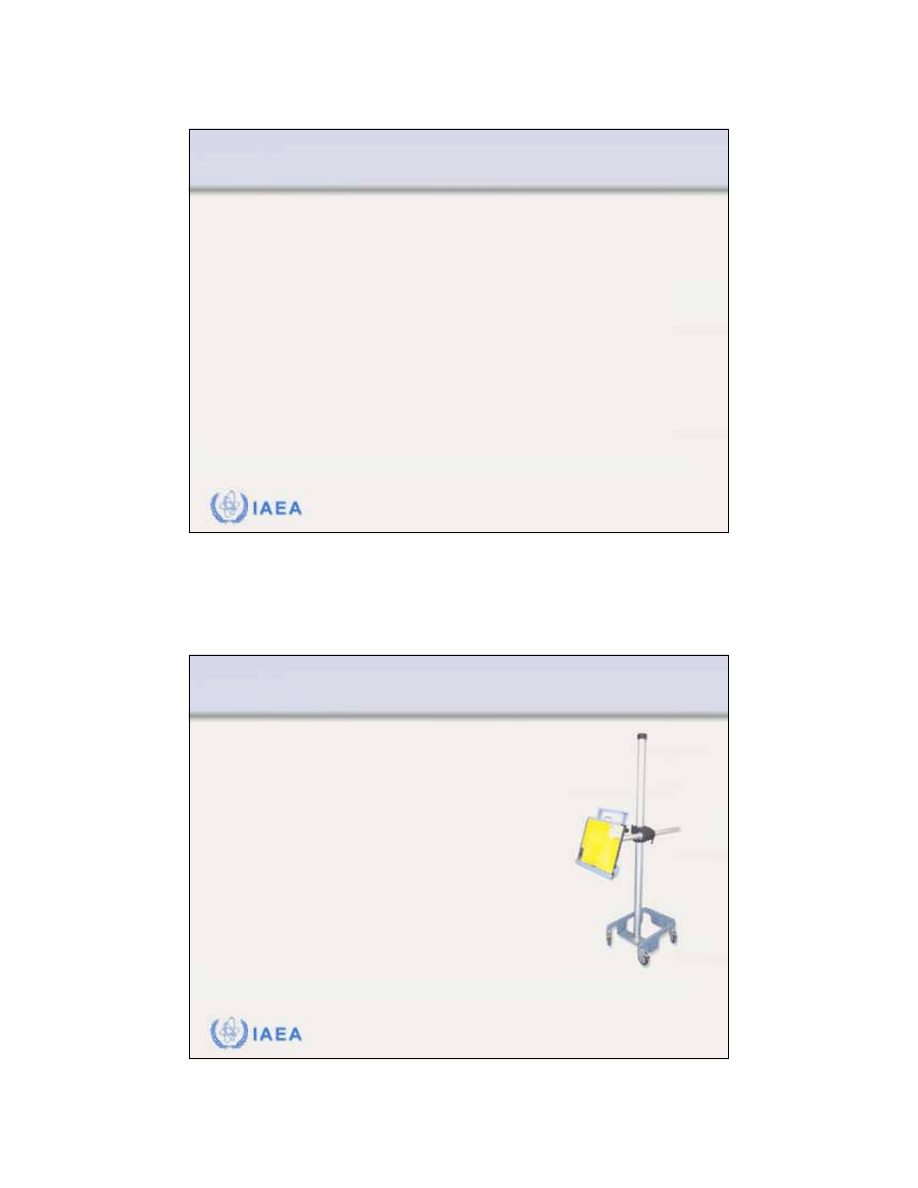
59
IAEA
Review of Radiation Oncology Physics: A Handbook for Teachers and Students - 12.4.2. Slide 1 (117/146)
12.4 TREATMENT DELIVERY
12.4.2 Portal imaging
As an accuracy requirement in radiotherapy, it has been
stated that figures of 5–10 mm (95% confidence level) are
used as the tolerance level for the
geometric uncertainty
.
The geometric accuracy is limited by:
•
Uncertainties in a particular patient set-up.
•
Uncertainties in the beam set-up.
•
Movement of the patient or the target volume during treatment.
Portal imaging
is frequently applied in order to check geo-
metric accuracy of the patient set-up with respect to the
position of the radiation beam
IAEA
Review of Radiation Oncology Physics: A Handbook for Teachers and Students - 12.4.2. Slide 2 (118/146)
The purpose of portal imaging is in
particular:
•
To
verify the field placement
,
characterized by the isocenter or
another reference point,
relative to
anatomical
structures
of the patient,
during the actual treatment.
•
To
verify that the beam aperture
(blocks
or MLC) has been properly produced
and registered.
Portal film device
12.4 TREATMENT DELIVERY
12.4.2 Portal imaging
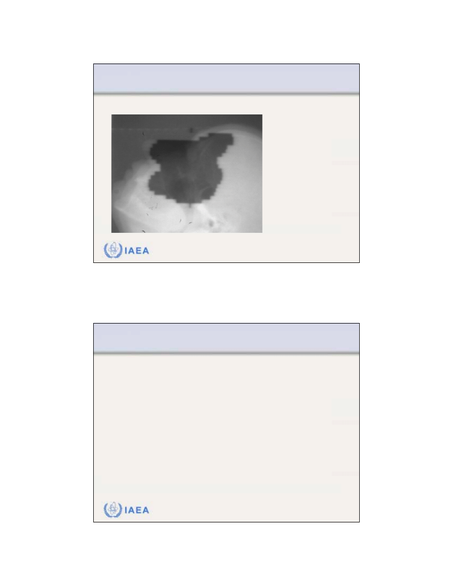
60
IAEA
Review of Radiation Oncology Physics: A Handbook for Teachers and Students - 12.4.2. Slide 3 (119/146)
Port film for a lateral
irregular MLC field
used in a treatment of
the maxillary sinus.
This method allows to
visualization of both
the treatment field and
the surrounding
anatomy.
Example for portal imaging: Port film
12.4 TREATMENT DELIVERY
12.4.2 Portal imaging
IAEA
Review of Radiation Oncology Physics: A Handbook for Teachers and Students - 12.4.2. Slide 4 (120/146)
Disadvantage of the film technique is its
off-line character
,
which requires a certain amount of time before the result
can be applied clinically.
For this reason
on-line electronic portal imaging devices
(EPIDs)
have been developed.
Three methods are currently in clinically use:
1.
Metal plate–phosphor screen combination
is used to convert the
photon beam intensity into a light image. The screen is viewed by
a sensitive video camera.
2.
Matrix of liquid filled ionization chambers.
3.
Amorphous silicon flat panel systems.
12.4 TREATMENT DELIVERY
12.4.2 Portal imaging
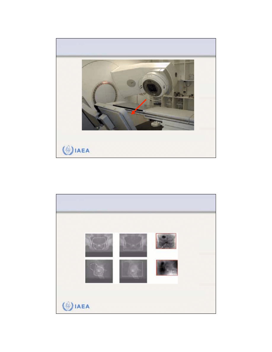
61
IAEA
Review of Radiation Oncology Physics: A Handbook for Teachers and Students - 12.4.2. Slide 5 (121/146)
Amorphous silicon type of EPID installed on the gantry of a linac.
12.4 TREATMENT DELIVERY
12.4.2 Portal imaging
IAEA
Review of Radiation Oncology Physics: A Handbook for Teachers and Students - 12.4.2. Slide 6 (122/146)
DRRs from treatment fields and large fields to verify the position of
isocentre and the corresponding EPID fields
.
Comparison between digitally reconstructed radiograph
(DRR) and image obtained with EPID
DRR treatment fields
DRR EPID fields
EPID images
12.4 TREATMENT DELIVERY
12.4.2 Portal imaging

62
IAEA
Review of Radiation Oncology Physics: A Handbook for Teachers and Students - 12.4.2. Slide 7 (123/146)
As part of the QA process, portal imaging may lead to
various strategies for
improvement
of positioning accuracy,
such as:
•
Improvement of patient immobilization.
•
Introduction of correction rules.
•
Adjustment of margins in combination with dose escalation.
•
Incorporation of set-up uncertainties in treatment planning.
12.4 TREATMENT DELIVERY
12.4.2 Portal imaging
IAEA
Review of Radiation Oncology Physics: A Handbook for Teachers and Students - 12.4.2. Slide 8 (124/146)
QA in portal imaging
•
Process control requires that local protocols must be
established to specify:
•
Who has the
responsibility
for verification of portal images (generally a
clinician), and
•
What
criteria
are used as the basis
to judge the acceptability of
information
conveyed by portal images.
12.4 TREATMENT DELIVERY
12.4.2 Portal imaging

63
IAEA
Review of Radiation Oncology Physics: A Handbook for Teachers and Students - 12.4.3. Slide 1 (125/146)
12.4 TREATMENT DELIVERY
12.4.3 In-vivo dose measurements
There are many steps in the chain of processes which
determine the
dose delivery to a patient
undergoing
radiotherapy and each of these steps may introduce an
uncertainty.
It is therefore worthwhile, and maybe even necessary
for specific patient groups or for unusual treatment
conditions to use
in-vivo dosimetry
as an ultimate check
of the actual treatment dose.
IAEA
Review of Radiation Oncology Physics: A Handbook for Teachers and Students - 12.4.3. Slide 2 (126/146)
In-vivo dose measurements
can be divided into
•
Intracavitary dose measurements (frequently used).
•
Entrance dose measurements (less frequently used).
•
Exit dose measurements (still under investigation).
Diodes applied for
intracavitary
in vivo
dosimetry.
12.4 TREATMENT DELIVERY
12.4.3 In-vivo dose measurements

64
IAEA
Review of Radiation Oncology Physics: A Handbook for Teachers and Students - 12.4.3. Slide 3 (127/146)
In-vivo dose measurements
12.4 TREATMENT DELIVERY
12.4.3 In-vivo dose measurements
IAEA
Review of Radiation Oncology Physics: A Handbook for Teachers and Students - 12.4.3. Slide 4 (128/146)
Examples of typical application of in-vivo dosimetry:
•
To check the
MU calculation
independently from the programme
used for routine dose calculations.
•
To trace any
error
related to patient set-up
, human errors in the
data transfer during the consecutive steps of the treatment
preparation, unstable accelerator performance and inaccuracies
in dose calculation, e.g., of the treatment planning system.
•
To determine the
intracavitary dose
in readily accessible body
cavities, such as the oral cavity, oesophagus, vagina, bladder,
and rectum.
•
To
assess the dose to organs at risk
(e.g., eye lens, gonads and
lungs during TBI) or situations where the dose is difficult to
predict (e.g., non-standard SSD or using bolus).
12.4 TREATMENT DELIVERY
12.4.3 In-vivo dose measurements
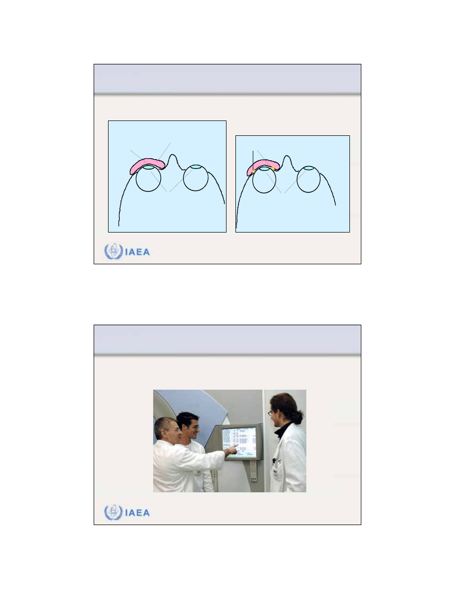
65
IAEA
Review of Radiation Oncology Physics: A Handbook for Teachers and Students - 12.4.3. Slide 5 (129/146)
Example for TLD in vivo dosimetry: Lens dose measurements
lens of
eye
arangement in lateral radiation fields
TLD
detectors
lens of
eye
7 mm of wax bolus
to mimick the position
of the lens under the lid
arangement in AP or PA
radiation fields
TLD detector
12.4 TREATMENT DELIVERY
12.4.3 In-vivo dose measurements
IAEA
Review of Radiation Oncology Physics: A Handbook for Teachers and Students - 12.4.4. Slide 1 (130/146)
12.4 TREATMENT DELIVERY
12.4.4 Record-and-verify systems
A computer-aided
record-and-verify system aims to compare
the set-up parameters with the prescribed values.

66
IAEA
Review of Radiation Oncology Physics: A Handbook for Teachers and Students - 12.4.4. Slide 2 (131/146)
Patient identification data, machine parameters and
dose prescription data are entered into the computer
beforehand
.
At time of treatment, these parameters are identified at
the treatment machine and,
if there is no difference
, the
treatment can
start
.
If discrepancies are present, this is indicated, the para-
meters concerned are highlighted,
and the treatment
cannot start
until the discrepancies are corrected or
overridden.
12.4 TREATMENT DELIVERY
12.4.4 Record-and-verify systems
IAEA
Review of Radiation Oncology Physics: A Handbook for Teachers and Students - 12.4.4. Slide 3 (132/146)
Discrepancies can be indicated only when tolerance
values are exceeded.
Tolerance values must be therefore established before.
•
Tolerances for verification of machine parameters
should be
provided by the manufacturer.
•
Clinical tolerance tables
must also be defined locally in the
department for each set of techniques to allow for patient/set-
up variations day-to-day.
•
Record-and-verify systems
must have the flexibility to be
overridden. This feature must be used with care and only
when reasons are clear and properly documented.
12.4 TREATMENT DELIVERY
12.4.4 Record-and-verify systems

67
IAEA
Review of Radiation Oncology Physics: A Handbook for Teachers and Students - 12.4.4. Slide 4 (133/146)
QA of Record-and-verify systems
•
Treatment delivered, if relying on record-and-verify system setting
or verifying the parameters, is only as good as the information input
to the system.
•
Therefore, it is vital that the data in the record-and-verify system is
quality-controlled, using independent (redundant) checking to verify
the input and to sanction its clinical use.
•
Performance of the record-and-verify system should be included in
an appropriate QA program.
•
Details of such QA tests will be specific to the system in question.
12.4 TREATMENT DELIVERY
12.4.4 Record-and-verify systems
IAEA
Review of Radiation Oncology Physics: A Handbook for Teachers and Students - 12.5.1. Slide 1 (134/146)
12.5 QUALITY AUDIT
12.5.1 Definition
Definition of Quality Audit
•
Quality audit is a systematic and independent examination to
determine whether or not:
•
Quality activities and results comply with planned arrangements.
•
Arrangements are implemented effectively and are suitable to
achieve the stated objectives

68
IAEA
Review of Radiation Oncology Physics: A Handbook for Teachers and Students - 12.5.1. Slide 2 (135/146)
12.5 QUALITY AUDIT
12.5.1 Definition: Parameters of quality audits
Quality audits:
•
Can be conducted for internal or external purposes.
•
Can be applied at any level of a QA program.
•
Are performed by personnel not directly responsible for the
areas being audited, however in cooperative discussion with
the responsible personnel.
•
Must be against pre-determined standards, linked to those
that the QA program is trying to achieve.
•
Evaluate the need for improvement or corrective action if
those standards are not met.
IAEA
Review of Radiation Oncology Physics: A Handbook for Teachers and Students - 12.5.1. Slide 3 (136/146)
12.5 QUALITY AUDIT
12.5.1 Definition: Parameters of quality audits
Quality audits:
•
Should be regular and form part of a quality feedback loop to
improve quality.
•
Can be mainly
procedural
, looking at QA procedures, proto-
cols, QC programs, QC and QA results and records, etc.
•
Can be mainly
practical
to verify the effectiveness or perfor-
mance of a quality system.
•
May be voluntary and co-operative, or may be regulatory (e.g.,
for accreditation of the department or hospital, for QS
certification, etc.).
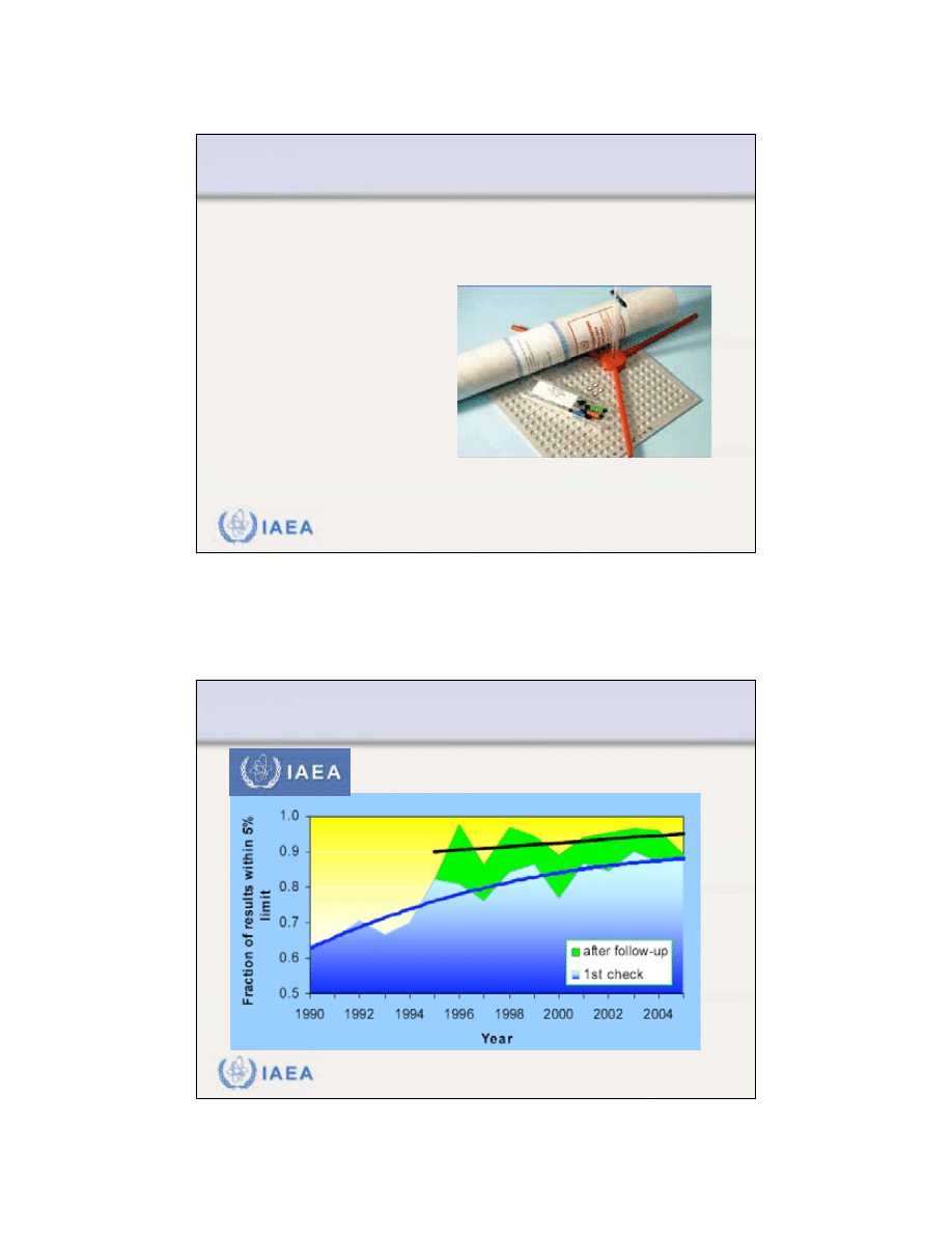
69
IAEA
Review of Radiation Oncology Physics: A Handbook for Teachers and Students - 12.5.2. Slide 1 (137/146)
12.5 QUALITY AUDIT
12.5.2 Practical quality audit modalities
A good example for an external audit is the simple but
very effective dosimetry audit organized as postal audit
with mailed dosimeters (usually TLD).
These are generally
organized by SSDLs
or agencies, such as the
IAEA, Radiological Physics
Center (RPC) in the U.S.,
ESTRO (EQUAL), national
societies, national quality
networks, etc.
Material used in IAEA/WHO TLD audits
IAEA
Review of Radiation Oncology Physics: A Handbook for Teachers and Students - 12.5.2. Slide 2 (138/146)
12.5 QUALITY AUDIT
12.5.2 Practical quality audit modalities
TLD results within the 5% limit

70
IAEA
Review of Radiation Oncology Physics: A Handbook for Teachers and Students - 12.5.3. Slide 3 (139/146)
12.5 QUALITY AUDIT
12.5.3 What should be reviewed in a quality audit visit?
The content of a quality audit visit must be pre-defined.
It will depend on the purpose of the visit:
•
Is it a routine regular visit within a national or regional quality
audit network?
•
Is it regulatory or co-operative between peer professionals?
•
Is it a visit following a possible misadministration?
•
Is it a visit following an observed higher-than-expected deviation
in a mailed TLD audit program that the centre cannot explain?
IAEA
Review of Radiation Oncology Physics: A Handbook for Teachers and Students - 12.5.3. Slide 4 (140/146)
12.5 QUALITY AUDIT
12.5.3 What should be reviewed in a quality audit visit?
Example of content of a
comprehensive quality audit visit:
Check infrastructure
•
Equipment.
•
Personnel.
•
Patient load.
•
Existence of policies and procedures.
•
Quality assurance program in place.
•
Quality improvement program in place.
•
Radiation protection program in place.
•
Data and records, etc.

71
IAEA
Review of Radiation Oncology Physics: A Handbook for Teachers and Students - 12.5.3. Slide 5 (141/146)
12.5 QUALITY AUDIT
12.5.3 What should be reviewed in a quality audit visit?
Example of content of a
comprehensive quality audit visit:
Check documentation
•
Content of policies and procedures
•
QA program structure and management
•
Patient dosimetry procedures
•
Simulation procedures
•
Patient positioning, immobilization and treatment delivery
procedures
•
Equipment acceptance and commissioning records
•
Dosimetry system records
•
Machine and treatment planning data
•
QC program content
•
Tolerances and frequencies, QC and QA records of results and
actions
•
Preventive maintenance program records and actions
•
Patient data records
•
Follow-up and outcome analysis etc.
IAEA
Review of Radiation Oncology Physics: A Handbook for Teachers and Students - 12.5.3. Slide 6 (142/146)
12.5 QUALITY AUDIT
12.5.3 What should be reviewed in a quality audit visit?
Example of content of a
comprehensive quality audit visit:
Carry out check measurements of
•
Beam calibration
•
Depth dose
•
Field size dependence
•
Wedge transmissions (with field size), tray, etc. factors
•
Electron cone factors
•
Electron gap corrections
•
Mechanical characteristics
•
Patient dosimetry
•
Dosimetry equipment comparison
•
Temperature and pressure measurement comparison, etc.

72
IAEA
Review of Radiation Oncology Physics: A Handbook for Teachers and Students - 12.5.3. Slide 7 (143/146)
12.5 QUALITY AUDIT
12.5.3 What should be reviewed in a quality audit visit?
Example of content of a
comprehensive quality audit visit:
Carry out check of training programs
•
Academic program.
•
Clinical program.
•
Research.
•
Professional accreditation.
•
Continuing Professional Education.
IAEA
Review of Radiation Oncology Physics: A Handbook for Teachers and Students - 12.5.3. Slide 8 (144/146)
12.5 QUALITY AUDIT
12.5.3 What should be reviewed in a quality audit visit?
Example of content of a
comprehensive quality audit visit:
Carry out check measurements on other equipment
•
Simulator
•
CT scanner, etc.
Assess treatment planning data and procedures.
Measure some planned distributions in phantoms.

73
IAEA
Review of Radiation Oncology Physics: A Handbook for Teachers and Students - 12.5.3. Slide 9 (145/146)
12.5 QUALITY AUDIT
12.5.3 What should be reviewed in a quality audit visit?
Example of a
comprehensive international external audit:
The
QATRO
(Quality Assurance Team for Radiation Oncology)
project developed by the IAEA.
Based on:
•
Long history of providing assistance for dosimetry audits in radio-
therapy to its Member States.
•
Development of a set of procedures for experts undertaking
missions to radiotherapy hospitals in Member States for the on-site
review of the dosimetry equipment, data and techniques, and
measurements, and training of local staff.
•
Numerous requests from developing countries to perform also
comprehensive audits of radiotherapy programs.
IAEA
Review of Radiation Oncology Physics: A Handbook for Teachers and Students - 12.5.3. Slide 10 (146/146)
12.5 QUALITY AUDIT
12.5.3 What should be reviewed in a quality audit visit?
In response to requests from member states, the IAEA
convened an expert group, comprising of radiation onco-
logists and medical physicists, who have developed
guidelines for the IAEA audit teams to initiate and perform
such audits and report on them.
•
The guidelines have been field-tested by IAEA teams performing
audits in radiotherapy programs in hospitals in Africa, Asia, Latin
America and Europe.
•
QUATRO procedures are endorsed by the European Society for
Therapeutic Radiology and Oncology (ESTRO), the European
Federation of Organizations for Medical Physics (EFOMP) and
the International Organization for Medical Physics (IOMP).
Wyszukiwarka
Podobne podstrony:
24 G23 H19 QUALITY ASSURANCE OF BLOOD COMPONENTS popr
24 G23 H19 QUALITY ASSURANCE OF BLOOD COMPONENTS popr
7 Modal Analysis of a Cantilever Beam
6 Quality Assurance Plan
8 Harmonic Analysis of a Cantilever Beam
9 Transient Analysis of a Cantilever Beam
Pearson Process Quality Assurance For Uml Based Projects (2003)
89 1268 1281 Tool Life and Tool Quality Summary of the Activities of the ICFG Subgroup
Quality Assurance Plan
3 NonLinear Analysis of a Cantilever Beam
Mechanical failure of external fixator during hip joint distraction for Perthes disease
71 1021 1029 Effect of Electron Beam Treatment on the Structure and the Properties of Hard
Gorban A N singularities of transition processes in dynamical systems qualitative theory of critica
Pomological and quality traits of mulberry (Morus spp )
Graveyard of Dreams H Beam Piper
DIN 61400 21 (2002) [Wind turbine generator systems] [Part 21 Measurement and assessment of power qu
Ministry of Disturbance H Beam Piper
więcej podobnych podstron