
Small mammal, exotic animal and wildlife nursing
171
Small mammal,
exotic animal and
wildlife nursing
Sharon Redrobe and Anna Meredith
This chapter is designed to give information on:
• The principal aspects of hospitalization of the exotic pet and wildlife patient
• The common diseases of these animals
• The main points of perioperative care of these species
• Zoonoses of these species and how to minimize the risks associated with their handling
• The correct administration of medicines to these species
8
8
Introduction
This chapter will deal with the group of small animals
commonly presented for veterinary treatment that are
‘not cats or dogs’. This includes common pet small
mammals, birds and reptiles. The reptile group includes
snakes, lizards and chelonians. The term chelonian refers
to those reptiles that possess a shell (turtles, terrapins
and tortoises). Some native UK wild animals that are
brought into the veterinary surgery by the public will
also be considered.
All these animals require a different approach to inpatient
care from that given to dogs and cats. Correct veterinary
nursing forms a vital part of the care of these patients and
affects whether treatment is successful or otherwise.
Hospitalization
•
Weigh patients daily to evaluate body condition and
clinical progress and to ensure accurate treatment dosage
•
Handle correctly to minimize stress, trauma and injury to
both handler and animal
•
Minimize handling to reduce stress (tame social species are
an exception)
•
Offer correct feed to stimulate the animal to eat and to
prevent gastrointestinal upset and dietary deficiencies
• Ensure that each individual animal can be identified
from the moment it is admitted to the veterinary
surgery. A description of the animal is sufficient in
some cases; stickers with names may be affixed to
reptile shells; and cages should be clearly labelled.
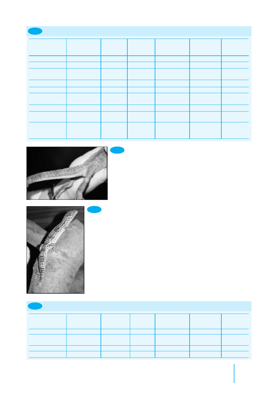
Some species may be microchipped for permanent
identification (Figure 8.1).
Animal
Suggested site
Fish
Midline, anterior to dorsal fin
Amphibians
Lymphatic cavity
Reptiles
It is recommended that tissue glue is
placed over the needle entry site in all
reptiles
Chelonians
Subcutaneously in left hindleg
(intramuscularly in thin-skinned
species)
Subcutaneously in the tarsal area in giant
species
Crocodilians
Cranial to nuchal cluster
Lizards
Left quadriceps muscle, or
subcutaneously in this area (all species)
In very small species, subcutaneously on
the left side of the body
Snakes
Subcutaneously, left nape of neck placed
at twice the length of the head from
the tip of the nose
Birds
Left pectoral muscle
Exceptions: ostriches – pipping muscle;
penguins – subcutaneously at base of
neck
Mammals
Large: left mid neck subcutaneously
Medium and small: between scapulae
8.1
Suggested sites for identification
microchip
(based on guidelines of the British
Veterinary Zoological Society)
Clinical parameters
It is important to be able to distinguish the normal from the
abnormal animal. The level of activity or stress should be

172
Manual of Advanced Veterinary Nursing
taken into account when evaluating whether the rates for vital
signs are within the normal range.
It is also important to examine the animal and gain an
appreciation of body condition (e.g. obese, very thin) rather
than rely on absolute figures for body weight.
Mammals
Examples of the clinical parameters of common mammal
species are presented in Figure 8.2.
Birds
Examples of the clinical parameters of common bird species
are presented in Figure 8.3. The body condition of a bird may
be gauged by feeling for the prominence of the breastbone
(keel) and giving a condition score ranging from 0 to 5:
•
A very prominent keel with no muscle cover is given a
score of 0
•
If the keel can only be palpated with pressure, due to
prominent muscles and fat, the score is 5
•
Most birds in good condition have a score of 3–4 and tend
to be leaner if they have free flight.
Reptiles
Examples of the clinical parameters of common reptile species
are presented in Figure 8.4.
•
The snout–vent length (SVL) is an important
measurement in the examination of a reptile. This is the
straight distance from the nose to the vent
•
Weight will obviously depend upon the size of the animal;
for example, a young boa constrictor with an SVL of 10 cm
might weigh only 15 g, compared with an older boa with
an SVL of 2 m which might weigh 15 kg
•
The body length of chelonians is taken as the straight-line
distance between the front and back edge of the shell, not
including the head or tail.
Mammal
Weight range (g)
Rectal temperature (
°
C)
Approximate pulse
Approximate respiratory
rate/minute
rate/minute
Badger
10000–15000
38–39
50–80
15–45
Chipmunk
100–250
38
200
100
Chinchilla
400–600
35.4–38
100
45–65
Ferret
500–2000
38.8
180–250
30–36
Fox
5000–10000
38
40–80
30
Guinea-pig
500–1100
38
230–380
70–100
Hamster
85–120
37–38
280–500
50–120
Hedgehog
800–1100
35.1
100–250
40–60
Mouse
20–60
37.4
300–700
150–200
Rabbit
1000–5000
38.5–40
130–320
30–60
Gerbil
50–90
39
260–600
70–120
Rat
250–400
38
300–500
80–100
8.2
Clinical parameters of common mammal species (adults)
Bird
Approx. weight
Rectal temperature (
°
C)
Approximate pulse
Approximate respiratory
range (g)
rate/minute
rate/minute
African Grey Parrot
300–400
40–42
100–300
15–45
Blue-fronted Amazon
300–500
40–42
125–200
15–45
Parrot
Budgerigar
30–60
40–42
260–400
60–100
Canary/finch
12–30
40–42
300–500
60–100
Chicken
2000–4000
40–42
80–100
20–50
Cockatiel
100–180
40–42
150–350
40–50
Umbrella Cockatoo
450–750
40–42
100–300
15–40
Lesser Sulphur-Crested
250–400
40–42
100–300
15–45
Cockatoo
Duck
2000–3000
40–42
100–150
15–30
Kestrel
150–300
40–42
150–350
15–45
Lovebird
50–70
40–42
250–400
60–100
Blue and Gold Macaw
900–1300
40–42
115–250
15–30
Greenwinged Macaw
1000–1500
40–42
100–250
15–30
Pennant’s Parakeet
180–200
40–42
150–300
30–60
Peregrine Falcon
550–1500
40–42
100–200
30–60
Pigeon
260–350
40–42
150–300
30–50
Quail
20–40
40–42
300–600
60–100
Sparrowhawk
150–300
40–42
150–350
15–45
Sparrow
25–30
40–42
250–600
100–150
Swan
5000–7000
40–42
60–100
15–30
Clinical parameters of common bird species (adults)
8.3
Small mammal, exotic animal and wildlife nursing
173
Common name
Species
Typical SVL
Weight
Environmental
Approx. pulse
Approx.
(cm)
range (g)
temperature
rate/minute
respiratory
range (
°
C)
rate/minute
Boa Constrictor
Boa constrictor
200–400
10000–18000
25–30
30–50
6–10
Cornsnake
Elaphe guttata
100–180
150–250
25–30
40–50
6–10
Day Gecko
Phelsuma
10–15
15–40
23–30
40–80
6–10
cepediana
Garter Snake
Thamnophis sp.
50–120
50–100
22–26
20–40
6–10
Green Iguana
Iguana iguana
100–150
900–1500
26–36
30–60
10–30
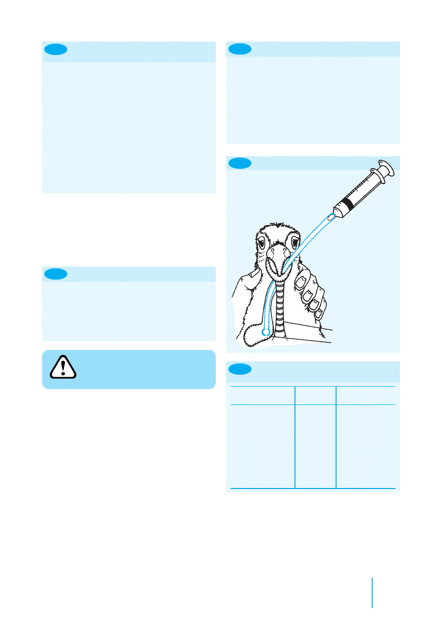
Leopard Gecko
Eublepharus
10
25–50
23–30
40–80
20–50
macularius
Royal Python
Python regius
80–150
400–800
25–30
30–50
6–10
Red-eared Terrapin
Trachemys scripta
20 (shell
800–1200
20–30
40–60
2–10
elegans
length)
Mediterranean
Testudo graeca
20–30 (shell
1000–2500
20–35
40–60
2–10
(spur-thighed)
length)
Tortoise
8.4
Clinical parameters of common reptile species (SVL = snout–vent length of adult)
When calculating drug dosages, the whole weight of the
chelonian is used. A common mistake is to attempt to deduct
the weight of the shell. The shell is part of the skeleton –
trying to ignore this weight is similar to trying to deduct the
weight of a dog’s skeleton from its body weight when
calculating doses and is clearly not sensible.
Body condition is estimated from the soft tissue
(muscle) covering the pelvis and tail bones – these bones
should be barely visible. Figure 8.5 illustrates the tail of an
emaciated green iguana. Some animals store fat in the tail
(e.g. leopard gecko) and so the tail base should be thicker
than the pelvis width if the animal has adequate fat storage
(Figure 8.6).
Reptiles regulate their internal body temperature by
moving between hot and cool areas in their enclosure. The
temperatures listed reflect the normal temperature range to
which the animals should have access in order to regulate
successfully.
Note the high variation in ‘normal’ rates; for example,
these are low when basking but higher when exercising or
stressed. The level of activity or stress should be taken into
account when evaluating whether the rates are within the
normal range.
Amphibians
Amphibians can tolerate a wide range of environmental
temperatures but the lower temperatures may be
immunosuppressive. Clinical parameters for common pet
amphibian species are given in Figure 8.7.
Common name
Species
Typical SVL
Weight
Environmental
Approx. pulse
Approx.
(cm)
range (g)
temperature
rate/minute
respiratory
range (
°
C)
rate/minute
Crested Newt
Triturus cristatus
10
5–15
18–22
40–80
10–40
Tiger Salamander
Ambyostoma
10
100–150
15–25
40–80
5–40
tigrinum
Leopard Frog
Rana pipiens
8
50
15–25
60–80
50–80
Tree Frog
Hyla arborea
3
20–50
15–25
60–80
50–80
Clinical parameters of common amphibian species (SVL = snout–vent length of adult)
8.7
8.5
The tailbones are
readily visible in
this emaciated
Green Iguana.
The tail of this well-fed Leopard
Gecko is wider than the pelvis.
8.6

174
Manual of Advanced Veterinary Nursing
Special techniques
Bandaging techniques
Bandages are not required to cover lesions or wounds in all
cases. They should be used only after due consideration of the
advantages and disadvantages of bandage application in a
particular situation.
In some cases the use of dressing or bandages can create a
problem: for example, the stress of repeated restraint to
perform regular bandage changes can be detrimental to the
welfare of a captive wild animal. Some animals will
consistently chew a bandage but would not interfere with the
underlying lesion if it were left uncovered.
Mammals
Many of the bandaging techniques used for domestic
mammals can be applied to exotic mammals. Some individuals
will not tolerate bandaging and will self-traumatize in an effort
to remove the bandage. Certain rabbits will tolerate an
Elizabethan collar, whereas others will not; the use of these
appliances should be judged on a case-by-case basis.
Birds
Most birds will not remove subcutaneous sutures and so
bandaging may not be necessary. Bandages should be placed so
as not to restrict chest movements, or respiration will be
compromised. The use of strong adhesive tape on the skin
should be avoided as avian skin is easily torn.
Amphibians
Bandaging of amphibians is impractical and adhesive tapes will
easily damage the thin skin. The use of human oral ulcer
barrier creams on the skin will protect underlying lesions and
seal the skin to prevent secondary infection.
Fish
Bandaging of fish is impractical. The use of human oral ulcer
barrier creams on the skin will protect the underlying lesions
and reduce osmotic stress on the fish.
Assisted feeding and oral therapy
If the animal is bright and alert, warmed oral fluids may be
given. Oral rehydration fluids may be given daily equal to
4–10% of body weight initially. Liquidized feed may be used
once the animal is rehydrated. The general points concerning
assisted feeding of animals are:
•
A small amount of the food should also be available to
tempt the animal to self-feed
•
To prevent digestive disturbances, an appropriate food
substance should be used – i.e. vegetable-based diets for
herbivores, meat-based diets for carnivores.
Mammals
Most mammals have a strong chewing response and will
readily feed from a syringe placed gently into the corner
of the mouth. Appropriate food substances should be
given; feeding the incorrect diet can lead to digestive
disturbances that may severely compromise the health
of an already sick animal.
The use of a nasogastric tube for assisted feeding is a
useful technique in the supportive care of larger mammals
that tolerate an amount of handling (e.g. rabbits, ferrets)
(Figure 8.11).
Rabbits are obligate nose breathers, so avoid
placing a nasogastric tube in those animals
already showing signs of respiratory distress,
or they may be further compromised.
•
Swimming upright
•
Smooth scales
•
No evidence of skin lesions
•
No rubbing
•
No petechiation
8.8
Signs of health in common
ornamental fish
Fish
Fish should be examined initially in the tank or pond, where
their behaviour should be noted. For closer examination,
individual fish may then be transferred with some water into a
small clear plastic bag.
Checking the water quality is an important part of the
investigation of disease in fish. Clinical parameters evaluated
in the examination of fish are presented in Figures 8.8 and
8.9 along with the water parameters required to ensure fish
health.
Reptiles
Most reptiles tolerate bandages well. Care should be taken
to use lightweight materials in animals with poor skeletal
density (e.g. cases of metabolic bone disease), since fractures
may be caused by the weight of the bandages. Snakes provide
a unique challenge to bandaging technique but finger or
stockinette bandage materials may be used. Plastic drapes
or condoms with the tips cut off make useful occlusive
bandages (Figure 8.10). Strong adhesive tape should not be
used on the thin-skinned geckos as the skin may easily tear
on removal of the bandage.
Water quality for common
ornamental fish
8.9
Group
pH
Temperature
Ammonia
(
°
C)
level (mg/l)
Cold water
6.5–8.5
10–25
Tropical
6.5–8.5
23–26
Marine
6.5–8.5
< 0.05
Salmonids
6.5–8.5
< 0.002
Non-salmonids
6.5–8.5
< 0.01
8.10
An occlusive bandage in a snake.
Small mammal, exotic animal and wildlife nursing
175
1. Sedate the animal or restrain safely
2. Instil topical local anaesthetic drops into the nose and
allow to take effect
3. Measure the distance from the nose to the position of
the stomach externally and mark the length on the tube
4. Lubricate the tube with lubricant gel
5. Gently introduce the tube into the ventral medial
aspect of the nostril and advance it into the nose
6. If resistance is detected: stop, withdraw the tube,
relubricate and reposition
7. Gently advance the tube until it is in the stomach as
indicated by the mark on the tube
8. Check the tube is in place
9. Glue the tube to the head using a flap of tape and tissue
glue
10. Some animals will require a restriction collar to prevent
them pulling out the tube.
How to place a nasogastric tube
in mammals
8.11
Never administer fluids into any nasogastric tube without
first checking that it is in place. Many sick rabbits will passively
inhale the tube. Check that the tube is in the stomach: either
use radiography (if a radiopaque feeding tube has been used) or
quickly inject 5 ml of air into the tube whilst listening over the
stomach area with a stethoscope for a ‘pop’ noise. Figure 8.12
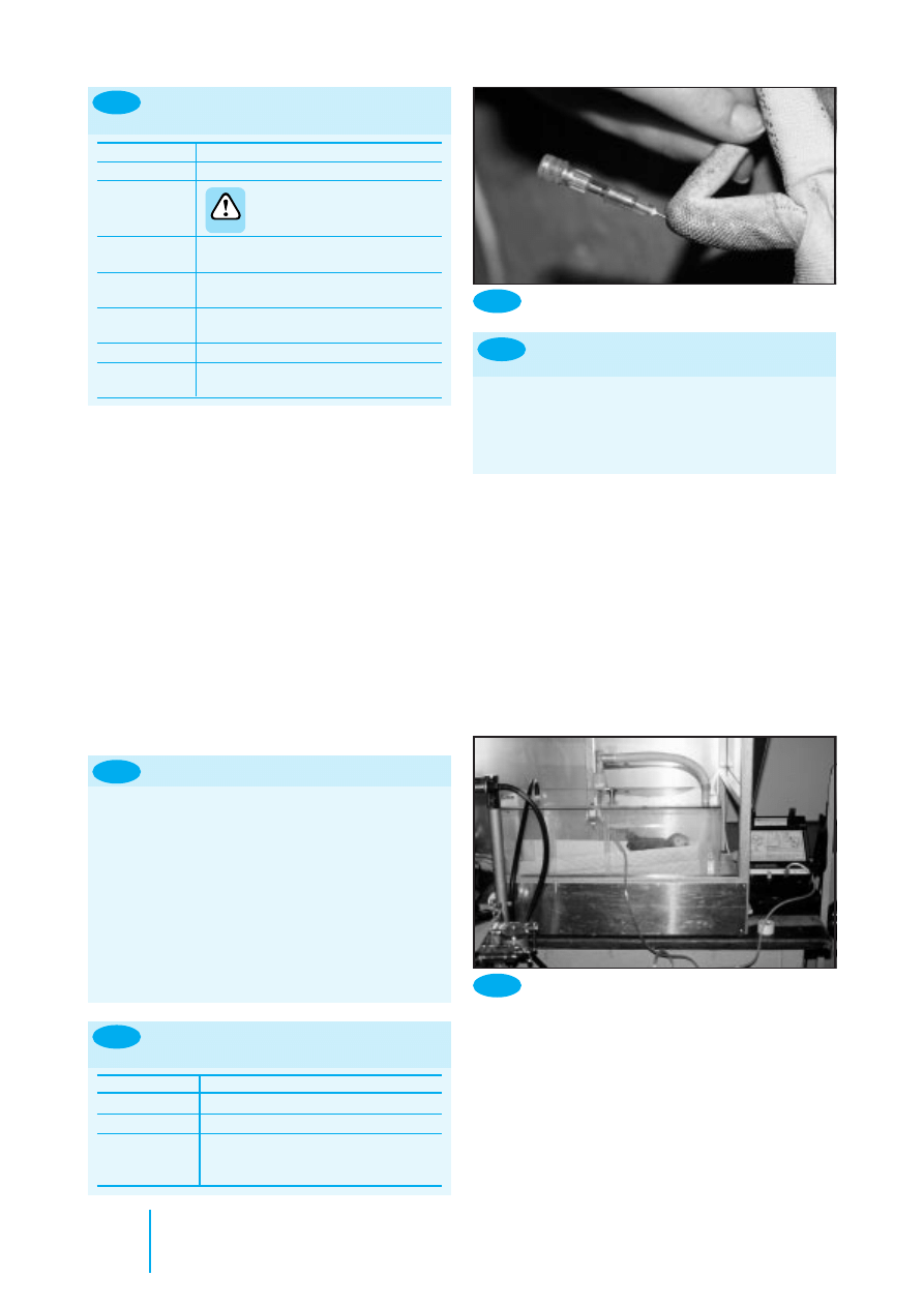
describes how to use a nasogastric tube safely.
1. Warm fluids to 38–40
°
C
2. Restrain bird upright
3. Extend neck
4. Insert gag if using plastic crop tube, or use metal crop tube
5. Insert crop tube into mouth at left oral commissure and
angle into right side of neck
6. Palpate placement in crop
7. Infuse fluid slowly
8. Check during infusion for regurgitation, if seen release
bird immediately and allow bird to swallow
8.13
How to crop tube a bird
Birds
With birds, oral tube feeding is often called crop tubing, as the
liquid is instilled into the distal oesophagus or crop, not the
equivalent of the stomach. However, not all birds possess a
true crop. Those with a well defined crop include parrots,
pigeons, and raptors; those with a poorly defined crop include
most waterfowl.
•
Parrots have strong beaks and large fleshy tongues that can
make inserting the gag difficult
•
Pigeons have small tongues and the beak can be held open
with a finger
•
Raptors have small tongues but it is wise to use a gag to
keep the beak open
• Do not fight with a struggling patient: many of the birds
are very ill and easily stressed. It is possible to injure the
choana (the slit on the roof of mouth) or crop if the bird is
not restrained properly.
The technique for crop tubing is described in Figure 8.13
and illustrated in Figure 8.14. The approximate volumes and
frequency of crop tubing will vary with the size of bird;
guidelines are given in Figure 8.15.
Bird species
Volume
Frequency
(ml)
(times per day)
Finch
0.1–0.5
6
Budgerigar
0.5–3
4
Lovebird
1–3
4
Cockatiel
1–8
4
Small conure
3–12
4
Large conure
7–24
3–4
Amazon parrot
5–35
3
African grey parrot
5–35
3
Cockatoo
10–40
2–3
Macaw
20–60
2–3
Suggested volumes and frequency
of crop tubing of selected species
8.15
Reptiles
Liquids may be instilled directly into the reptile stomach.
Figure 8.16 gives the method for stomach tubing, Figure 8.17
suggests appropriate tube sizes and Figure 8.18 describes the
position of the stomach in reptiles.
When the head of a tortoise is retracted, the oesophagus has
an S-bend. Thus the neck of a tortoise must be fully extended
before a tube is introduced (Figure 8.19) or the tube may be
accidentally pushed through the wall of the oesophagus.
Rabbits are unable to vomit and their stomach
is relatively non-distensible. It is possible to
rupture the stomach by giving too large a
volume of fluid.
•
Carefully calculate the safe volume to instil each time
•
If the animal shows signs of discomfort: stop, withdraw
some fluid and inform the attending veterinary surgeon
•
Many sick rabbits develop ileus (gastrointestinal stasis),
thus decreasing stomach emptying time. This will
require a reduction in oral fluid volumes and medical
therapy to treat the ileus.
8.12
How to use a nasogastric tube safely
8.14
Crop tubing a parrot
RIGHT
SIDE
Crop
tube
LEFT
SIDE
Crop
Trachea

176
Manual of Advanced Veterinary Nursing
8.16
1. Select flexible feeding tube of appropriate size and length (Figure 8.19)
2. Measure distance to stomach so that appropriate length is inserted, to ensure end of tube is in stomach (Figure 8.18)
3. Lubricate tube well (a small amount of lubricant can also be placed at the back of the mouth)
4. Insert gag gently into mouth (avoid damaging the delicate teeth of snakes and lizards)
5. The reptile glottis and trachea lie rostrally in the floor of the mouth thus the whole of the back of the oral cavity is oesophagus
6. Insert tube to stomach distance – stop if resistance is detected
7. Slowly infuse warmed fluid
8. If fluid is seen coming back into the mouth: stop immediately and note the volume already given (for future reference)
9. Once the animal starts to eat or drink by itself, less will be required by stomach tube
10. The stressed reptile will regurgitate food immediately after instillation. Tube the animal whilst holding it vertically and
hold it so for about a minute after tubing to prevent immediate regurgitation
11. Avoid handling the reptile for 24 hours to prevent regurgitation.
How to stomach tube a reptile
8.17
Species
Bodyweight
Size of feeding tube (Fr)
Approx. volumes (ml) twice daily
Mediterranean tortoises
> 1 kg
8
10
Juvenile iguanas
100–400 g
6–8
2–8
Adult cornsnake
200 g
8–10
5–10
Reptile stomach tube: suggested sizes and volumes
8.18
Reptile
Stomach position
Method of orally dosing
Lizard
Caudal edge of the ribcage
Some will take fluids straight from the syringe
Many will open their mouths defensively if the snout is tapped
Gentle traction on the dewlap (if present) may also be used
Chelonian
Middle of the abdominal shield
Fully extend the neck
of the plastron (lower shell)
To extract the head, push in the rear limbs and tail, placing the fingers
around the back of the mandibles, and maintain traction
Snake
At the beginning of the second
The snake may disarticulate its jaws if the mouth is prised open; this is a
third of the body length
normal response
Position of stomach and methods of orally dosing reptiles
1. Choose a food item equivalent to the diameter of the
snake
2. Lubricate the food item with a water-based lubricant
3. Hold the snake vertically
4. Gag open the mouth
5. Gently introduce the food item to the back of
the mouth
6. ‘Milk’ the food item to the stomach region
(approximately halfway down snake)
7. Retain snake in vertical position for 1 minute
8. Gently return snake to vivarium.
8.20
How to force feed a snake
•
When orally dosing or force feeding reptiles, the use of
sharp objects to push the item into the mouth should be
avoided as they may lacerate the oesophagus
• If the operator is scratched by a reptile’s teeth, the hands
should be thoroughly washed and the incident reported
appropriately
• Care should be taken to avoid damaging a reptile’s teeth, as
this may lead to osteomyelitis of the jaw. If a snake’s teeth are
damaged, ensure that the animal is checked again 2 weeks
and 4 weeks later and that appropriate therapy is initiated
• If tube feeding is required over a long period, consider
placing a temporary pharyngostomy tube to prevent
oesophageal damage from repeated stomach tube placement
•
For most snakes, force feeding means using a whole dead
rodent of appropriate size (Figure 8.20)
• A general guide to the type of food to be force fed to
reptiles is given in Figure 8.21
• The amount and frequency of feeding required in reptiles
depends on the age, size and species of the reptile;
guidelines are given in Figure 8.22.
Measuring the mouth-to-
stomach distance in a
tortoise.
8.19

Small mammal, exotic animal and wildlife nursing
177
Species
Products for assisted feeding
Group
Examples
Diet
Examples
Snakes
Boas, pythons, rat snakes, gopher snakes, bull
Meat-based
Proprietary liquid meat products for
snakes, vipers, garter snakes, water snakes,
dogs or cats
racers, vine snakes
Lizards
Herbivores
Green iguanas
Vegetable-based
Purees, baby food
Carnivores
Monitors, geckos, anoles, skinks, chameleons
Meat-based
Meat-based products
Chelonians
Carnivores
Turtles and terrapins
Meat-based
Meat-based products
Herbivores
Tortoises
Vegetable-based
Purees, baby food
8.21
Type of food for assisted feeding of reptiles
Reptile
Frequency
Small snakes and lizards
Once or twice/week
Young of boas and pythons
Three times/week
Herbivores
Daily
Large snakes
Once/2–4 weeks
Guide to feeding frequency for
reptiles
8.22
Animal
Site(s)
Small mammal
Jugular (ferret, rabbit, chinchilla)
Marginal ear vein (rabbit)
Cephalic (rabbit, chinchilla, guinea-pig)
Lateral tail vein (rodents)
Bird
Jugular, brachial, medial metatarsal
Snakes
Ventral tail vein, jugular vein, cardiac
Lizard
Ventral tail vein, jugular vein
Chelonian
Jugular vein, dorsal tail vein
Amphibian
Central ventral abdominal vein, cardiac
Fish
Caudal vein
Intravenous injection and blood
sampling sites
8.23
Amphibians
Food may be placed directly into the mouth of an amphibian.
It will usually be swallowed if it is placed at the back of the
mouth. Care must be taken not to damage the delicate skin
when attempting to open the mouth to introduce feed.
Administration of medicines
Oral route
The methods described above for assisted feeding are also
applicable to individual oral dosing of animals. These are the
most accurate methods of oral administration of drugs.
Administering drugs in the drinking water is of limited
use. Success of treatment using this method depends upon:
•
The amount of water consumed – most psittacines and
reptiles drink too little to make this a useful option
•
The oral bioavailability of the drug – if the drug is not
absorbed from the gastrointestinal tract then it can only be
used to treat gut infections using this method.
Birds
Proprietary medicated seed is available to treat birds. It is
difficult to assess an accurate dose for the bird as not all the
seed offered may be eaten. This method is obviously not
suitable for the anorexic or non-seed-eating bird.
Amphibians
Amphibians may be dosed from a syringe placed directly into
the mouth.
Fish
Some types of medicated fish feed are commercially available.
Homemade medicated feed can be produced by combining
fish flakes and the required drug with gelatine. A dose of
medicine per fish is calculated and the amount of food the
fish will eat is assessed. The concentration of drug to be used
in the feed is then calculated. The necessary amount of
gelatine is made up with water to which the drug has been
added. Fish flakes are then added to the liquid mixture; the
mixture is allowed to set and then grated for feeding to the
fish at the required dose.
Intravenous route
Figure 8.23 lists accessible intravenous sites. These may be
used for the introduction of fluids and drugs or for
withdrawing a blood sample.
Mammals
The use of the intravenous site to deliver fluids or drugs
in small mammals is arguably only practical in the rabbit,
where access to the marginal ear veins is relatively simple
(Figures 8.24 and 8.25). It is useful to apply a local
anaesthetic cream to the skin prior to venepuncture to
minimize discomfort.
Birds
Three main intravenous sites are used in birds. The brachial
vein is readily identified in the medial elbow (Figure 8.26) but
is prone to haematoma formation after sampling. The medial
metatarsal vein (Figure 8.27) is less fragile and can be used in
larger birds. The right jugular vein is larger than the left. Each
jugular vein is located in a featherless tract on the neck and so
is easily visualized.

178
Manual of Advanced Veterinary Nursing
1. Shave the lateral ear over the vein and prepare the
site aseptically
2. Apply local anaesthetic cream and leave for
appropriate amount of time
3. Insert catheter of suitable size and glue in
place, using cyanoacrylate adhesive
4. Flush with heparin saline
5. Pack inside of ear with roll of gauze and tape in place
6. Connect catheter to giving set or mini extension set
7. Apply Elizabethan collar to the rabbit if required.
How to place an intravenous
catheter in a rabbit
8.24
Rabbit with an
intravenous infusion
line in place.
8.25
8.28
1. Extend the neck fully by using continuous traction,
placing fingers behind head. Sedation may be required
for strong patients
2. The vein runs from the tympanic membrane to the base
of the neck
3. The vein may be raised by placing a finger at the base of
the neck
4. Insert the needle parallel to the neck into the vein
5. After access, apply pressure to the site for a few
minutes to limit haematoma formation.
How to access the jugular vein in
the chelonian
1. Restrain the animal and hold the tail with the ventral
aspect facing the operator
2. Insert the needle in the exact midline at a point distal to
the vent and hemipenes (if present)
3. Advance to touch ventral aspect of the tail vertebra (at
right angles to tail in snake, at 45 degree angle in lizard)
4. Aspirate slowly and withdraw slightly until blood is
seen in the hub of the needle.
8.30
How to access the ventral tail vein
in a lizard or snake
1. Fully extend the tail
2. Insert the needle into the exact midline of the dorsal
tail close to the shell
3. Advance the needle to touch the vertebrae
4. Aspirate the syringe and withdraw it slightly until
blood is seen in the hub of the needle.
8.33
How to access the dorsal tail vein
in a tortoise
Reptiles
The choice of vein used for the intravenous sites depends
upon the type of reptile under consideration. The jugular vein
is useful in chelonians (Figures 8.28 and 8.29), but access to
this vein requires a surgical cut-down in snakes and lizards; the
ventral tail vein is useful in snakes and lizards (Figures 8.30,
8.31 and 8.32). Care must be taken if injecting into the dorsal
tail vein of a chelonian (Figure 8.33) as the injection may be
inadvertently placed in the epidural space and may produce
hindlimb paresis or paralysis. Intracardiac catheters may be
placed in snakes to access the circulation if the peripheral veins
are too small for ready access. Aseptic technique is required
when accessing the veins or heart.
Metatarsal vein of a
swan.
8.27
Brachial vein of a
pigeon.
8.26
Obtaining a jugular
blood sample from a
tortoise.
8.29
Obtaining a ventral
tail vein blood sample
from an iguana.
8.32
Obtaining a ventral
tail vein blood sample
from a snake.
8.31

Small mammal, exotic animal and wildlife nursing
179
Amphibians
The only accessible vein in amphibians is the central ventral
abdominal vein. The heart may be accessed for blood sampling
in the anaesthetized animal. The lymphatic system is a useful
site for injection in amphibians and appears to be effective in
delivering parenteral therapy. The site is dorsal, just off the
midline of the body.
Fish
The caudal vein in fish is accessed on the ventral aspect on
the midline, just cranial to the tail and caudal to the anal fin.
The vein lies immediately ventral to the vertebral column. The
method is similar to accessing the ventral tail vein of the
snake or lizard.
Intramuscular route
Figure 8.34 suggests sites for intramuscular injection.
Reptiles
Reptiles have a renal portal venous circulation. This means
that, in theory, blood from the caudal half of the body can
flow through the kidneys before returning to the heart. Thus
drugs injected into the hindlegs or tail may be lost via the
kidneys before being distributed around the body, or may
damage the kidneys if the drugs are potentially nephrotoxic.
There is still debate as to whether this significantly affects
drug distribution. It is generally accepted, however, that
injections should be given in the cranial half of the body
whenever possible.
Care needs to be taken in giving intramuscular injections
to reptiles:
•
Some lizards can shed their tails and so the injection of
substances into the tail should be avoided
•
It is good practice to alternate sides or sites where possible
•
Some chameleons may show a temporary or permanent
colour change at the injection site.
Amphibians
Amphibians also possess a renal portal system (see
considerations for reptiles, above). The front limb muscles
may be injected but these are usually small and so large
volumes should be avoided.
Fish
Abscess formation and drug leakage out of the needle track is
common in fish after intramuscular injection.
Subcutaneous route
The subcutaneous route (Figure 8.36) is an impractical route in
chelonians. Larger volumes may be given via the subcutaneous
route than intramuscularly in small lizards and snakes.
Animal
Site(s)
Small mammal
Quadriceps (rabbit, ferret)
Lumbar (rabbit, ferret)
Bird
Breast (pectoral) muscles
Snake
Intercostal muscles of body in middle
third of snake: insert needle just deep
enough to cover bevel, shallow angle
Lizard
Triceps (forelimb), quadriceps
(hindlimb), tail muscles in some
species (not geckos)
Chelonian
As lizard, also pectoral muscle mass at
angle of forelimb and neck. A short
needle should be used and the head
extended to avoid injecting the
structures of the neck or penetrating to
the lung/heart. The needle should only
be inserted to the depth of the bevel
Amphibian
(Fore)limb muscles
Fish
Dorsal lateral musculature
8.34
Intramuscular injection sites
1. Palpate and identify the breast bone (keel) as a ridge
running down the centre of the two breast muscles and
identify the edge of the sternum
2. Divide the breast muscles into four imaginary parts
(top right, top left, bottom right, bottom left)
3. Inject deeply into the muscle in alternate sites
4. After injection, place a finger over the puncture site for
a minute to minimize bleeding. Normally, there should
be no or very little bleeding.
8.35
How to give a bird an intramuscular
injection
Mammals and birds
A relatively large volume injected into the muscle causes
unnecessary pain to small animals. Drug reactions and
myositis have been associated with this route in rabbits and
rodents. Studies have also shown that the uptake of
subcutaneous or intraperitoneal injections in small rodents is
as fast as from the intramuscular site.
This is, however, a useful site for dosing birds (Figure 8.35).
8.36
Animal
Site(s)
Small mammal
Dorsal body (scruff)
Bird
Dorsal body between wings
Snake
Dorsal lateral third of snake, over ribs
Lizard
In loose lateral skin fold over ribs
Chelonian
Some loose skin on limbs
Amphibian
Dorsal area over shoulders
Fish
Not used
Subcutaneous injection sites
Intraperitoneal/intracoelomic route
This route (Figure 8.37) generally allows for a large volume to
be given. The fluids must be warmed to the body temperature
appropriate to the species.
Birds
The peritoneal space in birds is merely a potential one and
cannot be accessed for injection unless ascites is present.
Attempted injection into the abdominal space in birds will
usually result in injection into the air sacs, severely
compromising respiration, and is often fatal.
Reptiles
Reptiles do not possess a diaphragm and so the injection of
large volumes of fluid into the coelom can compromise
respiration.
180
Manual of Advanced Veterinary Nursing
Intraosseous route
The intraosseous route is a useful one for parenteral therapy,
especially in small animals, because:
•
Placing an intraosseous catheter or needle into a bone
enables fluids to be given into the medullary cavity, where
absorption is as rapid as the intravenous route
•
Small veins are fragile and easily lacerated by catheters or
‘blown’ when introducing fluids, whereas an intraosseous
catheter is stable in bone
•
If the animal displaces or damages the intraosseous
catheter, it is unlikely to haemorrhage from this site
compared with intravenous catheterization.
Figure 8.38 describes how to place an intraosseous
catheter; suggested sites for intraosseous catheters are given in
Figure 8.39 and illustrated in Figure 8.40. The management of
an intraosseous catheter (Figure 8.41) is similar to the
technique used to manage an intravenous catheter.
Animal
Site
Small mammal
Proximal femur, proximal tibia
Bird
Distal radius, proximal tibiotarsus
Reptile
Proximal or distal femur, proximal tibia;
bridge between carapace and plastron
in chelonians
Suggested sites for intraosseous
catheters
8.39
•
Use aseptic technique when giving drugs/fluids
•
To prevent clot formation, fill catheter with heparin or
heparinized saline between use
•
Flush three times daily with heparinized saline if not
used for drug or fluid administration.
8.41
How to manage an intraosseous
catheter
8.37
Animal
Site(s)
Small mammal
Off midline, caudal to level of umbilicus
Bird
Snake
Immediately cranial to vent on lateral
body wall
Lizard
Off midline, caudal to ribs, cranial to
pelvis
Chelonian
Extend hindlimb, inject cranial to
hindlimb in fossa
Amphibian
Ventrolateral quadrant
Fish
Immediately rostral to vent on ventral
surface
Intraperitoneal/intracoelomic
injection sites
Not possible in healthy
animal – avoid as attempts
may drown animal
1. Prepare site aseptically
2. Inject local anaesthesia into site (unless animal is under
general anaesthesia)
3. Introduce spinal needles or plain needles of appropriate
size into the bone (needle size sufficient to enter
medullary cavity, based on knowledge or guided by
radiographic image of cavity)
4. Flush with heparinized saline to ensure patency
5. Secure in place with surgical cyanoacrylate adhesive or
suture
6. Attach short extension tube
7. Bandage area to maintain cleanliness and reduce
mobility of limb.
How to place an intraosseous catheter
8.38
Nebulization
This is a useful technique for delivering drugs to the respiratory
system. Drugs given by nebulization are not systemically
absorbed and so potentially nephrotoxic or hepatotoxic drugs
may be used relatively safely. This technique is especially useful
in the treatment of respiratory tract disease in birds and reptiles,
where adequate drug levels may not reach the respiratory tract
following oral or parenteral dosing. It also minimizes the stress
of handling and potential damage caused by repeated injections.
The animal is placed in a chamber and nebulized with the drug
for an appropriate length of time (Figure 8.42). The nebulizer
must generate particles of less than 3 microns in order to enter
the lower respiratory tract of birds.
Via the water environment
This route can be used for fish, amphibians and aquatic
invertebrates.
•
Antibiotics should not be administered via the water if a
biological filtration system is in use
•
The calcium present in hard water may chelate some
antibiotics and so reduce their availability
•
Many of the drugs used are toxic in high doses
•
Calculations of water volume and drug required must be
made accurately
8.40
Intraosseous catheter in femur of a Green Iguana.
8.42
Bird in nebulization chamber.

Small mammal, exotic animal and wildlife nursing
181
• If possible, test the solution using a few animals before
dosing a large number
•
Mix the water thoroughly to ensure that the drug is evenly
dispersed
•
Starve the animal for 24 hours before treatment.
There are two methods of administering drugs using
the water:
•
Dipping the animal into a strong solution for a short
period (usually administered in a separate ‘hospital tank’,
then the animal is returned to its home environment)
•
Bathing the animal in a weaker solution for a longer
period. If the animal shows any signs of distress, the
treatment should be stopped. This may be performed in
the home tank to minimize disturbance, or in a separate
‘hospital tank’.
Topical application of medicine
Mammals
Mammals commonly groom off any topical treatment,
reducing its effectiveness. Any medication applied to the skin
should be non-toxic if ingested. Collars may be used to
prevent the animal from removing the topical medication.
Birds
Topical medication should be applied to the skin, not feathers,
of a bird. Collars may be tolerated by some animals and can be
used to prevent ingestion of the medicine.
Weight of animal (g)
Maximum safe volume of
blood to take (ml)
500
5
200
2
100
1
50
0.5
8.43
Guide to small animal weights and
maximum blood volume that may
be taken safely
Reptiles
Most reptiles will tolerate topical therapy without grooming
or licking the medicine. It is useful to bandage the area after
application to prevent the animal rubbing the medicine off;
this is especially important in snakes.
Amphibians
Most topically applied medications will be systemically
absorbed by amphibians and so any wound dressings should be
applied with care. This route may therefore be used to
administer medicines. The dose should be carefully calculated.
Ophthalmic drops are often used for this purpose.
Fluid therapy
•
Volumes required are usually 1–2% of bodyweight
•
The advantages and disadvantages of subcutaneous,
intramuscular and intraperitoneal routes have been
described above
•
Placement and maintenance of intravenous catheters is as
for larger domestic animals (see Figure 8.23 for description
of accessible veins)
•
Intraosseous catheters are useful to administer fluids to
smaller animals or those in which a vein is not readily
accessible (see Figure 8.39 for suggested sites and Figure
8.38 for method of placement).
Blood sampling
See Figure 8.23 for blood sampling sites.
•
Up to 10% of the blood volume may be safely taken from
an animal. This must be carefully calculated using an
accurate weight when dealing with small animals (Figure
8.43 gives examples)
•
EDTA may lyse some avian and reptile cells
•
A fresh blood smear is useful when examining cell
morphology and checking for blood parasites
•
The laboratory should be contacted for guidance on
(minimum) sample volume and tubes required.
Common diseases
Common diseases for various animals, along with their causes
and treatment, are described in Figures 8.44–8.50.
Problem
Species
Possible causes
Treatment
Comment
Anorexia
All
Urolithiasis
Surgery when stable
All
Renal disease, liver disease
Supportive
Especially older animals
Guinea-pig, young
Change in diet or
Reduce stress
Very common in new pets
rabbit, hamster
environment
Probiotics
Guinea-pig,
Dental disease
Burring/removal of
Usually due to lack of
chinchilla, rabbit
affected teeth
dietary fibre or genetic
factors
Guinea-pig, rabbit
Pregnancy toxaemia
Corticosteroids
Especially in obese animals
Dextrose
Emergency surgery
Rabbit
Viral haemorrhagic disease
None (fatal)
Routine vaccination
Vaccinate in-contact
recommended
animals
8.44
Common conditions of small mammals
Figure 8.44 continues
▼

182
Manual of Advanced Veterinary Nursing
Problem
Species
Possible causes
Treatment
Comment
Diarrhoea
All
Dietary change, stress,
Increase fibre intake
Address underlying cause
enteritis (bacterial,
Probiotics
fungal, viral)
Antibiotics
Fluid therapy
Rabbit
Lack of fibre, coccidiosis
Increase fibre intake
Look for and prevent
Probiotics
associated myiasis
Coccidiostats
Fluid therapy
Hamster (‘wet tail’
Campylobacter jejuni
Oral antibiotics and fluid
Very common
or proliferative
Escherichia coli
therapy
Poor prognosis
ileitis)
Chlamydia tracheomatis
Predisposing factors:
Desulfovibrio spp.
stress, dietary change
Guinea-pig, rabbit,
Inappropriate antibiotics
Increase fibre intake
Avoid penicillins,
hamster
Probiotics
cephalosporins
Stop antibiotics
Fluid therapy
Respiratory
All
Viral, bacterial
Supportive therapy
‘Chronic respiratory
disease
Appropriate antibiotics
disease’ in rats may
Mucolytics
require long-term
treatment
Dermatitis
All
Ectoparasites
Ivermectin
Bacterial
Antibiotics
Fungal (e.g.
Griseofulvin
dermatophytosis)
Viral
None
Self or cagemate trauma
Separate animals
(barbering)
Hamster
Neoplasia (lymphoma;
Euthanasia
mycosis fungoides)
Guinea-pig
Scurvy
Vitamin C
Always add vitamin C to
the diet and/or water
Myiasis (fly
Rabbit
Maggots
Removal of maggots,
Investigate underlying
strike)
ivermectin, antibiotics,
cause of debilitation (e.g.
corticosteroids (shock)
obesity, arthritis, dental
disease)
Haematuria
All
Urolithiasis
Surgery
Cystitis
Antibiotics
Neoplasia, bladder
Surgery/none
Neoplasia, uterus
Surgery (spay)
Renal infection
Antibiotics
Rabbit
Normal red pigments
None required
Neurological
Rabbit
Pasteurellosis (middle ear
Antibiotics
signs
or brain)
Rabbit, ferret
Parasites in brain
Supportive/none
(Encephalitozoon cuniculi,
aberrant migration of
nematodes, Toxoplasma)
All
Trauma
Supportive/none
Rabbit, ferret
Heat stroke
Supportive/cool slowly
All
Lead toxicity
Drugs to chelate lead,
Usually due to ingested
surgery to remove source
lead foreign body (i.e.
if lead ingested
lead in gut); rarely results
from lead shot in muscle
tissue
Ferret
Insulinoma
Glucose, surgery
Lymphoma
Cancer therapy
8.44
Common conditions of small mammals
continued
Figure 8.44 continues
▼
Treat underlying cause and
in-contact animals
Separate animals
Investigate individual
cause
Lameness,
weakness

Small mammal, exotic animal and wildlife nursing
183
Problem
Species
Possible causes
Treatment
Comment
Ferret continued
Anaemia
Specific therapy
Common in entire
unmated female ferrets
who develop persistent
oestrus. May not respond
to mating with
vasectomized male
Aleutian disease (viral)
None, supportive
Canine distemper
None, supportive
Vaccinate with canine
vaccine
All species,
Pododermatitis
As above, husbandry,
May progress to
especially rat and
(‘bumblefoot’)
bandaging feet
amyloidosis and renal
rabbit
failure
All species,
Arthritis (limbs, spine)
Analgesia,
especially rat and
anti-inflammatories
rabbit
All
Fractures, intervertebral
Supportive, surgery if
disc protrusion
fractures, euthanasia if
spinal
Subcutaneous
Rabbit, rodents,
Abscess
Lance, drain, antibiotics,
Facial abscesses in rabbit
masses
ferret
treat underlying cause
often related to dental
infection or osteomyelitis
Guinea-pig
Cervical adenitis
Surgical removal of
(Streptococcus
infected lymph node(s),
zooepidemicus)
antibiotics, euthanasia
All
Lipoma, other neoplasia
Surgery
Rabbit
Myxomatosis
Supportive, vaccinate
Usually fatal
other animals in contacts
Guinea-pig
Sebaceous adenoma
Surgery
Corneal ulcer
All
Trauma, entropion
Antibiotics, surgery
Rodents
Viral infection of
None; supportive (eye
lachrymal glands (SDAV)
may perforate)
Rodents
Calcification of cornea
None
Ferret
Distemper, influenza
Supportive
Ocular
Rabbit
Dacryocystitis (infection
Flush ducts, antibiotics
Check molar roots not
discharge
of tear duct)
impinging on duct
(radiography required to
evaluate)
Chinchilla, rabbit
Overgrown molar teeth
Dental treatment
Poor prognosis
roots impinging on duct
Red staining
All
Stress, concurrent
Treat underlying cause
Known as porphyria/
tears
disease
chromodacryorrhoea
8.44
Common conditions of small mammals
continued
Lameness,
weakness
continued
Problem
Common clinical condition
Treatment
Skin/face
Periocular swelling
Ocular or sinus disorder
Investigate and treat appropriately
Epiphora, conjunctivitis
Ocular or sinus disorder, partial lid
Investigate and treat appropriately
paralysis (cockatiel), psittacosis (cockatiel,
duck)
Scabs, scars, pustules
Pox virus
Vaccination of in-contacts
Brown hypertrophy of cere
Endocrinopathy (budgerigars)
None
Hyperkeratosis
Cnemidocoptes spp. (mites)
Ivermectin
Crusting of cere
8.45
Common conditions of birds
Figure 8.45 continues
▼

184
Manual of Advanced Veterinary Nursing
Problem
Common clinical condition
Treatment
Nares
Discharge (rhinitis)
Sinusitis, air sacculitis
Based on sensitivity, flush out sinuses,
infuse antibiotics
Rhinoliths
Hypovitaminosis A
Vitamin A therapy
Enlarged orifice
Severe rhinitis (bacterial, fungal), atrophic
Improve diet
rhinitis(African greys)
Rhinoliths: remove with needle point,
treat underlying cause
Oral cavity
Excessive moisture
Inflammation
Investigate and treat appropriately
Blunting choanal papillae
Hypovitaminosis A
Vitamin A therapy
Improve diet
White plaques (removable)
Hypovitaminosis A
Vitamin A therapy
Improve diet
White/yellow fixed plaques
Pox, bacterial ulceration, Candida,
Investigate and treat appropriately
Trichomonas
Feathers
Dystrophic
Psittacine beak and feather disease (PBFD)
None
virus, polyoma virus
Broken, matted, chewed,
Self-trauma (discomfort, psychological);
Investigate and treat appropriately
plucked, missing
cage too small, seizures, by cagemate
(bullying, mating), endocrinopathy
Beak
Overgrowth, malocclusion
Cnemidocoptic mange, PBFD,
Investigate and treat appropriately
hypovitaminosis A
Crop
Dilatation
Thyroid hyperplasia (budgerigars); bird
Iodine deficiency if fed cheap loose
‘clicks’ and sits forward to breathe
seed
Add iodine to water and give good diet
Thickening
Inflammation – Candida, Trichomonas
Antifungal therapy
spp.
Regurgitation
Behavioural
Bonded to owner or toy/mirror – remove
toy
Proventricular dilation syndrome
Supportive
Abdominal enlargement
Enlargement
Liver enlargement, egg retention, excess
Investigate and treat appropriately
fluid, neoplasia or granuloma of internal
organ (gonad, liver, spleen, intestines)
Miscellaneous
Abnormal position of limbs
Neoplasm, fracture (require radiography to
Investigate and treat appropriately
differentiate), trauma
Distortion of limbs
Distortion may be due to incorrect diet,
Investigate and treat appropriately
fracture, neoplasia, arthritis, articular gout
External vent – soiled
Gastrointestinal tract disease; differentiate
Investigate and treat appropriately
between prolapse, impaction and tumour
Papillomatosis, cloacoliths
Increased size of preen gland
Squamous cell carcinoma, adenoma, abscess
Surgery
(note: gland absent in some birds)
Nails overgrown, deformed
Hypovitaminosis A, liver disease
Correct diet, investigate cause
Digits – necrosis, abnormal shape
Constriction by wire, etc: frostbite,
Amputation, ivermectin, antibiotics,
cnemidocoptic mange, bumblefoot
bandaging, surgery as appropriate
8.45
Common conditions of birds
continued

Small mammal, exotic animal and wildlife nursing
185
Toxicity
Diagnosis and signs
Treatment
Typical source(s)
Zinc
Feather chewing, green
EDTA
New wire, new cages, coins,
> 2 ppm probable
diarrhoea
jewellery
> 10 ppm commonly
toxic level
Warfarin
History
Vitamin K
Access to rodenticide or
Bleeding
poisoned rodents
Vitamin D toxicity
Dietary history
Charcoal, fluid therapy,
Access to rodenticide
Cholecalciferol rodenticide
frusemide, calcitonin,
Oversupplementation of diet
Mineralization of soft tissues
prednisolone, low calcium diet
PTFE (Teflon®)
Collapse
Oxygen therapy, prednisolone,
Overheated ‘non-stick’ pans,
Seizure activity
dexamethasone, fluids,
oven papers
History of cooking in house
antibiosis
Often presents as acute death
Lead
CNS signs, green diarrhoea
EDTA, surgical removal,
Ingestion of foreign body, e.g.
> 0.2 ppm suggestive
Radiographic findings
D-penicillamine
fishing weight, curtain
> 0.5 ppm very likely
weight, lead shot/pellets
(rarely from shot in muscle
tissue)
8.46
Common toxicities of birds
8.47
Common conditions of reptiles
Figure 8.47 continues
▼
Problem
Clinical signs
Possible causes
Treatment
Comment
Anorexia
Not eating
Most diseases, stress,
Fluid therapy with
Requires rapid diagnosis and
inappropriate
glucose, force
treatment to avoid hepatic
husbandry, seasonal/
feeding, treat
lipidosis
physiological
underlying cause
Number of feeds missed is
decrease in appetite
more important than total
time anorexic with regard to
assessing nutrient deficit
Dysecdysis
Dull skin, incomplete
Most diseases, stress,
Soak animal in warm
Take care with retained
(slough
shedding (snakes),
inappropriate
water and rub off
spectacle to avoid damaging
retention)
retained spectacle
husbandry (including
loose skin with wet
underlying cornea
(snakes), loss of
low humidity),
towel. May require
Can lead to loss of digits or
digit (geckos)
seasonal/physiological
several soakings
tail (dry gangrene of
decrease in appetite
over 4–6 days
extremities)
Treat underlying
cause
Infectious
Oral petechiation,
Aeromonas hydrophila
Early cases: topical
May progress to pneumonia,
ulcerative
excess salivation,
(and other Gram-
povidone–iodine
osteomyelitis
stomatitis
oral abscessation
negative bacteria)
solution
May be associated with
Advanced cases:
oral trauma
correct antibiotic
selection
Vitamins A and C for
healing
Abscesses
Subcutaneous
Trauma. Check for
Inspissated pus
Commonest cause of swellings
swelling
underlying cause,
produced in reptiles
in reptiles
especially septicaemia
requires surgical
removal
Burns
Open wounds,
Access to unguarded
Clean, debride, suture
Reptiles will lie on extremely
necrotic tissue
heat source
where necessary
hot surfaces and sustain deep
Fluid therapy,
burns (even penetrating
antibiosis,
coelom). Must be prevented
antifungals, analgesia
access to heaters
Plastic adhesive drape
useful to keep site
clean and avoid
excessive water loss

186
Manual of Advanced Veterinary Nursing
8.47
Common conditions of reptiles
continued
Problem
Clinical signs
Possible causes
Treatment
Comment
Nutritional
Pathological fractures
Calcium deficiency
Correct diet and
Educate owner in proper
osteodystrophy/
Lameness, weakness
Improper
husbandry, minimal
husbandry of animal
metabolic bone
Fibrous
calcium:phosphorous
handling, calcium
disease
osteodystrophy
ratio
injections with fluid
Muscle tremors
Lack of vitamin D
3
therapy
Seizures
Lack of ultraviolet light
Tetany
Protein deficiency
(disease of kidneys,
liver, small intestine,
thyroid or
parathyroid – rare)
Vitamin A
Swollen eyes
Deficient diet (meat
Vitamin A (correct
Common in terrapins
deficiency
only)
dose for weight)
Renal damage may be fatal
Correct diet
Overdosage results in skin
sloughing
Vitamin B
1
Neurological signs
Deficient diet (e.g. fed
Thiamine
Common in garter snakes
deficiency
(fitting, twitching)
frozen fish without
Correct diet
Nervous system damage may
supplementing with
be fatal
B
1
)
Cardiomyopathy may develop
Respiratory
Nasal discharge,
Poor husbandry
Appropriate
Reptiles do not possess
disease
open-mouth
Lack of exercise
antimicrobial
diaphragms so cannot cough
breathing, extended
Poor ventilation
Nebulization
to expel debris
neck/head, cyanosis
Incorrect temperature
Coupage (hold upside
Bacterial, fungal
down and tap body
to expel debris from
lungs)
Correct husbandry
Dystocia
Straining, lethargy
Lack of nesting site
Stabilize
Common in captivity (lack of
Cloacal discharge
Oviduct infection
Provision of nest site
nesting site, poor husbandry)
Oversized eggs
Calcium
Debilitation
Oxytocin if not
oversized egg
Surgery
Pre-ovulatory
Swollen abdomen,
(Unknown)
Supportive care in
Common problem in captive
follicular stasis
constipation,
Lack of nesting site
early stages and
iguanas and some other
anorexia
Poor nutritional status
animal may ovulate,
lizards
Poor husbandry for
Advanced cases:
Prophylactic ovariectomy to
nesting
stabilize and
be recommended for these
ovariectomize
species
Shell disease
Pitted shell to large
Poor husbandry
Debride, appropriate
Extensive defects must be
shell defects with
Trauma
antimicrobial,
repaired with acrylic
underlying
Infection (bacterial,
bandage, fibreglass
osteomyelitis
fungal)
reconstruction
Correct husbandry
Cloacal prolapse
Part of distal
Calculi
Treat underlying
intestinal tract
Parasitism
cause
everted
Polyps
Clean and replace
Infection
prolapse
Diarrhoea
Amputate necrotic
Obstruction of the
tissue
lower intestinal tract
Retaining sutures
Post-hibernation
Anorexia on
Any concurrent disease
Glucose saline i.p.,
PHA is not a diagnosis
anorexia
emergence from
Frost damage to retina
i.v. or i.o.
Requires further investigation
hibernation
Aural abscess
Treat underlying
to find underlying cause
Rhinitis
cause
Pneumonia

Small mammal, exotic animal and wildlife nursing
187
Problem
Clinical signs
Possible causes
Treatment
Bloat
Swollen body
Gastric fermentation
If air: remove by aspiration
Air swallowing
If fluid: treat underlying cause
Peritoneal effusions (infection, neoplasia)
Cloacal prolapse
Organ protruding from
Foreign body, parasites, masses,
Treat underlying cause
vent
gastroenteritis
Replace prolapse
Diarrhoea
Increased faecal output
Bacterial infection
Treat underlying cause
Parasites
Supportive care
Toxins (e.g. lead, rancid feed)
Masses
Masses in skin or
Parasites
Investigate cause
internal organs
Bacteria
Surgery or medical therapy
Mycobacterium
Spontaneous tumours caused
Neoplasia
by Lucke tumour herpes virus
Corneal oedema
Cloudy eye(s)
Poor water quality
Improve husbandry
Trauma
Treat underlying cause
Ocular infection
Corneal keratopathy White patches on
Lipid keratopathy (high fat diet)
Evaluate diet and husbandry
cornea
Trauma
and amend as required
Poor water quality
Metabolic bone
Curved limb bones
Poor diet (low calcium,
Correct diet and husbandry
disease
Spinal deformities
calcium:phosphorus imbalance,
Poor growth
vitamin D deficiency)
Fractures
Lack of UV light
Poor condition
Weight loss
Parasites
Treat underlying cause
Poor growth
Bacterial/fungal systemic infection
8.48
Common conditions of amphibians
Problem
Clinical signs
Possible causes
Treatment
Cataract
Opacity of lens
Nutritional deficiency (e.g. zinc, copper,
None – treat underlying cause
selenium)
Eye fluke
Corneal opacity
Eye appears cloudy
Trauma
Treat underlying cause
Gas bubble trauma
Poor water quality
Nutritional imbalance
Eye fluke
Exophthalmia
Enlarged eye
Spring viraemia of carp (see below)
None – treat underlying cause
Swim bladder inflammation
Systemic infection
Vertebral deformity
Deviation in spine, fish
Nutritional deficiency (e.g. phosphorus,
None – treat underlying cause
swimming in circles
vitamin C)
Respiratory distress
Gasping, crowding at
Low dissolved oxygen
Treat underlying cause
inlets
Gill disease
Toxins in the water
Anaemia
Skin irritation
Jumping, rubbing
Ectoparasites
Treat underlying cause
Toxins in water
White spots or
As described
Ichthyophthirius infection
Treat underlying cause
cotton wool
Saprolegnia infection
patches on skin
Cytophagia infection
Skin ulceration
Loss of scales, deep or
Nutritional imbalance
Treat underlying cause
superficial defect,
Trauma
Surgically debride ulcer, apply
underlying muscles
Ectoparasite
barrier cream and administer
exposed
Bacterial/ fungal infection (Aeromonas
parenteral antimicrobials as
salmonicida)
required
Systemic infection
Common conditions of fish
8.49
Figure 8.49 continues
▼

188
Manual of Advanced Veterinary Nursing
Problem
Clinical signs
Possible causes
Treatment
‘Hole in the head
Large erosions in head
Hexamita
Metronidazole
disease’
Fin rot
Ragged fins, loss of fins
Trauma
Treat underlying cause
Cytophagia infection
Saprolegnia infection
Aeromonas/Pseudomonas infection
Ectoparasite
Nutritional imbalance
Spring viraemia of
Lethargy, dark skin,
Virus (Rhabdovirus carpio)
None. Notifiable in UK under
carp
respiratory distress,
the Diseases of Fish Act 1937
loss of balance,
(as amended)
abdominal distension,
petechial haemorrhages
Common conditions of fish
continued
8.49
Problem
Clinical signs
Possible causes
Treatment
Trauma
Lost or damaged limbs
Mishandling
If losing haemolymph, surgical
Damaged body
Attacks by others
glue can be used to seal the
defect
Limbs may regenerate
Minor injuries will heal at the
next slough
Alopecia
Loss of hairs (especially
Overhandling
Reduce handling
spiders)
Stress
Provide hiding places in
Incorrect husbandry
enclosure
Correct husbandry
Infectious disease
Larvae become wet
Bacteria
Isolation of diseased stock
Adults have diarrhoea,
Fungi
Improve husbandry
exudates, discharges
Viruses
Quarantine new arrivals
Parasites
Weight loss
Parasitic wasps and flies
Improve husbandry
‘Eaten alive’ by parasites
Nematodes
Use effective barriers
Death
Mites
Mite treatment licensed for
bees
Nutritional
Weight loss
Incorrect food
Provide correct feed and
Death
Too little food
conditions
Poor growth
Incorrect humidity, temperature
Toxicity
Death
Accidental use of insect sprays or powders
Remove toxin by ventilation,
near invertebrates
dust off animal, give bathing
facilities
Common conditions of invertebrates
8.50
Zoonoses
Diseases that can be transmitted from animal to human
(zoonoses) are found in common domestic as well as ‘exotic’
species. It is therefore wise to adopt appropriate precautionary
measures with all species. Note that an animal can appear
perfectly healthy but be carrying a disease that may affect
humans. Figure 8.51 lists some zoonoses and their symptoms
in animals and humans.
Steps to decrease the risks of exposure to potential
zoonoses include the following.
• Appropriate protective clothing (e.g. hats, masks, gloves)
should be worn
• Animals should not be ‘petted’ unnecessarily
• Hands should be washed after handling an animal or its faeces
• Care should be taken to rinse thoroughly any cuts, scratches
or bites incurred and they should be reported appropriately
• It should be ensured that staff tetanus and other
appropriate vaccinations are up to date
• The doctor should be made aware of staff contact with animals.
If an animal is suspected of, or confirmed to have, a
zoonotic disease:
•
Euthanasia of the animal for public health reasons may
be considered and submission of its body for post
mortem to check for the zoonotic disease under
consideration
•
The animal may be treated (only after careful
consideration of the first point)
•
A minimal number of people should have contact with
that animal
•
Only suitably trained staff should have contact with
that animal
•
Appropriate precautions should be taken when in contact
with that animal
•
If a zoonosis in a human is suspected, or staff have been
in contact with a zoonosis, the doctor should be informed
as soon as possible
•
Some diseases must be reported to the appropriate
authorities.

Small mammal, exotic animal and wildlife nursing
189
8.51
Disease
Causative agent
Common
Signs in animal
Symptoms in humans
P
recautions required
animal hosts
Ringworm
Microsporum canis
Hedgehog
Scaly patches, hair loss
Scaly patch of skin, may be
W
ear gloves, change clothes between animals
T
richphyton gypseum
Hamster
pruritic
(All rodents, rabbits)
F
erret
Scabies
Sarcoptes scabiei
F
erret, fox,
Dermatitis, pruritus
Dermatitis, pruritus
W
ear gloves
rodents
Cestodiasis/tapeworm
Hymenolepis
spp.
Mouse,
young
rat
W
eight loss, constipation
Diarrhoea, constipation
Caution when handling animal or its faeces
Salmonellosis
Salmonella
spp.
Reptiles
None, diarrhoea
Diarrhoea
Caution when handling animal or its faeces
F
ox, badger
, ferret
Birds
Invertebrates
Cryptosporidiosis
Cryptosporidium
spp.
Reptiles
None, diarrhoea
Diarrhoea
Caution when handling animal or its faeces
F
erret
Thickening of stomach
(especially if human is immunocompromised)
mucosa causing
regurgitation in snakes
Giardiasis
Giardia
spp.
Reptiles
None, diarrhoea
Diarrhoea, abdominal pain,
Caution when handling animal or its faeces
Birds
septicaemia
F
erret
P
sittacosis
Chlamydia psittaci
Birds
None, respiratory
, lethargy
Headache, fever
, confusion,
W
ear mask/respiratory apparatus, gloves,
myalgia, non-productive
change of clothing
cough,
lymphadenopathy
Reportable in some areas
Influenza (’flu)
Orthomyxovirus
F
erret
Sneezing, nasal discharge,
Sneezing, nasal discharge, fever
Mask
fever
,
lethargy
lethargy
More commonly from human to ferret
Leptospirosis
Leptospira
spp.
F
erret, rodents
None
Severe ’flu-like symptoms
A
void contact with urine
Amphibians
W
ear mask and gloves
T
uberculosis
Mycobacterium bovis
,
F
erret, badger
, deer
None, wasting, pneumonia
P
neumonia, cough
W
ear mask and gloves
M. tuberculosis
F
ish
Notifiable
Amphibians
L
ymphocytic
Arenavirus
Rodents
None, respiratory signs,
’Flu-like, choriomeningitis
V
ery rare
choriomeningitis
CNS
signs
W
ear mask and gloves
Hantavirus
Hantavirus genus
Small mammals,
None
F
ever
, vomiting, haemorrhages,
V
ery rare
rodents
renal failure
Reported in wild rats in UK
R
abies
Rhabdovirus
All mammals
CNS signs
CNS signs
Not endemic in UK
None
V
accinate staff if at risk
F
ull barrier protection if suspected
Notifiable
Campylobacteriosis
Campylobacter
Birds
None, diarrhoea
Diarrhoea
Caution when handling animal or its faeces
Common zoonoses

190
Manual of Advanced Veterinary Nursing
Perioperative care
Preoperative care
•
Every effort should be made to minimize the anaesthetic
time
•
Prior to anaesthetizing the animal, all equipment,
personnel and drugs should be prepared
•
The postoperative recovery area should be set up in
advance.
Anaesthesia is required for humane restraint, muscle
relaxation and analgesia. There are particular factors to be
taken into account when considering anaesthetizing exotic
and wild animals. These factors include species, age, weight,
percentage of body fat, environmental temperature, and the
presence of concurrent cardiovascular or respiratory disease.
Any animal that is compromised by dehydration, blood
loss, cachexia, anorexia or infection will pose a greater
anaesthetic risk than a clinically normal animal. Complete
preanaesthetic assessment and stabilization are therefore
especially important for wild animals for which no prior
history is available.
•
A thorough clinical examination is carried out to ensure
that the animal is free from clinical disease, especially with
regard to respiratory and cardiovascular function
•
Food and water intake should be measured preoperatively
and used to assess postoperative recovery
•
An intravenous or intraosseous catheter may be pre-placed
for intraoperative and postoperative care
•
The patient should be weighed immediately before surgery
to enable the correct dosing of the animal
•
The patient should be handled correctly to minimize
trauma and stress.
Mammals
•
Preanaesthetic fasting is not required in rodents as they do
not vomit and there is a risk of hypoglycaemia with
prolonged starvation
•
Food (not water) may be withheld from rabbits and
guinea-pigs for 3–6 hours to reduce the amount of
ingesta in the gut
•
Fasting may significantly alter the body weight of the
animal
•
It is beneficial to administer subcutaneous fluids as a
routine at a rate of 10 ml/kg Hartmann’s fluid before
surgery.
Birds
•
Assessment of the hydration status, blood glucose level
and liver function is particularly important
•
Preanaesthetic starvation is restricted to the time required
to empty the crop (in those species that have one). This
can be easily palpated as full or empty. In emergency cases,
the crop can be manually evacuated once general
anaesthesia has been induced.
Reptiles
• Premedication is not considered necessary
• Reptiles should be maintained at their correct
temperatures prior to anaesthesia and during recovery
•
Fluid therapy is essential to maintain hydration, especially
if the recovery period is prolonged (e.g. following
ketamine anaesthesia)
•
Preoperative starvation is generally not considered
necessary, provided no food is present in the oesophagus
or live insects in the stomach
•
Larger chelonians and lizards may be starved for 18 hours,
snakes for 72–96 hours, to ensure digestion is completed.
Amphibians and fish
Amphibians and fish should be starved for 24–48 hours prior
to anaesthesia.
Anaesthetic agents and methods of
administration
Inhalation anaesthesia
Inhalation is a relatively simple method of anaesthetic
induction and maintenance of most species. Rapid variations
in depth and rapid recoveries are possible. Induction of
anaesthesia can be achieved via a face mask or by placing the
whole animal in an anaesthetic chamber. Endotracheal
intubation should be used whenever possible to allow
scavenging of waste gases, to reduce the amount of gas used
and to allow positive pressure ventilation if required. In
general, isoflurane is the preferred agent, at 4% for induction
and 1–2% for maintenance of general anaesthesia. Many
reptiles can breath-hold, making induction by mask or
chamber impractical.
Mammals
The technique of endotracheal intubation in the larger
mammals is essentially similar to that for a similar-sized
domestic animal (e.g. badger and dog). Endotracheal
intubation, however, is technically difficult in rabbits and small
rodents: these animals have a relatively large tongue and big
teeth, small oral cavities and a small deep larynx that make
visualization of the laryngeal opening difficult.
•
Techniques for endotracheal intubation in the rabbit are
given in Figures 8.52 (visual technique) and 8.53 (blind
technique). Tube sizes and equipment required are given in
Figure 8.54
•
Unsuccessful intubation attempts can produce
laryngospasm in rabbits, which is often fatal. The animal
should be sufficiently anaesthetized so that swallowing and
coughing reflexes are abolished
•
Most rodents can be intubated using the blind technique
(Figure 8.53). Endotracheal tubes may be made out of
infusion set tubing or plastic intravenous catheters.
Birds
•
An uncuffed tube should be used, as birds possess
complete tracheal rings that may be ruptured by inflation
of a cuff
•
Ensure that the bird is anaesthetized by mask inhalation or
an injectable regime before attempting intubation
•
Use a gag to keep the beak open in those with powerful
beaks (e.g. parrots). A finger may be used to keep open
the mouth of some birds (e.g. pigeons)
•
Visualize the glottis (Figure 8.55). This is easy to see in
passerines and raptors but difficult in psittacine species,
due to their fleshy tongue – use a tongue depressor to
allow visualization of the glottis.

Small mammal, exotic animal and wildlife nursing
191
Tip
Endotracheal tubes for birds and reptiles may be made
from appropriate gauge intravenous plastic catheters or
intravenous drip tubing.
Reptiles
•
An uncuffed tube should be used, as reptiles possess
complete tracheal rings that may be ruptured by inflation
of a cuff
•
A gag should be used to keep the mouth open
•
The glottis of the snake is easily visualized on the floor of
the mouth
•
The lizard glottis (Figure 8.56) is positioned at the back of
the tongue and is sometimes difficult to visualize in
animals with a large fleshy tongue. To aid visualization,
pressing beneath the chin externally may raise the glottis
• The chelonian possesses a large fleshy tongue that
obscures the view of the glottis. Pressing upwards below
the chin raises the glottis; fully extending the head will aid
visualization
• Many chelonians have a very short trachea. A long
endotracheal tube should not be used, as intubation of
one bronchus may occur – resulting in ventilation of only
one lung.
1. Place the animal in sternal recumbency with the head
lifted up and extended, or in dorsal recumbency with
the neck extended
2. Use a laryngoscope or an otoscope to visualize the
larynx
3. Place an introducer (e.g. 4 Fr cat urinary catheter) into
the trachea, thread the endotracheal tube over it into
the trachea and remove the introducer.
8.52
The visual method of endotracheal
tube placement in rabbits
8.53
1. Estimate externally the position of the larynx
2. Advance the endotracheal tube until it is at the position
of the laryngeal opening
3. Listen for the breath sounds and advance the
endotracheal tube into the larynx on inspiration
4. Alternatively, use a transparent endotracheal tube – this
will show condensation within the tube when it is near
the larynx, when each expiration will fog the tube.
Advance the tube on inspiration.
The ‘blind’ method of endotracheal
tube placement in rabbits
8.54
Weight of
Size of endotracheal
Type of laryngoscope
rabbit (kg)
tube (mm O/D)
1–3
2–3
Wisconsin blade
No. 0
3–7
3–6
Wisconsin blade
No. 1
Endotracheal tube sizes and
laryngoscope types required for
rabbit intubation
Amphibians
Amphibians may be intubated using plastic tubing of an
appropriate size.
Injectable agents of anaesthesia
Agents of anaesthesia for the various animals are described in
Figures 8.57–8.62. If an injectable agent is used to induce
anaesthesia it is always good practice, and in some cases essential,
to provide supplementary oxygen via mask or endotracheal
tube, with or without the addition of gaseous anaesthesia.
Via the water
This method is used for amphibians, fish and aquatic
invertebrates.
•
Two containers of water should be available – one to make
up the anaesthetic solution and one to recover the animal
•
The animal should be anaesthetized and recovered in water
taken from its tank or pond, to prevent any stress due to
temperature, pH or other differences
•
The anaesthetic agent is added to the water at a low dose
initially and mixed thoroughly
•
The animal is introduced to the anaesthetic mixture
•
Once the righting reflex is lost, the animal may be taken
out of the anaesthetic solution and placed on a wet towel
8.55
The glottis of a raptor.
Courtesy of N. Forbes.
8.56
Glottis of an
iguana.

192
Manual of Advanced Veterinary Nursing
8.57
Drug
Dose per species and route
Duration of anaesthesia
Mouse
Rat
Guinea-pig
Rabbit
Fentanyl/fluanisone
0.2–0.5 ml i.m.
As mouse
0.2–0.4 ml
Sedation only 30–45
(Hypnorm; Janssen)
0.3–0.6 mg/kg i.p.
Fentanyl/fluanisone
0.4 ml/kg
0.3 ml/kg
1 ml/kg i.m.
0.3 ml/kg i.m.
45–60
(Hypnorm; Janssen)/
5 mg/kg
2.5 mg/kg
2.5 mg/kg
2 mg/kg i.p.
diazepam
Fentanyl/fluanisone
10 ml/kg
a
2.7 ml/kg
a
8 ml/kg
a
0.3 ml/kg i.m.
45–60
(Hypnorm; Janssen)/
0.5–1 ml/kg
midazolam
a
i.v.
Ketamine/medetomidine
200 mg/kg
90 mg/kg
40
35
20–30
0.5 mg/kg
0.5 mg/kg
0.5
0.5
Propofol
26 mg/kg i.v.
10 mg/kg i.v.
–
10 mg/kg i.v.
5
Atipamazole
1 mg/kg i.m., i.p., s.c., i.v., to reverse any combination using medetomidine
Anaesthetic agents for use in mammals
a One part fentanyl/fluanisone (Hypnorm; Janssen), one part midazolam (5 mg/ml), two parts water
(minutes)
8.58
Anaesthetic
Dosage (mg/kg)
Comments
Isoflurane
Induction 4%, maintenance 2%
Swift induction, rapid recovery
Halothane
Induction 1%, increase to 3%, maintain at 1.5–3%
Cardiac failure if too rapid induction,
unexpected deaths commonly reported
Ketamine + diazepam
25 ketamine; 2.5 diazepam or midazolam i.m.
20–30 min deep sedation
or midazolam
Ketamine/medetomidine
Raptors 3–5 Ket/50–100 Med i.m.
Reversed by atipamazole 250–380
µ
g/kg i.m.
Psittacines 3–7 Ket/75–150 Med i.m.
Propofol
3–5 i.v.
Wears off very quickly
Care with transfer to gaseous anaesthetic
Anaesthetic agents for use in birds
8.59
Drug
Dosage (mg/kg)
Site
Alphaxalone/alphadolone (Saffan;
6–9
i.v.
Coopers Pitman Moore)
9–15
i.m.
Ketamine
20–100 (larger dose to smaller
s.c. i.m. i.p.
animals)
Propofol
Tortoises 14
i.v. (agent of choice for induction)
Lizards 10
Snakes 10
Halothane
1–4%
Inhalation
Isoflurane
1–6%
Inhalation (agent of choice for maintenance)
Anaesthetic agents for use in reptiles
8.60
Anaesthetic agent
Dosage for amphibians
Comments
Tadpoles,
Frogs,
Toads
newts
salamanders
Methanesulphate (MS222)
200–500 mg/l
500–2000 mg/l
1–3g/l
To effect (begin with low concentration)
Ethyl-4-aminobenzoate
50 mg/l
200–300 mg/l
200–300 mg/l
Must be dissolved in methanol then added to
(benzocaine)
water, as not very soluble. Stock solution may
be kept in dark bottle for up to 3 months
Ketamine
50–150 mg/kg
Isoflurane, halothane
4–5% bubbled through water
Animals may be intubated using small
tubing and placed on moistened towels
Doxapram hydrochloride
Empirical dosage (one drop)
Useful to stimulate breathing
Anaesthetic agents for use in amphibians

Small mammal, exotic animal and wildlife nursing
193
•
Fish and amphibians should be handled with wet gloves at
all times
•
Anaesthesia may be maintained by syringing the stock
anaesthetic solution over the gills in fish or over the skin
in amphibians, as required.
To recover, the fish is placed into the clean water and
moved in a slow circle until voluntary swimming movements
commence. Fish should never be dragged backwards through
the water as this will damage the gills.
Amphibians may be recovered in a similar way, or by
running the clean water over the animal until it regains
voluntary and respiratory movements.
Monitoring anaesthesia
Monitoring anaesthesia in fish and amphibians is limited to
observing the heart beat and gill movements. Monitoring in
invertebrates is limited to observations of movements.
Temperature
A common reason for perianaesthetic deaths in small animals
is hypothermia. A decreased core temperature leads to
prolonged recovery times, increases the potency of
anaesthetics and may lead to death during anaesthesia or on
recovery. The heat sources should be monitored to avoid
hyperthermia or burns. All electronic monitoring equipment
must be able to measure the heart rate, respiratory rate and
volume and core temperature of the particular species being
monitored. The standard equipment used for dogs and cats
will often not accurately measure these parameters in small
mammals (Figure 8.63), birds or reptiles (Figure 8.64).
Methods to minimize heat loss
•
Heat loss via respiration and a cold flow of gas should be
avoided by using humidifiers and warming the air in the
anaesthetic circuit
•
Hair/feather removal over surgical area should be
minimized
•
Excessive wetting of the patient should be avoided
•
The use of alcohol-based antiseptics should be avoided, as
these will chill the animal
•
Anaesthetic time should be minimized by adequate
preparation; prolonged surgery should be avoided
•
Areas of the body away from the surgical site should be
insulated
•
A regulated heat source should be provided
•
Core temperature should be monitored constantly.
8.61
Anaesthetic agent
Dosage (into water)
Comments
Methanesulphate (MS222)
100 mg/ml
Only licensed product in UK
Ethyl-4-aminobenzoate
40 g into 1 l methanol; 11 ml of this
Must be dissolved in methanol then added to water, as
(benzocaine)
solution into 9 l water
not very soluble
Stock solution may be kept in dark bottle for up to 3
months
Anaesthetic agents for use in fish
8.62
Anaesthetic agent
Dosage
Comments
Inhalational anaesthesia in
Halothane (5–10%)
Recovery may take hours but is well tolerated
induction chamber or bubbled
Carbon dioxide (10–20%)
through water
Tricaine
100 mg/l water
Recover in fresh water
Methanesulphate (for aquatic
species)
Benzocaine (for aquatic species)
Dissolve in acetone, add 100 mg/l water
Recover in fresh water
Anaesthetic agents for use in invertebrates
8.64
Small mammal under general anaesthesia.
Avoid excessive feather removal in birds, as many
only moult once or twice a year. The extent of
feather loss is especially important when
assessing whether wild birds are fit for release.
8.63
Reptile under anaesthesia.

194
Manual of Advanced Veterinary Nursing
Assessment of anaesthetic depth
Figure 8.65 offers a guide to monitoring the depth of
anaesthesia in animals.
Monitoring respiratory and cardiovascular systems
The respiratory rate, depth and pattern may be monitored by
direct observation of chest wall, movement of reservoir bag
or electronic monitors. The heart rate can be monitored by
direct observation of the beating heart or palpation of a pulse
(Figure 8.66), using an ECG (Figure 8.67) or indirectly by
using a pulse oximeter (Figure 8.68). Capillary refill times,
mucous membrane colour and a peripheral pulse may be used
to assess cardiac output and tissue perfusion as in larger
domestic animals.
General management of animals under
general anaesthesia
Mammals
Intraoperative care is as for domestic mammals.
Birds
•
Rapid induction/recovery is possible with gaseous
anaesthetic agents
•
Restriction of ribs/sternal movement by weight on the
sternum (e.g. surgeon’s hands, instruments, heavy drapes,
bandages) can lead to suffocation
•
The bird should be positioned in sternal (ideal) or lateral
recumbency, as dorsal recumbency compromises
respiration by 10–60%
•
Force ventilate with 100% oxygen every 5 minutes, as
birds easily become hypercapnic (excess carbon dioxide)
•
Rapid position changes of the anaesthetized bird should
be avoided, as this can lead to a severe drop in blood
pressure
•
If the bird has ascites, it should be placed in upright or
head-elevated position to avoid impairment of respiration
and fluid entering the lung during surgery
•
Some birds become apnoeic after approximately 30
minutes of anaesthesia and require positive pressure
ventilation and careful monitoring during this period.
8.65
Depth
Small mammals
Reptiles and
Birds
Fish
Invertebrates
amphibians
Light plane
– Absence of
– Absence of righting
– Absence of
– Erratic swimming
Loss of righting
righting reflex
reflex
righting reflex
– Loss of reactivity
reflex
– Absence of tail
– Intact pedal
– Intact corneal
pinch reflex
withdrawal
palpebral and
– Intact pedal
– Snakes still respond
pedal reflexes
withdrawal
to stroking of
ventral surface
Surgical plane
Absence of pedal
– Absence of tongue
– Eyelids closed
– Absence of
No response to
withdrawal
withdrawal (snake)
– Pupils dilated
righting reflex
surgical
– Absence of limb
stimulus
withdrawal
– Absence of
palpebral reflex
Too deep
Rabbit – palpebral
– Fixed dilated pupils
– Loss of corneal
– Very shallow
Difficult to
reflex lost
– Slow heart rate
reflex
opercular
assess
– Slow shallow
movements
respiration
– Gasping
– Respiratory arrest
– Cessation of
operculum
movements
Monitoring depth of anaesthesia
Site
Mammals
Reptiles
Birds
Amphibians
Fish
Chelonians
Snakes
Lizards
Carotid artery
✓
✓
✓ (rare)
✓
✓
Heart beat
✓
✓
✓
✓
✓
Other arteries
Ear (rabbit)
Medial metatarsal
Mandibular
Tongue
Femoral
8.66
Sites for manual monitoring of heart rate/pulse

Small mammal, exotic animal and wildlife nursing
195
Reptiles
•
Many reptiles can maintain apnoea for a prolonged period
when conscious; thus induction by inhalation anaesthetic
is not recommended
•
Many reptiles will require intermittent positive pressure
ventilation (IPPV) continuously throughout the
operation, as apnoea is common
•
The respiratory rate required to maintain gaseous
anaesthesia is often greater than the normal respiratory
rate of the conscious animal, but should be based on this
rate initially and the depth of anaesthesia monitored
•
If the reptile had been maintained or induced with a
long-acting injectable agent (e.g. ketamine), the animal
may take hours to regain consciousness completely
• IPPV with oxygen should not be stopped until the reptile
has begun to breathe spontaneously.
Tip
The careful use of dry heat (e.g. from a hairdryer) on the
recovering reptile will speed the time taken to regain
spontaneous breathing and voluntary movement. Monitor
the heat to avoid overheating the reptile.
•
Apply to:
–
Tongue, ears, tail, nail bed and footpads in mammals
and reptiles
–
Wing web or tibiotarsal bone in birds
•
Not validated for reptiles and so the trend rather than
absolute figures should used to monitor the patient
•
Allows measurement of the oxygen saturation of the
blood and is an indication of respiratory depth,
respiratory obstruction or equipment failure
•
Displays the pulse rate to give an indication of
cardiovascular depression (if low and at a fast rate, may
indicate that anaesthetic plane is too light)
•
Pulse signal is also evidence that blood is flowing
through the tissues
8.68
Pulse oximeter sites and application
8.69
Causes
•
Overdose of anaesthetic
•
Blocked or displaced endotracheal tube
•
Equipment failure
•
Lack of oxygen
• Pain
•
Laryngeal spasm (rabbits)
•
Weight on thorax (e.g. surgeon’s hands).
Signs
•
Respiratory rate less than 40% of conscious rate
•
Cyanosis of mucous membranes (iris in albino animals)
(note that oxygen saturation must fall to < 50% before
cyanosis is seen in mammals)
•
If oxygen saturation falls by:
> 5%
= mild hypoxia
> 10% = emergency
> 50% = severe life-threatening hypoxia.
Action
• If under gaseous anaesthesia, check oxygen is still
supplied, check patency of circuit, check endotracheal
tube is not blocked, decrease the plane of anaesthesia
• If using injectable anaesthesia, reverse anaesthesia if at
convenient stage of procedure, provide oxygen by
endotracheal tube (preferable) or face mask
• In all cases:
–
Provide oxygen
–
Begin chest compressions to aid ventilation
–
Administer doxapram (respiratory stimulant) every
15 minutes as required
• Rocking or gently swinging the small animal is often an
effective method of ventilating, especially in small mammals
• If stable, continue anaesthesia; if not, continue manual
ventilation and recover animal.
Respiratory failure
Causes
•
Overdose of anaesthesia
•
Hypoxia/hypercapnia
•
Blood loss (15–20% = hypovolaemia and shock)
•
Hypothermia (body temperature of < 25
°
C leads to
cardiac arrest in mammals).
Signs
•
Increased capillary refill time, cyanosis, pallor
•
Decreased body temperature (slow change)
•
Gradual decrease in blood pressure or pulse rate
•
Change in heart rate/rhythm.
Action
•
Administer 100% oxygen via endotracheal tube or mask
and ventilate
•
Administer fluids at a rate of:
–
10–15 ml/kg per hour for maintenance, or
–
50 ml/kg over 1 hour in emergency due to
hypovolaemia
• If cardiac arrest, start chest compressions at rate
appropriate for heart rate of animal
• Reverse anaesthesia.
8.70
Cardiovascular failure
8.67
In general
Red electrode – place on the right foreleg
Yellow electrode – place on the left foreleg
Green electrode – place on the left hindleg
Black electrode – place on the right hindleg.
Special considerations
Large mammals – attach to body wall
Small mammals – attach to feet
Birds – attach pads or clips to wing web and feet
Reptiles – attach to the feet or space out along length of a
snake.
Lead attachment sites for ECG
monitor
Care must be taken with interpretation: the electrical
impulse does not always equate with an adequate
cardiac output.
Anaesthetic emergencies
Figures 8.69 and 8.70 describe how to recognize and treat
respiratory and cardiovascular failure, respectively.

196
Manual of Advanced Veterinary Nursing
Postoperative care
•
The animal should be monitored until full recovery is
noted
•
Animals should always be recovered individually in a quiet
dimly lit area
•
The recovery area should be at the correct temperature for
the species
•
The animal’s core temperature should be monitored until
it has recovered fully
•
Fluids (including glucose) should be administered if the
animal does not begin to eat and drink within a reasonable
period for the species
• Analgesia should be administered routinely after a procedure
or if assessment on recovery indicates pain (Figure 8.71
describes signs of pain or discomfort in animals)
•
The animal should always be given the benefit of the
doubt. Analgesics administered appropriately will not
harm the animal.
Postoperative analgesia is often overlooked when exotic
animal or wildlife surgery is conducted. This is not a humane
approach. Animals in pain will reduce their food and water
intake and suffer from stress-related disorders. Inadequate
analgesia can seriously compromise postoperative recovery.
Figure 8.72 suggests analgesic regimes in animals.
8.71
Aggression
Overgrooming/lack of
grooming
Inactivity
Hiding at back of cage
Hunched posture
Increased respiratory
rate
Polydipsia
Anorexia
Hyperthermia/
hypothermia
Tooth grinding
Self-trauma over painful
area
(Note: vocalizing is rare)
Signs of pain or discomfort
Immobility
Anorexia
Abnormal
locomotion or
posture
Increased aggression
Dull colouration
Small mammals
Reptiles
Birds
Amphibians
Fish
Immobility, collapse
Increased aggression
Abnormal posture or
locomotion
Less ‘talking’ or
singing
Less response to
human if previously
tame and interactive
Picking or plucking
over painful area
Immobility
Anorexia
Abnormal locomotion
or posture
Increased aggression
Dull colouration
Loss of appetite
Hollow sides or
underparts
Fins folded
Poor skin colour
Sluggish swimming
Unusual swimming
action, e.g. jerkiness,
imbalance
Rubbing on stones or
ornaments
Drug
Small mammals
Larger mammals
(e.g. rat)
(e.g. rabbit, badger)
Dosage
Route
Frequency
Dosage
Route
Frequency
(mg/kg)
(hours)
(mg/kg)
(hours)
Buprenorphine
0.05–0.1
s.c.
6–8
0.01–0.05
s.c.
6–8
Butorphanol
1–5
s.c.
4–6
0.1–0.5
s.c.
4–8
Carprofen
5
s.c.
8–12
1–5
s.c.
8–12
Meloxicam
0.2
s.c.
12–24
0.1–0.2
s.c.
24
Drug
Birds
Reptiles
Dosage
Route
Frequency
Dosage
Route
Frequency
(mg/kg)
(hours)
(mg/kg)
(hours)
Buprenorphine
0.02
i.m.
2–4
Not established (use mammalian dosage?)
Butorphanol
3
i.m.
1–4
Not established (use mammalian dosage?)
Carprofen
5–10
s.c.
4–8
5
s.c.
12–24
Meloxicam
0.2
s.c.
12–24
0.2
s.c.
24
8.72
Analgesia (many of these doses are anecdotal and approximate and may not be
licensed for the species)

Small mammal, exotic animal and wildlife nursing
197
Additional considerations
for the wildlife patient
Many of the aspects of treating wild animal species can be
adapted from the techniques used to treat their domestic
counterparts. Poisoning is perhaps seen more often in wildlife
but can also occur in captive species (see Figure 8.46). This
section will deal with the extra information needed to treat
wildlife effectively, safely and legally.
Assessment
On accepting a wildlife patient, an assessment should be made
as soon as possible as to whether the animal should be treated
or humanely euthanased. This aspect of treating wildlife is
perhaps the most difficult, but for the animal’s sake this hard
decision should be made as soon as possible.
Questions to consider when assessing the wildlife casualty
are:
•
Will the animal benefit from any form of medical or
surgical therapy?
•
Will it ever be fit for release?
•
Will the prolonged rehabilitation period in itself cause
suffering to the animal?
It is important to record, in as much detail as possible,
where and when the animal was found. This will aid its release
to an appropriate area and will also help to gather information
on the prevalence of native wildlife in certain areas.
The animal should be correctly identified as to species
and age so that the appropriate husbandry can be provided.
Some species are covered by legislation that may require
specific action or may affect how or if the animal is to be
released.
An assessment should be made of whether the practice
facilities and staff are able to deal with the species concerned.
It is useful to make contacts with local wildlife centres and
discuss which facility would best deal with certain situations.
Nursing
Important points when nursing the wildlife casualty are:
•
Accurate daily records should be kept of body weight,
amount eaten and drunk, passage of faeces and urine
•
Handling and interaction with the animal should be
minimized
–
To minimize stress
–
To avoid habituating the animal to humans
•
The progress of the animal should be assessed daily with
regard to continuation of treatment, fitness for release or
requirement of euthanasia.
•
Do not euthanase animals by chilling or
freezing. This is not a humane approach:
research has shown that animals perceive
freezing as painful
•
Do not use ether to anaesthetize or euthanase animals.
It is an irritant substance to the animal and to humans.
It is also a fire hazard
•
Do not attempt to perform an intraperitoneal injection in
a bird. The peritoneal cavity is only a potential space in
the healthy bird. Injection into the body cavity will result
in injection into the air sac and will drown the bird.
Method of euthanasia
Mammals
Reptiles
Birds
Amphibians
Fish
Invertebrates
Overdose of anaesthetic via:
Intravenous route (conscious or
✓
✓
✓
✓
✓
–
sedated animal)
Intraperitoneal route
✓
✓
–
✓
✓
–
Intrarenal or intrahepatic
✓
✓
–
✓
✓
–
injection
Intrahepatic injection only
–
–
✓
–
–
–
Intraosseous route
✓
✓
✓
✓
–
–
Cervical dislocation (< 500 g
✓
–
–
–
–
–
body weight only)
Overdose of inhalational
✓ (not
–
✓ (not
–
–
✓ (terrestrial
anaesthetic in chamber
diving
diving
species)
species)
species)
Overdose of anaesthetic in water
–
–
–
✓
✓
✓ (aquatic
species)
Concussion by striking back of
–
–
–
✓
✓
✓
head, followed by destruction of
the brain
Overdose of anaesthetic via
✓
✓
✓
✓
✓
–
intracardiac injection after
sedation or induction of
anaesthesia by other methods
8.73
Methods of euthanasia
Methods of euthanasia
The various methods used to euthanase animals humanely are
described in Figure 8.73.

198
Manual of Advanced Veterinary Nursing
Legislation
Wildlife and Countryside Act 1981 (as amended 1988,
1991)
This makes it illegal to kill, injure, take, possess or sell
certain UK native wild animals. An exception is made for
those taking and possessing sick or injured animals, or
euthanasing injured animals. The burden of proof falls on
the person in possession of the animal, and so accurate and
up-to-date records must be kept.
Section 8 of the Act states that birds should be kept in
cages large enough for them to stretch their wings fully. A
smaller cage may be used for transport or while undergoing
veterinary treatment.
If diurnal birds of prey are taken under this Act, they must
be ringed and registered if kept for more than 6 weeks; if for
less than 6 weeks they may be held under an exemption for
veterinary surgeons.
Non-indigenous species may not be released into the wild,
unless they are listed in the Act as already established.
Dangerous Wild Animals Act 1976 and (Modification)
Order 1984
A licence is required to keep certain species of venomous snakes,
lizards and all crocodilians. This also includes all primates (except
marmosets) and some poisonous spiders and scorpions. UK
wildlife included are the wild cat and the adder. An exception
is made if the animal is in a veterinary surgery for treatment.
Protection of Animals Acts 1911, 1988; Protection of
Animals (Scotland) Acts 1912, 1988
This legislation makes it illegal to cause unnecessary suffering
– which may include failure to provide food, water or
veterinary treatment. Killing an animal is not an offence unless
it is carried out inhumanely.
Abandonment of Animals Act 1960
This states that animals should not be abandoned in
circumstances likely to cause them suffering. This is especially
relevant when considering the release of a wildlife casualty.
Animal Health Act 1981; Transit of Animals Order 1973
(as amended 1988)
This states that animals (including invertebrates) must be
transported without causing unnecessary suffering. Appropriate
containers and vehicles must be used and adequate food, water,
ventilation and temperature must be provided.
Veterinary Surgeons Act 1966
This Act restricts the veterinary treatment of mammals, birds
and reptiles to veterinary surgeons and practitioners. Fish,
amphibians and invertebrates may be treated by anyone,
provided the Protection of Animals Acts are complied with.
Owners may give minor treatment to their own animals.
Anyone may give emergency first aid to an animal.
Medicines legislation: Medicines Act 1968; Medicines
(Veterinary Drugs) (Prescription Only) Order 1985;
Misuse of Drugs Act 1971; Misuse of Drugs
Regulations 1985
Prescription-only drugs (POMs) must only be supplied by a
veterinary surgeon to ‘animals under his care’. These
regulations apply to any animal for which the drugs are
supplied – even the species that do not come under the
Veterinary Surgeons Act.
Health and Safety at Work etc. Act 1974
Staff, volunteers or students working with non-domesticated
species must be provided with additional safety procedures,
depending upon risks involved. This includes training,
working protocols and protective equipment.
Animals Act 1971
Those in possession of non-domesticated species (whether
owned by them or not) that are likely to cause serious damage
must ensure that damage to property and injuries to people are
prevented.
Further reading
Beynon PH and Cooper JE (1991) BSAVA Manual of Exotic
Pets. British Small Animal Veterinary Association,
Cheltenham
Beynon PH, Forbes NA and Lawton MPC (1996) BSAVA
Manual of Psittacine Birds. British Small Animal Veterinary
Association, Cheltenham
Beynon PH, Lawton MPC and Cooper JE (1992) BSAVA
Manual of Reptiles. British Small Animal Veterinary
Association, Cheltenham
Butcher (1992) BSAVA Manual of Ornamental Fish. British
Small Animal Veterinary Association, Cheltenham
Wyszukiwarka
Podobne podstrony:
Chemotherapy Dose Calculation and Administration in Exotic Animal Species
Bingo animals and colours
activity-animals-and-parts-of-the-body, Filologia angielska, Praktyki
Antibody manufacture in transgenic animals and comparisons
37 Animals and birds
islcollective worksheets beginner prea1 kindergarten elementary school spelling anim pictures animal
Farm Animals and their Terrain Wordsearch
animals and their terrain wordsearch
VERDERAME Means of substitution The use of figurnes, animals, and human beings as substitutes in as
Kay, Legible skins Animals and the
White Houseman The Navigability of strong ties small worlds tie strength and network topology
2009 6 NOV Small Animal Parasites Biology and Control
2012 2 MAR Common Toxicologic Issues in Small Animals
Comparison of Human Language and Animal Communication
Cruelty of Animal Testing Analysis of Animal Testing and A
1 2 3 Draw Pets and Farm Animals Freddie Levis
Producing proteins in transgenic plants and animals
2012 3 MAY Small Animal Theriogenology
więcej podobnych podstron