
Fate of Ingested
Clostridium difficile
Spores in Mice
Amber Howerton, Manomita Patra, Ernesto Abel-Santos*
Department of Chemistry, University of Nevada - Las Vegas, Las Vegas, Nevada, United States of America
Abstract
Clostridium difficile infection (CDI) is a leading cause of antibiotic-associated diarrhea, a major nosocomial complication. The
infective form of C. difficile is the spore, a dormant and resistant structure that forms under stress. Although spore
germination is the first committed step in CDI onset, the temporal and spatial distribution of ingested C. difficile spores is
not clearly understood. We recently reported that CamSA, a synthetic bile salt analog, inhibits C. difficile spore germination
in vitro and in vivo. In this study, we took advantage of the anti-germination activity of bile salts to determine the fate of
ingested C. difficile spores. We tested four different bile salts for efficacy in preventing CDI. Since CamSA was the only anti-
germinant tested able to prevent signs of CDI, we characterized CamSa’s in vitro stability, distribution, and cytotoxicity. We
report that CamSA is stable to simulated gastrointestinal (GI) environments, but will be degraded by members of the natural
microbiota found in a healthy gut. Our data suggest that CamSA will not be systemically available, but instead will be
localized to the GI tract. Since in vitro pharmacological parameters were acceptable, CamSA was used to probe the mouse
model of CDI. By varying the timing of CamSA dosage, we estimated that C. difficile spores germinated and established
infection less than 10 hours after ingestion. We also showed that ingested C. difficile spores rapidly transited through the GI
tract and accumulated in the colon and cecum of CamSA-treated mice. From there, C. difficile spores were slowly shed over
a 96-hour period. To our knowledge, this is the first report of using molecular probes to obtain disease progression
information for C. difficile infection.
Citation: Howerton A, Patra M, Abel-Santos E (2013) Fate of Ingested Clostridium difficile Spores in Mice. PLoS ONE 8(8): e72620. doi:10.1371/
journal.pone.0072620
Editor: Adam Driks, Loyola University Medical Center, United States of America
Received March 23, 2013; Accepted July 10, 2013; Published August 30, 2013
Copyright: ß 2013 Howerton et al. This is an open-access article distributed under the terms of the Creative Commons Attribution License, which permits
unrestricted use, distribution, and reproduction in any medium, provided the original author and source are credited.
Funding: This material is based upon work supported by the National Science Foundation under grant number 0957400. The funders had no role in study
design, data collection and analysis, decision to publish, or preparation of the manuscript.
Competing Interests: EAS received a grant from the National Science Foundation (grant number 0957400). These funds partially paid for this work. Materials
described in this manuscript have been submitted for protection by a provisional patent application. A Provisional Patent entitled ‘‘Reducing Risk of Contracting
Clostridium difficile Associated Disease’’ was filed on 8/13/2012 with the USPTO. The Application Number is 61682505. This patent application does not alter the
authors’ adherence to all the PLOS ONE policies on sharing data and materials.
* E-mail: ernesto.abelsantos@unlv.edu
Introduction
Clostridium difficile infection (CDI) is the major identifiable cause
of antibiotic-associated diarrhea in hospitals [1]. In the US alone,
CDI develops in over 500,000 patients with up to 20,000 deaths
per year [2]. The yearly health care burden has been estimated to
be greater than
$
3 billion.
The infective agent of CDI is the C. difficile spore, a hardy
structure formed under nutrient deprivation [3]. In a healthy gut,
indigenous microbes form a protective barrier against C. difficile
colonization of the gastrointestinal (GI) tract, but this protective
function can be weakened by antibiotic therapy [4]. Under these
favorable conditions, C. difficile spores interact with small molecule
germinants, triggering a series of events committing the spore to
germinate into toxin producing bacteria [5].
Since spore germination is the first committed step in CDI,
understanding the behavior of spores in the GI tract of the host is a
necessary first step in infection control [1]. Taurocholate, a natural
bile salt, and glycine, an amino acid, were shown to activate C.
difficile spore germination [6]. We have reported that C. difficile
spores bind taurocholate and glycine through a complex
mechanism [7]. Using kinetic analysis, we showed that unknown
receptor homo- and heterocomplexes are formed. Others and we
also showed that chenodeoxycholate, another natural bile salt, is a
competitive inhibitor of C. difficile spore germination [7,8,9,10].
These findings strongly implicate the presence of unidentified
proteinaceous germination receptor(s) that C. difficile uses to bind
small molecules to activate spore germination.
Analogs of taurocholate and glycine were used as chemical
probes to determine structure activity relationships for germinant
binding and activation of germination of C. difficile spores in vitro
[8]. The putative germination machinery of C. difficile seems to
contain unique binding sites for alkyl, aromatic, and basic amino
acids as co-germinants whereas the binding region for bile salts is
restricted to taurocholate analogs [8]. We reported that a meta-
benzene sulfonic acid derivative of taurocholate, CamSA, is a
strong competitive inhibitor of taurocholate-mediated C. difficile
spore germination in vitro. Even more, a single 50 mg/kg dose of
CamSA prevented CDI in mice without any observable toxicity
[11]. Our results support a mechanism whereby the anti-
germination effect of CamSA is responsible for preventing CDI
signs.
Although the germination of C. difficile spores has been studied
in vitro, the in vivo fate of ingested spores is not clear [1].
Determining the timing of ingested spore germination will allow
assessing the time window when patients are at risk of developing
CDI. Furthermore, determining the transit time of ingested spores
through the GI tract will allow defining whether ingested spores
contribute to CDI relapse.
Understanding the fate of ingested spores has been hampered
by the rapid CDI progression from spore challenge to clinical
PLOS ONE | www.plosone.org
1
August 2013 | Volume 8 | Issue 8 | e72620
endpoint in the hamster model of CDI [12]. This is further
complicated by the ability of C. difficile vegetative cells to re-
sporulate in the intestine of the animal host [13]. Indeed, previous
works have not been able to distinguish between ingested spores
and spores formed in the gut of infected animals [14].
In the current study, we tested the ability of four bile salt analogs
as in vivo inhibitors of C. difficile spore germination. Since CamSA
had the best biological activity, we further characterized CamSA’s
stability, distribution and cytotoxicity in vitro. Finally, we used
CamSA as a probe to estimate the transit time of ingested C.
difficile spores and the timing of CDI onset in mice. With this
information we proposed a model that describes the spatial and
temporal fate of ingested C. difficile spores.
Results
CamSA had no Observable Adverse Effects on Mice
To determine the acute toxicity of CamSA to mice, we used the
fixed dose procedure [15]. No physical adverse effects or weight
loss were observed when CamSA was administered for three
consecutive days at doses up to saturating 300 mg/kg (Fig. S1). A
300 mg/kg dose of chenodeoxycholate caused immediate death,
probably due to observed precipitation of chenodeoxycholate
upon interaction with mouse saliva and gastric juice. Chenodeox-
ycholate at 50 mg/kg did not cause any observable side effects.
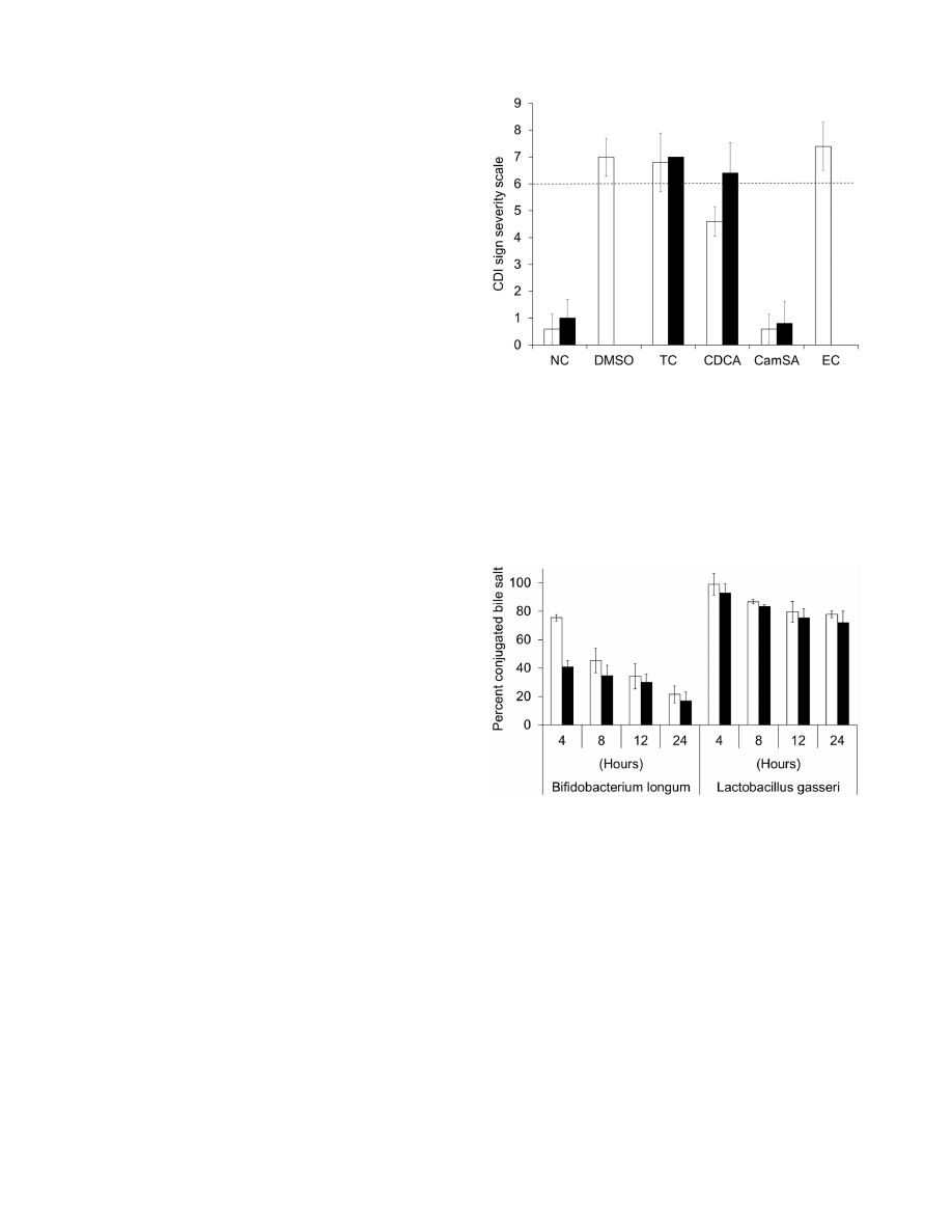
Prevention of CDI by Bile Salt Analogs
As previously reported, when mice were challenged with
10
8
CFU of C. difficile spores, severe CDI signs developed and
all animals reached clinical endpoint by 48 hours post-challenge
[16]. The large (10
8
CFUs) inoculum of spores ensured synchro-
nized CDI onset and fast CDI sign progression. Mice treated with
up to 300 mg/kg taurocholate or ethyl cholate also developed
severe CDI and signs were undistinguishable from control
DMSO-treated animals (Fig. 1 and Fig. S2). Mice treated with
50 mg/kg chenodeoxycholate developed moderate to severe signs
of CDI, but onset was delayed by 24 hours (Fig. S3). In contrast,
all animals treated with 50 or 300 mg/kg CamSA showed no sign
of CDI and were undistinguishable from non-challenged animals
[11]. All asymptomatic animals remained free of CDI signs for at
least 14 days post-challenge.
Stability of CamSA
CamSA is a taurocholate analog with an amide bond linking
cholic acid to meta-aminobenzene sulfonic acid. To be effective,
CamSA must survive the changing environments of the GI tract.
To test for stability, CamSA was incubated in artificial gastric juice
and intestinal juice. No degradation of CamSA was evident even
after 24 hours incubation under both conditions (data not shown).
Bacterial bile salt hydrolases (BSHs) deconjugate primary and
secondary bile salts [17]. B. longum and L. gasseri are two intestinal
bacteria commonly used as test strains for BSH production. After
incubation with a culture of B. longum for 24 hours, CamSA and
taurocholate are both hydrolyzed to cholic acid at similar rates
(Fig. 2). CamSA and taurocholate are less sensitive to degradation
by BSHs secreted by L. gasseri. Less than 30% of either CamSA or
taurocholate was hydrolyzed to cholic acid after 24 hours (Fig. 2).
E. coli does not produce BSH and both CamSA and taurocholate
were stable after 24 hour incubation with E. coli cultures (data not
shown). CamSA was not degraded in growth medium alone.
Caco-2 Permeability of CamSA
To prevent C. difficile spores from germinating, CamSA needs to
be retained in the intestinal lumen. Caco-2 monolayers serves as
an in vitro surrogate assay for intestinal permeability, absorption,
and metabolism [18]. CamSA was studied in a Caco-2 perme-
ability assay and displayed an apical to basolateral apparent
permeability coefficient (P
app
) of ,10
26
cm/s and basolateral to
apical P
app
of 10.9610
26
cm/s. The efflux ratio suggests that
CamSA is a substrate for active transport (Table S1). In both
assays, CamSA was recovered at 100% indicating low binding,
accumulation, and metabolism by Caco-2 cells.
Figure 1. CamSA protects mice from CDI. Comparison of CDI sign
severity after 48 hours (white bars) and 72 hours (black bars) of animals
challenged with C. difficile spores and treated with DMSO, 300 mg/kg
taurocholate (TC), 50 mg/kg chenodeoxycholate (CDCA), 50 mg/kg
CamSA, or 300 mg/kg ethyl cholate (EC). Non-challenged (NC) animals
were used as controls. Clinical endpoint was set as .6 in the CDI sign
severity scale (dashed line). None of the animals in the DMSO and EC
groups survived to 72 hours post-challenged. Standard deviations
represent at least five independent measures.
doi:10.1371/journal.pone.0072620.g001
Figure 2. Stability of CamSA and taurocholate towards bile salt
hydrolases. CamSA (white bar) and taurocholate (black bars) were
incubated with cultures of B. longum or L gasseri. Percent conjugated
bile salts were derived by dividing the intensity of TLC spots obtained at
different times by the intensity of the TLC spot obtained at the
beginning of incubation (time 0). Time 0 was set at 100% and is not
shown for clarity. Standard deviations represent at least five indepen-
dent measures.
doi:10.1371/journal.pone.0072620.g002
Fate of Clostridium difficile Spores
PLOS ONE | www.plosone.org
2
August 2013 | Volume 8 | Issue 8 | e72620
Effect of CamSA on Bacterial Growth
E. coli, B. longum, and L. gasseri are indigenous mammalian gut
bacteria and are continuously exposed to bile salts [17]. As
expected, growth of these bacteria was unaffected by the presence
of CamSA in the growth medium. C. difficile cells also grew
normally in the presence of CamSA (Fig. S4).
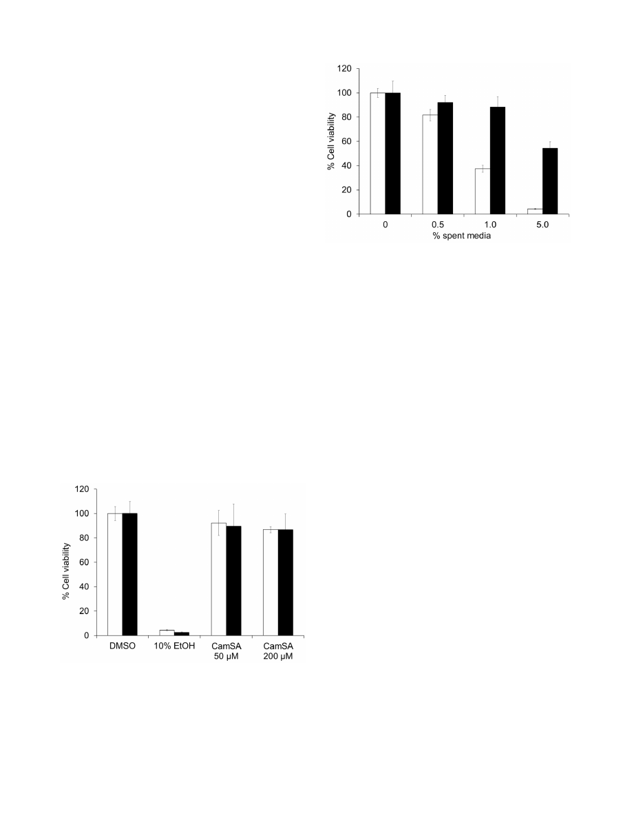
Cytotoxicity of CamSA
Cell viability was qualitatively determined by visual observation
of rounded/detached cells and trypan blue staining. CamSA-
treated Vero, Caco-2, and macrophage cells appeared healthy and
were undistinguishable from DMSO-treated cells (Fig. S5). Cell
viability was also quantitatively determined by ATP production.
Vero and Caco-2 cells treated with 50 or 200 mM CamSA
produced ATP at similar levels to healthy control cells (Fig. 3).
CamSA Protection of Vero and Caco-2 Cells
Spent media from outgrowing C. difficile spores killed Vero cells
in a dose-dependent manner (Fig. 4). These data are consistent
with previous reports indicating that vegetative C. difficile secretes
cell-killing toxins during growth [19]. When C. difficile spores were
incubated in medium containing 200 mM CamSA, bacterial
growth was reduced but not eliminated. As expected, spent media
from CamSA-treated bacterial cultures were less effective at killing
epithelial cells. Similar results were observed for Caco-2 cell
cultures (data not shown).
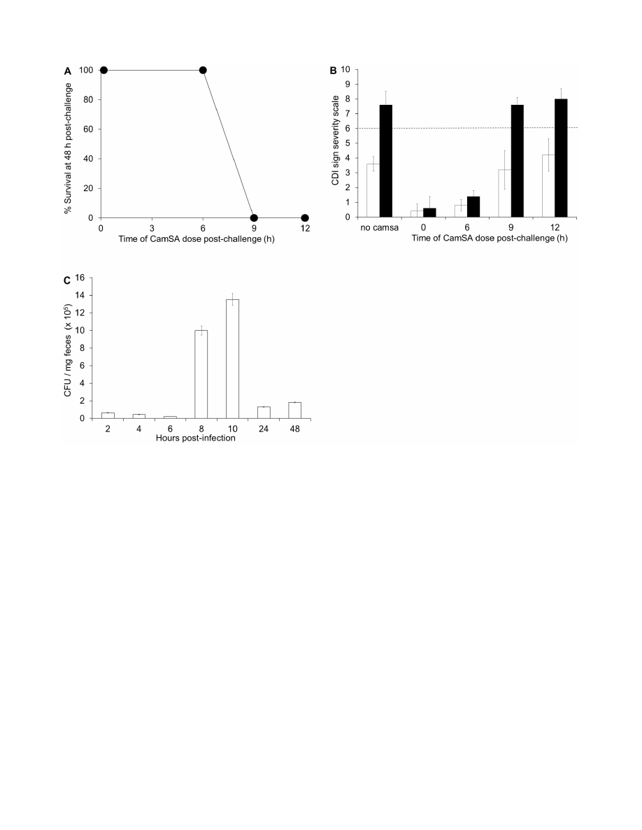
Timing of CDI Onset
To determine the onset of CDI in mice, animals were
challenged with C. difficile spores and treated with 300 mg/kg
CamSA between 0 and 12 hours post-challenge. All animals
treated with CamSA up to 6 hours post-challenge were fully
protected from CDI. In contrast, all animals treated with CamSA
at 9 or 12 hours post-challenge developed severe CDI undistin-
guishable from untreated mice and reached the clinical endpoint
48 hours post infection (Figs. 5A and 5B).
Similar to previous reports, GI contents from animals with CDI
signs contained almost exclusively C. difficile vegetative cells [13].
These animals started to excrete large amounts (.10610
5
CFUs)
of vegetative cells reaching a maximum between 8 and 10 hours
post spore challenge (Fig. 5C). Although some C. difficile spores
were excreted by diseased animals, the amounts were negligible
(,10% of vegetative CFUs) compared to the high amount of
excreted vegetative cells.
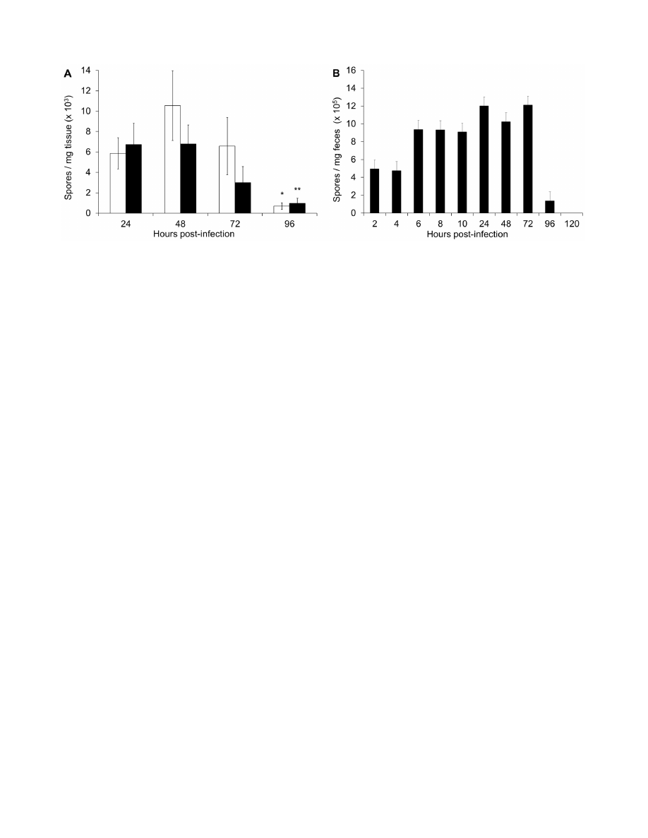
Recovery of C. difficile Cells and Spores from Intestines
and Feces of CamSA-treated Mice
Similar to the hamster CDI model [13], ingested C. difficile
spores narrowly localized to the cecum and colon of CamSA
treated mice at every time point tested. A negligible amount of C.
difficile was discovered in the small intestine and stomach (Fig. S6).
C. difficile spores remained in the cecum and colon for 72 hours
after spore challenge (Fig. 6A). By 96 hours, the amount of spores
recovered from the cecum and colon of CamSA treated animals
decreased almost tenfold, from greater than 12610
5
to less than
2610
5
CFUs.
Consistent with the results from intestinal content, the feces of
CamSA-treated animals contained almost exclusively spores
(Fig. 6B). In these animals, excretion of ingested C. difficile spores
started 2 hours post-challenge and continued until at least 96
hours post-challenge. In fact, by 120 hours post-challenge, the sum
of excreted C. difficile spores was quantitatively identical to the
number of spores given by gavage.
Discussion
Germination of C. difficile spores is believed to be the first step in
establishing CDI [1]. However, determining the fate of ingested C.
difficile spores is challenging since spores can germinate and the
resulting cells can then re-sporulate perpetuating a cycle of disease.
Anti-germinants prevent CDI by effectively freezing ingested
spores in their dormant state. We took advantage of this anti-
Figure 3. Cytotoxicity of CamSA. Vero cells (white bars) or Caco-2
cells (black bars) were incubated overnight with 10% DMSO, 10% EtOH,
50
mM CamSA or 200 mM CamSA. Cell viability was determined with the
CellTiter Glo viability kit. The luminescence signal from DMSO-treated
cells was undistinguishable from untreated cells and was set as 100%
cell viability. Percent survival for other conditions was calculated
relative to untreated cells. Error bars represent standard deviations from
at least five independent measurements.
doi:10.1371/journal.pone.0072620.g003
Figure 4. Inhibition of
C. difficile
toxin production by CamSA
treatment. C. difficile spores were incubated overnight in media
containing 0
mM CamSA (white bars) or 200 mM CamSA (black bars).
The resulting spent media were added to Vero cell cultures and
incubated for 24 hours. Cell viability was determined with the CellTiter
Glo viability kit. The luminescence signal from untreated cells was set as
100% cell viability. Percent survival for other conditions was calculated
relative to untreated cells. Error bars represent standard deviations from
at least five independent measurements.
doi:10.1371/journal.pone.0072620.g004
Fate of Clostridium difficile Spores
PLOS ONE | www.plosone.org
3
August 2013 | Volume 8 | Issue 8 | e72620
germinant property to study the temporal and spatial distribution
of ingested C. difficile spores.
Following our success using the anti-germination properties of
CamSA to protect mice from CDI, we tested taurocholate (a
natural germination enhancer), chenodeoxycholate (a natural
germination inhibitor), and ethylcholate (a commercially available
germination inhibitor) as prophylactics of CDI. Ethyl cholate
inhibits C. difficile spore germination with a half maximal inhibitory
concentration (IC
50
) of 8.2 mM, seven-fold more potent than
CamSA in vitro (data not shown). Chenodeoxycholate, on the other
hand, inhibits C. difficile spore germination with an IC
50
of
235 mM, approximately five-fold less potent than CamSA [20].
As previously reported, CamSA was a prophylactic of murine
CDI [11]. In this study, we were able to prevent CDI with
saturating concentrations of CamSA without any overt toxic
effects. This is consistent with the low toxicity observed for
taurocholate and cholate [21]. In fact, CamSA had anti-CDI
activity at concentrations that were at least six times lower than its
toxic threshold. Furthermore, CamSA-treated animals did not
show signs of CDI even 14 days post-challenge. At 300 mg/kg
concentration, chenodeoxycholate showed acute toxicity probably
due to low solubility in biological fluids. At lower concentrations,
chenodeoxycholate was not able to prevent CDI. Animals treated
with 50 mg/kg chenodeoxycholate show the same CDI sign
patterns as animals treated with 5 mg/kg CamSA [11]. Another
potent inhibitor of C. difficile spore germination in vitro, ethylcho-
late, did not protect mice from CDI even at 300 mg/kg. Similarly,
taurocholate, a germinant, was unable prevent CDI. CamSA was
the only compound tested that was effective in preventing CDI.
Hence, we further characterized CamSA’s in vitro pharmacological
properties.
To be effective as in vivo probes, anti-germination compounds
must be stable to the variable GI tract environments. CamSA is
stable to all tested GI tract microenvironments except incubation
with the BSH-producing bifidobacteria. Antibiotics disrupt the
normal microflora dynamics of the gut allowing for outgrowth of
C. difficile [22]. A recent study shows that antibiotic cocktails shift
the normal murine gut bacterial population to a preponderance of
Lactobacilli [23]. Since BSHs produced by Lactobacilli were not
Figure 5. CDI is established between 6 and 9 hours post-infection. (A) Survival of infected mice at 48 hours after challenge with C. difficile
spores. Mice were treated with 300 mg/kg CamSA at 0, 6, 9, or 12 hours post-challenge. (B) Comparison of CDI severity after 24 hours (white bars) and
48 hours (black bars) for animals challenged with C. difficile spores and treated with 300 mg/kg CamSA at 0, 6, 9, or 12 hours post-challenge. Clinical
endpoint was set as .6 in the CDI sign severity scale (dashed line). (C) C. difficile vegetative cell count in feces of untreated, diseased animals. Feces
were collected from cages housing five untreated mice challenged with C. difficile spores. Open bars represent C. difficile vegetative cells. The amount
of C. difficile spores excreted by untreated animals was negligible (,10% of vegetative cell counts). Standard deviations represent at least five
independent measures. Recovered CFU and recovered spores represent mean values from pools of five animals.
doi:10.1371/journal.pone.0072620.g005
Fate of Clostridium difficile Spores
PLOS ONE | www.plosone.org
4
August 2013 | Volume 8 | Issue 8 | e72620
effective in hydrolyzing CamSA, CamSA should remain stable in
the gut of antibiotic-treated mammals.
Since CDI is an intestinal infection, anti-germination com-
pounds require low oral bioavailability and high GI tract stability
for maximum efficacy. In vitro assays suggest that CamSA will be
retained in the intestinal tract of mammals (Table S1). The efflux
ratio of CamSA suggests that it is a substrate for Pgp [24]. Hence,
the efficacy of CamSA might be in part due to retention in the GI
tract due to Pgp mediated excretion of the drug back into the
intestinal lumen. The poor bioavailability of CamSA will also
reduce toxic effects to other organs since CamSA is unlikely to
circulate outside of the intestinal lumen. Tox-ADME analyses also
suggest low metabolism of CamSA by intestinal epithelial cells and
further supports that CamSA will remain stable in the intestinal
lumen.
Because the natural microbiota is key to resist C. difficile
infection, anti-germination compounds should not damage this
natural barrier [22]. Indeed, CamSA did not affect growth of
commensal bacteria, B. longum, L. gasseri, or E. coli. Although
CamSA inhibits spore germination in vitro, it does not affect C.
difficile vegetative growth (Fig. S4). These data support the
proposed mechanism that CamSA inhibits spore germination
and does not act as an antibacterial agent. Anti-germination
compounds should also show low toxicity toward mammalian
cells. Indeed, CamSA did not affect the viability of two different
mammalian epithelial cell lines nor did it affect macrophage
immune cells.
Since toxin is only secreted by metabolically active C. difficile
cells, halting germination should result in less toxin production.
Tissue culture protection experiments rely on decreasing the
accumulation of toxins secreted into media during C. difficile
vegetative growth [19]. CamSA inhibits C. difficile spore germina-
tion in vitro and hence protects mammalian cells by reducing the
number of toxin-producing bacteria.
Previous works have not been able to distinguish between
ingested C. difficile spores and spores that were produced after
colonization of the host’s GI tract. Indeed, enumeration of C.
difficile in feces and GI content from infected hamsters yields
mixtures of vegetative cells and spores [13,14]. Furthermore,
enumerated spore loads were higher than the original inoculum.
These data suggest that ingested C. difficile spores germinated and
re-sporulated during colonization of the hamster gut. Due to the
fast progression of CDI in untreated mice, we could only
determine bacterial loads for the first 48 hours after challenge.
Even then, we could not distinguish whether spores recovered
from diseased animals came from the original inoculum or from
re-sporulation in the intestines.
Since CamSA shows favorable in vitro pharmacological proper-
ties and can block C. difficile spore germination in vivo, we were able
to follow the fate of ingested C. difficile spores without interference
from germination and/or re-sporulation. CamSA was effective in
preventing CDI when administered up to six hours following spore
challenge. In contrast, CamSA was ineffective when administered
nine hours post-challenge, even at the highest concentration
tested. This narrow three-hour window correlates C. difficile spore
germination with maximum C. difficile vegetative cell shedding in
symptomatic mice. These data suggest that a fraction of
germinated C. difficile cells are excreted soon after germination,
while the remaining C. difficile vegetative cells lead to CDI onset.
Human CDI shows more heterogeneous symptoms and longer
disease progression than rodent models [12]. CamSA treatment
allowed us to observe the behavior of ungerminated spores for a
period extended beyond the normal clinical endpoint of CDI-
diseased mice. Ingested C. difficile spores were quantitatively
recovered from feces, cecum, and colon contents of CamSA-
treated mice. Interestingly, ingested C. difficile spores started to be
shed soon after challenge, but part of the population remained in
the lower GI for up to four days.
The mechanism of dormant C. difficile spore accumulation in the
lower intestine is not understood, but suggests that ingested C.
difficile spores can form a transitory reservoir that is slowly released
from the lower intestine. A possibility is that a small fraction of C.
difficile spores enter a superdormant state that helps them to be
retained in the intestine [25]. Although the amount of unattached
spores in the intestines is small, it is tempting to speculate that
these spore reservoirs serve as a focal point for CDI relapse.
CamSA’s anti-germination activity can be used to address
mechanistic details about CDI initiation (Fig. 7). The sum of our
Figure 6.
C. difficile
spores accumulate in the cecum, colon, and feces of CamSA-treated animals. (A) Amount of C. difficile spores
recovered at different time points following spore challenge from the cecum (white bars) and colon (black bars) of mice treated with 50 mg/kg
CamSA. Student’s unpaired t-test was used to determine the significance of difference of means. *indicates recovered spores significantly below 72
hour levels (P = 0.019; Student’s t-test). **indicates recovered spores significantly below 72 hour levels (P = 0.049; Student’s t-test). (B) Feces were
collected from cages housing five mice challenged with C. difficile spores and treated with 50 mg/kg CamSA. Closed bars represent C. difficile spores.
The amount of C. difficile vegetative cells in CamSA-treated animals was negligible (,10% compared to spore counts). Standard deviations represent
at least five independent measures. Recovered CFU and recovered spores represent mean values from a pool of five animals.
doi:10.1371/journal.pone.0072620.g006
Fate of Clostridium difficile Spores
PLOS ONE | www.plosone.org
5
August 2013 | Volume 8 | Issue 8 | e72620
data suggests that ingested spores rapidly transit through the GI
tract (Fig. 6B) and accumulate in the lower intestine (Fig. 6A and
Fig. S6). Six to nine hours after ingestion enough C. difficile spores
germinate to establish infection (Fig. 5A). C. difficile vegetative cells
start shedding almost immediately after germination and continue
throughout the infection (Fig. 5C). In contrast, dormant C. difficile
spores are slowly shed over a four day period (Fig. 6B). The timing
of C. difficile spore germination and the persistence of ungermi-
nated spores in the lower intestine can have profound implication
in the prophylactic treatment of CDI.
Materials and Methods
Materials
Bile salts were purchased from Sigma-Aldrich Corporation (St.
Louis, MO) or were synthesized in the Abel-Santos laboratory [8].
All bile salts were dissolved in DMSO prior to use. Artificial gastric
juice, intestinal juice and PRO disks were purchased from Fisher
Scientific (Pittsburg, PA). Thin layer chromatography (TLC) silica
gel 60 F
254
plates were purchased from EMD Chemicals
(Gibbstown, NJ). Cell Titer-Glo luminescent cell viability assay
kit was obtained from Promega Corporation (Madison, WI).
Clostridium difficile selective agar plates were purchased from BD
Biosciences (Franklin, Lakes, NJ). All bacterial strains used in this
study were purchased from ATCC (Manassas, VA) and grown as
suggested.
Animals
This study was performed in strict accordance with the
recommendations in the Guide for the Care and Use of
Laboratory Animals of the National Institutes of Health. The
protocol was reviewed and approved by the Institutional Animal
Care and Use Committee at the University of Nevada, Las Vegas
(Permit Number: R0411-266). Weaned Female C57BL/6N mice
were purchased from Harlan laboratories (Indianapolis, IN).
Animals were housed in groups of 5 mice per cage in the
University of Nevada, Las Vegas animal care facility. All cages,
water, food and bedding were autoclaved prior to contact with
animals. Upon arrival, mice were allowed to acclimate for one
week prior to experimentation. Post-challenge animal manipula-
tions were performed in a biosafety level 2 laminar flow hood.
Acute Toxicity of CamSA in Mice
To determine acute toxicity of CamSA, we used the Fixed Dose
Procedure (FDP) [15]. CamSA was dissolved in DMSO to a
concentration of 100 mg/ml. Groups of five mice were treated by
oral gavage for three consecutive days with 50 mg/kg body weight
of CamSA or chenodeoxycholate. A control group was adminis-
tered DMSO. Weight changes were recorded daily and mice were
observed for adverse reactions such as vomiting, diarrhea, hair
loss, hunched posture, weight loss, and lethargy. Other groups of
mice were treated with 300 mg/kg CamSA or chenodeoxycholate
and observed as above.
Prevention of CDI by Bile Salt Analogs
C. difficile 630 (ATCC BAA-1382) were prepared as previously
described [11]. Purified C. difficile spores of 630 strain were used to
challenge mice as published [16,26]. Briefly, an antibiotic cocktail
containing kanamycin (0.4 mg/ml), gentamycin (0.035 mg/ml),
colistin (850 U/ml), metronidazole (0.215 mg/ml), and vancomy-
cin (0.045 mg/ml) was prepared in autoclaved water and sterile
filtered. For three consecutive days, mice were allowed to drink the
antibiotic cocktail ad libitum. The antibiotic water was refreshed
daily. After three days of antibiotic water, all mice received
autoclaved water for the remainder of the experiment. A single
dose of clindamycin (10 mg/kg) was administered by intraperito-
neal (IP) injection on the fourth day (24 hours before C. difficile
challenge). At this time, groups of five antibiotic-treated mice
received neat DMSO, 300 mg/kg CamSA, 50 mg/kg chenodeox-
ycholate, 300 mg/kg taurocholate, or 300 mg/kg ethylcholate
oral gavage. The day of challenge, animals received 10
8
CFUs of
C. difficile spores by oral gavage. One hour post challenge, animals
received a second dose of the corresponding bile salt or DMSO. A
third dose of bile salt or DMSO was administered 24 hours post-
challenge. All animals were observed twice daily for signs of CDI.
Disease signs were scored using the following rubric: pink
anogenital area (score of 1), red anogenital area (score of 2),
lethargy (score of 1), diarrhea/increase in soiled bedding (score of
1), wet tail (score of 2), hunchback posture (score of 2), 8-15% loss
of body weight (score of 1), .15% loss of body weight (score of 2).
Animals scoring 2 or less were undistinguishable from non-infected
controls and were considered non-diseased. Animals scoring 3–4
were considered to have mild CDI with signs consisting of pink
anogenital area, lethargy, an increase of soiled bedding and minor
weight loss. Animals scoring 5–6 were considered to have
moderate CDI with signs consisting of mild CDI signs plus red
anogenital area and hunchback posture. Animals scoring .6 were
considered to have severe CDI and were immediately euthanized.
These animals displayed signs described above plus wet tail and
severe weight loss. Asymptomatic animals were monitored for up
to 14 days post-challenge to monitor CDI onset delay.
Stability of CamSA in Artificial Gastric and Intestinal
Juices
CamSA was analyzed for stability in simulated gastric and
intestinal juices as published [27]. Briefly, 100 mg CamSA was
added to 1 ml of either artificial intestinal or artificial gastric juice
and incubated at 37
uC. Aliquots were taken at 4, 8, 12, and 24
hours. Samples (1 ml) were spotted on silica TLC plates and
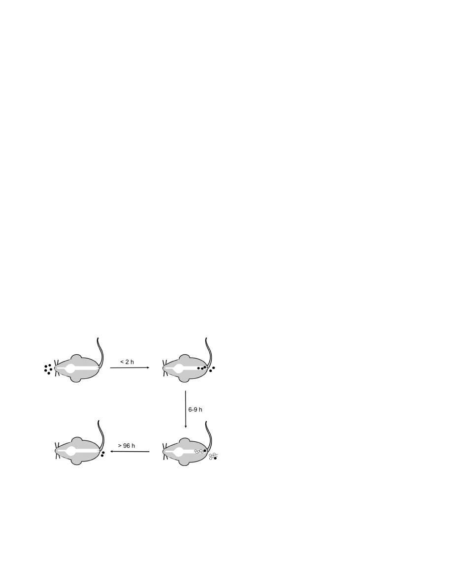
Figure 7. Time line model for CDI onset in mice. C. difficile spores
(black circles) are ingested by the host. Spores rapidly transit through
the upper GI tract and colonize the colon and cecum. Spore shedding
begins less than 2 hours post-ingestion. Between 6 and 9 hours after
ingestion sufficient numbers of spores germinate to establish infection.
The outgrowing C. difficile cells (white circles) proliferate in the lower
intestine, are shed, and can re-sporulate. A small amount of ingested
spores remain in the lower intestine for more than 96 hours post
ingestion.
doi:10.1371/journal.pone.0072620.g007
Fate of Clostridium difficile Spores
PLOS ONE | www.plosone.org
6
August 2013 | Volume 8 | Issue 8 | e72620

allowed to air dry. Plates were developed with 75% ethyl acetate/
methanol. TLC plates were visualized by spraying with a 10% wt/
vol phosphomolybdic acid (PMA)/ethanol solution followed by
heating at 100
uC for 2 minutes. Quantification of CamSA was
determined using a GE Healthcare Typhoon 9410 Variable Mode
Imager and analyzed using ImageQuant TL 5.2 software.
Stability of CamSA after Incubation with Bile Salt
Hydrolase (BSH) Producing Bacteria
Following previous procedures [28], Escherichia coli DH5a,
Bifidobacterium longum (ATCC BAA-999) and Lactobacillus gasseri
(ATCC 33323) were incubated for 24 hours at 37
uC. Bacterial
cultures were then adjusted to an optical density of 1.0 with fresh
media supplemented with 6 mM CamSA or taurocholate and
incubated at 37
uC. Samples of spent media were taken at 4, 8, 12,
and 24 hours. Bile salt concentration was monitored by TLC as
above. Percent conjugated bile salts were derived by dividing the
intensity of TLC spots obtained at different times by the intensity
of the TLC spot obtained before incubation.
In vitro Permeability Assays
Caco-2 permeability assays of CamSA were performed by
Apredica, LLC (Watertown, MA). Briefly, CamSA was dissolved
in DMSO and added to Caco-2 cell cultures to 10 mM final
concentration. CamSA was analyzed for both apical to basolateral
permeability and basolateral to apical permeability across a Caco-
2 cell monolayer. After a 2 hour incubation, CamSA concentra-
tions in the apical and basolateral sides of the Caco-2 monolayers
were determined by HPLC-MS. An in vitro ADME-Tox test was
also conducted to estimate the percent recovery of CamSA from
either the apical to basolateral permeability or basolateral to apical
permeability.
Effect of CamSA on Bacterial Growth
Laboratory strains of E. coli DH5a, B. longum, L. gasseri, and C.
difficile were individually inoculated from freezer stock onto
appropriate agar medium as directed by ATCC. Plates were
incubated overnight at 37
uC either aerobically (L. gasseri and E.
coli) or anaerobically (C. difficile and B. longum). Anaerobic
conditions consisted of a 5% CO
2
, 10% H
2
, and 85% N
2
environment. Single cell clones were carefully selected and used to
inoculate 5 mL of liquid medium. Inoculated broth was shaken at
37
uC for approximately four hours until optical density at 580 nm
reached 0.8 representing exponential phase of growth. Bacteria
were sub-cultured (1:100) into fresh media and individually
supplemented with 10 mM CamSA or taurocholate. An increase
of optical density at 580 nm (OD
580
) was used to measure
exponential bacterial growth. The OD
580
was recorded at 0, 1, 2,
3, 4, 6, and 8 hours post subculture inoculation. Growth inhibition
was determined by comparing optical density of bile salt-treated
cultures with untreated control cultures.
Cytotoxicity of CamSA
Vero cells, Caco-2 cells, and murine macrophages J774A.1 were
seeded in complete medium (minimum essential medium (MEM),
10% fetal bovine serum (FBS), and 1% penicillin/streptomycin).
Cells were grown at 37
uC with 5% CO
2
. Cells were detached by
incubation with 1 mM EDTA-trypsin for 5 minutes. Complete
medium was then added and monolayers lifted with a cell scraper.
Cells were recovered at 8006g for 5 minutes at room temperature
and the cell pellet was resuspended in fresh complete medium. A
sample of cell suspension was treated with trypan blue to
determine background non-viable cells [29]. Culture cells were
plated in 12-well or 96-well tissue culture plates at a density of 10
5
cells/ml and allowed to attach overnight. Spent medium was
removed and fresh complete medium supplemented with 50 or
200 mM CamSA was added. As negative control, cells were
treated with complete media supplemented with DMSO. As
positive control, cells were treated with complete media supple-
mented with 10% EtOH. Plates were incubated overnight as
described above.
The 12-well plates containing cell cultures were used as a
qualitative method to determine cytotoxicity by visual observation
of morphological changes, such as cell rounding. Cell viability was
also determined by trypan blue dye exclusion staining. The 96-well
plates cultured with mammalian cells were used as a quantitative
method to determine cytotoxicity using the CellTiter Glo
Luminescent cell viability assay. This assay quantitates the
concentration of ATP, which indicates metabolically active cells
[30]. After overnight CamSA-treatment, the 96-well plates were
equilibrated to room temperature for 30 minutes before addition
of the CellTiter-Glo reagent. Luminescence was read with an
integration time of 1 second per well using a Tecan Infinite 200
plate reader and iControl software. The luminescence signals from
cell cultures supplemented with DMSO only were set as 100% cell
viability. Percent survival for other conditions was calculated
relative to these untreated cells.
C. difficile Toxin-induced Cell Death
C. difficile strain 630 spores were washed five times with
nanopure water, heat activated at 68
uC for 30 minutes and
washed five more times with water. Spore pellets were resus-
pended in 0.1 M sodium phosphate buffer supplemented with
0.5% sodium bicarbonate (pH 6.0) to an OD
580
of 1.0. Spores
were diluted five-fold in BHI broth supplemented with 6 mM
taurocholate/12 mM glycine (germination medium), germination
medium supplemented with 50 mM CamSA, or germination
medium supplemented with 200 mM CamSA. Spore suspensions
were incubated anaerobically at 37
uC overnight. The following
day, C. difficile cells and spores were removed by centrifugation and
spent media filter sterilized. In parallel, Vero and Caco-2 cells
were cultured in 96-well plates, as described above. The
mammalian cell cultures were then exposed to varying concen-
trations of sterile spent media for 24 hours. Cell viability was
determined as before with the CellTiter Glo viability kit. The
luminescence signals from cell cultures supplemented with 0%
spent media were set as 100% cell viability. Percent survival for
other conditions was calculated relative to these untreated cells.
Onset of CDI Signs in Mice
Antibiotic treated mice were challenged by oral gavage with
10
8
CFUs of C. difficile strain 630 spores. Individual groups of five
mice were treated with a single 300 mg/kg dose of CamSA at 0, 6,
9 or 12 hours post-spore challenge. A second 300 mg/kg dose of
CamSA was administered 24 hours after the first dose. Mice were
observed for signs of CDI twice daily and scored accordingly.
Enumeration of C. difficile Vegetative Cells and Spores
Spore challenged animals were treated with 0 or 300 mg/kg
CamSA. Cages from infected animals were changed and feces
were collected at different time points. Selected animals were
sacrificed at different time points and their GI tract observed for
signs of disease. Gastrointestinal tracts were removed in blocks and
GI tract contents were flushed with autoclaved water. Feces and
intestinal contents were weighed and homogenized in autoclaved
water. Aliquots of the fecal suspensions were heated to 68
uC for 30
minutes. Heated and unheated feces and GI tract contents were
Fate of Clostridium difficile Spores
PLOS ONE | www.plosone.org
7
August 2013 | Volume 8 | Issue 8 | e72620

serially diluted in water and plated on Clostridium difficile selective
agar (CDSA). Plates were incubated anaerobically for 48 hours
and colonies were counted to enumerate colony-forming units
(CFUs). CFUs obtained from unheated samples represent the sum
of C. difficile vegetative cells and spores. CFUs obtained from
heated samples represent the number of C. difficile spores only. The
presence of C. difficile was verified by PRO disk.
Statistical Analysis
Standard deviations represent at least three independent
measures, unless otherwise stated. Recovered CFU and recovered
spores represent mean values from a pool of five animals. Student’s
unpaired t-test was used to determine the significance of difference
of means.
Supporting Information
Figure S1
Mice treated with CamSA or chenodeoxycholate
shows no weight changes. Groups of five mice were treated with
DMSO (%), 50 mg/kg chenodeoxycholate (e), 50 mg/kg
CamSA (D), or 300 mg/kg CamSA (#). Animal weight was
obtained daily.
(TIFF)
Figure S2
Protection of mice from CDI by different bile salts.
Kaplan-Meier survival plot for C. difficile infected mice treated with
DMSO (e), 300 mg/kg taurocholate (D), 50 mg/kg chenodeox-
ycholate (#), 50 mg/kg CamSA (¤), or 300 mg/kg ethylcholate
(6). Non-infected animals were used as control (%).
(TIFF)
Figure S3
Figure 3. Signs severity for C. difficile infected animals
treated with different bile salts. Non-infected animals were used as
control (panel A). Animals challenged with C. difficile spores were
treated with three doses of DMSO (panel B), taurocholate (panel
C), chenodeoxycholate (panel D), CamSA (panel E), or ethylcho-
late (panel F). The severity of CDI signs was scored using the
Rubicon scale discussed above.
(TIF)
Figure S4
CamSA does not affect vegetative bacterial growth. E.
coli DH5a (%), B. longum (#), L. gasseri (D), and C. difficile (e) were
incubated in media supplemented with 0 or 10 mM CamSA. The
OD
580
was recorded at 0, 1, 2, 3, 4, 6, and 8 hours. Growth
inhibition was determined by subtracting optical density of
CamSA-treated cultures from untreated control cultures.
(TIFF)
Figure S5
CamSA is not toxic to mammalian cells. Murine
macrophages J774A.1 were treated with DMSO (panel A), 10%
ethanol (panel B), or 200 mM CamSA (panel C). Cell viability was
determined by trypan blue dye exclusion staining
(TIFF)
Figure S6
Distribution of C. difficile spores in the GI tract of
CamSA-treated animals. The stomach (St), duodenum (Du),
jejunum (Je), and ileum (Il) showed negligible amounts of spores
compared to the cecum (Ce) and colon (Co).
(TIFF)
Table S1
CamSA permeability across Caco-2 cell monolayer
a
.
(DOCX)
Author Contributions
Conceived and designed the experiments: EAS AH. Performed the
experiments: AH MP. Analyzed the data: EAS AH MP. Contributed
reagents/materials/analysis tools: MP AH. Wrote the paper: EAS AH MP.
References
1. Cohen S, Gerding D, Johnson S, Kelly CP, Loo V, et al. (2010) Clinical practice
guidelines for Clostridium difficile infection in adults: 2010 update by the Society
for Healthcare Epidemiology of America (SHEA) and the Infectious Diseases
Society of America (IDSA). Infection Control and Hospital Epidemiology 31:
431–455.
2. Kachrimanidou M, Malisiovas N (2012) Clostridium difficile infection: A
comprehensive review. Critical Reviews in Microbiology 37: 178–187.
3. Speight S, Moy A, Macken S, Chitnis R, Hoffman PN, et al. (2011) Evaluation
of the sporicidal activity of different chemical disinfectants used in hospitals
against Clostridium difficile. Journal of Hospital Infection 79: 18–22.
4. Rolfe RD, Helebian S, Finegold SM (1981) Bacterial interference between
Clostridium difficile and normal fecal flora. Journal of Infectious Diseases 143: 470–
475.
5. Setlow P (2003) Spore germination. Current Opinions in Microbiology 6: 550–
556.
6. Sorg JA, Sonenshein AL (2008) Bile salts and glycine as cogerminants for
Clostridium difficile spores. Journal of Bacteriology 190: 2505–2512.
7. Ramirez N, Liggins M, Abel-Santos E (2010) Kinetic evidence for the presence
of putative germination receptors in C. difficile spores. Journal of Bacteriology
192: 4215–4222.
8. Howerton A, Ramirez N, Abel-Santos E (2011) Mapping interactions between
germinants and Clostridium difficile spores. Journal of Bacteriology 193: 274–282.
9. Sorg JA, Sonenshein AL (2009) Chenodeoxycholate is an inhibitor of Clostridium
difficile spore germination. Journal of Bacteriology 191: 1115–1117.
10. Sorg JA, Sonenshein AL (2010) Inhibiting the initiation of Clostridium difficile
spore germination using analogs of chenodeoxycholic acid, a bile acid. Journal of
Bacteriology 192: 4983–4990.
11. Howerton A, Patra M, Abel-Santos E (2013) A new strategy for the prevention
of Clostridium difficile infections. Journal of Infectious Diseases 207: 1498–1504.
12. Best EL, Freeman J, Wilcox MH (2012 ) Models for the study of Clostridium
difficile infection. Gut Microbes 3: 145–167.
13. Buckley AM, Spencer J, Candlish D, Irvine JJ, Douce GR (2011) Infection of
hamsters with the UK Clostridium difficile ribotype 027 outbreak strain
R20291. Journal of Medical Microbiology 60: 1174–1180.
14. Freeman J, Baines SD, Jabes D, Wilcox MH (2005) Comparison of the efficacy
of ramoplanin and vancomycin in both in vitro and in vivo models of
clindamycin-induced Clostridium difficile infection. Journal of Antimicrobial
Chemotherapy 56: 717–725.
15. Walum E (1998 ) Acute oral toxicity. Environmental Health Perspectives 106:
497–503.
16. Chen X, Katchar K, Goldsmith JD, Nanthakumar N, Cheknis A, et al. (2008) A
Mouse Model of Clostridium difficile-Associated Disease. Gastroenterology 135:
1984–1992.
17. Begley M, Gahan CG, Hill C (2005 ) The interaction between bacteria and bile.
FEMS Microbiological Reviews 29: 625–651.
18. Teksin Z, Seo P, Polli J (2010) Comparison of drug permeabilities and BCS
classification: Three lipid-component PAMPA system method versus Caco-2
monolayers. The AAPS Journal 12: 238–241.
19. Sutton PA, Li S, Webb J, Solomon K, Brazier J, et al. (2008) Essential role of
toxin A in C. difficile 027 and reference strain supernatant-mediated disruption of
Caco-2 intestinal epithelial barrier function. Clinical and Experimental
Immunology 153: 439–447.
20. Liggins M, Ramirez N, Magnuson N, Abel-Santos E (2011) Progesterone
analogs influence germination of Clostridium sordellii and Clostridium difficile spores
in vitro. Journal of Bacteriology 193: 2776–2783.
21. Klaassen CD (1973) Comparison of the toxicity of chemicals in newborn rats to
bile duct-ligated and sham-operated rats and mice. Toxicology and Applied
Pharmacology 24: 37–44.
22. Vollaard EJ, Clasener HA (1994) Colonization resistance. Antimicrobial Agents
and Chemotherapy 38: 409–414.
23. Reeves AE, Theriot CM, Bergin IL, Huffnagle GB, Schloss PD, et al. (2011) The
interplay between microbiome dynamics and pathogen dynamics in a murine
model of Clostridium difficile Infection. Gut Microbes 2: 145–158.
24. Takano M, Hasegawa R, Fukuda T, Yumoto R, Nagai J, et al. (1998)
Interaction with P-glycoprotein and transport of erythromycin, midazolam and
ketoconazole in Caco-2 cells. European Journal of Pharmacology 358: 289–294.
25. Setlow P, Liu J, Faeder JR (2012) Heterogeneity in Bacterial Spore Populations.
In: Abel-Santos E, editor. Bacterial Spores: Current Research and Applications.
Norfolk: Caister Academic Press. 199–214.
26. Sun X, Wang H, Zhang Y, Chen K, Davis B, et al. (2011) Mouse Relapse Model
of Clostridium difficile Infection. Infection and Immunity 79: 2856–2864.
27. Yan R, Ko NL, Li S-L, Tam YK, Lin G (2008) Pharmacokinetics and
Metabolism of Ligustilide, a Major Bioactive Component in Rhizoma
Chuanxiong, in the Rat. Drug Metabolism and Disposition 36: 400–408.
28. Tanaka H, Doesburg K, Iwasaki T, Mierau I (1999) Screening of lactic acid
bacteria for bile salt hydrolase activity. Journal of Dairy Science 82: 2530–2535.
Fate of Clostridium difficile Spores
PLOS ONE | www.plosone.org
8
August 2013 | Volume 8 | Issue 8 | e72620

29. Strober W (2001) Trypan Blue Exclusion Test of Cell Viability. Current
Protocols in Immunology: John Wiley & Sons, Inc. pp. Appendix 3B.
30. Crouch SP, Kozlowski R, Slater KJ, Fletcher J (1993 ) The use of ATP
bioluminescence as a measure of cell proliferation and cytotoxicity. Journal of
Immunological Methods 160: 81–88.
Fate of Clostridium difficile Spores
PLOS ONE | www.plosone.org
9
August 2013 | Volume 8 | Issue 8 | e72620
Wyszukiwarka
Podobne podstrony:
Nature of bacterial colonization influences transcription of mucin genes in mice during the first we
The Fate of Psychiatrie Patients in Belarus During the German Occupation
1999 The past and the future fate of the universe and the formation of structure in it Rix
The Wannsee Conference, the Fate of German Jews, and Hitler s Decision in Principle to Exterminate A
Effect of Cistus laurifolius L leaf extracts and flavonoids on acetaminophen induced hepatotoxicity
The Fate of Psychiatrie Patients During the Nazi Period in Styria Austria; Part I, German Speaking
The Fate of Psychiatrie Patients During the Nazi Period in Styria Austria; Part 2, The Yugoslav Reg
Clostridium difficile, Ratownicto Medyczne, MIKROBIOLOGIA
16 197 208 Material Behaviour of Powder Metall Tool Steels in Tensile
The History of the USA 6 Importand Document in the Hisory of the USA (unit 8)
Existence of the detonation cellular structure in two phase hybrid mixtures
Clostridium difficile, Mikrobiologia
fitopatologia, Microarrays are one of the new emerging methods in plant virology currently being dev
Nukariya; Religion Of The Samurai Study Of Zen Philosophy And Discipline In China And Japan
79 1111 1124 The Performance of Spray Formed Tool Steels in Comparison to Conventional
Clostridium difficile Polish FINAL
Mossbauer study of the retained austenitic phase in
Lord of the Flies Character Changes in the Story
Taming of the Shrew, The Theme?velopment in the Play
więcej podobnych podstron