netter75

Lungs
lawaiHŁUMMiasraMPrai
Medial Surface of Kight Lung
Medial Surface of Left Lung
Croove for subdavian artery.
Area for trachea Apex and esophagus
.Groove for ' subclavian artery
Groovc for brachiocephalic (innominate) vein v
Area for esophagus
Groove for superior vena i
,R. upper lobe bronchus ✓ Upper lobe
Groove for arch of aorta >
Upper lobe.
Groove for brachiocephalic (innominate) vei
Area for Ihymus and mediastinal fatty tissue -
■ Obliguc fissure ■ ^Lower lobe—
-Area for
thymus and
mediastinal
■ Cul edge of pleura-
tatty tissue
■Pulmonary arteries—— •Bronchial arteries —‘
I lori/ontal fissure--
•Bronchi
•Superior
pulmonary
L Cardiac
Cardiac
depression'
lobe^
Obliqu
fissure^ yr Lower lobe^ Diaphragmatic surface
' Lymph nodes -•Interior pulmonary s
Groove for descending
-Cardiac
notch
Oblique
fissure
• Lingula
/ Groove for esophagus Pulmonary ligament
aorta-
Lower lobe
Pulmonary ligament <
2 tor esophagus Ł F/*
Diaphragmatic surface
IB\
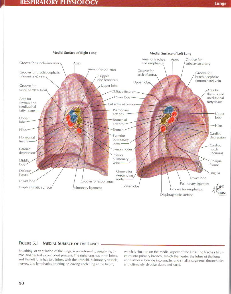
Figurę 5.1 Medial Surface of the Lungs
Brealhing, or ventilation of the lungs. is an automatic. usually rhyth-mic, and centrally controlled process. The right lung has three lobes. and the left lung has two lobes. with the bronchi, pulmonary ve$sels. nerves, and lymphatics enlering or leaving each lung at the hilum.
which is situated on the medial aspect of the lung. The trachea bifur-cates into primary bronchi, which then enter the lobes of the lung and further subdivide into smaller and smaller segmenłs (bronchioles and ultimately alveolar ducts and sacs).
90
Wyszukiwarka
Podobne podstrony:
netter156 GASTROINTESTINAL PHYSIOLOGY Overvievv of Cii Trać! Fluid and Electrolyte Transport Ingest
netter158 GASTROINTESTINAL PHYSIOLOGY Digestion of Carbohydrales Maftose Pancreatic amyiase
netter19 Limbie SysteiNEUROPHYSIOLOGY Genu of torpus caRosum Head of caudate nudeus Body of fornix T
netter53 CARDIOVASCULAR PHYSIOLOGY Overview of thc Cardiovascular System Pulmonary
netter5 NEUROPHYSIOLOCYSynaptic Transmission: Morphology of Synapses Numęrous boutons (synaptic knob
76657 netter188 ENDOCRINE PHYSIOLOGYHormonal Kugulation of thc Monstru.il Cyde Hormonal Reculation o
page7 (16) • The running surface of the new bushes for the clutch release shaft is coated with a pla
P5140082 Shin Splint or Medial Tibial Stress Syndrome The treatment for shin splints is as voried as
62048 page7 (16) • The running surface of the new bushes for the clutch release shaft is coated with
Jacek PRZEPIÓRKA , Marian SZCZEREK An effect of the free surface energy on tribological charact
więcej podobnych podstron