P1120988
Fig. 1.4: Transverse section through a uterine hom of a marę. The peritoneai borders are indicated by arrows. Analogous to the section reprcsented by A in Fig. 1.5.
Fig, 1.5: Schematic presentation of a transverse section through a uterine hom (A) and a longitudinal section through the uterine body (B).

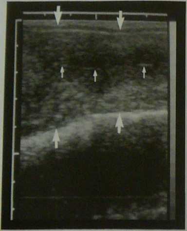
Fig. 1.6: Longitudinal section through the uterine body of a marę equivalent to the section illustratcd by B in Fig. 1.5. The dorsal and ventral uterine borders arc dcmarcatcd by large arrows. The opposing surfaces of the endometrium form un echoic linę (smali arrows).

Fig. 1.7:Trunsvcrse section through a uterine hom (arrows) of u nonpregnant marę. The uterus is positioned above 3 urched saeculations of the lefl dorsal colon. The diftercncc in imped-ance bclwecn the intestinal wali and the fcccs causc total rcflection of the nltrasound vvaves along the echoic saccula* tlons of the colon.
Wyszukiwarka
Podobne podstrony:
78187 P1130020 Fig. 147: Sagittal cross seetion through the uterine hom of a marę in diestrus. The h
P1130054 Fig- 1.105: Transverse section through the uterine hom (arrows) of a marę with chronię endo
P1130047 1.3.3.3.2 Ribs, trunk and stornach In order to determine the increase in the size of the tr
DSCN2472 (3) Despite INVASION into, or even through, the uterine wali or to the vaginaf there are st
Geography of the British Isles The British Isles are surrounded by the North Sea to the north and ea
Ask Me EverythingS Tell me morę: fog desert f* The desert climate of •4*4 the Namib is created
bbcn14?ck The Doctor and Martha go in search of a real live dodo, and are transported by the TARDIS
152 J. Ohnishi et al. Fig. 3. Cross-sectional view of a newly designed sex-tupole magnet. X(cm) Fig.
więcej podobnych podstron