78187 P1130020
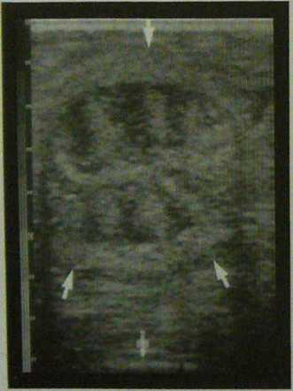
Fig. 147: Sagittal cross seetion through the uterine hom of a marę in diestrus. The hypoechoic peritoneal border (large ar-rows) and the Iransitkm frora myometriurn to endometrium (smali arrows) can be scen.

Fig. 1.48: Cross seetion through uterine hom during e< Due to the endometrial edema alternating areas of h echoic and morę echoic tissues can be seen. This causes typical spoke-wheel appearance of the uterine hom du estrus. Arrows indicate the transition from myo- to en. metrium.

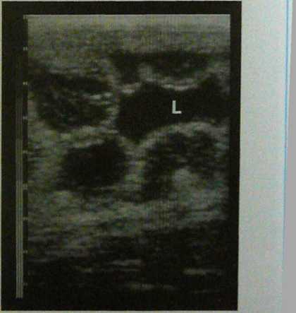
Fig. 1.49: Prominent spoke-wheel patiem of the uterus during Fig. 1.50: Extensive edema of the endometrial fołds dureu:
estrus. Arrows indicate the peritoneal border of the uterus. estrus. The endometrial folds with their echoic basc, hjpo
echoic edematous central area and hyperechoic surface bu^e into the uterine lumen (L) filled with seeretions.
Wyszukiwarka
Podobne podstrony:
P1120988 Fig. 1.4: Transverse section through a uterine hom of a marę. The peritoneai borders are in
P1130005 Fig. 1.24: Intense echogenicily (arrows) at the site of the est-rous follicle one day after
11427 P1130012 Fig. L33: Anovulatory, luteinizing follkle (between the cros-ses) witb a network of i
33227 P1130034 Fig. 1.75: Twin pregnancy on Day 28. Dorsalty, a chister of mu)ople endometrial cysłs
P1130030 Fig. 1.65: Pregwinty on Day 32. The cmbiyo lloats in the up-ptr third of the embryonic vesi
P1130054 Fig- 1.105: Transverse section through the uterine hom (arrows) of a marę with chronię endo
67607 P1130021 1J Uterine structures in the marę 1.3.1 Non-pregnant uterus A sagittal cross section
P1130046 Fig. 1-93: Eęample of takmg measurements of 3 rib cross sec-doiis with their corrssponding
więcej podobnych podstron