227 (21)

22 : Diagnostic approach to otitis extema


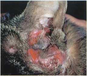
Figurę 22 :17: Invasive mast celi tumour in the ear canal
Figurę 22 :18: Ceruminous gland carcinoma in a cat presenting with chronic suppurative otitis

|
<>■ - " " |
Figurę 22:19: Otodectes cynotis (microscopy ofsample prepared in chloral lactophenol, X100)
Figurę 22:20: Demodex cati (microscopy of cerumen prepared in chloral lactophenol, X100) (courtesy of D.N. Carlotti)


Figurę 22:21: Malassezia pachydermatis (microscopy of cerumen, stained withDiff-Quik,X 1000)
Figurę 22:22: Cocci and neutrophils with many examples of phagocytosis (microscopy ofpus, stained with Diff-Quik, X1000) (courtesy ofD. Pin)


Figurę 22 : 24: CT scan ofa cat with an invasive auricular tumour: notę imasion of the brain by the tumour (courtesy ofF. Delisle)
22.7
Wyszukiwarka
Podobne podstrony:
225 (20) 22 : Diagnostic approach to otitis extema Figurę 22:9: Erythematous, scaling lesions on the
221 (22) Diagnostic approach to otitis extema Otitis extema is an acute or chronię inflammation of t
223 (25) 22 : Diagnostic approach to otitis externa Figurę 22:1: Parasitic erythemato-ceruminous oti
235 (21) 23 : Diagnostic approach to facial dermatoses Figurę 23:9: Erythema, erosions and crusting
247 (20) 24 : Diagnostic approach to feline pododermatoses Figurę 24 :18 : Crusting on the undersurf
7 (1239)
233 (20) 23 : Diagnostic approach to facial dermatoses Figurę 23 : 1 : Depigmentation of the nasal p
243 (19) 24 : Diagnostic approach to feline pododermatoses Figurę 24: 2 : Alopecia and rudimentary c
245 (17) 24 : Diagnostic approach to feline pododermatoses Figurę 24:10: Multiple paronychia with cr
249 (17) 24 : Diagnostic approach to feline pododermatoses Figurę 24:26: Multiple bacterial paronych
6 (1069) g18 Diagnostic approach to pruritic dermatoses Z. Alhaidari The diagnostic approach to feli
00024 ?b995e2ba34fa28ca2a7269f112a0d5 23 A Rule-Based Approach to Multiple Statistical Test Analysi
10 18 Peroneus brevis attachment to 5th metatarsal Figurę 10-18 Anatomy of peroneus brevis Exter»sor
cover2 THE OFFICIAL GUIDE TOUFOs From 1239 A.D. to modern times, man has been ob-serving startling p
info Eosy to add new pin drawings! Just place pin configuration drawings in the same folder as the p
CIIAPTER 8 comments we offer students need to appear helpful and not censorious. Sometjmcs they will
więcej podobnych podstron