CCF20130319�018

Fluorides - Modę of Action and Recommendations for Use Research and S

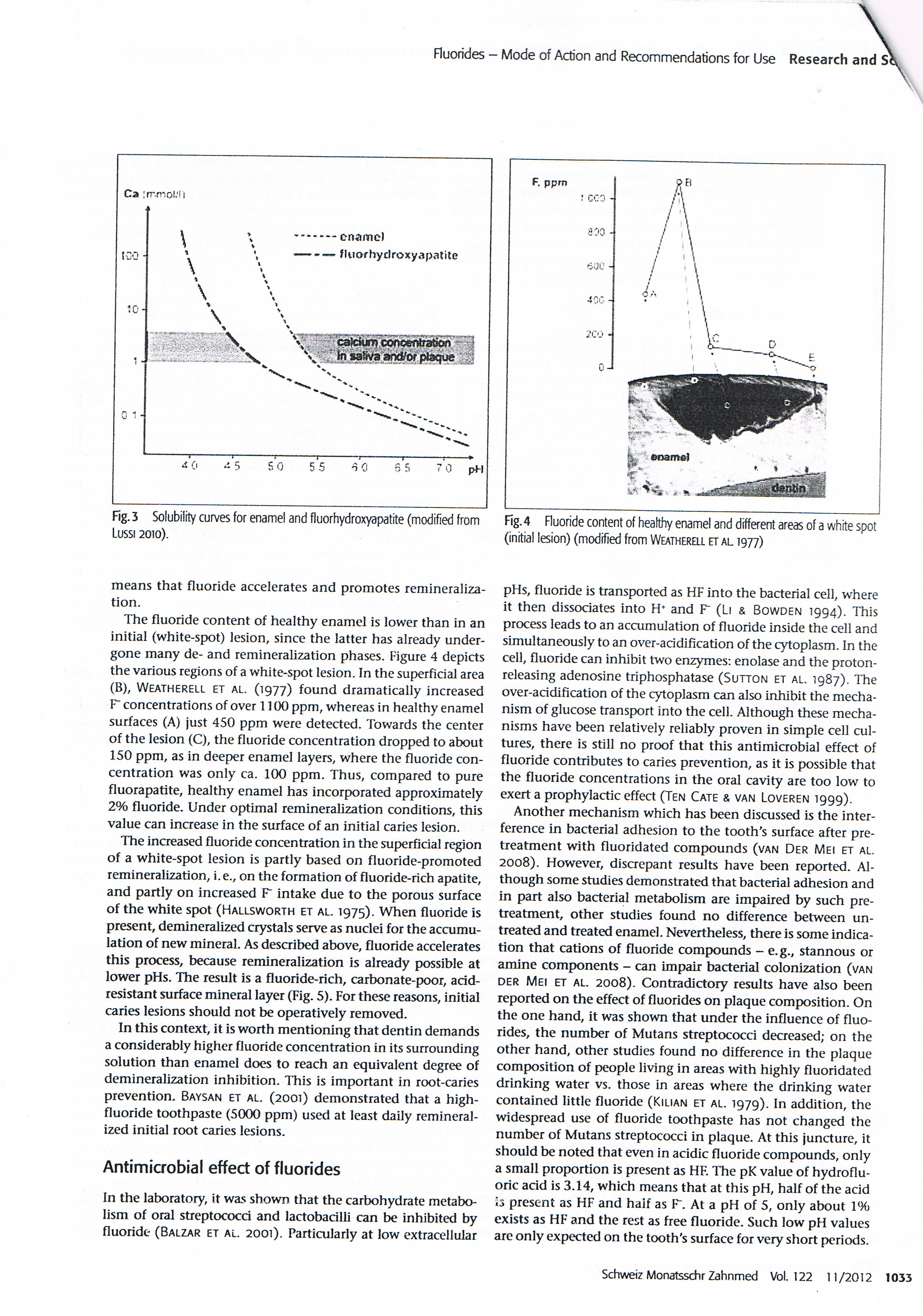
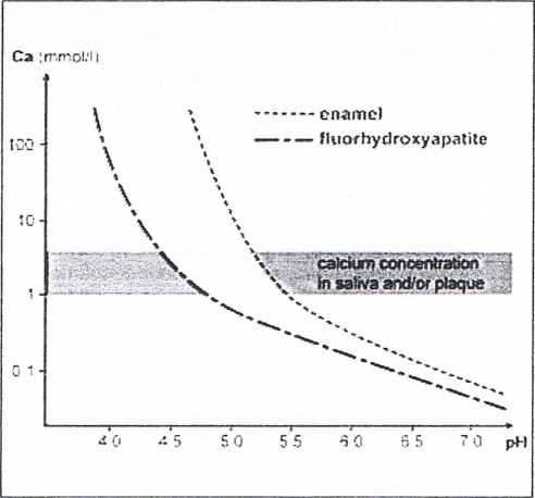
Fig. 3 Solubility curves for enamel and fluorhydroxyapatrte (modified from Lussi 2010).

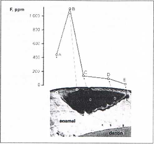
Fig. 4 Fluoride content of healthy enamel and different areas of a white spot (initial lesion) (modified from Weatherrl et al 1977)
means that fluoride accelerates and promotes remineraliza-tion.
The fluoride content of healthy enamel is lower than in an initial (white-spot) lesion, sińce the latter has already under-gone many de- and remineralization phases. Figurę 4 depicts the various regions of a white-spot lesion. In the superficial area (B), Weatherell et al. (1977) found dramatically increased F concentrations of over f 100 ppm, whereas in healthy enamel surfaces (A) just 450 ppm were detected. Towards the center of the lesion (C), the fluoride concentration dropped to about 150 ppm, as in deeper enamel layers, where the fluoride concentration was only ca. 100 ppm. Thus, compared to pure fluorapatite, healthy enamel has incorporated approximately 2% fluoride. Under optimal remineralization conditions, this value can increase in the surface of an initial caries lesion.
The increased fluoride concentration in the superficial region of a white-spot lesion is partly based on fluoride-promoted remineralization, i.e., on the formation of fluoride-rich apatite, and partly on increased F" intake due to the porous surface of the white spot (Hallsworth et al. 1975). When fluoride is present, demineralized crystals serve as nuclei for the accumu-lation of new minerał. As described above, fluoride accelerates this process, because remineralization is already possible at lower pHs. The result is a fluoride-rich, carbonate-poor, acid-resistant surface minerał layer (Fig. 5). For these reasons, initial caries lesions should not be operatively removed.
In this context, it is worth mentioning that dentin demands a considerably higher fluoride concentration in its surrounding solution than enamel does to reach an equivalent degree of demineralization inhibition. This is important in root-caries prevention. Baysan et al. (2001) demonstrated that a high-fluoride toothpaste (5000 ppm) used at least daily remineral-ized initial root caries lesions.
Antimicrobial effect of fluorides
In the laboratory, it was shown that the carbohydrate metabo-Iism of orał streptococci and lactobacilli can be inhibited by fluoride (Balzar et al. 2001). Particularly at Iow extracellular pHs, fluoride is transported as HF into the bacterial celi, where it then dissociates into H* and F (Li & Bowden 1994). This process leads to an accumuiation of fluoride inside the celi and simultaneously to an over-acidification of the cytoplasm. In the celi, fluoride can inhibit two enzymes: enolase and the proton-releasing adenosine triphosphatase (Sutton et al. 1987). The over-acidification of the cytoplasm can also inhibit the mecha-nism of glucose transport into the celi. Although these mecha-nisms have been relatively reliably proven in simple celi cul-tures, there is still no proof that this antimicrobial effect of fluoride contributes to caries prevention, as it is possible that the fluoride concentrations in the orał cavity are too Iow to exert a prophylactic effect (Ten Cate & van Loveren 1999).
Another mechanism which has been discussed is the inter-ference in bacterial adhesion to the tooth's surface after pre-treatment with fluoridated compounds (van Der Mei et al. 2008). However, discrepant results have been reported. Although some studies demonstrated that bacterial adhesion and in part also bacterial metabolism are impaired by such pre-treatment, other studies found no difference between un-treated and treated enamel. Nevertheless, there is some indica-tion that cations of fluoride compounds - e.g., stannous or aminę components - can impair bacterial colonization (van der Mei et al. 2008). Contradictory results have also been reported on the effect of fluorides on plaąue composition. On the one hand, it was shown that under the influence of fluorides, the number of Mutans streptococci decreased; on the other hand, other studies found no difference in the plaąue composition of people living in areas with highly fluoridated drinking water vs. those in areas where the drinking water contained little fluoride (Kilian et al. 1979). In addition, the widespread use of fluoride toothpaste has not changed the number of Mutans streptococci in plaąue. At this juncture, it should be noted that even in acidic fluoride compounds, only a smali proportion is present as HF. The pK value of hydroflu-oric acid is 3.14, which means that at this pH, half of the acid is present as HF and haif as F. At a pH of 5, only about 1% exists as HF and the rest as free fluoride. Such Iow pH values are only expected on the tooth's surface for very short periods.
Schweiz Monatsschr Zahnmed Vol. 122 11/2012 1033
Wyszukiwarka
Podobne podstrony:
CCF20130319�016 Fluorides - Modę of Action and Recommendations for Use Research ano. source &n
CCF20130319�020 Fluorides - Modę of Action and Recommendations for Use Research and Sciencpossible a
8. Herman A, Herman AP. Caffeine’s mechanisms of action and its cosmetic use.
Advantages and disadvantages of digital education 25 (for this purpose it is recommended to use the
screenshot 1 Recommended for you Homemade Angel Food Cake On Monday, Graham turned one, and the four
CCF20101207�001 lic American Socicty for Information Science and Technology”— USA) oraz :elni (Royal
CCF20100223�013 indicate the starting points, a circle ° stands for the targ^ggt p0jnt, and arrows m
CCF20121215�000 The phonemic alphabet: PracticePhonemic symbols for vowels See page 192 for a list o
CCF20121215�000 IThe phonemie alphabet: PracticePhonemic symbols for vowels See page 192 for a list
CCF20121215�000 IThe phonemie alphabet: PracticePhonemic symbols for vowels See page 192 for a list
skanuj0173 „Strategie mangement is a stream of decisions and actions which leads to the development
220 Boris KemBIBLIOGRAFIA Balteiro I., 2011, Awareness of LI and L2 Word-formation Mechanisms for th
AGH Uniyersity of Science and Technology UST Academic Cenlre for Materials and Nano-technology, whic
A welcome from Poland Poland is a modern and dynamie country fuli of life-time opportunities for you
Production Systems expectations evolved with changes in consciousness of clients and their awareness
Information for Authors Send to: Archives of Civil and Mechanical Engineering Poli
więcej podobnych podstron