628996288
1698 CHAUVEAU etal
BLOOD. 1 SEPTEMBER 2006 • VOLUME 106. NUMBER5
3). Double staining showed that DC-L.AMP* cells were HO-1 (Figurę 3).
An iirelevant isotype-matched mAb did not show any reactivity with either tissue (Figurę S4).
Thus, analysis of human tissues shows that iDCs express HO-1 in vivo. whereas maturę DCs do not.
HO-1 induction renders rat and human DCs refractory to LPS-induced maturation
Next. we investigated whether HO-1 activity during maturation could play a functional role in the regulation of DC' responses to pathogens.
To examine the effect of HO-1 activity on LPS-induced DC maturation. we pulsed-treated human iDCs and rat iBMDCs with CoPP or SnPP. CoPP is a strong ind u cer of U()-l gene transcrip-tion.20"22 whereas SnPP irreversibly binds and inactivates HO-1 enzymatic activity.::
CoPP and SnPP48-hour pulse treatments were not toxic to DCs. as detennined by flow cytometric analysis of physical parameters and TO-PRO-3 staining (Figure S5). Poiphyrin-pulsed DCs were cultured for 16 hours in the absence or presence of LPS. Expression of HO-1 in human (Figure 4A) and rat (data not shown) DCs was deterniined by Western biot analysis 24 hours laler. HO-1 expres-sion was strongly increased following treatment with CoPP and treatment with LPS paitially abrogated this increase. SnPPslightly increased HO-1 levels. likely through a feedback mechanism due to inhibition of HO-1 activity. as previously described in other cells.'1
Analysis of mRNA levels by quantitative RT-PCR showed that CoPPalone induced a 70- to 124-fold increase in HO-1 transcript levels versus those in iDCs, and a 29- to 69-fold increase when associated with LPS in rat DCs. A strong increase in HO-1 mRNA was also detected in CoPP-treated human DCs. Transcript levels increased 18- to 99-fold with CoPP alone and 29- to 67-fold when associated with LPS.
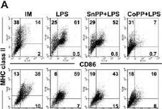
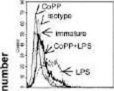
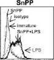
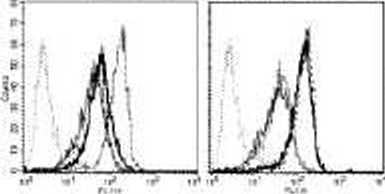
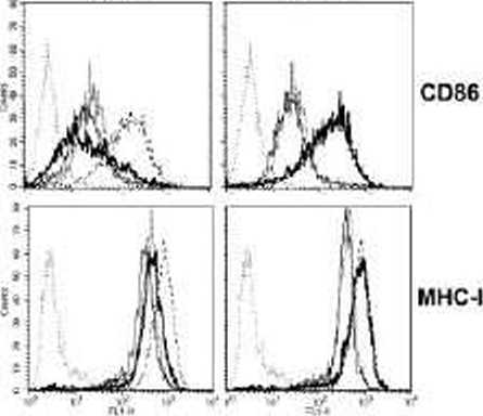
CoPP-pulsed human and rat DCs were refractory to LPS-induced maturation. as assessed by phenotypic analysis. A statisti-cally significant inhibition of celi surface marker expression was observed in CoPP-pretreated rat and human iDCs compared to untreated and SnPP-pretreated cells incubated with LPS (Figure 4B-C: Table S1).
SnPP inhibition of HO-1 activity had no effect on DC maturation. suggesting that inhibition of HO-1 activity does not act as an inducer of DC maturation in the absence of specific DC maturation stimuli. The absence of an effect of SnPP on LPS-induced DC maturation is probably due to the inhibition of HO-1 expression on LPS stimulation.
Thus. induction of HO-1 in DCs resulted in a blockade of DC phenotypic maturation induced by LPS.
HO-1 induction inhibits the secretion of proinflammatory cytokines and maintains IL-10 producdon by DCs exposed to an inflammatory signal
[XT maturation results in secretion of diverse cytokines that
mediate many of the functional effects of DCs on other celi *
— ICAM-1 —
32 kDa
GO kDo __ — —— -

MHC class II
CoPP
a>
O
CD83
CD80
Fluorcsccnce Intensity
CoPP
SnPP
Fluorescence intensity
Figur-? 4. Induction of HO-1 renders DCs refractory to LPS-induced phenotypic maturation. (A) Western blol analysisof HO-1 levels in human iDCs Ireated or not with SnPP or CoPP for 2 bours. cultured lor 16 hours. and then for a further 24 hours in the preserce or absence of LPS. Anti-p lubulin was used as a loading control. (G) Flow cylometry analysis showing thephenolype ol imma-ture (IM) rat BMCCs trealed or not with SnPP or CoPP tor 2 hours. cultured for 16 bours. and then for a lurlher 24 hours with or withoul LPS for 24 hcors. Expression ol MHC class II. CD80. CD86. and inlercellular adhesion molecule 1 (ICaM-1) was assessed. Similar results were cbtained in 5 independent experimenls. Numbers within graph quadrants are percentage ol posilive cells. (C) Flow cylometry analysis showing thephenolype of imma-ture human DCs Irealed or not with SnPP a CoPP for 2 hours. cultured tor 16 hours. and then stimulated or nol with LPS for 24 hours. Thin gray linę indicates untrealed immature DCs: dashed linę. untreated DCs slimulaled with LPS: Ihin black linę. Ireatrrenl wilh CoPP or SnPP: bold linę. treatment wilh CoPP or SnPP and slimulaled wilh LPS: and dotted linę. isotype. Expression ol CD83. CD9D. CD86. and MHC class I was assessed. Similar results were oblained in 3 independent experiments.
Wyszukiwarka
Podobne podstrony:
1702 CHAUVEAU ot al BLOOD. 1 SEPTEMBER 2005 • VOLUME 106. NUMBER5 32. Verhasselt V
HO-1 INHIBITSDC FUNCTION 1695 BLOOD. 1 SEPTEMBER 2005 • VOLUME 106. NUMBER 5 Mcylan, France). I.ow-d
HO-1 INHIBITSDC FUNCTION 1701 BLOOD. 1 SEPTEMBER 2005 • VOLUME 106. NUMBER 5 decrease T-cell prolife
1696 CHAUVEAU et al BLOOD. 1 SEPTEMBER 20C6 • VOLUME 106. NU MB ER 5Flow cytometry Rat !)Cs were sta
BLOOD. 1 SEPTEMBER 2005 • YOLUME 106. NUMBER 5 HO-1 INHIBITSDC FUNCTION 1697 Rat DCs ? IB
HO-1INHIBITSDC FUNCTION 1699 BLOOD. 1 SEPTEMBER 2005 • VOLUME106. NUMBER 5 populations. Production o
33/18 ARCHIWUM ODLEWNICTWA Rok 2006, Rocznik 6, Nr 18 (1/2) ARCHIVES OF FOUNDRY Year 2006,
76/18 ARCHIWUM ODLEWNICTWA Rok 2006, Rocznik 6, Nr 18 (1/2) ARCHIVES OF FOUNDRY Year 2006,
62/21 ARCHIWUM ODLEWNICTWA Rok 2006, Rocznik 6, Nr 21(2/2) ARCHIVES OF FOUNDARY Year 2006,
DSC06880 MINUSY - PLUSY + Ryc. 106. Mocne i słabe strony miejscowości (2006) Fig. 106. Strong and we
DSC06880 MINUSY - PLUSY + Ryc. 106. Mocne i słabe strony miejscowości (2006) Fig. 106. Strong and we
Lieber Jens, Kieł, den 13. September 2006 vielen Dank fOr deinen letzten Brief. Wie immer, wenn ich
big fsf September 2006 FEEL LUCKYŁUCK TananariueDiie Mictiael Handel Michael Liblmg
fsf January 2006 dog *7 decided to test the premise that im your best friend.
diagnostyka laboratoryjna Journal of Laboratory Diagnostics 2011 • Volume 47 • Number 2 • 197-203 Pr
więcej podobnych podstron