
FAST - Focused Assessment with Sonography for
Trauma
• FAST examines four areas for free
fluid:
– Perihepatic & hepato-renal space
– Perisplenic
– Pelvis
– Pericardium
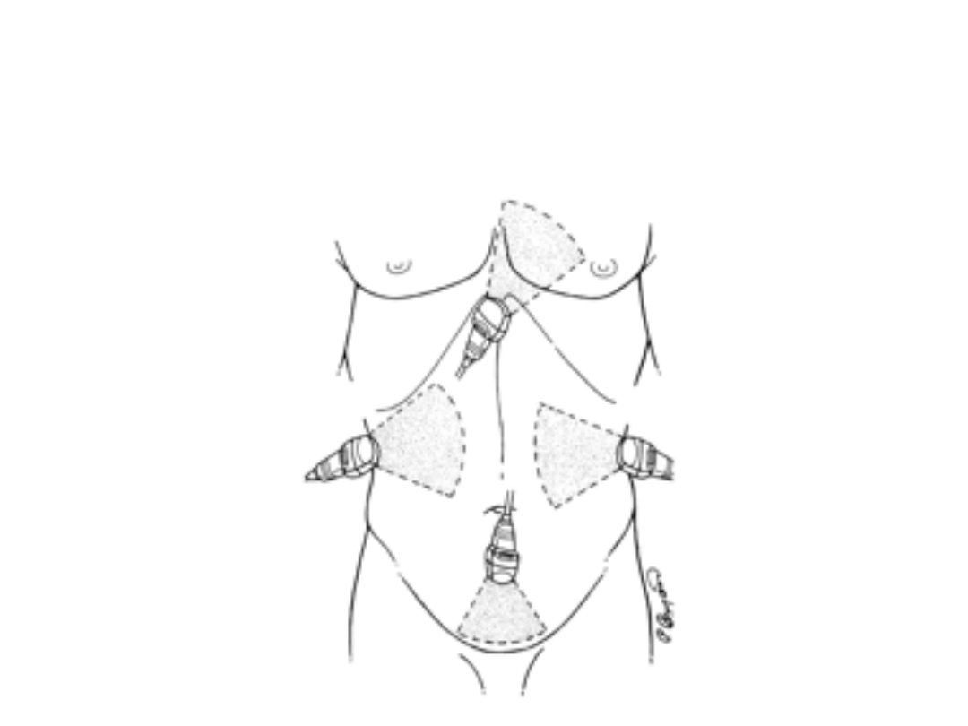
The 4 FAST views
The 4 FAST views

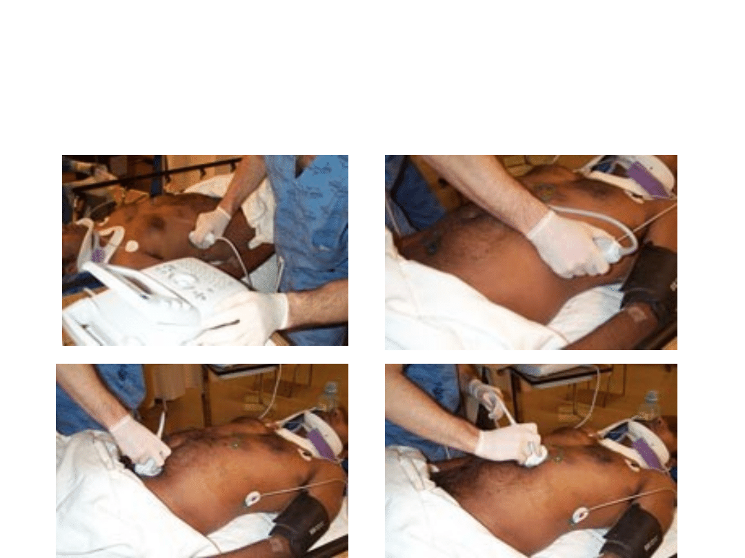
Views for FAST U/S
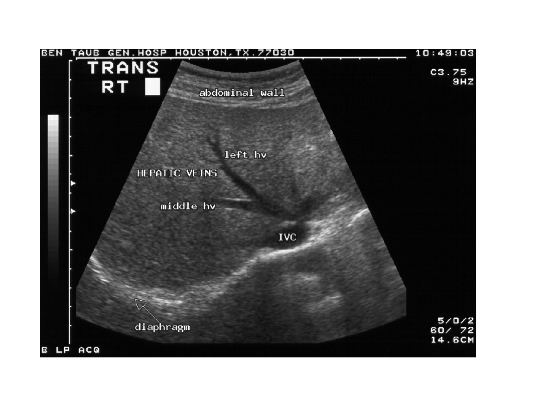
• RUQ view is the most important view for FAST
• 80% of hemoperitoneum detected on
hepatorenal view alone
• If blood collects in the RUQ, the fluid settles
between the kidney and liver
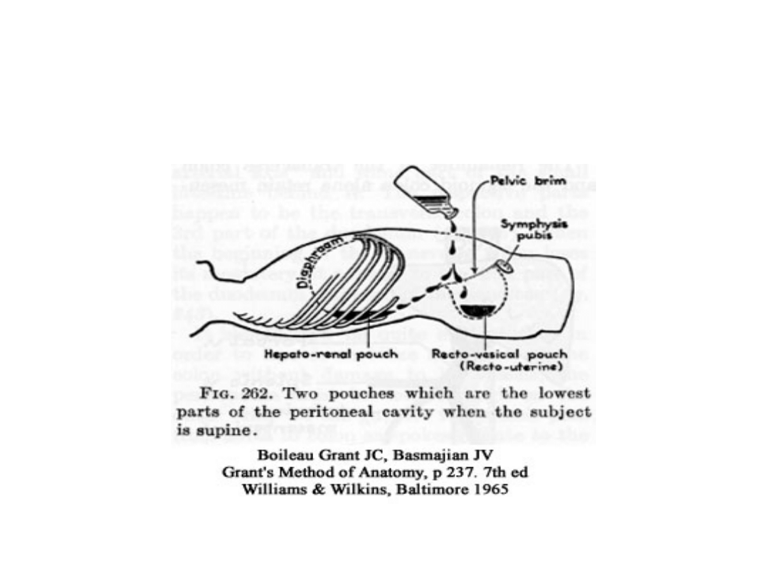
Morrison’s Pouch
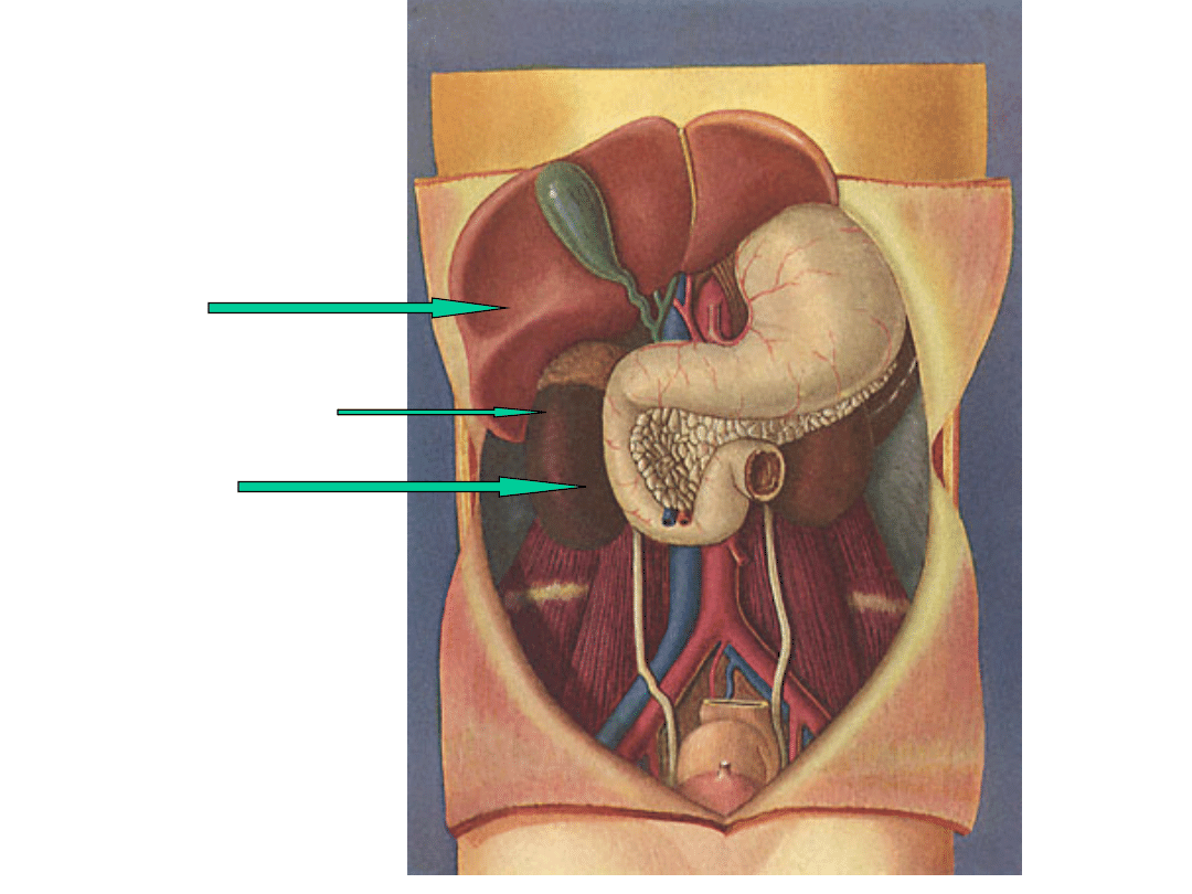
Liver
Morrison’s Pouch
Kidney
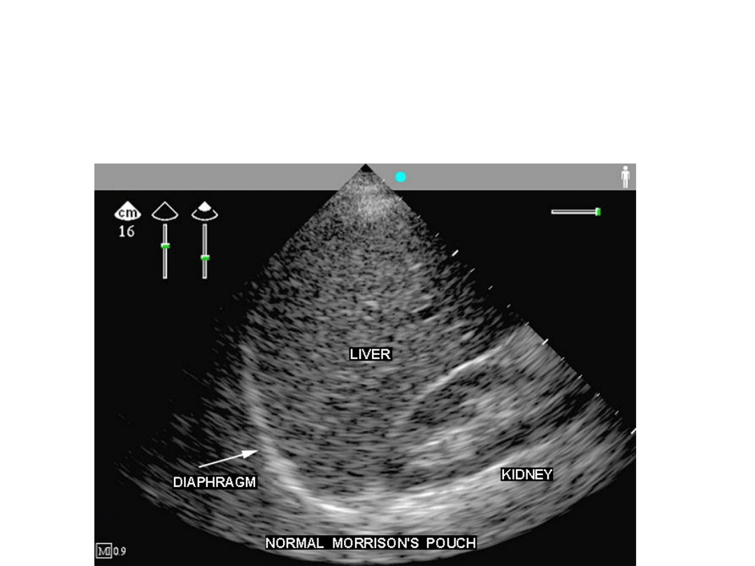
Normal Morrison’s pouch
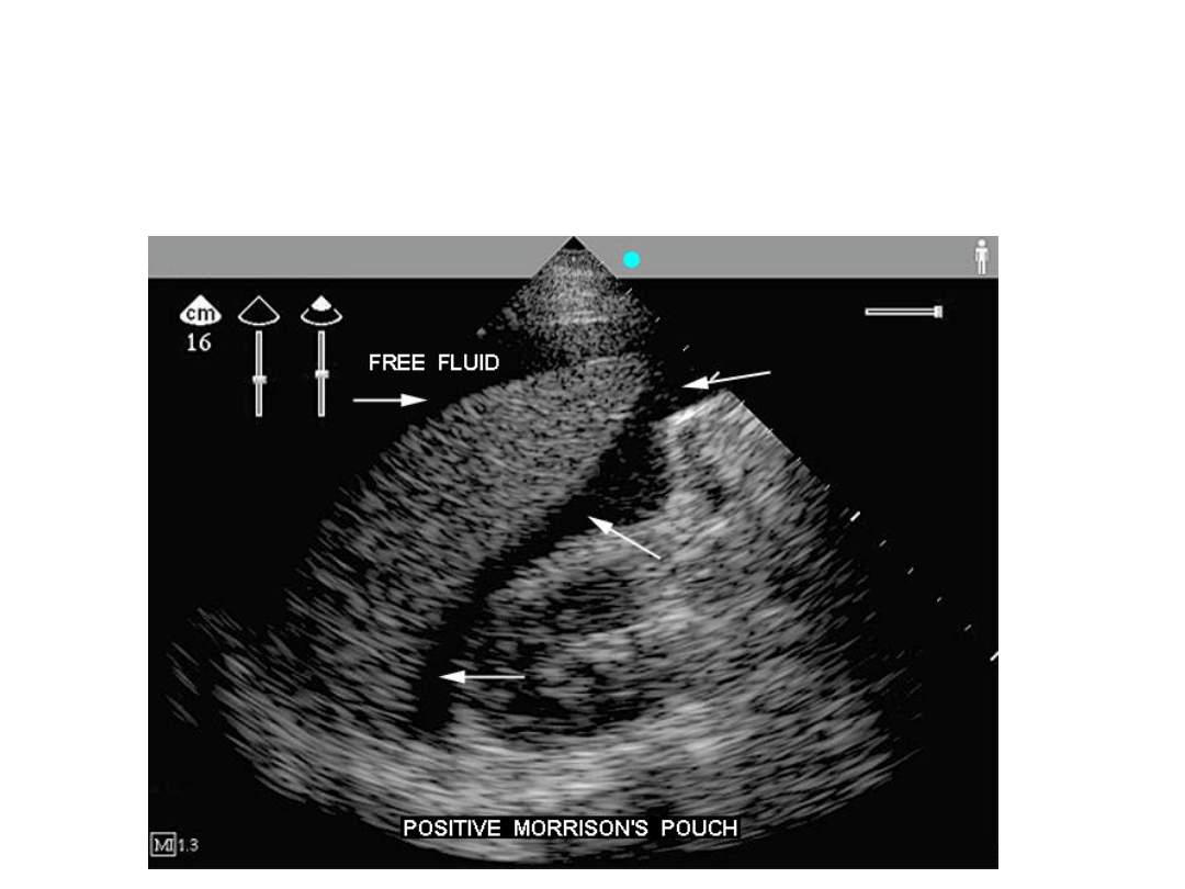
Abnormal Morrison’s

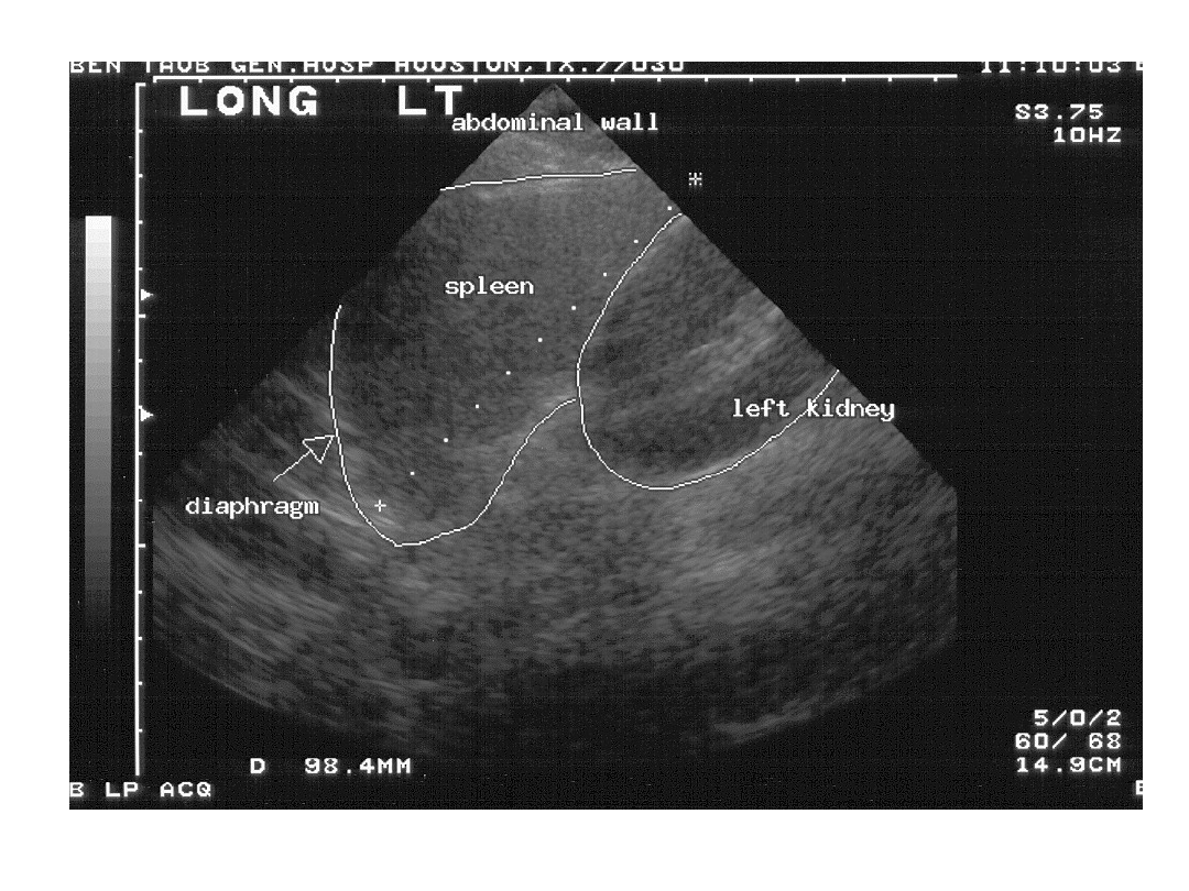
LUQ view
• In this view, looking for blood between the
– Spleen and the kidney OR
– Spleen and the diaphragm
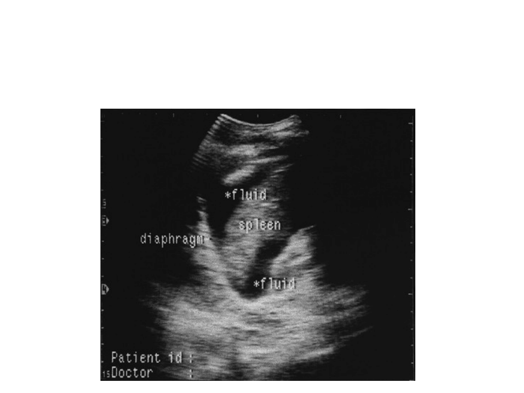
Abnormal LUQ

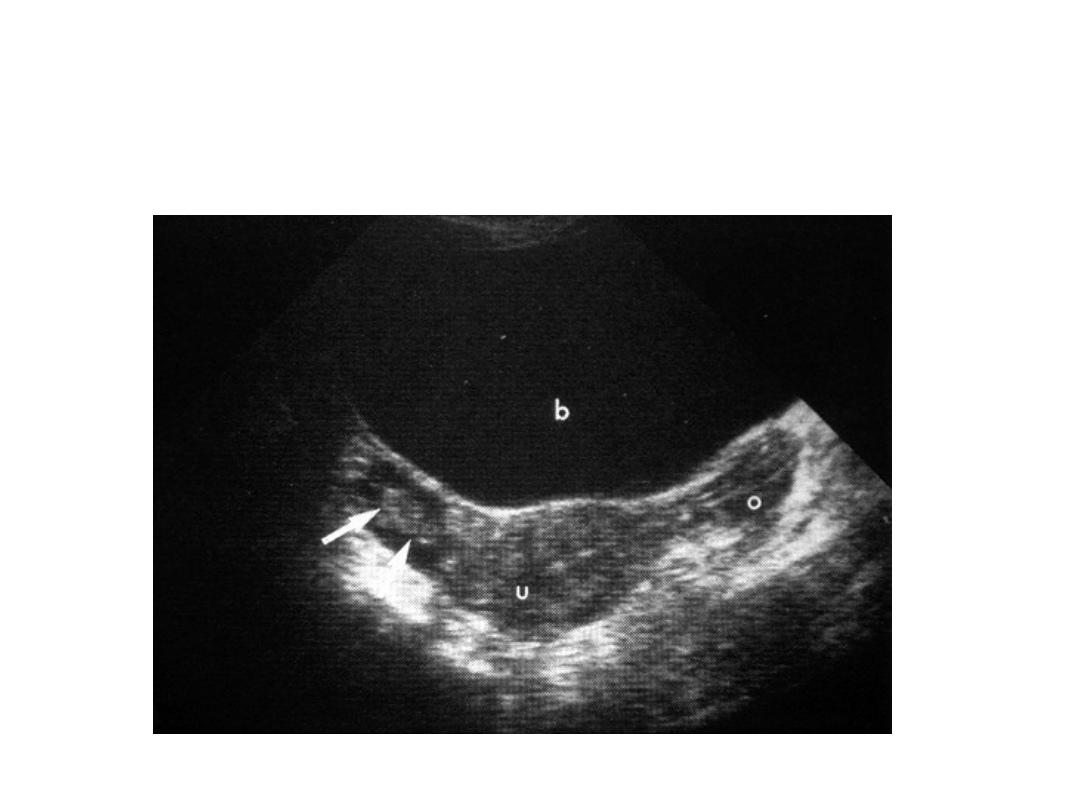
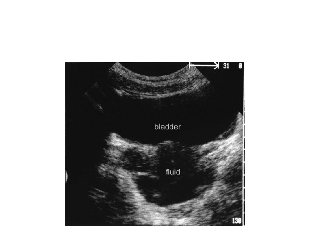
Pelvis view
• In this view, looking for blood in the
– Rectovesicular pouch (males)
– Rectouterine pouch aka pouch of Douglas (females)
Normal pelvis view
Abnormal pelvis view

Pericardial view
• In this view, looking for blood between the
pericardium and the heart
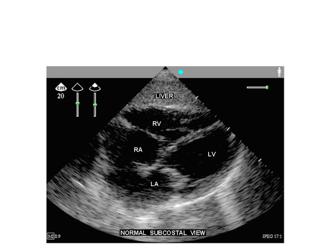
Normal pericardium
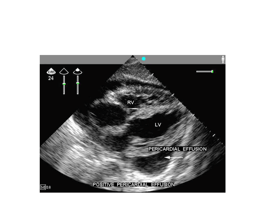
Abnormal pericardium

FAST vs DPL
FAST
DPL
Radiation
None
None
Rapid
++
+
Portable ++
+
Noninvasive Yes
No
Sensitive
Good
Excellent
Specificity
Good
Fair
Pericardial eval
Yes
No
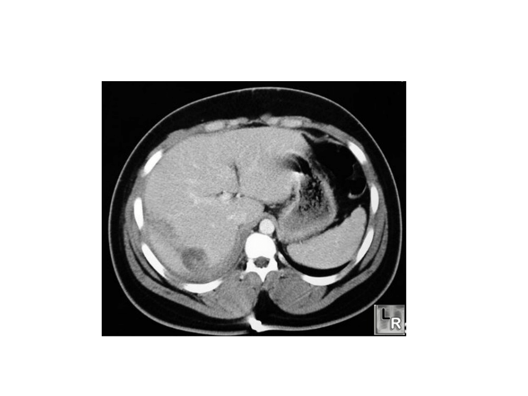
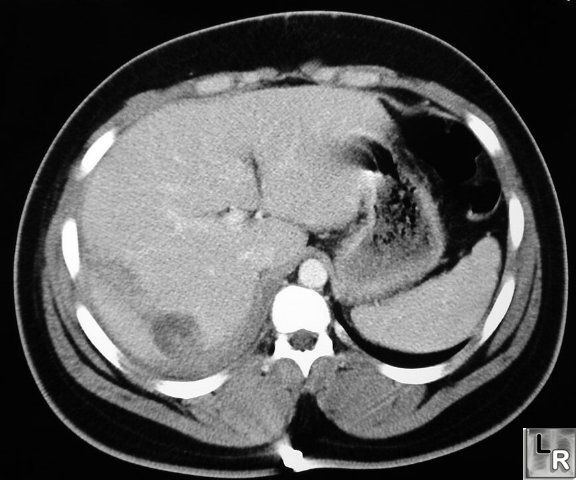
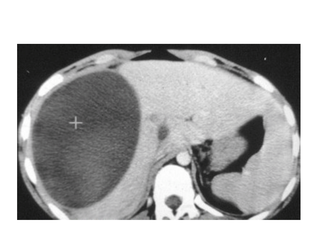
Contrast-enhanced CT of abdomen shows linear low-attenuation defectcrossing the
posterior aspect of the left lobe of the liver representing a laceration
Liver laceration
Liver laceration
Liver hematoma
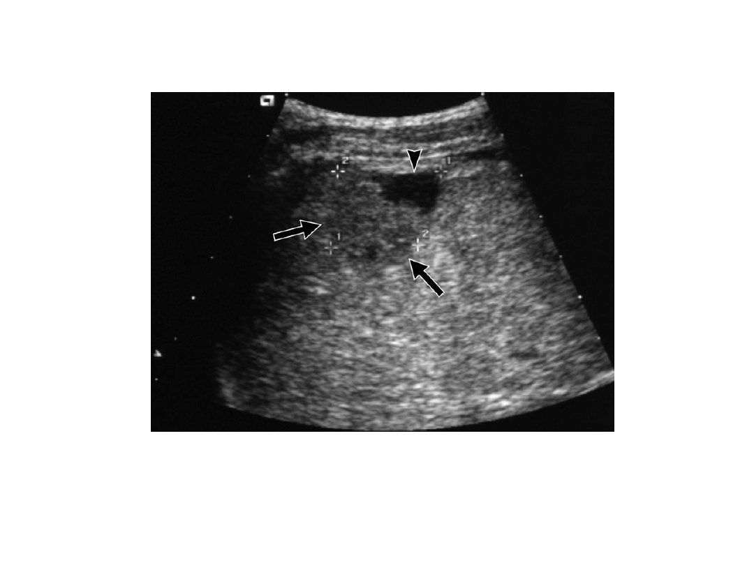
Ultrasound examination shows the laceration as a
relatively hypoechoic area (arrow) with a local anechoic
area (arrowhead) that represents a haematoma
Liver hematoma
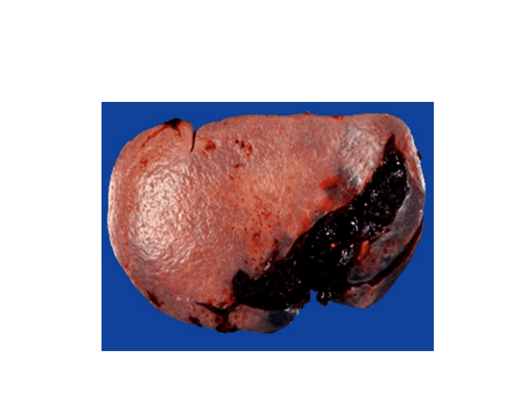
Spleen laceration
Spleen laceration and
hemoperitoneum
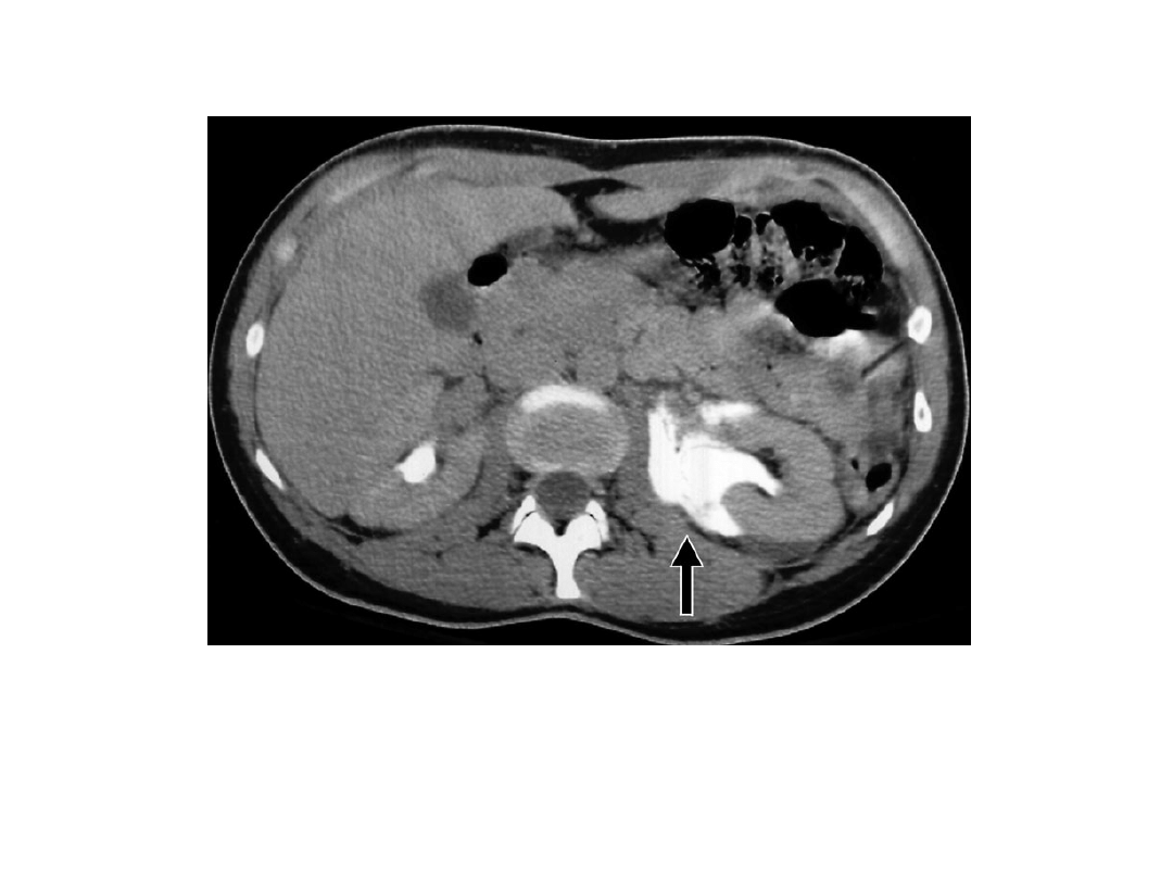
Kidney trauma
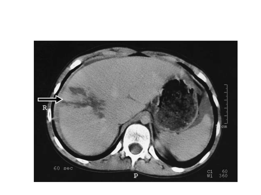
Axial contrast-enhanced CT section through
the kidneys showing extravasation of contrast
(arrow) from the left kidney, due to traumatic
forniceal rupture
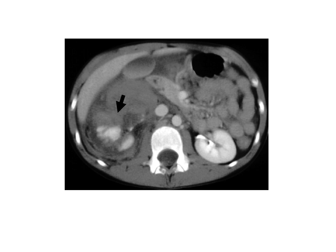
Kidney trauma
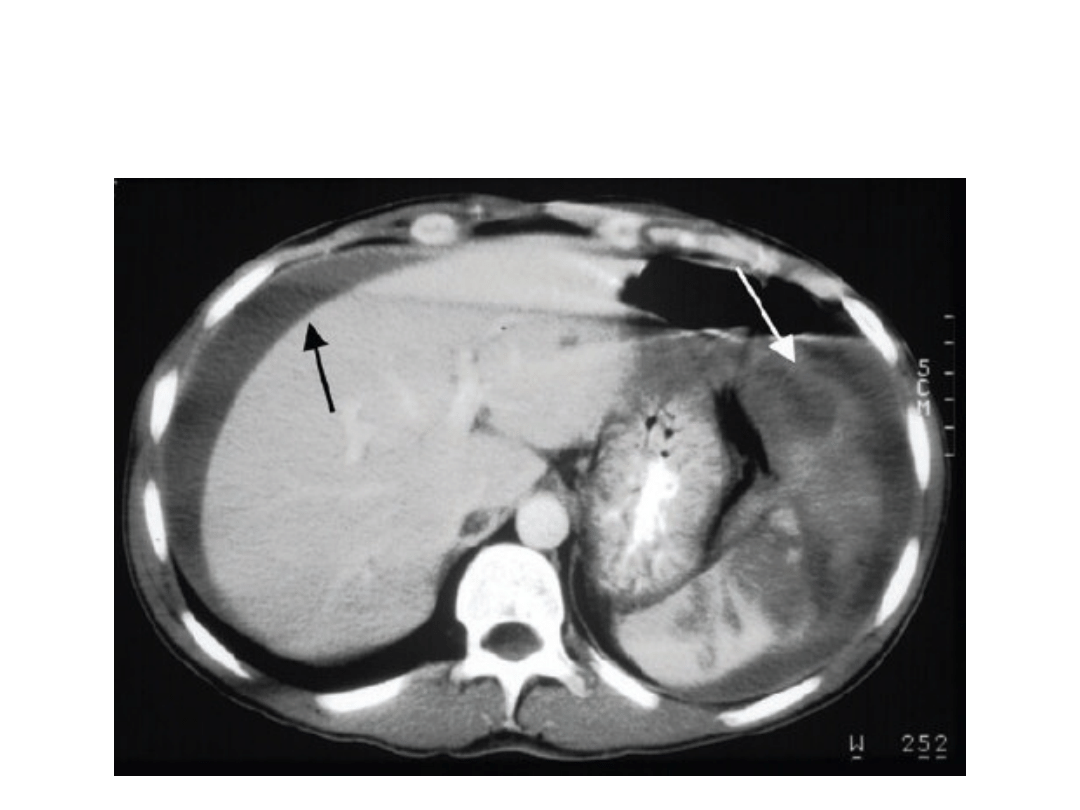
Axial contrast-enhanced CT section shows a
large haematoma surrounding the fractured
lower pole of the right kidney.
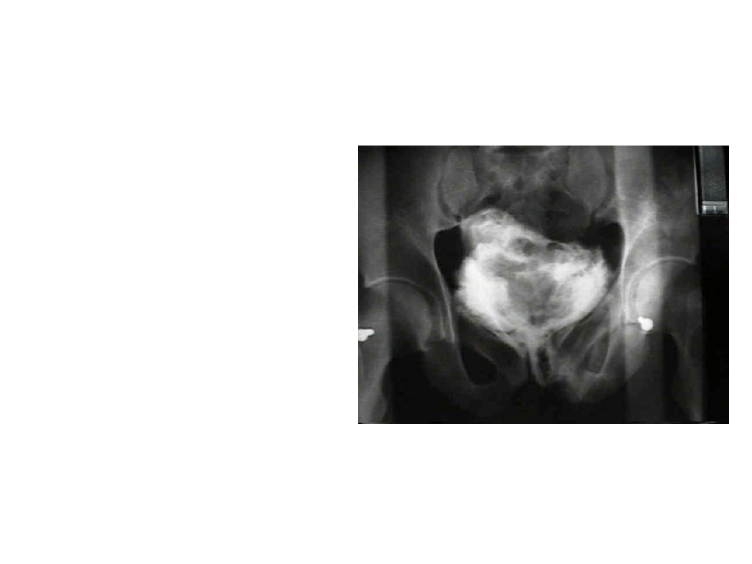
Urinary bladder rupture
- requires prompt diagnosis so as to
avoid hyperkalemia, hypernatremia,
uremia, acidosis, and peritonitis;
- can be extraperitoneal or
intraperitoneal (or both);
- extraperitoneal
rupture:
- most often, the rupture is anterior and
extraperitoneal;
- in rare may result from laceration from
sharp bone spike;
- in many cases, may be treated non
operatively w/ suprapubic drainage;
- intraperitoneal rupture:
- occurs in about 15% of major pelvic
fractures;
- most often occurs from contussion to
lower abdomen or to the symphyseal
region;
- may occurs w/o associated pelvic ring
disruptions as the result of a seatbelt or
steering wheel injury;
- usually requires operative correction;
Document Outline
- Slide 1
- Slide 2
- Slide 3
- Slide 4
- Slide 5
- Slide 6
- Slide 7
- Slide 8
- Slide 9
- Slide 10
- Slide 11
- Slide 12
- Slide 13
- Slide 14
- Slide 15
- Slide 16
- Slide 17
- Slide 18
- Slide 19
- Slide 20
- Slide 21
- Slide 22
- Slide 23
- Slide 24
- Slide 25
- Slide 26
- Slide 27
- Slide 28
- Slide 29
- Slide 30
- Slide 31
- Slide 32
- Slide 33
- Slide 34
- Slide 35
- Slide 36
Wyszukiwarka
Podobne podstrony:
Urazy brzucha
Tępe urazy brzucha
Urazy brzucha
urazy brzucha
TĘPE URAZY BRZUCHA
Urazy brzucha
Urazy brzucha
Urazy brzucha, Dokumenty Medyczne, MEDYCZNE
TST URAZY BRZUCHA I KLATKI PIERSIOWEJ- dla ucznia, ratownicto 2012 2013, ratownictwo medyczne, Testy
TST URAZY BRZUCHA I KLATKI PIERSIOWEJ, ratownicto 2012 2013, ratownictwo medyczne, Testy
Urazy brzucha, pierwsza pomoc Ratownictwo medyczne
Urazy brzucha spowodowane są przeważnie przez upadek
urazy brzucha (2)
Urazy brzucha
Urazy brzucha Bigos zjebane
URAZY BRZUCHA
Urazy brzucha, Ratownictwo Medyczne(1)
Tępe urazy brzucha
ZK IV Urazy III Urazy brzucha
więcej podobnych podstron