
1
Patomorfologia
Wykład 03
cracked by fazi
created by: sobatolog

2
Intracellular
Accumulations
• General principles
– Transient or permanent
– harmless or injurious
– Cytoplasm (lysosomes) or nucleus
– Synthesized by the affected cell or produced
elsewhere

3
Intracellular Accumulations
• General Principles
– Endogenous
• normal substance produced at normal or increased
rate / rate of metabolism inadequate for removal
(fatty liver)
• normal or abnormal substance cannot be
metabolized (storage diseases)

4
Intracellular Accumulations
• General Principles
– Exogenous
• cell cannot degrade substance (carbon)

5
Intracellular Accumulations
• Fatty Change (Steatosis)
– Causes
• alcohol abuse, oyher toxins, anoxia, obesity, protein
malnutrion
– Pathogenesis
• various steps involved
• egress of hepatic triglycerides requires complexing
with apoproteins to form lipoproteins
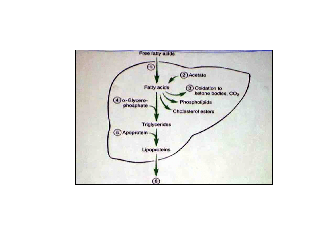
6

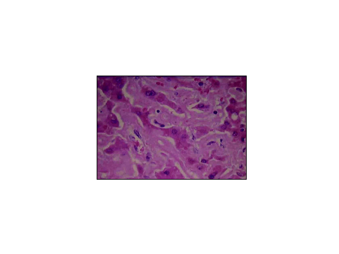
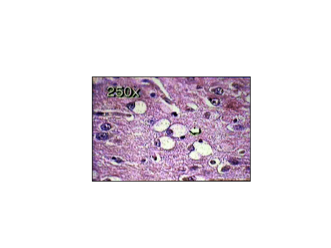
7
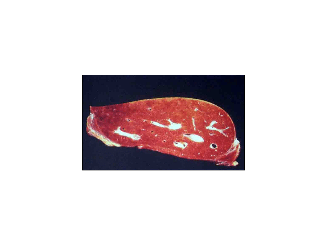
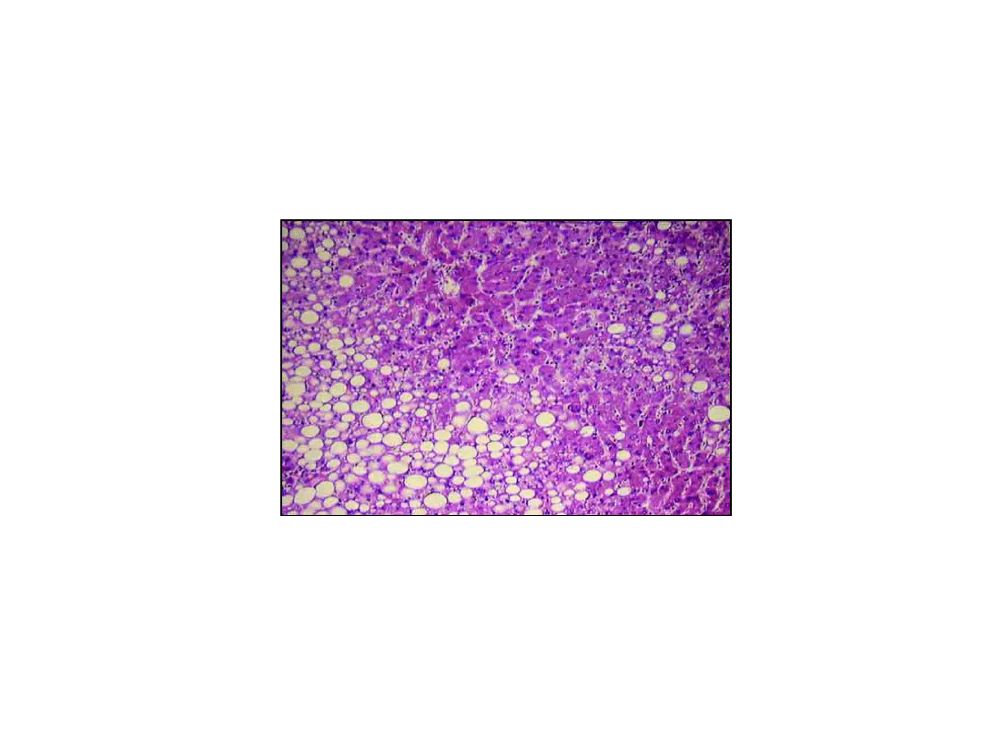
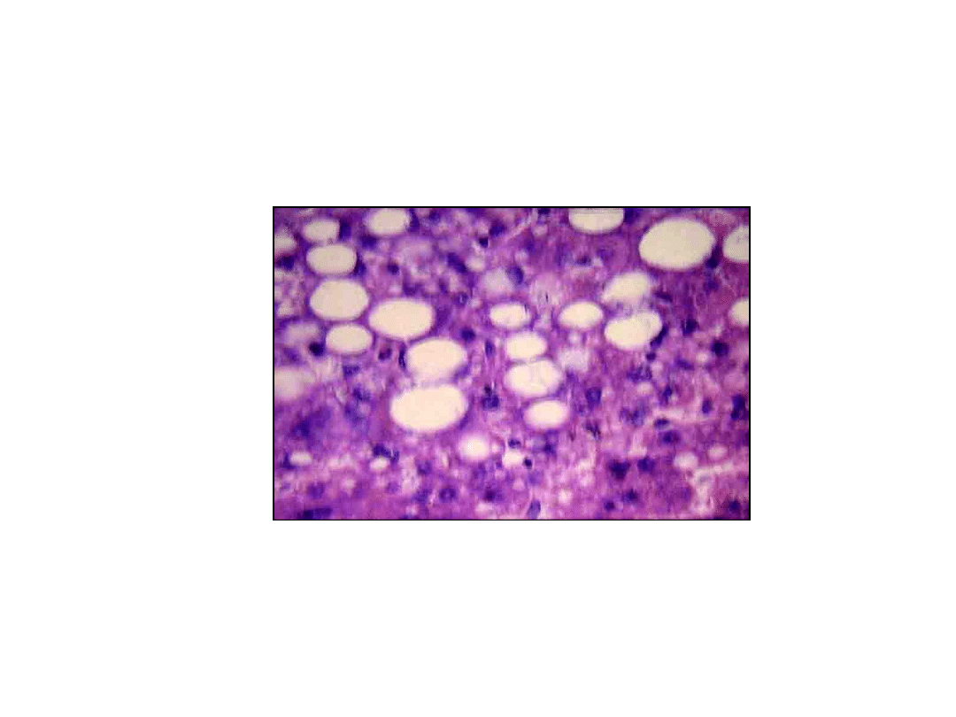
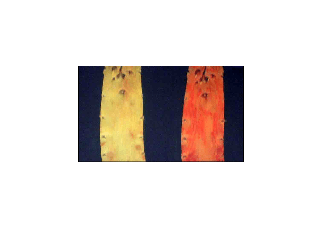
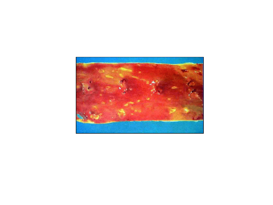
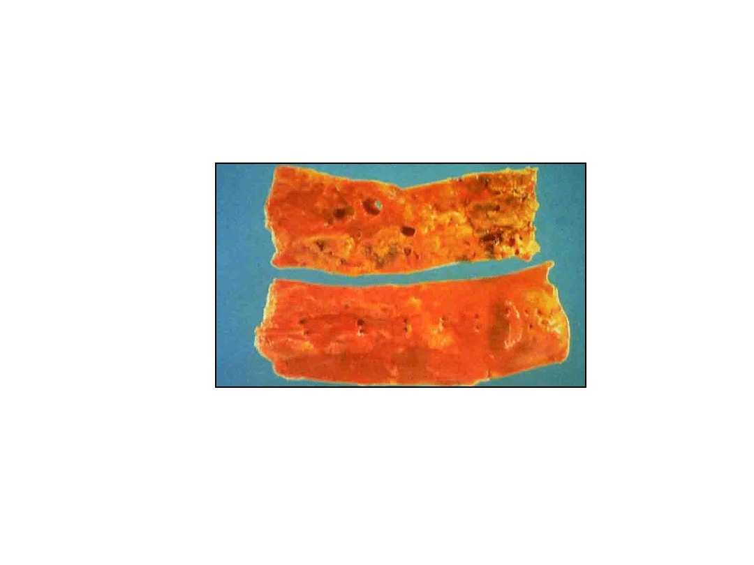
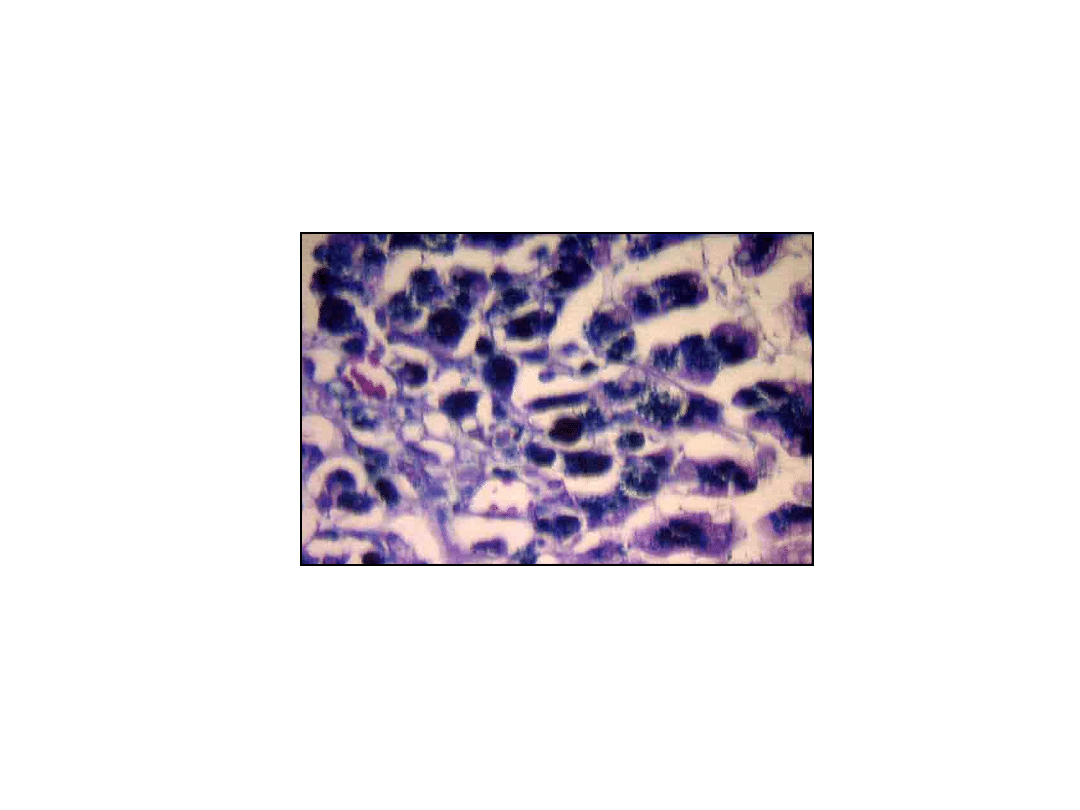
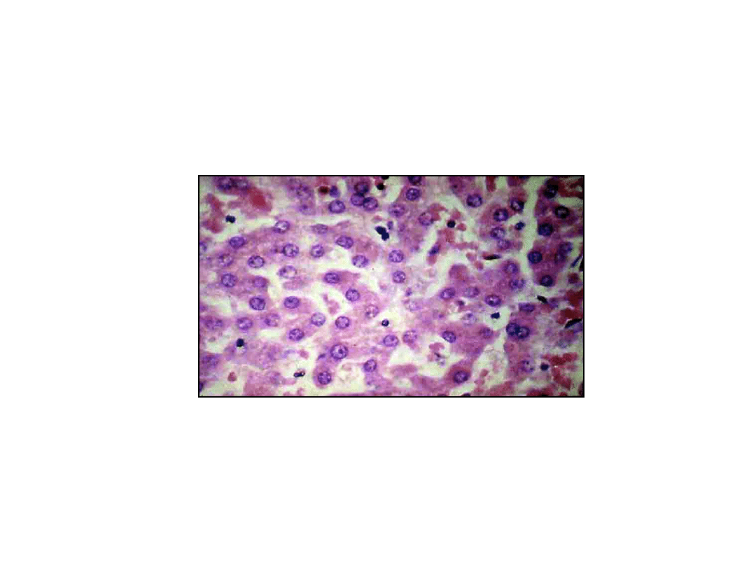
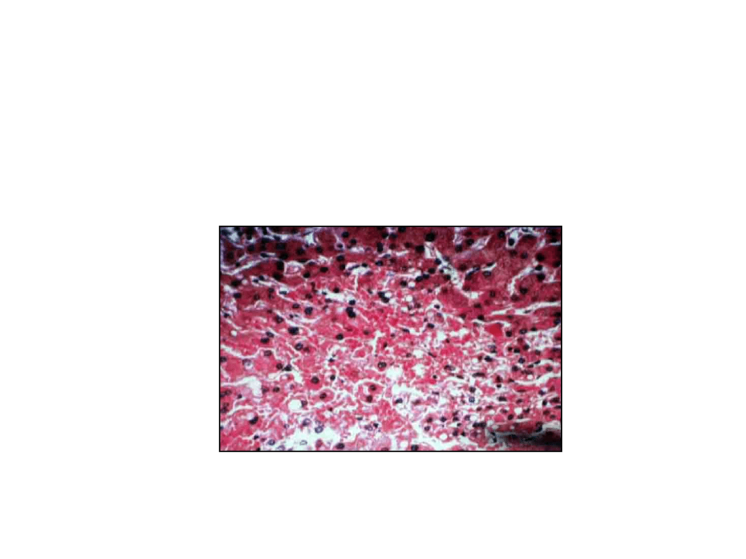
Intracellular Accumulations
• Fatty Change (Steatosis)
– Liver
• increased weight, yellow color
• fat vacuoles within cytoplasm of hapatocytes
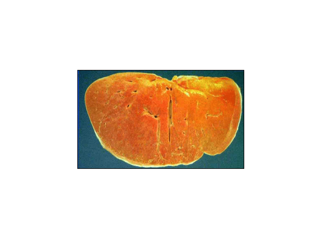
8
9
10
11

12
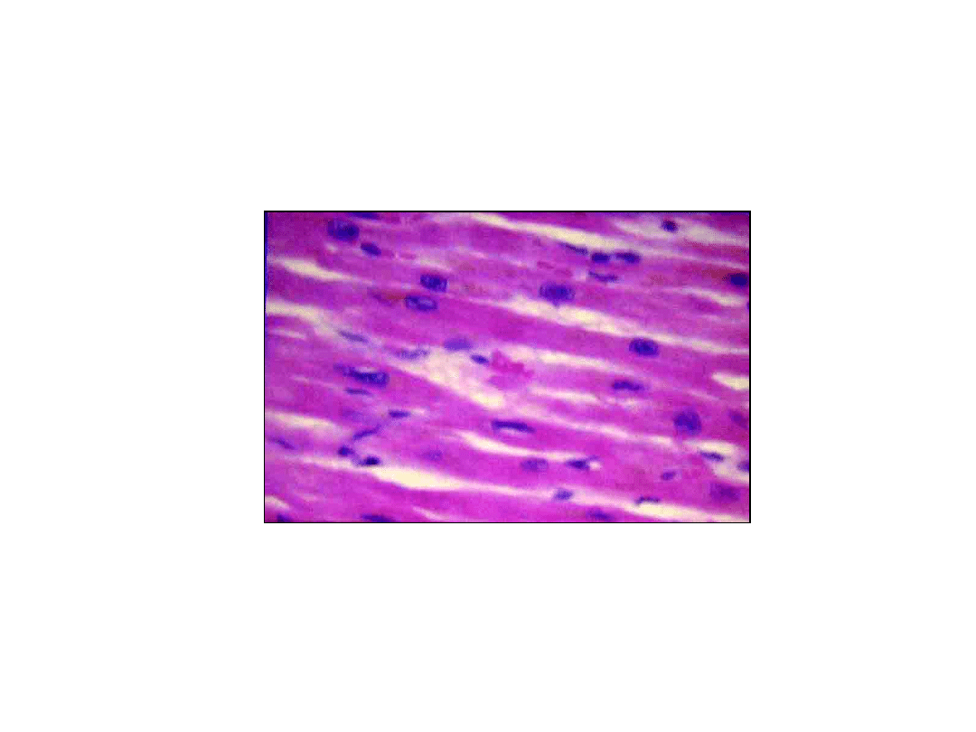
Intracellular Accumulations
• Fatty Change (Steatosis)
– Heart
• focal fat deposits in myocardium (anemia)
• diffuse fat deposits in myocardium (profund hypoxia,
diphtheric myocarditis)

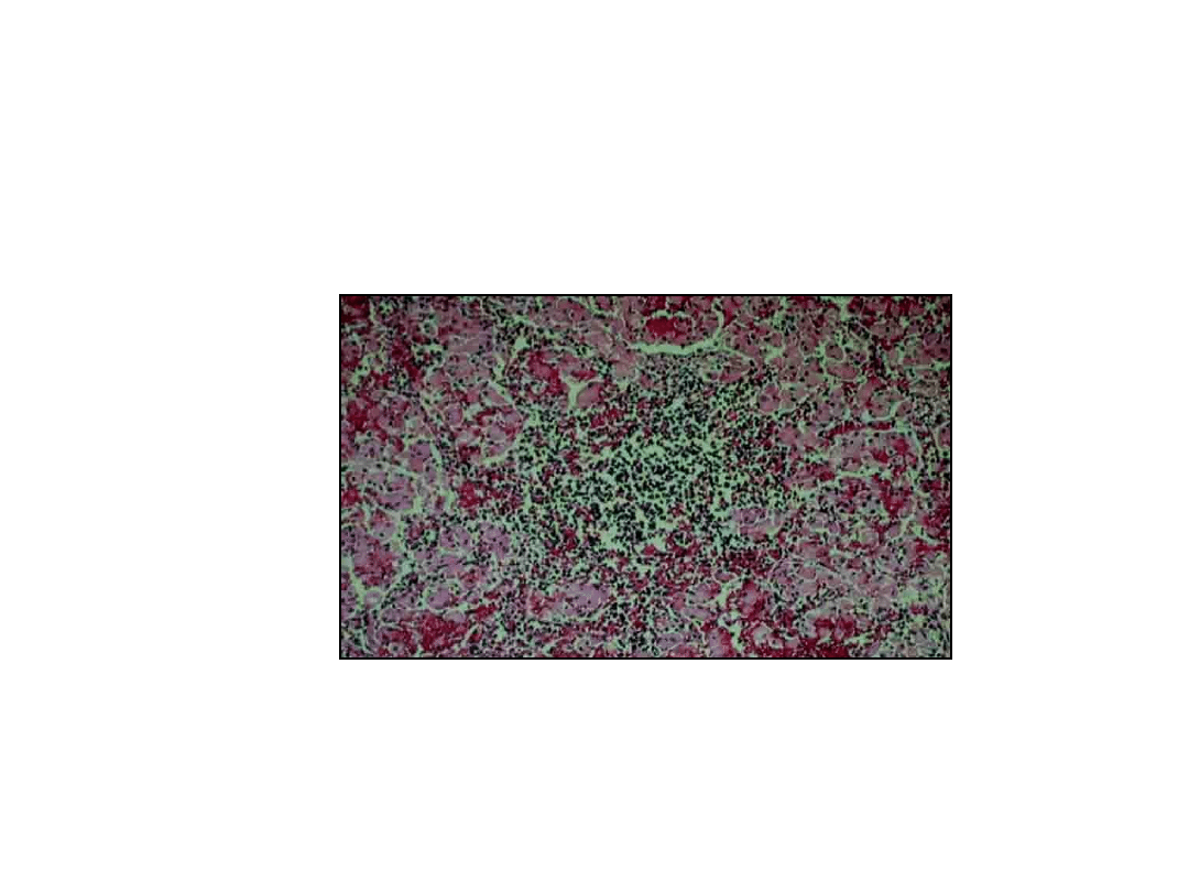
13
Intracellular Accumulations
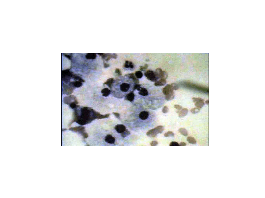
• Cholesterol and Cholesterol Esters
– Atherosclerosis
• macrophages and smooth muscle cells filled with
vacuoles
– Xanthomas
• macrophage accumulation / hereditary and acquired
hyperlipidemias
14
15
16
17

18
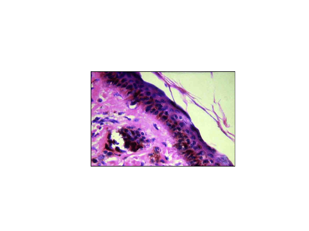
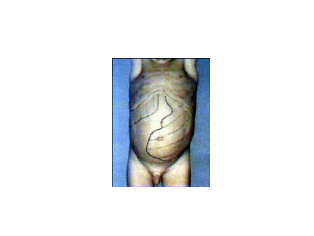
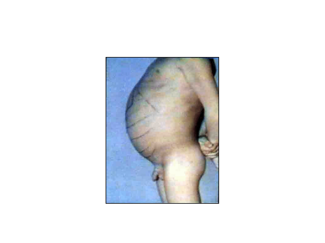
Intracellular Accumulations
• Proteins
– Renal tubular epithelium in proteinuria
– Plasma cells may accumulate
immunoglobulines (Russel bodiers)

19
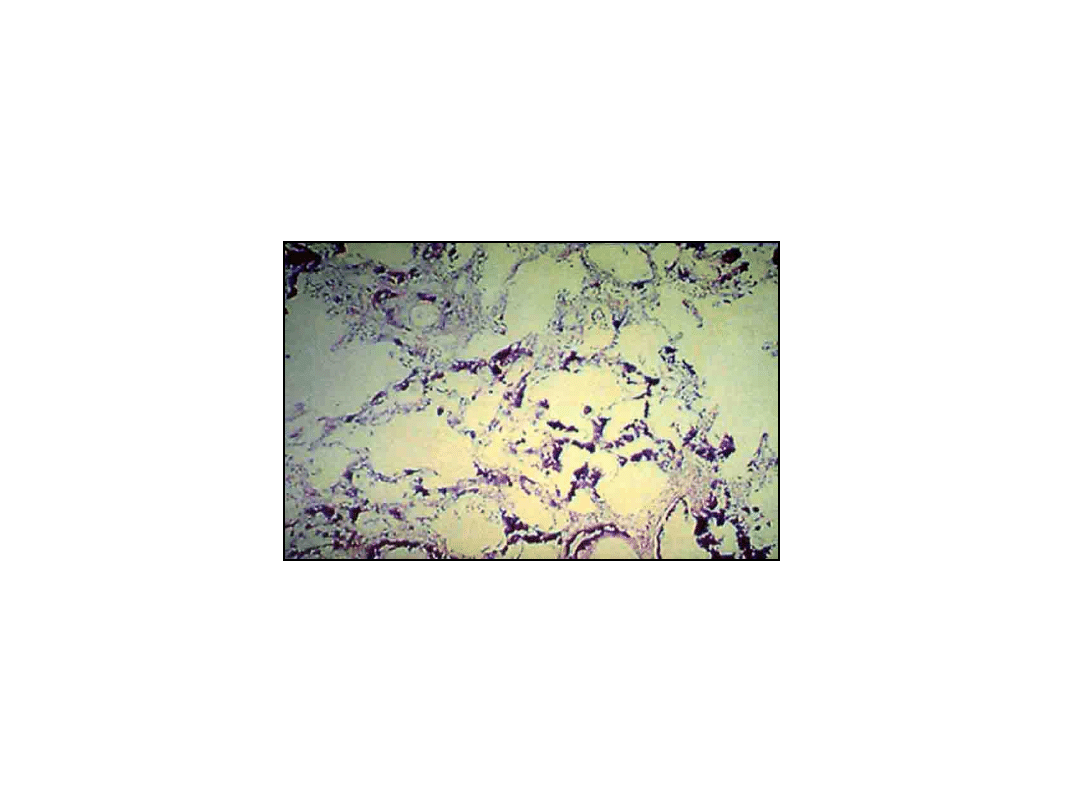
Intracellular Accumulations
• Glycogen
– Diabetes mellitus
• glycogen accumulation in renal tubular epithelium,
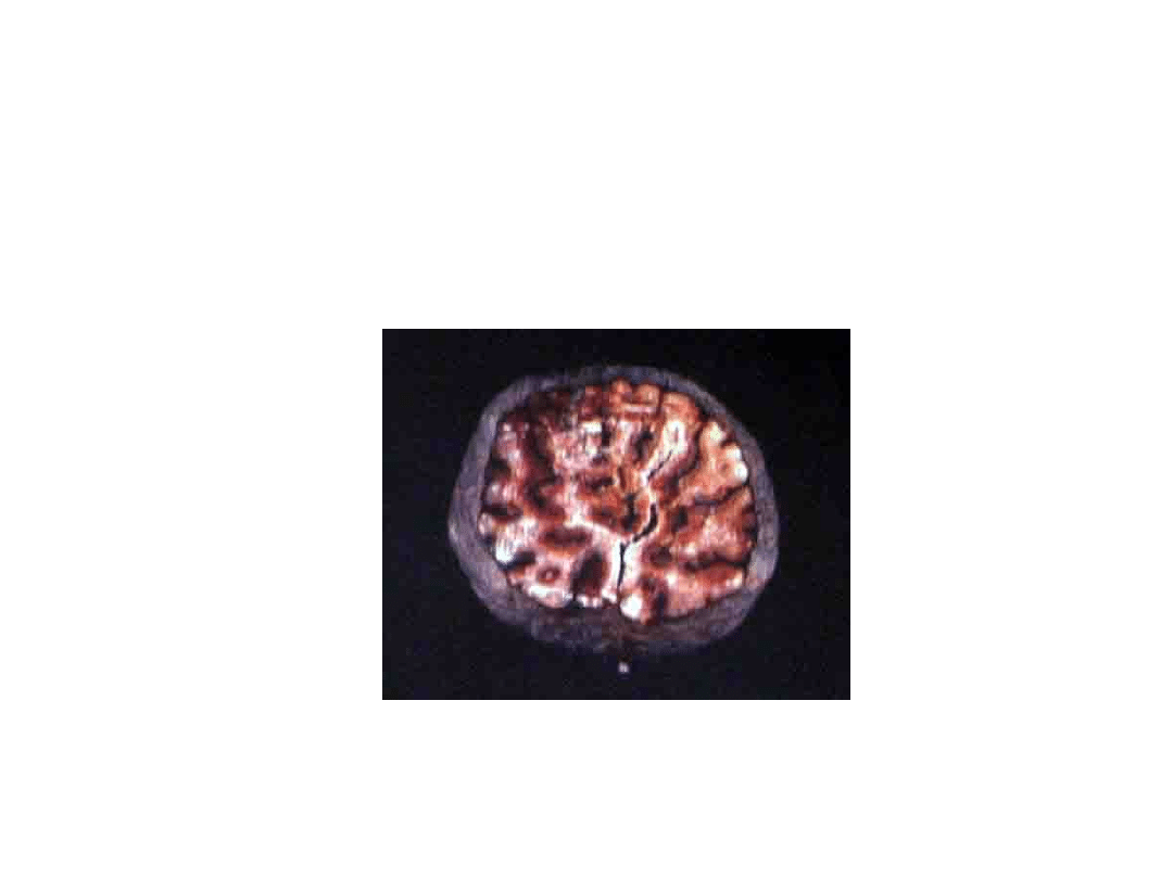
hepatocytes, cardiac myocytes, pancreatic beta cells
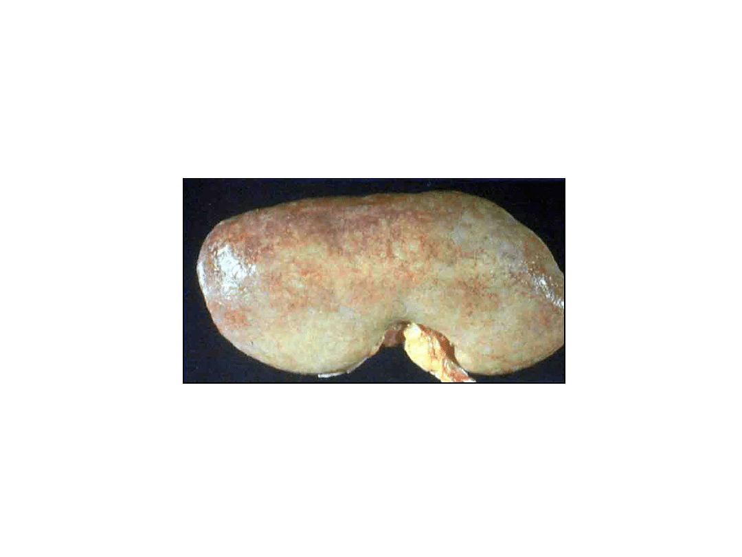
20

21
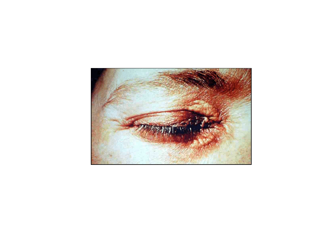
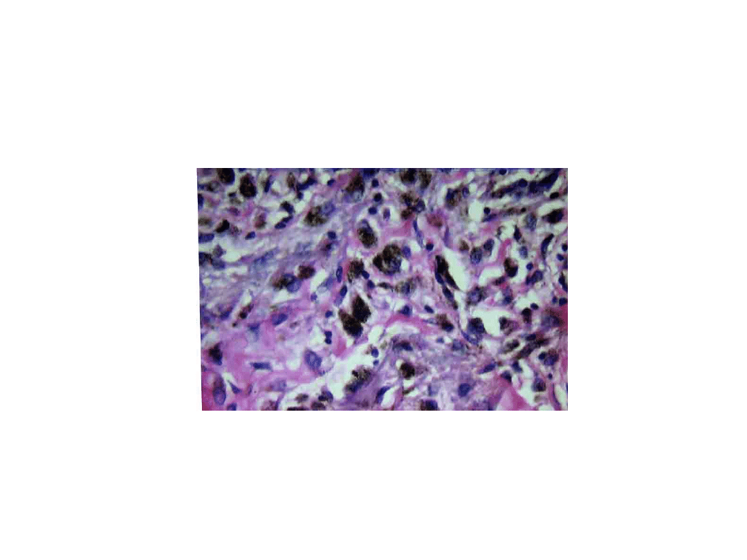
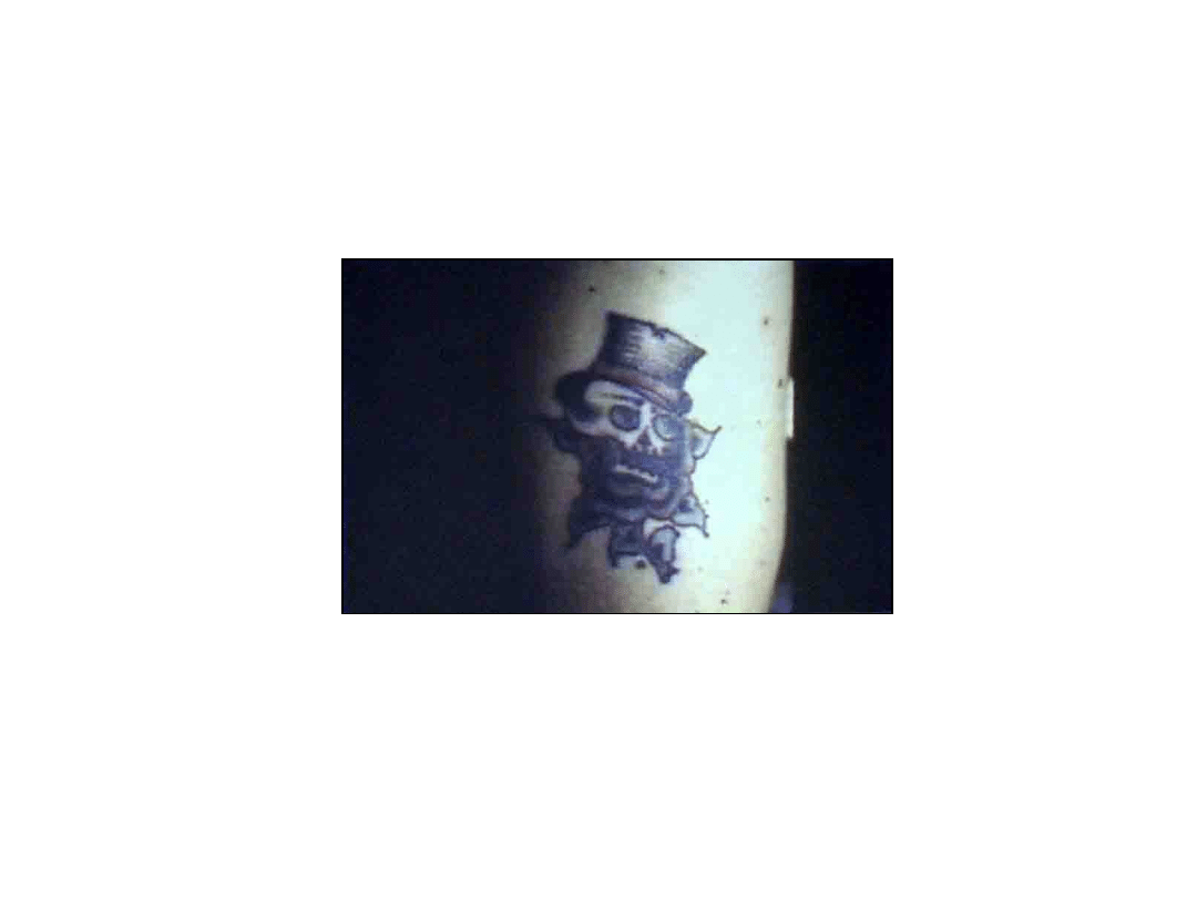
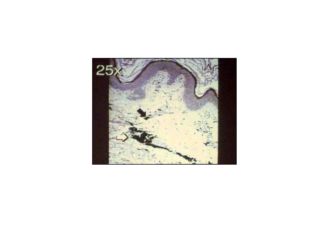
Intracellular Accumulations
• Exogenous Pigments
– Tattoos
• dyes phagocytosed by macrophages
22
23

24
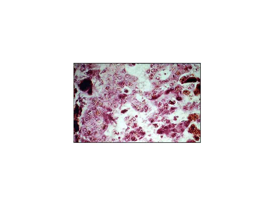
Intracellular Accumulations
• Endogenous Pigments
– Lipofuscin („wear and tear pigment”)
• brownish yellow especially in heart, liver, and brain –
function of age or atrophy („brown atrophy”)
• represents complexes of lipid / protein
• derived from free radical peroxidation of subcellular
membranes
25

26
Intracellular Accumulations
• Endogenous Pigments
– Melanin
• brown-black pigment derived from tyrosine
in melanocytes
• may also accumulate in basal keratinocytes
and dermal macrophages
27

28
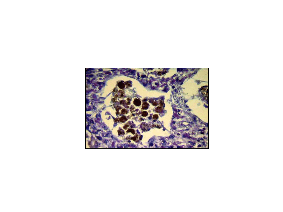
Intracellular Accumulations
• Endogenous Pigments
– Hemosiderin
• hemoglibin derived iron containing
golden-yellow pigment
• represents large aggregates of ferritin
micelles
• small amounts normal in phagocytic
cells of reticuloendothelial system

29
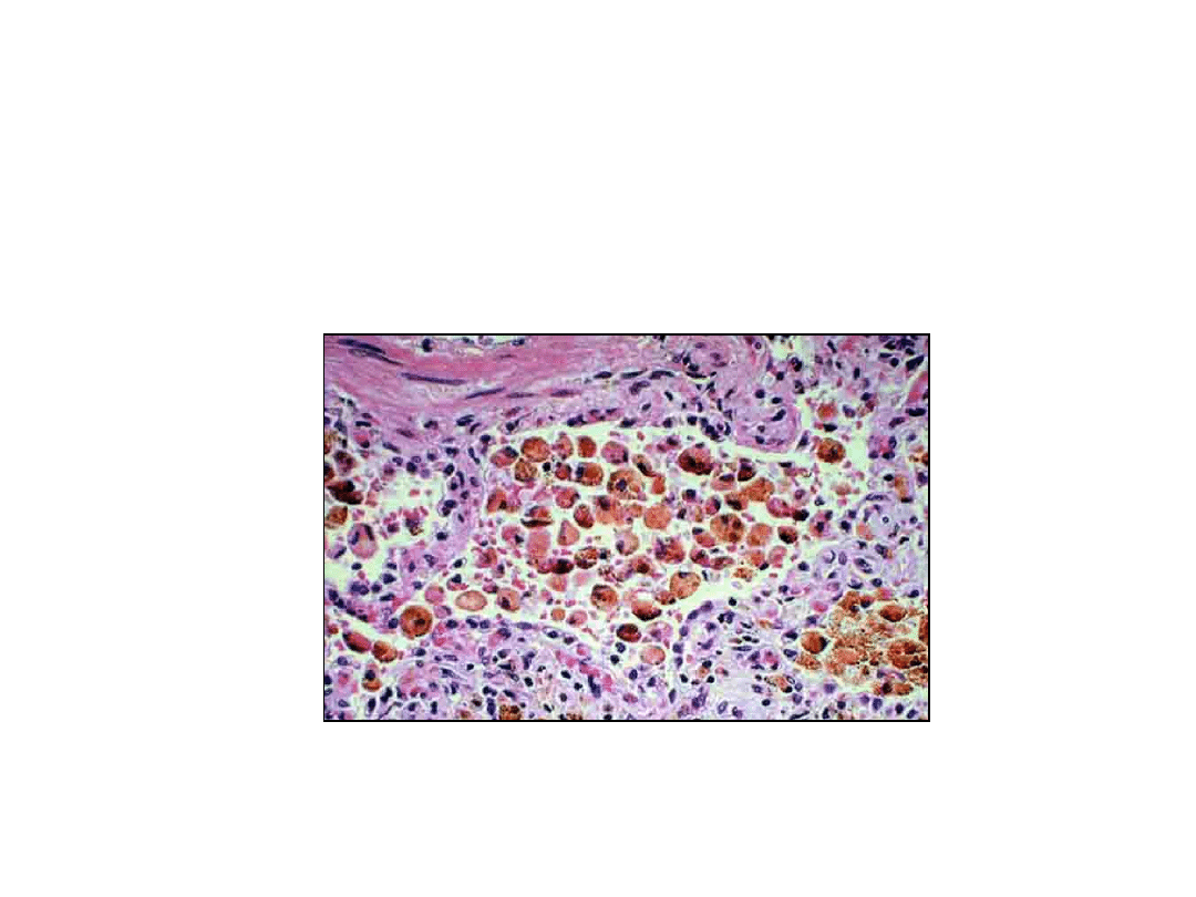
Intracellular Accumulations
• Endogenous Pigments
– Hemosiderin
• local excesses in focal hemorrhage
• systemic iron oberload (hemosiderosis)
– in macrophages and parenchyma mainly
in liver, pancreas, heart and endocrime
organs

30
Intracellular Accumulations
• Endogenous Pigments
– Hedmosiderin
• systemic iron overload (hemosiderosis)
– increases absorption or impaired
utilization of iron; hemolytic anemias;
transfusions
– ectensive accumulation –
hemochromatosis & organ fibrosis
31
32
33

34
Forms and Morphology of
Cell Injury
PATHOLOGIC CALCIFICATION

35
Pathologic Calcification
• Metastatic Calcification
– Occurs in normal tissue
– Occurs with hypercalcemia
٭hyperparathyroidism; bone catabolism with
tumors involving bone; vitamin D
intoxication, sacroidosis; renal failure
• Primary affects vessels, kineyes,
lungs ana gastric mucosa

36
Pathologic Calcification
• Dystrophic Calcification
– Normal serum calcium
– Areas of necrosis or injury
– Intracelular or extracellular
37
38

39
Amyloidosis
• Nature of Amyloid
– Abnormal proteinaceous substance
– Deposite between cells
– Not a single chemical entily
– Appears as a pink
translucentmaterial on H&E stain

40
Amyloidosis
• Chemical nature of Amyloid
– AL (amyloid light chain)
• associatrd with B-cell dyscrasis
• produced by immunoglobulin – secreting
cells

41
Amyloidosis
• Chemical nature of Amyloid
– AA (amyloid associated)
• non-immunoglobulin
• derived from SAA (serum amyloid –
associated precursor protein)
• associated with chronic inflammatory
diseases

42
Immunocyte Ddyscrasias With
Amyloidosis
• Characteristics
– Complete immunoglobulin light
chains (AL.) produced by abberant
monoclonal B-cells, such as in
multiple myeloma
– Serum M (myeloma) spike
– Bence Jones protein (either lambda
or kappa light chains)

43
Reactive Systemic Amyloidosis
• Characteristics
– AA protein deposits
– Occurs in setting of chronic
inflammation

44
Other Types of Amyloidosis
• Heredofamiliar Amyloidosis
– Familiar Mediterranean fever
• AA protein – may be due to reccurent
bounts of anflammation of joints and
serosal surfaces
– Familiar amyloid polyneuropathies
• mutant transthyretins deposited

45
Other Types of Amyloidosis
• Localized Amyloidosis
– Heterogenous chemical composition
and clinical presention
– Often associated with local infiltration
of plasma cells (AL type amyloid)
– Meduliary carcinoma of thyroid
(amyloid chemically related to
calcitonin – a hormone secreted by the
tumor cells

46
Other Types of Amyloidosis
• Amyloid of Aging
– Senile cardiac amyloidosis
• transthyretin
– Senile cerebral amyloidosis (in
Alzheimer’s disease)
• beta-2 amyloid protein

47
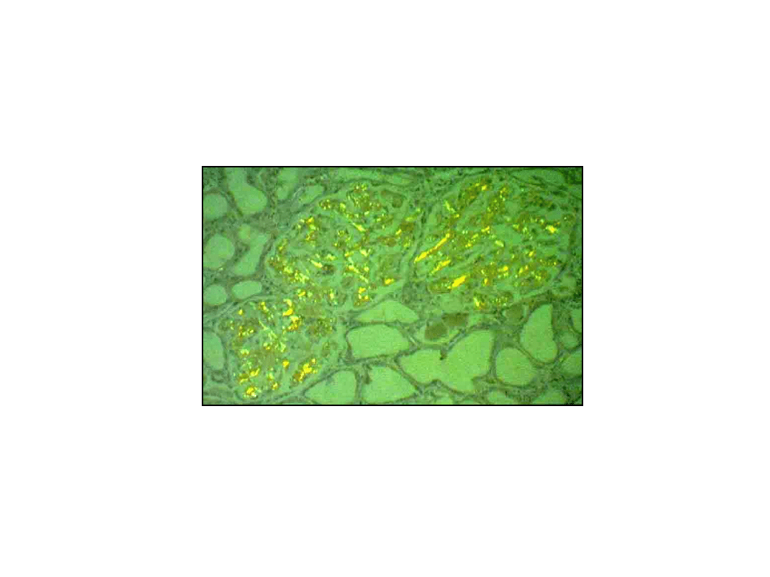
Morphology of Amyloidosis
• Histologic Apperance
– Pink staining intercellular substance
with H&E stain
– Red-orange staining with Congo red
• green birefringence under polarized light
– Often causes parenchymal cell
atrophy or drop out

48
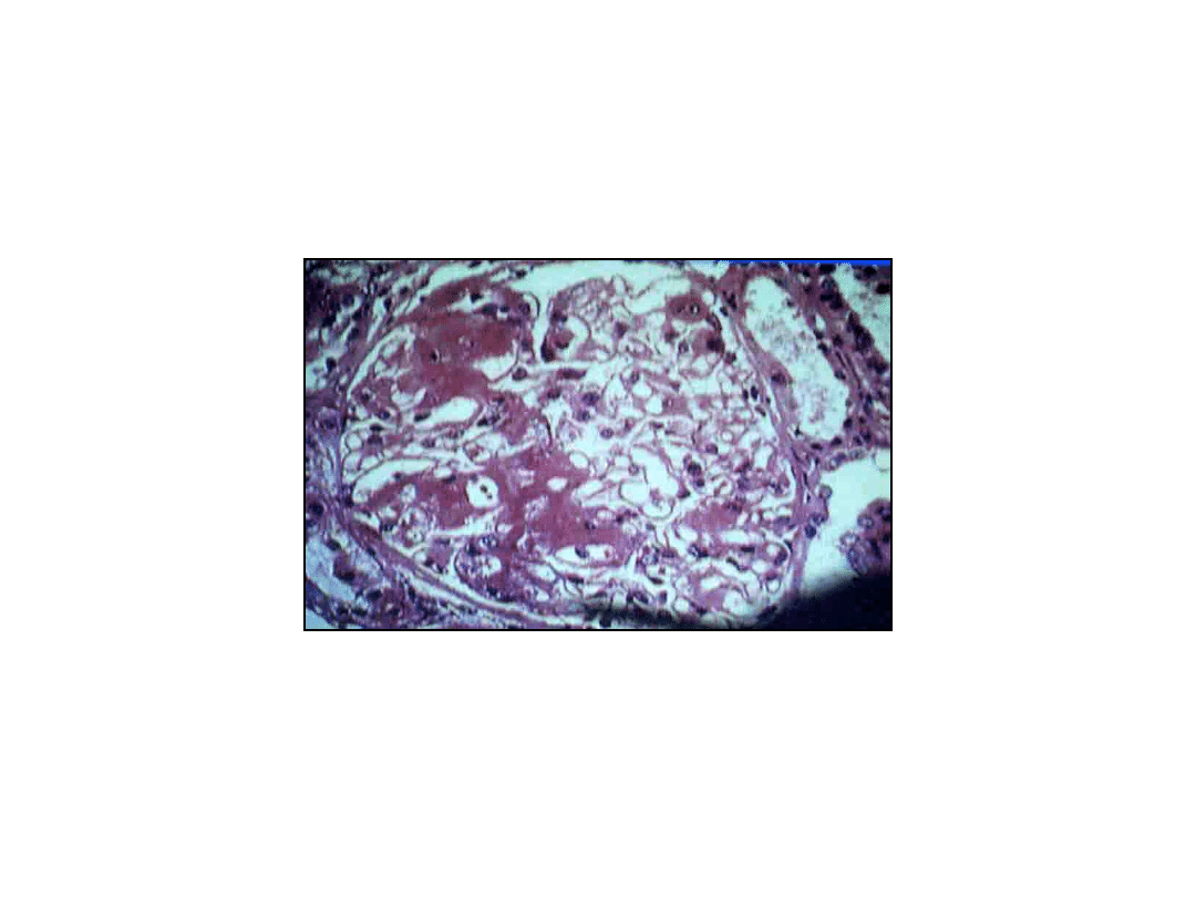
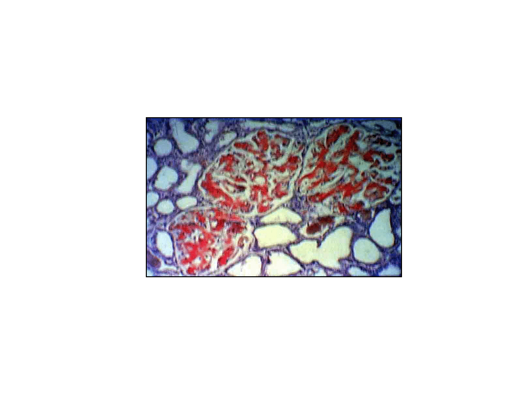
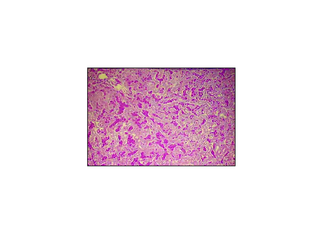
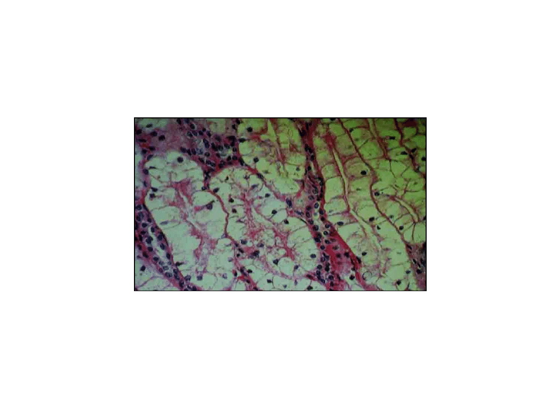
Amyloidosis of the Kidney
• Gross
– Unchanged or large and pale
• Microscopic
– Deposits mainly in glomeruli
• Also present in peritubular interstitium
and walls of blood vessels
49
50
51
52

53
Amyloidosis of Other Organs
• Spleen
– „Sago spleen” – splenic follicles
– „Lardaceous spleen” – splenic sinuses
&pulp
• Liver, Heart, Endocrine glands
– Enlarged
– Interstitial deposits of amyloid
– Pressure atrophy
54
55

56
Clinical Correlation
• Prognosis
– Poor
– Mean survival 1 to 3 years

57
Lysosomal Storage
Diseases

58
Sphingolipidoses
Tay-Sachs, Gaucher and Neimann-
Pick Diseases
59

60
Neimann-Pick Disease
• Characteristics
– Sphingomyelinase deficiency
– Acculmulation of sphingomyelin
– Involves phagocytic cells and neurons
– Spleen, liver, bone marrow, lympph
nodes & lungs as well as CNS affected
– Enlarged vacuolatescells
– Visceromegaly & neurologic defects

61
Gaucher Disease
• Characteristics
– Glucocerebrosidase dificiency
– Accumulation of glucocerebrosides
– Involves phagocytic cells
– Predominantly affects liver, spleen and
bone marrow; CNS in types 2 and 3
– Phagocytes enlarged with a fibrillar
:wrinkled tissue paper” cytoplasm
62
63
64
65

66
Gaucher Disease
• Types
– Type 1 (99%) hepatosplenomegaly and
absence of CNS involvement-longevity
somewhat shortened
– Type 2 severe CNS involvement; secondary
involvement of spleen / liver –highly lethal
– Type 3 involves brain and viscera with a
course intermediate to types 1 and 2

67
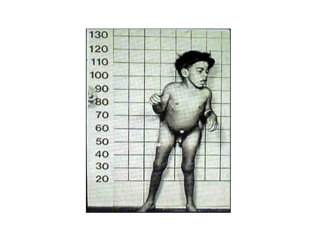
Mucopolysaccharidosis
• General Principles
– Progressive disorders
– Multiple organ invovlvment,
including liver, spleen, heart and
blood vessels
– Coarse facial features, clouding of
the cornea, joint stiffness, mental
retardation
68

69
Glycogen Storage Disorders
von Gierke, McArdle and Pomp
Diseases

70
von Gierke Disease
• Characterictics
– Glucose-6phosphatase deficiency
– Accumulation of glycogen in
cytoplasm
– Affects liver
– Hepatomegaly, hypoglycemia,
renomegaly, failure to thrive
– Mortality about 50%
71
72

73
Hemodynamic Disorders
Learning Objectives
• Explain active hyperemia and
passive congestion and give
clinically important examples of
each process
• Describe the fate of thrombi, with
special emphasis to clinical effects,
organization, recanalization and
embolization

74
Hemodynamic Disorders
Disorders of Perfusion (page 283)
„Hemodynamic disorders are
characterized by disturbed
perfusion that resultsin organ and
cellular injury.”

75
Hemodynamic Disorders
Hyperemia and Congestion
• Active (arterial) – augmented supply of
blood to an organ, usually physiologic
(exercise)
• Passive (venous) – engorgement of an
organ by venous blood, usually the
result of left ventricular heart failure,
which leads, in turn, to right
ventricular failure
76
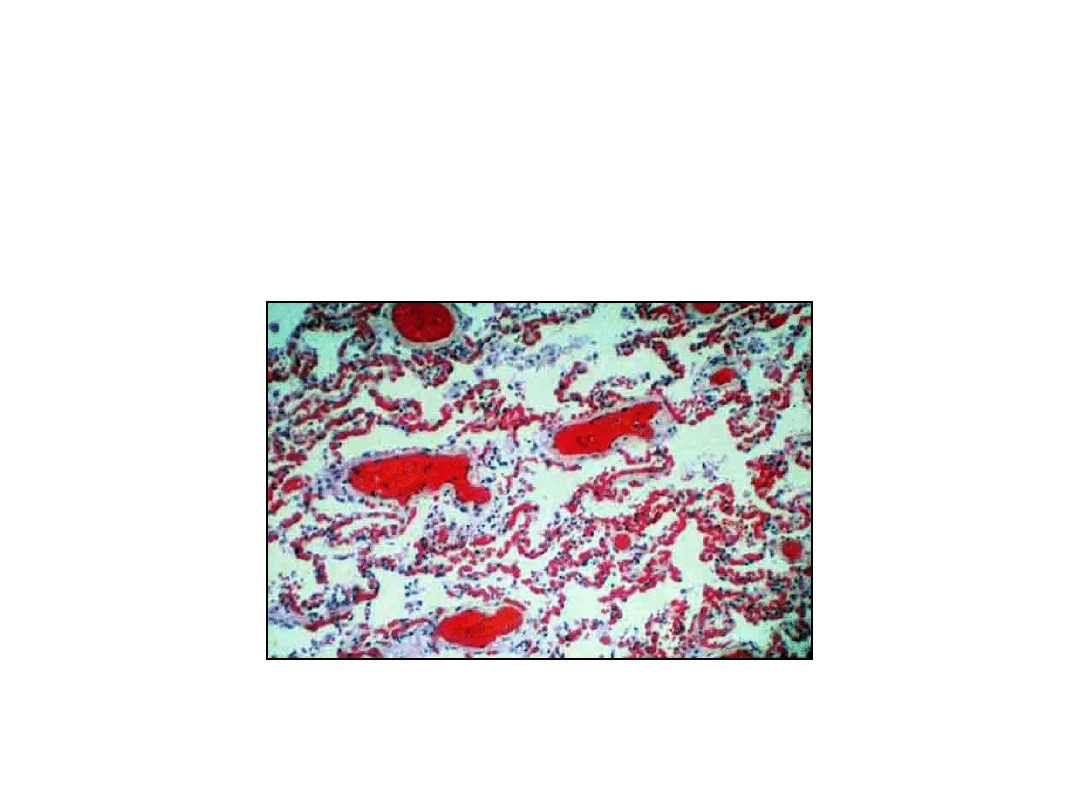
Hemodynamic Disorders
Passive Congestion, Lung
77
Hemodynamic Disorders
Pulmonary Edema, Gross
78
Hemodynamic Disorders
Pulmonary Edema, Micro
79
Hemodynamic Disorders
„Heart Failure Cells”, Lung, Micro
80
Hemodynamic Disorders
Kitchen Patology - Nutmeg
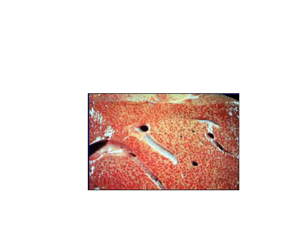
81
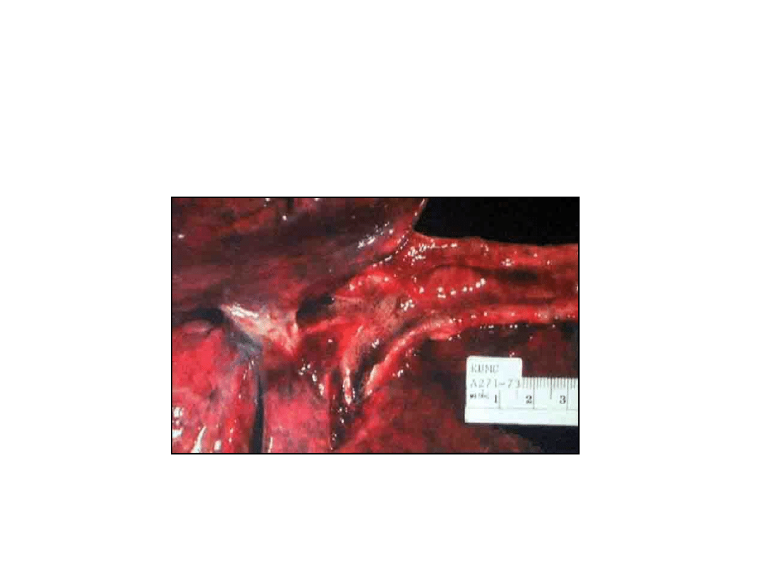
Hemodynamic Disorders
Nutmeg Liver (passive congestion)
82
Hemodynamic Disorders
Nutmeg Liver (centri-lobular
congestion)
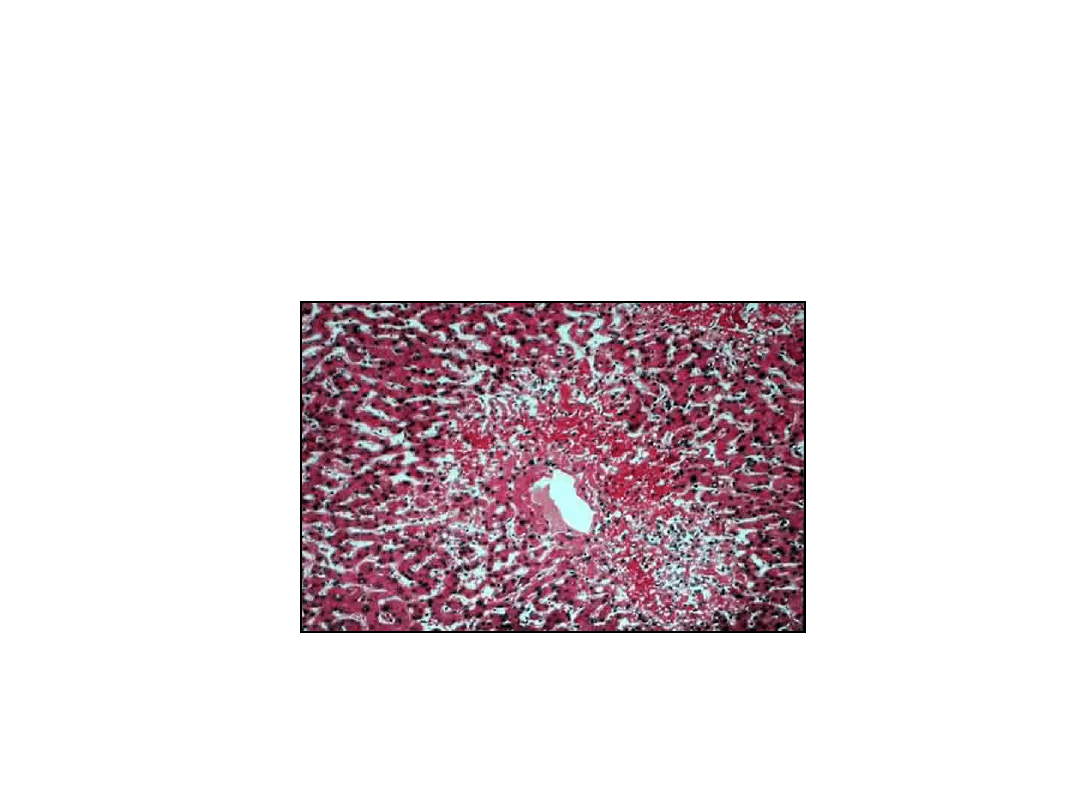
83
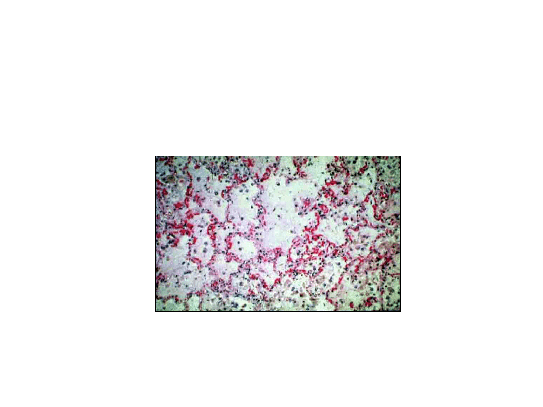
Hemodynamic Disorders
Liver, Passive Congestion, Cell Dropout

84
Hemodynamic Disorders
Hemorrhage
• Hemorrhage is a discharge of blood from the
vascular compartment to the exterior of the
body or into non-vascular body spaces,most
often caused by:
– Trauma (including surgeons)
– congenital defects (berry aneurysm)
– vessel wall defects (athreosclerosis, vasculitis)
– hypertension
– C

85
Hemodynamic Disorders
Hemorrhage - Classifitation
• Hematoma – collection of blood
within a tissue (often muscle)
• Hemopericardium
• Hemothorax
• Hemarthrosis
• Hemoperitoneum

86
Hemodynamic Disorders
Hemorrhage - Classification
• Petechia – pinpoint (capillary)
hemorrhage in the skin or elsewhere.
usually in conjunction with a
coagulophaty or vasculitis
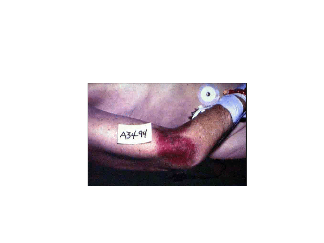
• Purpura – diffuse superficial hemorrhage
in the skin, up to 1cm in diameter
• Ecchymosis (bruise) – A superficial skin
hemorrhage > 1cm size
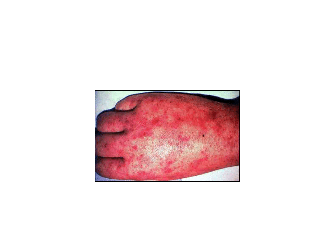
87
Hemodynamic Disorders
Hemorrhage – RMSF Petechial Rash
88
Hemodynamic Disorders
Ecchymosis, Gross
89
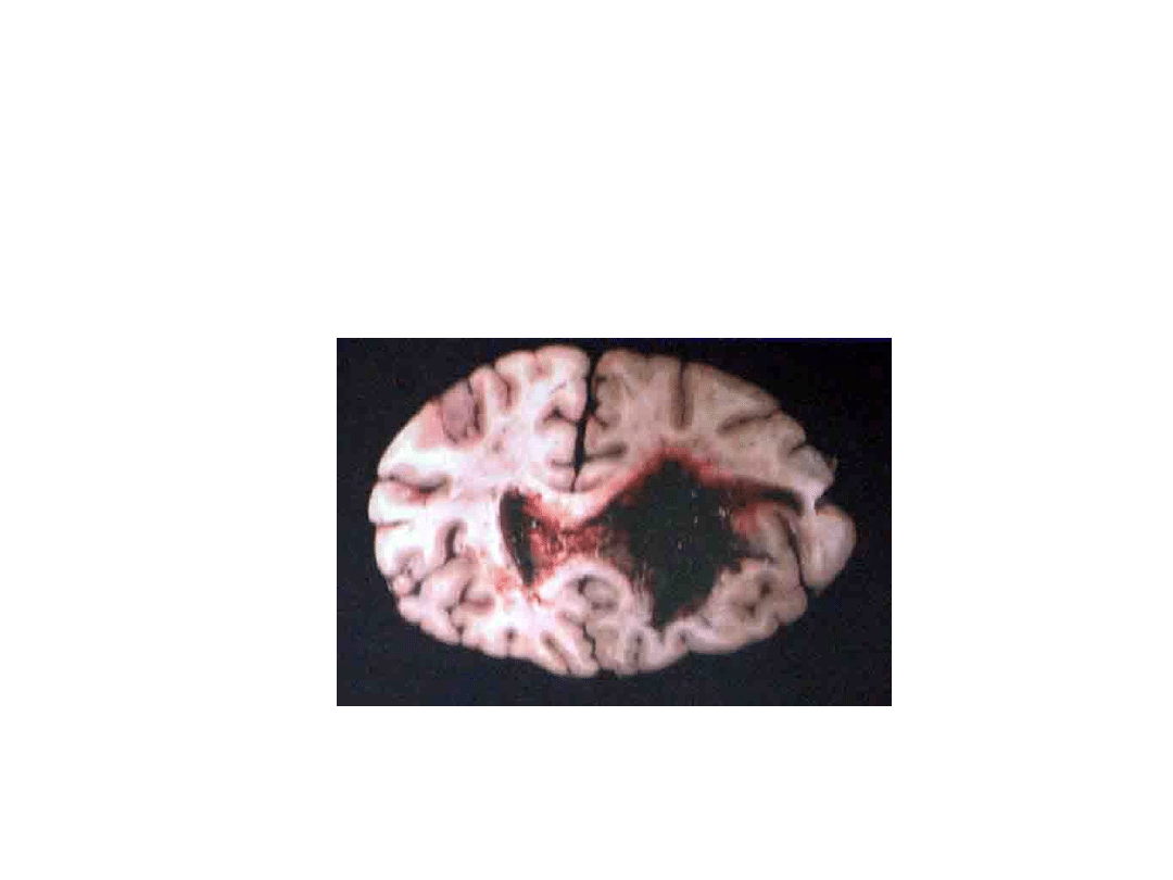
Hemodynamic Disorders
Hypertensive Hemorrhage, Gross

90
Hemodynamic Disorders
Thrombosis - Definition
• Thrombosis refers to the formation
within a vascular lumen of a
thrombus, defined as an aggregate
of coagulated blood containing
platelets, fibrin and entrapped
cellular elements. For all practical
purposes, the term „clot” is
synonymous.
91
Hemodynamic Disorders
Vascular Rheology – Laminar Flow
92
Hemodynamic Disorders
Vascular Rheology – Stenosis
93

94
Hemodynamic Disorders
Thrombosis – fate of thrombi
• Propagation
• Embolization
• Dissolution (lysis by the
thrombolytic system)
• Organization and re-canalization
95
Document Outline
- Slide 1
- Slide 2
- Slide 3
- Slide 4
- Slide 5
- Slide 6
- Slide 7
- Slide 8
- Slide 9
- Slide 10
- Slide 11
- Slide 12
- Slide 13
- Slide 14
- Slide 15
- Slide 16
- Slide 17
- Slide 18
- Slide 19
- Slide 20
- Slide 21
- Slide 22
- Slide 23
- Slide 24
- Slide 25
- Slide 26
- Slide 27
- Slide 28
- Slide 29
- Slide 30
- Slide 31
- Slide 32
- Slide 33
- Slide 34
- Slide 35
- Slide 36
- Slide 37
- Slide 38
- Slide 39
- Slide 40
- Slide 41
- Slide 42
- Slide 43
- Slide 44
- Slide 45
- Slide 46
- Slide 47
- Slide 48
- Slide 49
- Slide 50
- Slide 51
- Slide 52
- Slide 53
- Slide 54
- Slide 55
- Slide 56
- Slide 57
- Slide 58
- Slide 59
- Slide 60
- Slide 61
- Slide 62
- Slide 63
- Slide 64
- Slide 65
- Slide 66
- Slide 67
- Slide 68
- Slide 69
- Slide 70
- Slide 71
- Slide 72
- Slide 73
- Slide 74
- Slide 75
- Slide 76
- Slide 77
- Slide 78
- Slide 79
- Slide 80
- Slide 81
- Slide 82
- Slide 83
- Slide 84
- Slide 85
- Slide 86
- Slide 87
- Slide 88
- Slide 89
- Slide 90
- Slide 91
- Slide 92
- Slide 93
- Slide 94
- Slide 95
Wyszukiwarka
Podobne podstrony:
W01(Patomorfologia) II Lek
W02(Patomorfologia) II Lek
W01(Patomorfologia) II Lek
zagadnienia A, II lek, biofizyka
2.Organizacja i czynność pól czuciowych kory mózgowej, II lek, Fizjologia, !Fizjo, III
pytania- na biofizyke, II lek, biofizyka
patomorfa exam lek
1. Transport tlenu, II lek, Fizjologia, !Krew, III Transport gazów przez krew
W03 08 II
6.Okolice kojarzeniowe kory mózgu i ich znaczenie, II lek, Fizjologia, !Fizjo, III
1.Organizacja i czynność pól ruchowych kory mózgowej, II lek, Fizjologia, !Fizjo, III
10.Zróżnicowanie płciowe mózgu, II lek, Fizjologia, !Fizjo, III
1.Właściwości i czynności receptorów czuciowych, II lek, Fizjologia, !Fizjo, I
2 i 3.Przekazywanie impulsów czuciowych do OUN Prawa, II lek, Fizjologia, !Fizjo, I
8.Czynność bioelektryczna mózg; rodzaje fal mózgowych, II lek, Fizjologia, !Fizjo, III
CII opr, II lek, biofizyka
zag lek10, II lek, biofizyka
a2 pytania, II lek, biofizyka
więcej podobnych podstron