
1
Patomorfologia
Wykład 01
cracked by fazi
created by: sobatolog

2
Aspects of Disease
• Etiology
– cause of disease
• Pathogenesis
– mechanism of development
• Morphologic changes
– structural alterations within cells and organs
caused by a disease
• Clinical & Functional Significance
(Symptoms)
– consequences of morphologic changes

3
Disease Etiology -
VINDICATE
• Vascular
• Infections
– viral, bacterial, mycobacterial, fungal,
protozoall…
• Neoplastic
– malignant tumors…
• Drugs or toxins

4
Disease Etiology – VINDICATE,
cd.:
• Idiopathic
• Congetital
– chromosomal abnormalities, gene defects
• Autoimmune disorders
• Trauma or environmental
– heat, cold, vitamin deficiences, nutritional…
• Endocrine / metabolic
– diabetes, acidosois…

5
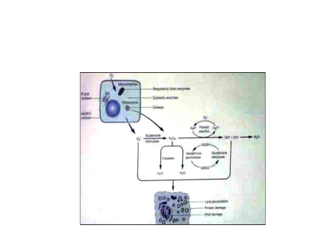
Morphologic Changes
• Morphologic changes in cell and
tissue structure may either be
characteristic or suggestive of the
disease process in question
• Morphologic changes which are
diagnostic of a specific disease are
pathognomic
6
Cell injury
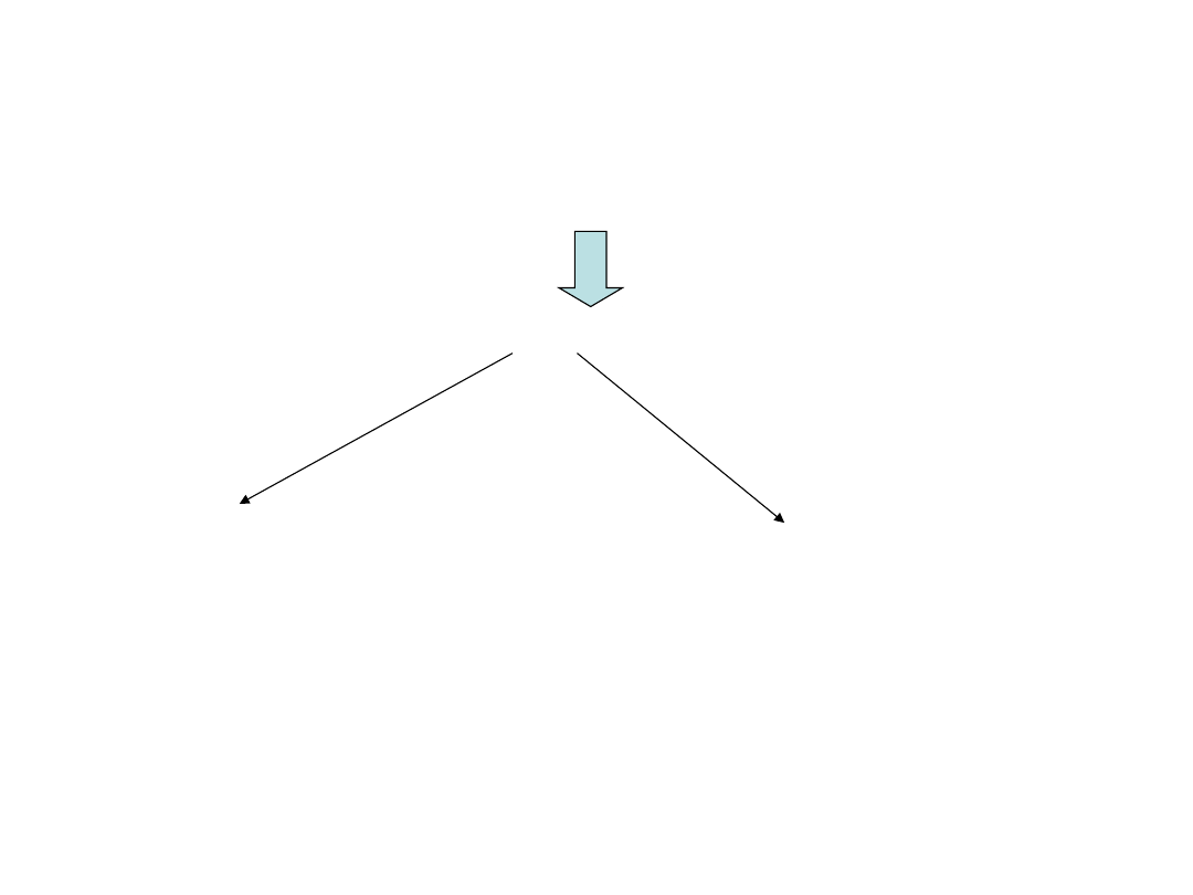
Adaptation
altered steady state
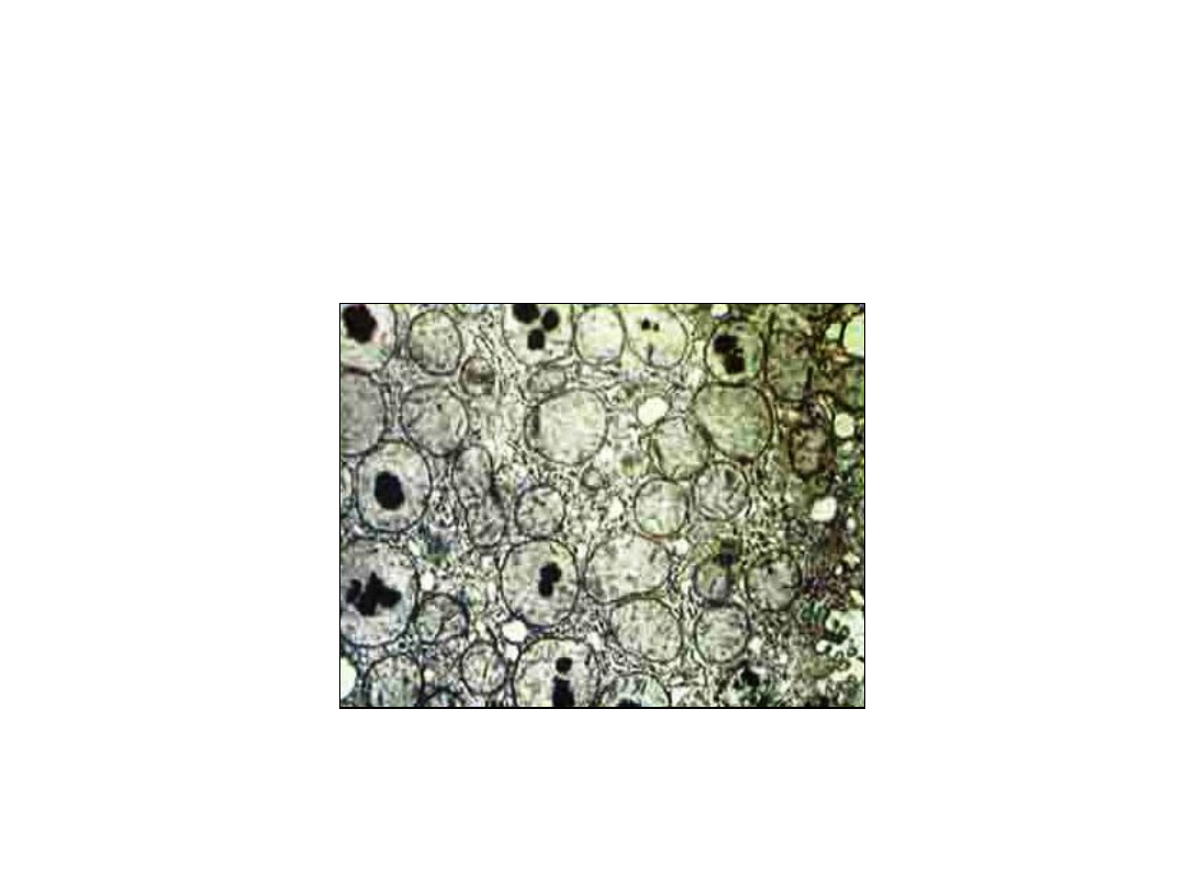
new homeostatis
(i.e.muscle
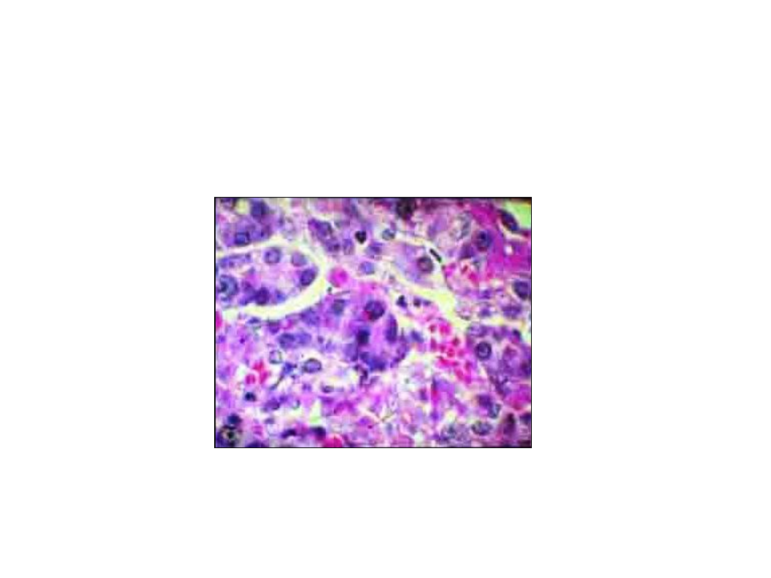
hypertrophy)
Cell injury
reversible or
irreversible cell
death (necrosis)
(myocardial infarct)
Cell in normal environment – homeostatic (steady state)
physiologic stress / pathologic stimull
adaptive capacity exceeded
7
Cell injury

8
Causes Cell Injury
• Hypoxia & ischemia
• Free-radical injury
• Physical agents (heat, cold, radiation,
trauma)
• Chemical agents & drugs
• Infectious organism
• Immunologie reactions
• Genetic derangements
• Nutritional imbalances

9
Principles of Cell Injury
• A cell / tissue’s response to injury is
dependent upon
– Injury factors
• type of infury (ischemia vs. toxic vs. infection
…)
• duration injury
• severity of the inciting injury
– Cellular factors
• cell type
• stage of cell cycle
• cell adaptability

10
Principles of Cell Injury
Susceptibility of cells to Ischemic
Necrosis
• High
– neurons
• 3-5 min. ischemia
• Intermediate
– myocardium, hepatocytes renal
epithelium
• 30 min-2hrs ischemia
• Low
– Fibroblasts, Epidermis Skeletal Muscles
• several hours ischemia

11
Principles of Cell Injury
• Systems hughly vulnerable to injury
– cellular membranes
– mitochondria (aerobic respiration)
– endoplasmic reticulum (protein synthesis)
– genetic apparatus (DNA, RNA)
• Injury at one focus often has a cascade
effect on multiple systems
– impaired aerobic respiration disruption Na
pump cell membrane

12
General Biochemical
Mechanisms of Cell Injury
• ATP depletion
• Oxygen free radicals
• Loss of calcium homestatis
• Membrane damage
• Irreversible mitochondrial damage

13
ATP DEPLETION
• ATP required for
– membrane transport, protein synthesis,
lipogenesis
• Production of ATP
– oxidative phosphorylation of ADP (major
pathway)
• aerobic respiration, requires O
2
– glycolytic pathway (minot pathway)
• does not require O
2

14
Oxygen-Derived Free
Radicals
• Reactive Oxygen Species (ROS)
– O•
2
(superoxide anion), H
2
0
2
(peroxide), OH
(hydroxyl radical)
– byproduct of mitochondrial respiration & other
causes
– radical scavenging system
• suproxide dismutase, catalase, glutathione
peroxidase
• neutralize ROS, prevent cell injury
– imbalance between ROS production and
scavenging system cell injury (oxidative stress)
• damage proteins, lipids, nucleic acids

15
Loss of Calcium (Ca)
Homeostasis
• Normal cell
– High extracellular Ca, Low intracytosolic
Ca
•intracellular Ca sequestered in
mitochondria (mito.) & endo plasmic
reticulum (ER)
•gradients maintained by membrane
associated energy (ATP) dependent
ATPases

16
Loss of Calcium (Ca)
Homeostasis
Cell Injury
• Cell injury (ischemia, certain toxins)
• Influx Ca into cytosol from extracellular
space, mitochondria & endoplasmic
reticulum
• Activates intracellular enzymes cell injury
– proteases damage membrane &
cytoskeleton proteins
– phospholipides damage membranes
– ATPases deplete ATP
– endonucleases damage DNA

17
Membrane Damage
• Loss of membrane selective
permeability
– Ca influx enzyme activation
• Causes
– ATP depletion
• effect energy dependent ATPases
– Direct damage
• bacteria, viruses, complement, neutrphile
enzymes & ROS, lymphocytes porphorins,
physical & chemical agent

18
Irreversible Mitochondrial
Damage
• Causes
– inc. cytoplasmic Ca, phospholipases, ROS …
• Formation of high conductance channels
in inner mitochondrial membrane
(mitochodrial permeability transitions)
– prevent maintenance of proton motive force
(potential) across membrane cessation of
oxidative phosphorylation
• begins at reversible change becomes irreversible

19
Irreversible Mitochondrial
Damage
• Leakage of cytochrome c into cytosol
apoptosis

20
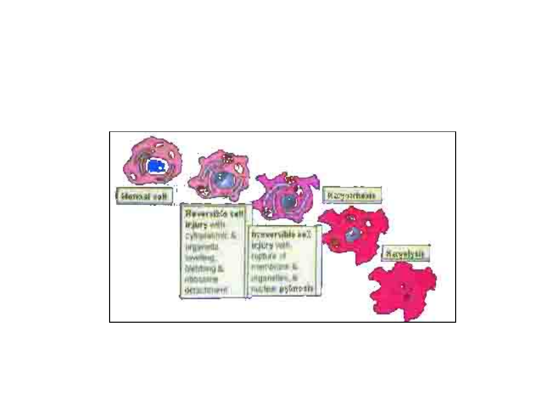
Cell Injury
Reversible vs. Irreversible Injury
• Cell injury is a continuum, and it is not
possible to precisely identify the exact
point at which injury becomes irreversible
– Certain ultrastructural & light microscopie
changes are associated with each type of
infury
• When irreversible injury occurs, the cell
undergoes
necrosis
, which is the light-
microscopie hallmark of cell death

21
Cell Injury
Reversible vs. Irreversible Injury
• Permanent organ injury is generally
associated with cellular necrosis
• The cellular response to persistent
sub-lethal injury reflects adaptation
of the cell to a hostile environment
– these changes are usually
reversible
on
discontinuation of the stress

22
Manifestation of reversible Cell
Injury (Degeneration)
Electron Microscopic Changes
• Cellular swelling (light microscopy)
• Endoplasmic reticulum swelling
• Ribosome detachment endoplasmic
reticulum
• Mitochondria swelling
• Blebs at cell surface
• Loss at microvilli
• Early clumping of nuclear chromatin
23
Reversible Cell Injury
Cell swelling – Light Microscopy
Normal
epithelium
Cellular Swelling
24
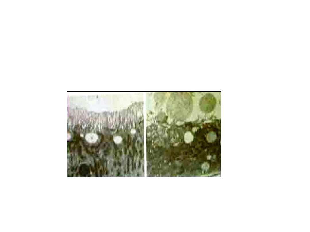
Reversible Cell Injury
Electron Microscopy
(EM)
Normal epithelium
Apical blebbing loss of microvilli

25
Manifestation of Irreversible
Cell Injury (Necrosis)
• Disruption of plasma & cell membranes (EM)
• Rupture of lysosomes (EM)
• Pyknosis – nuclear shrinkage &
condensation (EM & light)
• Karyorrhexis - nuclear shrinkage &
fragmentation (EM & light)
• Karyolysis – nuclear dissolution (EM & light)
• Mitochondrial Dense Bodies (calcium) (EM)

26
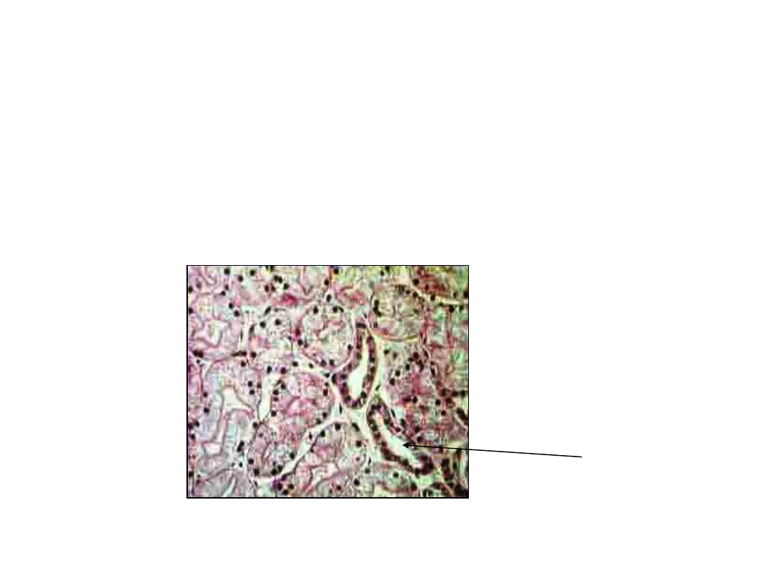
Irreversible Cell Injury
• Irreversible injury is classified into
two types depending on the
underlying mechanism
–
necrosis
&
apoptosis
– both look similar under the light
microscope
•can be distinguished by electron
microscopy
27
Irreversible Cell Injury
Pyknosis – Light Microscopy
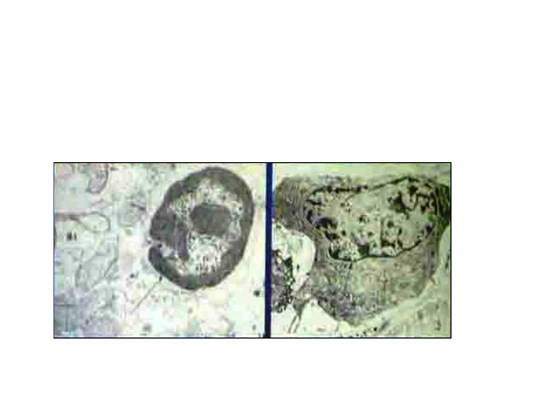
28
Cell Injury
Pyknosis - EM
Pyknotic nucleus
Normal nucleus
29
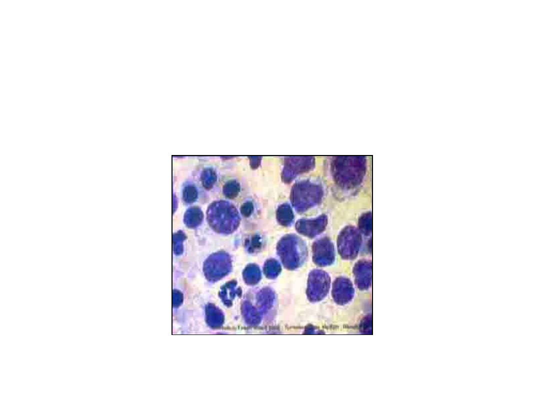
Cell Injury
Karyorrhexis – Light Microscopy
30
Cell Injury
Karyorrhexis - EM
31
Cell Injury
Karyorrhexis – Light Microscopy
32
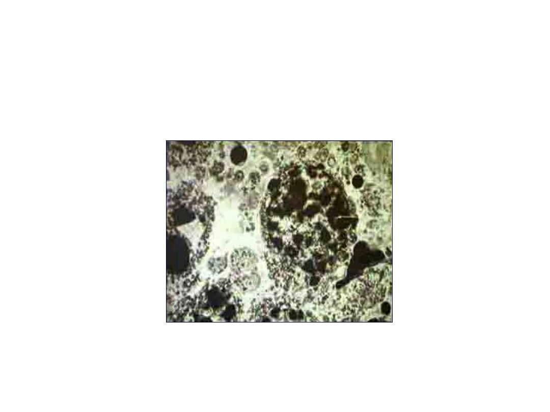
Cell Injury
Dense Bodies in Mitochondria
33
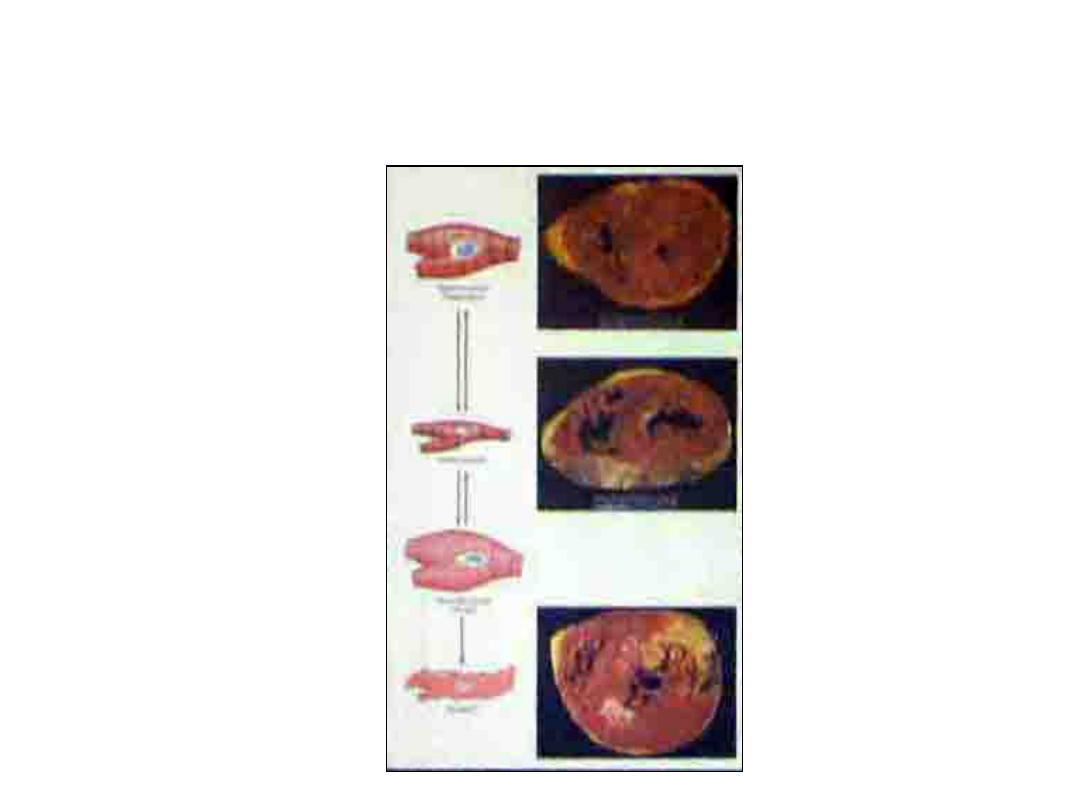
Overview of cell injury

34
Ischemia & Reperfusion
Injury
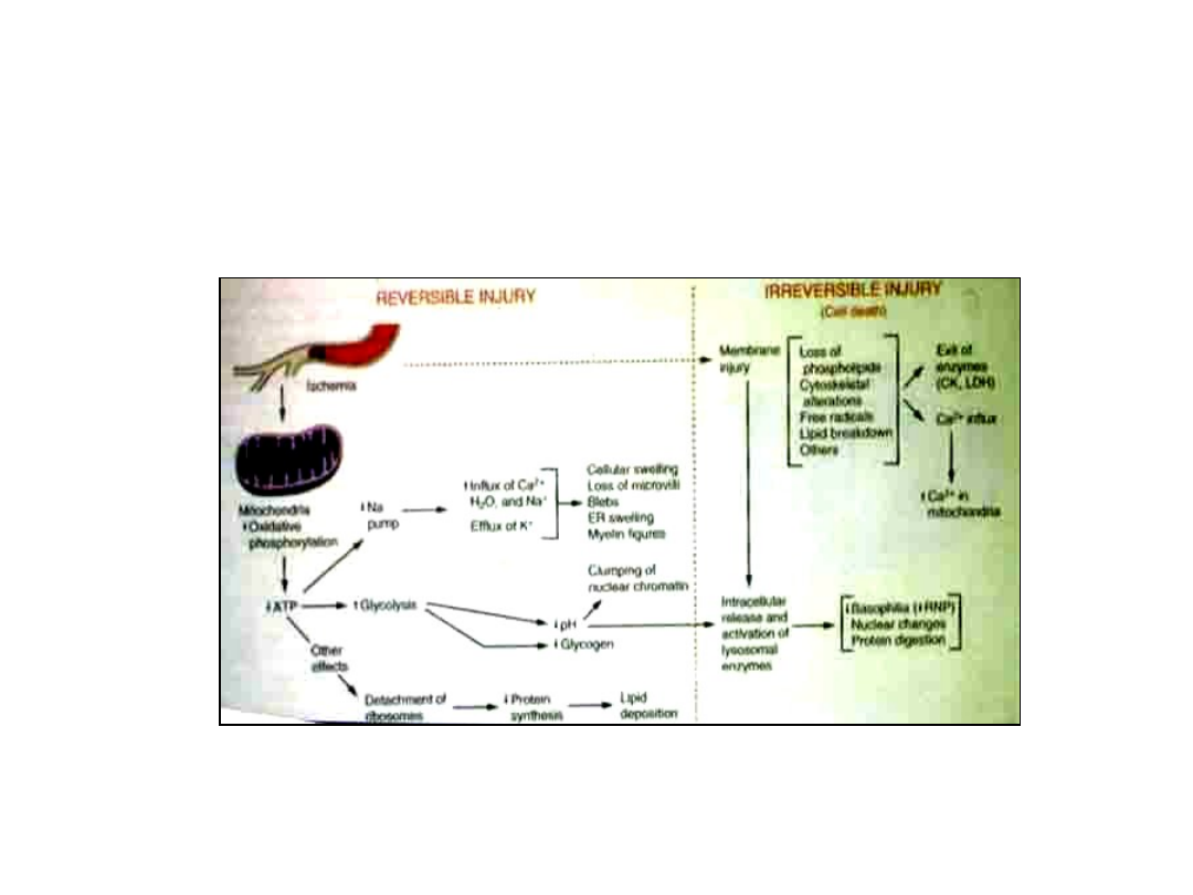
35
Ischemia – Reversible Cell
Injury

36
Ischemia – Reversible Cell
Injury
• Irreversible cell injury is consistently
charakterized by
– Irreversible mitochondrial damage
causing severe ATP depletion
– Severe disruption of membrane function

37
Mechanisms Reperfusion
Injury
• Increased production of ROS by
parenchymal cells, endothelium &
infiltrating leukocytes due to
– Oxidases derived from parenchyma,
endothelial cells & lekocytes
– Cells biochemically damaged by anoxia
• incomplete reduction of O
2
by mitochondria
• compromised radical scavenging system

38
Free Radical – Induced Cell
Injury

39
Free Radicals
• Chemical species (usually O
2
) with a
single unpaired electron in an outar
orbit
• Unstable configuration reactions with
adjacent molecules (proteins, lipids,
carbohydrates, nucleid acids)
• Can initiate autocatalytic reactions
– react with other molecules & convert them
into free radicals
– chain reaction pf auto-propagation
40
Formation of Free Radicals
• Cellular respiration (byproduct)
– O
2
mitochondria O
2
•- SOD H
2
O
2
• Activated PMNs (neutrophils)during
inflammation O
2
•-
• Transition metals (Fe, Cu) in intracellular
reactions donate or accept free electrons
– H
2
O
2
+ Fe
2
+
Fe
3
+
+ OH = OH
-
• Fenton reaction

41
Formation of Free Radicals
• Nitric Oxide (NO)
– produced by endothelial cells, macrophages,
neurons …
– can act as a free radical or be converted to
• ONOO
-
, NO
2
• & NO
3-
• Radiant energy (UV light, ionizing radiation)
• H
2
O OH• + H•
• Enzymatic metabolism of chemical or drugs
by
– CCl
4
P-450 oxidase CCl
3
•

42
Effect of Free Radicals
• Lipid peroxidation of membranes
– double bonds of unsaturated fatty acids
of membrane lipids are attacked by ROS
(especially OH•) H
2
O
2
autocatalytic
reaction severe damage

43
Effect of Free Radicals
• Oxidative modification of proteins
– oxidation of amino acid residues on protein
side chains protein-protein x-linkages
– oxidation of amino acid residues on protein
backbone fragmentation
– oxidative modyfication enhances protein
degradation by peroxisomes loss of
crical enzymes

44
Removal of Free Radicals
• Spontaneous degradation
• Antioxidants
– block formation or inactivate ROS
– Vit. E & A, ascorbic acid, glutathione
• Metal binding proteins
– transferrin, ceruloplasmin, lactoferin

45
Removal of Free Radicals
• Enzymes
– Catalase (within peroxisomes)
• 2H
2
O
2
2H
2
O + O
2
– Superoxide dismutase (SOD)
• 2O
2
• +2H H
2
O
2
+ O
2
– Glutathione peroxidase
• H
2
O
2
+ 2GSH GSSG = 2H
2
O
• 2OH• + 2GSH GSSG + 2H
2
O
46
Free Radicals

47
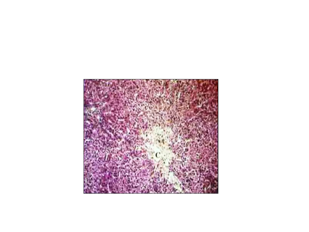
Acetaminophen (Tylenol)
Toxicity
• Large doses depletion GSH
– accumulation of ROS (H
2
O
2
, OH•)
oxidative damage, lipid peroxidation
• major cause of injury
– accumulation toxic metabolite
• destroys nucleophilic macromolecules, binds
proteins & nucleid acids
• Massive hepatic necrosis 3-5 days after
ingestion
– can be reduced by antuoxidant adminitration
48
Acetaminophen Toxicity
Liver

49
Reversible Cell Injury
(Degeneration)
vs. Necrosis

50
Reversible Cell Injury
(Degeneration)
Electron microscopie change
• Endoplastic reticulum swelling
• Ribosome detachment endoplasmic
reticulum
• Mitochondria swelling
• Blebs at cell surface
• Loss of microvilli
• Early clumping of nuclear

51
Reversible Cell Injury
(Degeneration)
Light Microscopy
• Cell swelling (hydropic change /
vacuolar degeneration)
– Cells incapable of maintaining ionic /
fluid homeostasis
– First manifestation of almost all forrms of
cell injury
– Microscopie
•swollen cell w / small clear vacuoles in
cytoplasm
– distended pinched-off ER
52
Cell swelling
swollen cells, vacuoles within cytoplasm

53
Reversible Cell Injury
(Degeneration)
Light Microscopy
• Fatty change
– Hypoxic, metabolic & toxic injury in cells
involvedon fat metabolism
• hepatocytes, myocardial cells
– Microscopie
• small & large lipid vacuoles in cytoplasma
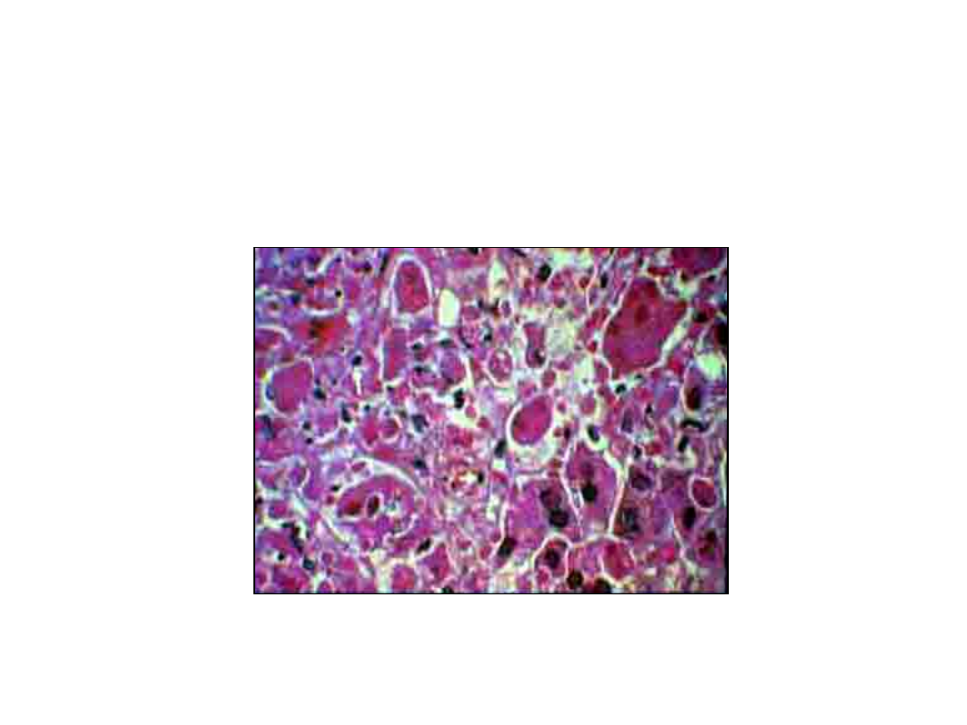
54
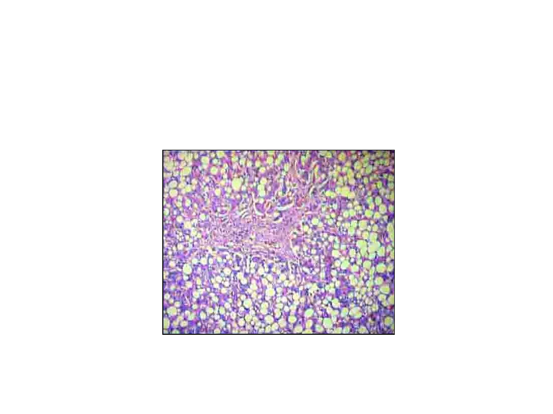
Fatty Change Liver
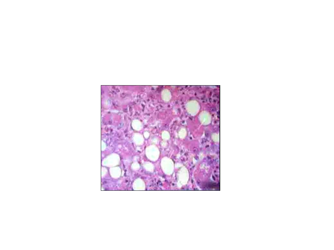
55
Fatty change liver
Document Outline
- Slide 1
- Slide 2
- Slide 3
- Slide 4
- Slide 5
- Slide 6
- Slide 7
- Slide 8
- Slide 9
- Slide 10
- Slide 11
- Slide 12
- Slide 13
- Slide 14
- Slide 15
- Slide 16
- Slide 17
- Slide 18
- Slide 19
- Slide 20
- Slide 21
- Slide 22
- Slide 23
- Slide 24
- Slide 25
- Slide 26
- Slide 27
- Slide 28
- Slide 29
- Slide 30
- Slide 31
- Slide 32
- Slide 33
- Slide 34
- Slide 35
- Slide 36
- Slide 37
- Slide 38
- Slide 39
- Slide 40
- Slide 41
- Slide 42
- Slide 43
- Slide 44
- Slide 45
- Slide 46
- Slide 47
- Slide 48
- Slide 49
- Slide 50
- Slide 51
- Slide 52
- Slide 53
- Slide 54
- Slide 55
Wyszukiwarka
Podobne podstrony:
W02(Patomorfologia) II Lek
W03(Patomorfologia) II Lek
zagadnienia A, II lek, biofizyka
2.Organizacja i czynność pól czuciowych kory mózgowej, II lek, Fizjologia, !Fizjo, III
pytania- na biofizyke, II lek, biofizyka
patomorfa exam lek
1. Transport tlenu, II lek, Fizjologia, !Krew, III Transport gazów przez krew
6.Okolice kojarzeniowe kory mózgu i ich znaczenie, II lek, Fizjologia, !Fizjo, III
1.Organizacja i czynność pól ruchowych kory mózgowej, II lek, Fizjologia, !Fizjo, III
10.Zróżnicowanie płciowe mózgu, II lek, Fizjologia, !Fizjo, III
1.Właściwości i czynności receptorów czuciowych, II lek, Fizjologia, !Fizjo, I
2 i 3.Przekazywanie impulsów czuciowych do OUN Prawa, II lek, Fizjologia, !Fizjo, I
8.Czynność bioelektryczna mózg; rodzaje fal mózgowych, II lek, Fizjologia, !Fizjo, III
CII opr, II lek, biofizyka
zag lek10, II lek, biofizyka
a2 pytania, II lek, biofizyka
3 i 4Czynność kory wzrokowej i słuchowej, II lek, Fizjologia, !Fizjo, III
więcej podobnych podstron