* Corresponding author. Tel.: #49-431-210-2660; fax: #49-431-
210-62660.
E-mail address: mohammed.es-souni@fh-kiel.de (M. Es-Souni).
Biomaterials 22 (2001) 2153}2161
On the transformation behaviour, mechanical properties
and biocompatibility of two NiTi-based shape
memory alloys: NiTi42 and NiTi42Cu7
Mohammed Es-Souni
*, Martha Es-Souni, Helge Fischer Brandies
Materials Testing and Joining, University of Applied Sciences, Fachhochschule Kiel, Grenzstrasse 3, D-24149 Kiel, Germany
Clinic of Orthodontics, Christian-Albrecht-University, Arnold Heller Strasse, D-24113 Kiel, Germany
Received 28 July 2000; accepted 20 November 2000
Abstract
The transformation behaviour, mechanical properties and cytotoxicity of a binary NiTi42 and a ternary NiTi42Cu7 alloy have been
investigated. The transformation temperatures were determined via di!erential scanning calorimetry, the mechanical properties have
been investigated in 3-point bending tests in the temperature range between 6 and 603C. The cytotoxicity tests were performed on
both alloys in cultured epithelial cells from human gingiva. The cytotoxicity investigations included both MTT tests and morphologi-
cal observations. It is shown that although the ternary alloy is characterised by a narrower hysteresis and superior mechanical
properties, including fatigue resistance, its cytotoxicity is higher than that of the binary alloy. This is thought to arise from the release
of copper ions in the medium, which upon atomic absorption spectroscopy measurements amount to approximately 2.8
g cm\ for
an incubation period of 7 days.
2001 Elsevier Science Ltd. All rights reserved.
Keywords: Shape memory alloys; NiTi; NiTiCu; Bending properties; Biocompatibility; Cytotoxicity
1. Introduction
NiTi-based shape memory alloys are being used in
many engineering and biomedical applications including
actuators, orthodontic arch wires, stents, etc.; more de-
tails about the mechanisms of the shape memory e!ect as
well as the potential applications of NiTi alloys may be
found in numerous review articles (e.g. [1}3]). The NiTi
shape memory alloys usually consist of binary alloys,
with Ni and Ti concentrations near the equiatomic com-
position. Their microstructures are generally processed
using complex thermomechanical treatments in order to
obtain suitable properties, e.g. thermal shape memory,
superelasticity, all-round memory e!ect, etc. However,
the binary alloys are known to be susceptible to thermal
cycling fatigue and the accompanying property modi"ca-
tion is a limiting factor in the long-term application of
such alloys [1,4]. A number of ternary NiTi-X alloys
have been introduced with the aim to improve the fatigue
properties and to decrease the thermal hysteresis range;
well-known ternary alloying elements are Cu and Fe
[1,4,5]. For a review on ternary NiTi-X and their proper-
ties see [1].
Particularly, Cu has been shown to dissolve in the B2
(austenite ) phase in a concentration up to 30 at%. How-
ever, NiTi}Cu solid solutions containing more than
10 at% are characterised by poor formability so that
alloys of technical interest usually contain Cu in the
range from 5 to 10 at%. The following e!ects of Cu on
the properties of NiTi-base alloys have been reported
[1,4,5]:
E Cu in the range from 5 to 15 at% changes the crystal-
lographic structure of the low-temperature phase. The
phase transformation of the austenite phase occurs
following the sequence: B2 (austenite, bcc)PB19
(martensite, orthorhombic)PB19
(martensite, mono-
clinic).
E The thermal hysteresis is reported to decrease from
40 K for the binary alloy to 11 K for NiTi}10 at%Cu.
E The stress of the superelastic hysteresis is decreased.
0142-9612/01/$ - see front matter
2001 Elsevier Science Ltd. All rights reserved.
PII: S 0 1 4 2 - 9 6 1 2 (0 0 ) 0 0 4 0 6 - 3

E The fatigue resistance during thermal cycling is
increased.
The (thermo)mechanical properties of NiTi-base alloys
certainly constitute the outstanding and most perceptible
attribute for their use. However, in biomedical applica-
tions involving contact with live tissues, biocompatibility
issues may prove as crucial (if not more) as mechanical
behaviour. The binary NiTi alloys have been reported to
exhibit good biocompatibility due to their high corrosion
resistance [5}10]. However, detailed investigations using
human cell cultures are still rare.
The aim of the present paper is to describe and discuss
comparatively the properties of a binary NiTi and a ter-
nary NiTi}Cu alloy in terms of their transformation
behaviour, mechanical properties and biocompatibility.
2. Materials and methods
The alloys studied are
`Neosentalloy F 80a (GAC),
hereafter designed NiTi, and
`Thermo Active Copper
353C
a (Ormco/A-Company), hereafter designed NiTiCu.
The samples were in form of arch wires with rectangular
sections of 0.55
;0.40 mm. For the di
!erent investiga-
tions the straight ends of the wires were used. The chem-
ical compositions of the alloys were determined in an
analytical scanning electron microscope using EDS anal-
ysis. The following compositions (in wt%) were found:
Ni57, 6Ti42, 4 for the binary NiTi and Ni50, 7Ti42, 4Cu6,
9 for the ternary NiTiCu alloy, respectively.
The transformation behaviour was investigated using
di!erential scanning calorimetry (DSC) in the temper-
ature range from !80 to #803C at a rate of 10 K/min.
The samples weighing approximately 10 mg were gently
cut and ultrasonically cleaned in acetone before testing.
A base line was recorded before each test run. Three
batches were tested for each alloy with however repro-
ducible results in the range of accuracy of the system
($1 K).
The mechanical properties were investigated in 3-point
bending tests using a beam length of 12 mm. The load vs.
de#ection curves were recorded at 6, 22, 37 and 603C.
Heating and cooling of the specimens were conducted in
an oil bath of constant viscosity in the range of the
temperature investigated. The temperature was control-
led to$0.53C using a closed-circuit cooler/heater (hetof-
rig, Heto). In order to show the e!ects of loading and
unloading cycles on the mechanical properties, "ve cycles
were generally performed at each temperature. The sam-
ples were changed for each temperature run.
The biocompatibility of the chosen alloys was charac-
terised by means of the in vitro MTT cytotoxicity test
as
well
as
morphological
investigations
in
the
SEM on cultured epithelial cells from explants of human
gingiva.
The following gives a detailed description of the pro-
cedures for cell culture and the biocompatibility tests:
2.1. Cell culture of gingival epithelial cells from clinically
healthy gingival explants
Tissue explants from "ve subjects (not treated for
orthodontic failures) aged between 33 and 59 years, have
been used for this purpose. A modi"ed method for the
primary culture [11] and subculture of gingival epithelial
cells was used. Right after excision, the explants were cut
into cubes of about 1 mm
, and cultured in -MEM
(supplemented with 10% FCS, 2.5
g/ml AmphotericinB,
100 U/ml Penicillin and 100
g/ml Streptomycin, BIO-
CHROM) in sterilised non-treated culture dishes (FAL-
CON) at 373C, and 5% CO. The medium was changed
every 72 hours beginning with the "fth day after seeding.
A week after seeding,
-MEM was replaced for
keratinocyte-SFM (GIBCO BRL, Life technologies).
Under these conditions, only epithelial cell growth was
observed.
Approximately 11 days after seeding, epithelial cell
colonies spread around each explant, covering an area of
about 1 cm
. At this stage, the cells were subcultured. The
colonies were removed by treatment with 1.5 ml Tryp-
sin/EDTA (0.05/0.02%, BIOCHROM) per dish for 5 min
at 373C and 5% CO. The trypsin activity was blocked
by addition of 1.5 ml
-MEM (containing 10% FCS) per
dish. Subsequently the suspension was centrifuged for
7 min at 120
;g. The supernatant was carefully aspirated
and the cells resuspened in fresh SFM. For the MTT test,
10
cells were seeded per microwell.
2.2. Immunohistochemical staining
Prior to biotoxicity tests, the cultured cells were
stained immuno-histochemically in order to prove their
epithelial character. For this purpose, they were tested
for their staining capacity by anti-human cytokeratin
AE1, AE3 (DAKO), a typical epithelial cell marker. The
indirect staining procedure was used [12]. The anti-
cytokeratin A1/A3 and the alkaline phosphatase-con-
jugated rabbit anti-mouse immunoglobulins were used in
a dilution of 1/100 and 1/10, respectively. Dilutions were
performed with the ready to use antibody diluent
(DAKO). The fast red substrate system (DAKO) was
used
as
substrate
and
chromogen
in
the
im-
munocytochemical staining procedure. Counterstaining
was performed with hematoxylin (DAKO). Prior to
staining, monolayers of cells were cultured on coverslips.
After "xation in 2.5% glutaraldehyde for 30 min and
washing in PBS pH 7.2, the cell cultures were treated
with 0.5% Triton X100 (MERCK) in order to promote
antibody access. After permeabilization the monolayers
were brought in contact with the primary antibody for
30 min. After thorough washing in PBS, the coverslips
2154
M. Es-Souni et al. / Biomaterials 22 (2001) 2153}2161

were covered with the alkaline phosphatase-conjugated
rabbit
anti-mouse
immunoglobulins
for
another
30 min and washed subsequently in PBS. The "ltered
fast red substrate solution (DAKO) was then allowed to
react with the bound phosphatase for 20 min. Counter-
staining was performed by incubation in an aqueous
solution containing hematoxylin for 1 min followed by
washing in warm tap water. Coversplips were mounted
on slides in an aqueous mounting medium (Faramount,
DAKO) and observed in an NIKON Optiphot 2 light
microscope.
2.3. Alloy substrates and dimensions
The tested alloys were cut into pieces of 5 mm length,
0.40 mm thickness and 0.55 mm width when used in the
MTT test, otherwise pieces of 21.5 mm length were pre-
pared. They were "rst cleaned ultrasonically in absolute
alcohol and then heat sterilised at 1203C for 20 min.
2.4. MTT cytotoxicity test
For the cytotoxicity determination of the tested alloys,
the method of Tada et al. [13] was applied. The test was
performed in sterile 96-microtiter plates (COSTAR) at
373C
and
5%
CO equipped with two pieces
(2
;9.9 mm active surface) of the tested alloys. The
"l-
tered MTT solution (100
l/well, 5 mg/ml) was added to
the cell cultures (i) 24 h, (ii) 48 h and (iii) 72 h after seeding.
4 h after the MTT addition, cell lysis and formazan
solubilization were started by adding 100
l of the lysing
bu!er (10% SDS in 0.01
N
HCl pH 3.5) and incubating
the plates for 20 h at 373C and 5% CO. Wells contain-
ing cells but no alloys were used for negative controls.
The positive controls consisted of wells containing cells,
where the addition of lysing bu!er preceded the addition
of the MTT solution. For each alloy, blanks containing
the metal, but no cells, were prepared. All experiments
were carried out in triplicate. Prior to the determination
of the formazan formation by absorption measurements
at 550 nm in a microplate reader (DYNATECH, MR
5000), 175
l supernatent of each well were transferred
into a new plate. The incoherent background was deter-
mined by absorption measurements at 630 nm. Coherent
background, due to the phenol red absorption of the
SFM was expressed in the remaining absorption of the
blanks. All absorption measurements were then correc-
ted for this background e!ect by subtraction.
2.5. Atomic absorption spectroscopy (AAS) measurements
Preliminary AAS measurements were conducted on
the ternary alloy in order to investigate Cu-ion release.
For this purpose, one piece of the ternary alloy with an
active surface of 0.38 cm
was incubated in cell free
SFM-medium for 7 days at 373C and 5% CO. After
incubation, the medium was "ltered through a sterile
millipore "lter with a pore size of 0.2
m and sub-
sequently analysed. As a blank measurement, a sample-
free medium was used.
2.6. Morphological investigations in the SEM
Two millilitre of a cell suspension containing 10
C/ml
were seeded in wells of sterile 12-well culture plates
(COSTAR). Prior to seeding, the wells were equipped
with sterilised coverslips (d"13 mm) and NiTiCu or
NiTi samples. Sample free cell cultures were used as
negative controls. After incubation for 72 h at 373C and
5% CO the cell cultures were washed three times with
PBS (phosphate-bu!ered saline, pH 7.2, BIOCHROM),
"xed for 1 h in 2.5% glutaraldehyde (MERCK), dehy-
drated in alcohol solutions of increasing concentrations
(each step 10 min) and "nally critical point dried in
a critical point dryer (BALZERS Union). Immediately
after drying, the coverslip cultures and the alloys were
gold-sputter-coated (EMITECH) and analysed in a SEM
(PHILIPS, XL30).
3. Experimental results
3.1. Transformation behaviour
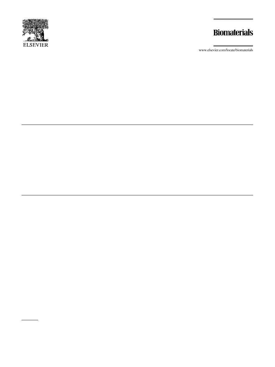
Fig. 1a and b show the DSC curves of both alloys
investigated. NiTiCu shows a direct transformation from
austenite to martensite, probably B19 whereas the binary
NiTi alloy transforms "rst to the trigonal R-phase before
the B19
transformation occurs at lower temperature.
The transformation temperatures are summarised in
Table 1.
The alloys show quite similar A temperatures, the
austenite}martensite hysteresis is however, as expected,
narrower in the case of the ternary alloy.
3.2. Mechanical properties
The stress}strain diagrams at di!erent temperatures
are shown in Figs. 2 and 3 as "ve loading and unloading
cycles . In Table 2, the results of the plateau stresses and
elastic moduli are summarised.
The overall deformation behaviour of both alloys is
quite similar: The strength increases drastically with tem-
perature due to the presence and/or thermal stabilisation
of the austenite phase. Furthermore both alloys show on
loading, particularly in the presence of the austenite
phase (temperature between A and A or above A)
a di!use stress peak followed by a yield drop before the
plateau stress, which denotes the progressive stress-in-
duced consumption of the austenite phase by the growing
martensite variants, is established. On unloading,
and before completion of the reverse transformation of
M. Es-Souni et al. / Biomaterials 22 (2001) 2153}2161
2155
Fig. 1. (a) and (b) DSC curves of the investigated alloys: NiTi (a) and NiTiCu (b). See also Table 1.
Table 1
Transformation temperatures of the alloys investigated. M: marten-
site-"nish; M: Martensite start; R: R-phase "nish; R: R-phase start;
A: Austenite start; A: Austenite "nish
Alloy
M (3C) M (3C) R (3C) R (3C) A (3C) A (3C)
NiTi42
!
59.0
!
23.4
15.3
21.0
21.8
32.7
NiTi42Cu7
!
4.0
14.2
*
*
17.8
34.0
martensite to austenite, a reverse peak stress is observed
at temperatures above A. However, at 603C, the peak
stress becomes more di!use and almost disappear, and
this is particularly true for the binary alloy, when the
specimen is subjected to loading/unloading cycles.
Nevertheless, a closer look at the stress}strain diagrams
reveals speci"c di!erences of the deformation behaviour
of the alloys investigated. At 63C, which is below the
R and above the M temperatures of the binary NiTi
alloy, the characteristic deformation behaviour of the
R-phase upon loading [14] can be seen. Following the
elastic deformation a stress plateau (though slightly in-
creasing) which is attributed to the movement of the
R-variants with preferential orientation to the stress axis
is established up to a strain of approximately 0.75%;
beyond that, the deformation continues elastically until
a smooth transition to a new plateau occurs which de-
notes the beginning of the stress-induced transformation
of the R-phase to the martensite. In contrast, the ternary
alloy shows only the martensitic
`plateaua which is in-
itiated at a relatively lower stress, and is characterised by
a slight, continuous increase of stress with increasing
deformation. At 223C, the deformation of the binary
alloy proceeds by the direct stress induced transforma-
tion of R to M, whereas the ternary alloy shows a defor-
mation behaviour proper to a martensitic}austenitic-
phase mixture with martensitic predominance. Further-
more the peak stress followed by a yield drop appears at
this temperature, which suggests that the peak stress is
closely related to the stress-induced transformation of
austenite to martensite. At 373C, both alloys are fully
austenitic and exhibit the superelastic deformation be-
haviour characterised by the superelastic hysteresis
which is however narrower in the case of the NiTiCu
ternary alloy. At this temperature, both alloys show
a good resistance against softening during loading}un-
loading cycles; the peak stresses remain practically un-
changed, and the plateau stresses, though somewhat
shifted towards lower values on cycling, are only margin-
ally a!ected. At higher temperatures, the properties of the
binary alloy are dramatically a!ected, particularly on
cycling. The peak stress disappears in the second
cycle,
the
remaining
deformation
increases
with
the cycle number and the superelastic properties are
gradually lost. In contrast the integrity of the properties
is only slightly a!ected in the case of NiTiCu alloy, apart
from a comparatively small amount of remaining defor-
mation which does not increase with increasing cycle
number.
Fig. 4 shows plots of the plateau stresses vs. temper-
ature where for both alloys a linear relationship between
stress and temperature is well veri"ed. This indicates that
the stress to move the martensite plates or to induce the
martensitic transformation is well described by the
Clausius}Clapeyron equation [1]:
d
d¹
"!
H
¹
,
(
1)
where
is the stress, H the transformation enthalpy,
a transformation strain and ¹ the absolute temper-
ature. The calculated slopes of the linear intercept lines
yield similar values of 12 and 12.6 MPa/3C for the stress
rates for NiTi and NiTiCu, respectively.
2156
M. Es-Souni et al. / Biomaterials 22 (2001) 2153}2161
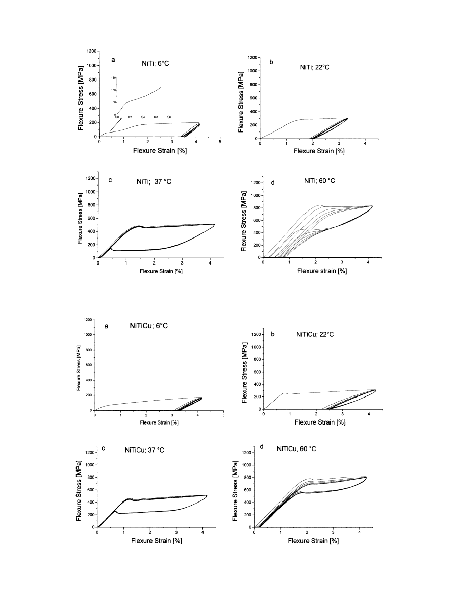
Fig. 2. Stress/strain diagrams of the binary NiTi alloy at di!erent temperatures: 63C (a), 223C (b), 373C (c) and 603C (d).
Fig. 3. Stress/strain diagrams of the ternary NiTiCu alloy at di!erent temperatures: 63C (a), 223C (b), 373C (c) and 603C (d).
M. Es-Souni et al. / Biomaterials 22 (2001) 2153}2161
2157
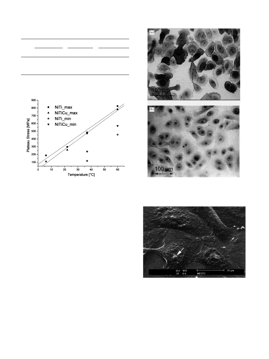
Table 2
Maximum and minimum plateau stresses as given by
o!set stresses
Alloy
max (MPa)
min (MPa) E-modul (GPa)
Temperature (3C)
Temperature (3C)
Temperature (3C)
6
22
37
60
6
22
37
60
6 22
37
60
NiTi
188 297 485 826
*
*
117 456
*
20
43
45.2
NiTiCu 110 258 471 782
*
*
237 570
17 33.4 41.6 42.9
Fig. 4. Plateau stresses versus temperature.
Fig. 5. (a) Cytokeratin AE1, AE3 stained epithelial cells. (b) Negative
control.
Fig. 6. Secondary electron micrograph (SE) of the epithelial cell mor-
phology in control cultures showing nuclei (bold arrow) and cytoplas-
mic projections ("ne arrow).
3.3. Biocompatibilty results
3.3.1. Morphological investigations
Monolayers of non-exposed gingival cell cultures (con-
trol culture) stained uniformly positive for cytokeratin,
a$rming that the cells are of epithelial origin. Light
microscopy (LM) investigations of the stains showed
a well-attached monolayer with the typical morphology
illustrated in Fig. 5.
The cells show distinct boarders and prominent nuclei.
SEM investigations con"rmed these facts: The cells
showed well-developed intercellular junctions, nuclei as
well as numerous cytoplasmic projections, Fig. 6. The
cells grown in the presence of the binary alloy adhered to
both metal and coverslip and exhibited a quite similar
morphology to the control cultures, Fig. 7a and b.
The cells grown in presence of the ternary alloy were
found to di!er in their morphological characteristics. The
cell growth and adhesion seemed to be poorer, e.g.
Fig. 8a, where detached cells can be seen. Furthermore,
a higher number of spherical cells with irregular plasma
membrane and missing cytoplasmatic projections were
found.
3.3.2. MTT in vitro cytotoxicity test
The MTT cytotoxicity tests were performed on
10
cells/100 l/well. The cells were exposed for 24, 48
and 72 h to the alloys.
2158
M. Es-Souni et al. / Biomaterials 22 (2001) 2153}2161
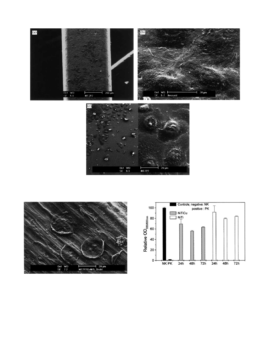
Fig. 7. (a) SE micrograph of the epithelial cells grown on the binary NiTi alloy. (b) Similar to Fig. 7a at higher magni"cation. (c) Dual magni"cation SE
micrograph of the epithelial cells grown on coverslip in the presence of the binary NiTi alloy.
Fig. 8. SE micrograph of the epithelial cells grown on the ternary
NiTiCu alloy.
Fig. 9. Relative OD of formazan formation for both alloys for
di!erent incubation periods of 24, 48 and 72 h. The "gure also shows
negative (NK) and positive (PK) controls. The error bars denote the
standard error.
Fig. 9 shows that there is a di!erent response to for-
mazan formation depending on the alloy used. It can be
seen that the binary alloy leads to a loss of approximately
10}15% of viability with respect to the negative control,
whereas the ternary alloy induces a loss of viability of
about 30}38%. Statistical evaluation of the results using
the one-way Anova test yields a signi"cant di!erence
between both populations with p"0.0145 at the signi"-
cance level of 0.05. Due to the fact that the ternary alloy
contains 6.9% Cu, and that Cu is well known for its
cytotoxic e!ects [15], it was thought, that the loss of
viability might be attributed to the release of Cu-ions in
M. Es-Souni et al. / Biomaterials 22 (2001) 2153}2161
2159

the medium. Preliminary AAS measurements were con-
ducted on the medium after incubation with NiTiCu. The
results show that Cu is released to a concentration of
approximately 5.3$1.0
g cm\ after an incubation
period of 7 days at 373C. However, when this result is
corrected by the blank measurement of 2.5$1.0, a value
of 2.8$2.0
g cm\ is obtained which is close to the
result reported by Gil et al. [5] who found approximately
1.8
g cm\ after 7 days incubation of a similar alloy in
arti"cial saliva.
Fig. 9 also shows that when testing the incubation time
dependence of the MTT transformation within a sample
group, there seems to be a signi"cant di!erence in the
results obtained from cultures incubated with the sam-
ples for 24 h and 48 h. A longer incubation period of 72 h
does not change signi"cantly the MTT results in com-
parison to those of 48 h.
4. Discussion
4.1. Transformation behaviour and mechanical properties
The transformation behaviour of the alloys investi-
gated is quite di!erent because of di!erent thermo-
mechanical histories and chemical compositions. The
presence of copper in the ternary alloy is expected to
decrease the hysteresis of the martensite
austenite
transformation temperatures. The R-phase transforma-
tion in the binary alloy points to a thermomechanical
treatment making use of cold deformation [1,4] which
leads to the B19
martensite transformation being shifted
to lower temperatures. A direct comparison of the hyster-
esis of the alloys investigated is therefore not meaningful.
Comparison of the mechanical properties shows the
superior behaviour of the ternary NiTiCu alloy, parti-
cularly on loading}unloading cycles. The addition of
copper reduces the superelastic hysteresis stress and
allows higher unloading plateaus to be obtained which is
of particular interest for medical applications. The resist-
ance to softening during loading and unloading cycles at
higher temperatures is also considerably improved.
These properties have been attributed to the ease of
interface movement in the Cu alloyed NiTi [1] in com-
parison to the binary alloy, and the mechanical proper-
ties reported in the present work seem to con"rm this.
The appearance of the yield points on loading and un-
loading at temperatures above A suggests that for both
alloys a critical stress has to be overcome before the
austenite
martensite transformations take place, and is
in many aspects similar to LuKders deformation [14,16].
As the testing temperature is increased, the yield points
are considerably attenuated (NiTiCu) or disappear (NiTi)
on loading and unloading cycles. This is also accom-
panied by considerable softening. Due to the fact that
loading was always con"ned to the plateau region, we
attribute this softening to the build-up of residual stresses
which at these temperatures may arise from the forma-
tion of interface dislocations. The presence of residual
stresses may also rise the transformation temperature,
and the increasing
`remaininga deformation with in-
creasing the number of cycles may be a further support
for this mechanism. The improved behaviour of NiTiCu
over the binary alloy lies in the easier movement of
interphase boundaries as mentioned above.
4.2. Biocompatibility
In contrast to other biotoxicity studies [17,18], we did
not use commercially available cell lines, but cultivated
monolayers of gingival epithelial cells from primary cul-
tures of gingival explants. Prior to any cytotoxicity test it
was shown by means of immuno-cytochemical tests, that
the cells used were of epithelial origin.
The SEM and LM studies of cells grown in the pres-
ence of the binary alloy did not show any signi"cant
morphological di!erence to the control sample. Adher-
ence and growth were similar on both coverslip and
metal, although the adherence on the metal seemed less
pronounced than on the coverslip. Nevertheless a normal
morphology of the cells adhering on the NiTi was ob-
served, which stresses the biocompatibility of this alloy.
The results of the MTT-biotoxicity test con"rms this fact.
The formazan formation, which denotes the dehydrogen-
ase activity and thus the intact mitochondrial content of
the cells tested, decreases only by 10% after a 24 h incu-
bation period, and by additional 5%, when incubated for
48 or 72 h. This decrease might be attributed to a release
of Ni ions which have been shown in a recent study to be
highest in the "rst 2 days [9].
The results of the ternary alloy tested di!er from those
obtained for the binary alloy. Cell adhesion and growth
are less pronounced in the presence of NiTiCu. SEM
investigations of cells grown on coverslips in the presence
of the metal show a higher number of spherical, some-
times detached cells. These e!ects are more pronounced
on the alloy. The MTT-test con"rms these "ndings. In
fact there is a 30}38% decline of the dehydrogenase
activity obtained for incubation periods of 24 and 48 or
72 h, respectively. Gil et al. [5] show that Cu ions are
released to a higher concentration than Ni and Ti ions
and that the release rate is maximum in the initial incuba-
tion phase. Therefore, it is thought that the higher
cytotoxicity of the ternary alloy arises from the release of
Cu ions which are known for their cytotoxic e!ects.
Evidence for the release of the Cu ions could be also
found in this work using AAS. However, it should be
mentioned, that for both alloys no signi"cant change was
observed between an incubation period of 48 and 72 h.
This points to a time-dependent build up of a passive
layer on the alloy surfaces, which usually follows a para-
bolic law. This explains why the MTT results remain
2160
M. Es-Souni et al. / Biomaterials 22 (2001) 2153}2161

almost constant with increasing incubation time. The
formation of this passive layer will be investigated in
more detail using electron spectroscopic surface analysis
methods.
The present results show that the Cu addition to the
NiTi shape memory alloys a!ects their biocompatibility
in vitro. This may appear to disagree with the results of
Wen et al. [10], who claim that Cu does not a!ect the
biocompatibility in vivo of these alloys, although their
results show unambiguously higher corrosion rates with
increasing Cu content (see Fig. 1 of their work). Further-
more, reinterpreting their
`histomorphometrica results,
there seems to be a signi"cantly thicker capsule of con-
nective tissue covering the implant with the highest Cu
content after one month of implantation which points to
a lower biocompatibility. Nevertheless, it is thought that
the signi"cantly higher cytotoxicity of the ternary alloy
observed in the present work may arise from the speci"c
response of the epithelial cell cultures in an in vitro
test [19].
5. Conclusion
The present work reports a comparative study of the
properties, including biocompatibility, of a binary
NiTi42 and a ternary NiTi42Cu7 shape memory alloy.
The following conclusions may be inferred:
E The alloys are characterised by a di
!erent transforma-
tion behaviour. The binary alloy shows a R-phase
transition due to cold working while the ternary alloy
shows a direct austenitic to martensitic transforma-
tion.
E The ternary alloy shows superior mechanical proper-
ties. The superelastic hysteresis stress is lower resulting
in higher unloading plateau stresses, and the resistance
to softening during loading}unloading cycles is better.
E This study shows a signi
"cant di!erence in the in vitro
biocompatibility between the binary and ternary alloy.
The presence of the binary alloy leads to few mor-
phological changes and slight reduction of dehydro-
genase activity of epithelial cell cultures, whereas the
e!ect of the ternary alloy appears to be more pro-
nounced, obviously due to releasing of copper ions.
The biocompatibility may, however, be improved by
using passivated wires. This will be the subject of
a forthcoming study.
References
[1] Saburi T. Ti-Ni shape memory alloys. In: Otsuka K, Waymann
CM, editors. Shape memory materials. New York: Cambridge
University Press, 1998. p. 49}96.
[2] Melton KN. General applications of SMA's smart materials. In:
Otsuka K, Waymann CM, editors. Shape memory materials. New
York: Cambridge University Press, 1998. p. 220}39.
[3] Duerig TW. Present and future applications of shape memory and
superelastic materials. In: George EP, Takahashi S, Trolier-
McKinstry S, Uchino K, Wun-Fogle M, editors. Materials for
smart systems. Pittsburgh: Materials Research Society, 1995.
p. 497}506.
[4] Suzuki Y, Horikawa H. Thermal hysteresis in NiTi and Ni-Ti-X
alloys and their applications. In: Liu CT, Kunsmann H,
Otsuka K, Wuttig M, editors. Shape memory materials and
phenomena * Fundamental aspects and applications. Materials
for smart systems. Pittsburgh: Materials Research Society, 1992.
p. 403}6.
[5] Gil FJ, Planell JA. E!ect of copper addition on the super-
elastic behaviour of NiTi shape memory alloys for ortho-
dontic
applications.
J
Biomed
Mater
Res
1999;48(5):
682}8.
[6] Gil FJ, Manero JM, Planell JA. Relevant aspects in the clinical
applications of NiTi shape memory alloys. J Mater Sci Mater
Med 1996;7:403}6.
[7] Wever DJ, Veldhuizen AG, Sanders MM, Schakenrad JM,
van
Horn
JR.
The
cytotoxic,
allergic
and
genotoxic
activity of a
nickel-titanium alloy. Biomaterials 1997;18:
1115}20.
[8] RyhaKnen J, Kallioinen M, Tuukkanen J, Lehenkari P, Junila J,
NiemelaK E, Sanvik P, Serlo W. Bone modelling and cell-material
interface
response
induced
by
nickel}titanium
shape
memory
alloy
after
periosteal
implantation.
Biomaterials
1999;20:1309}17.
[9] Wever
DJ,
Veldhuizen
AG,
de
Vries
J,
Busscher
HJ,
Uges DRA, van Horn JR. Electrochemical and surface
characterization
of
a
nickel}titanium
alloy.
Biomaterials
1998;19:761}9.
[10] Wen X, Zhang N, Li X, Cao Z. Electrochemical and histomor-
phometric evaluation of the TiNiCu shape memory alloy. Biomed
Mater Eng 1997;7(1):1}11.
[11] Judd
DA,
Battista
PJ,
Behm
DD.
Culture
of
human
keratinocytes in de"ned serum-free medium. Focus 1997;19:
1:2}5.
[12] Bourne JA. Handbuch I der Immunoperoxydase FaKrbemethoden.
DAKO corporation, Carpenteria, CA. 1997. p. 16.
[13] Tada H, Shibo O, Kuroshima K, Koyama M, Tsukamoto K. An
improved colorimetric assay for interleukin-2. Immunol Methods
1986;93:157}65.
[14] Duerig TW, Melton KN. Di!use yield drop and snap
action in a NiTi alloy. In: George EP, Takahashi S, Trolier-
McKinstry S, Uchino K, Wun-Fogle M, editors. Materials for
smart systems. Pittsburgh: Materials Research Society, 1995.
p. 159}64.
[15] Scarino ML, Poverini R, Di Lullo G, Biseses G. Inhibition of
protein synthesis after exposure of Caco2 cells to heavy metals.
ATLA 1992;20:325}33.
[16] Orgeas L, Favier D. Stress induced martensitic transformation of
a NiTi alloy in isothermal shear, tension and compression. Acta
Mater 1998;46:5579}91.
[17] Grant DM, Lo WJ, Parker KG, Parker TL. Biocompatibility and
mechanical properties of low temperature deposited quaternary
(Ti, Al, V)N coating on Ti6Al4 V titanium alloy substrate. J Mater
Sci Mater Med 1996;7:579}84.
[18] Prigent H, Pellen-Mussi P, Cathelineau G, Bonnaure-Mallet M.
Evaluation of the biocompatibility of titanium}tantalum alloy
versus titanium. J Biomed Mater Res 1998;39:200}6.
[19] Wintermantel E, Suk-Woo H. Biokompatible Werksto!e und
Bauweisen. Berlin: Springer, 1998. p. 119}20.
M. Es-Souni et al. / Biomaterials 22 (2001) 2153}2161
2161
Wyszukiwarka
Podobne podstrony:
40 549 563 On the Precipitation Behaviour in Maraging Steels
Report on the Sexual Behavior o Robert F Young(1)
Notes on the Alchemical Transformation of Mecury
Phillip G Zimbardo A Situationist Perspective On The Psychology Of Evil Understanding How Good Peo
Begault Direct comparison of the impact of head tracking, reverberation, and individualized head re
The impact of network structure on knowledge transfer an aplication of
Parzuchowski, Purek ON THE DYNAMIC
Enochian Sermon on the Sacraments
GoTell it on the mountain
Interruption of the blood supply of femoral head an experimental study on the pathogenesis of Legg C
CAN on the AVR
Ogden T A new reading on the origins of object relations (2002)
On the Actuarial Gaze From Abu Grahib to 9 11
91 1301 1315 Stahl Eisen Werkstoffblatt (SEW) 220 Supplementary Information on the Most
Pancharatnam A Study on the Computer Aided Acoustic Analysis of an Auditorium (CATT)
Newell, Shanks On the Role of Recognition in Decision Making
BIBLIOGRAPHY I General Works on the Medieval Church
Chambers Kaye On The Prowl 2 Tiger By The Tail
więcej podobnych podstron