Biomaterials 23 (2002) 1761–1768
Al–Cu–Fe quasicrystal/ultra-high molecular weight
polyethylene composites as biomaterials for
acetabular cup prosthetics
Brian C. Anderson
a,c
, Paul D. Bloom
b,c
, K.G. Baikerikar
b,c
, Valerie V. Sheares
b,c
,
Surya K. Mallapragada
a,c,
*
a
Department of Chemical Engineering, Iowa State University, 2114 Sweeney Hall, Ames, IA 50011-2230, USA
b
Department of Chemistry, Iowa State University, Ames, IA 50011, USA
c
Ames Laboratory, United States Department of Energy, Ames, IA 50011, USA
Accepted 2 September 2001
Abstract
Polymer composites of Al–Cu–Fe quasicrystals and ultra-high molecular weight polyethylene (UHMWPE) were investigated for
use in acetabular cup prosthetics. The wear properties of the Al–Cu–Fe/UHMWPE samples and a 440 steel ball counterface were
measured. The mechanical strength of the Al–Cu–Fe/UHMWPE composites was compared to UHMWPE and alumina/
UHMWPE. The biocompatibility of the composite material was tested using a direct contact cytotoxicity assay. Al–Cu–Fe/
UHMWPE demonstrated lower volume loss after wear and higher mechanical strength than UHMWPE. This composite material
also showed no increase in counterface wear or cytotoxicity relative to UHMWPE. These combined results demonstrate that
Al–Cu–Fe/UHMWPE composites are promising candidate materials for acetabular cup prosthetics. r 2002 Elsevier Science Ltd.
All rights reserved.
Keywords:
Quasicrystal; Reduced wear; Polymer composite; Joint replacement; Biocompatible
1. Introduction
Hip arthroplasty is an important medical procedure in
which the socket joints in the pelvic bone and/or the
femoral head are replaced with prosthetic devices. The
femoral head is usually replaced by a metal, attached by
a stem inserted into the femur. The acetabulum in the
pelvic bone is replaced by a prosthetic cup, typically
made of a polymeric biomaterial. Recent work in the
area of biomaterials for total hip arthroplasty has
involved investigations of ceramic, metal, and polymeric
materials for both the femoral head and acetabulum
prostheses [1–4].
The current technology for the acetabular prosthetic
cup is dominated by usage of ultra-high molecular
weight polyethylene (UHMWPE). This polymer, usually
in the molecular weight range of M
n
¼ 310
6
to 6 10
6
,
is both strong and bioinert. However, after prolonged
shear stress from a metallic surface, for example a
titanium femoral head prosthesis, the surface can wear
and leave debris in the body. These particulates have
been studied and have been found to cause osteolysis
and subsequent device loosening and failure [5–7]. It has
been reported that osteolysis does not usually occur in
patients with low acetabular cup wear rates [8].
Quasicrystals, first discovered in 1984 [9], are complex
metal alloys that possess physical properties such as low
thermal conductivity, lowcoefficients of friction, high
hardness, etc. The name quasicrystal stems from the
unusual rotational symmetries and aperiodic lattice
spacings found in these materials. Crystalline materials
that have periodically repeating unit cells that comple-
tely fill space must have two, three, four or six fold
rotational symmetries. All other rotational symmetries
are forbidden. Quasicrystals exhibit symmetries that are
*Corresponding author. Department of Chemical Engineering, Iowa
State University, 2114 Sweeney Hall, Ames, IA 50011-2230, USA. Tel.:
+1-515-294-7407; fax: +1-515-294-2689.
E-mail address:
suryakm@iastate.edu (S.K. Mallapragada).
0142-9612/02/$ - see front matter r 2002 Elsevier Science Ltd. All rights reserved.
PII: S 0 1 4 2 - 9 6 1 2 ( 0 1 ) 0 0 3 0 1 - 5

forbidden by classical crystallography, such as the six
fivefold axes found in icosahedral quasicrystals. In
addition to the required rotational symmetries, diffrac-
tion patterns from crystalline materials have periodically
spaced spots that can be used to determine lattice
parameters. In contrast, diffraction patterns from
quasicrystals contain aperiodic spacings [10]. The
relationship between quasicrystalline structures and
their novel physical properties is a current topic in
quasicrystal research. Since their discovery, several
hundred alloys are nowknown to exhibit quasicrystal-
line phases. The physical attributes of quasicrystals,
coupled with their availability in fine powder form and
potential lowcost, have made them ideal materials for
evaluation as reinforcing fillers in polymeric materials
[11,12].
Quasicrystal filled polymers have displayed novel
wear properties in addition to typical improvements in
other mechanical properties associated with rigid, low
aspect ratio fillers. Both thermoplastics and thermosets,
such as poly(p-phenylene sulfide) bisphenol A polyar-
ylene ether ketone, and Dowepoxy resins cured with
diethylenetriamine have been investigated. Composites
containing Al–Cu–Fe were determined to have en-
hanced wear resistance over unfilled plastics. In addi-
tion, the presence of the quasicrystalline powders in a
polymer matrix caused almost no abrasion to counter-
face materials during wear testing [13,14]. Therefore,
compared to its constituent metals and other abrasive,
rigid fillers such as SiC and alumina, which are known
to enhance the wear resistance of polymer composites
[15], Al–Cu–Fe quasicrystals are attractive, newfillers
for high-wear plastics.
The high wear resistance and low abrasive nature of
these polymer composites has lead us to investigate the
use of UHMWPE filled with quasicrystalline Al–Cu–Fe
as a material for hip arthroplasty femoral components.
These Al–Cu–Fe/UHMWPE composites have not been
fabricated in the past. The possibility of lower wear rates
on both the femoral head and acetabular cup is a valu-
able alternative to either the less wear resistant UHM-
WPE or more abrasive polymer composites. This is the
first study that investigates the suitability of Al–Cu–Fe/
polymer composites for biomaterial applications.
As with any potential biomaterial, a central issue is
the biocompatibility of this material. UHMWPE is
commonly used as a negative control for direct contact
cytotoxicity tests, and any substitute for this material in
acetabular prosthetics would have to maintain this low
level of cytotoxicity. To test the quasicrystal/polyethy-
lene samples for cytotoxicity, a direct contact assay was
performed, as described by an ASTM standard [16] to
determine whether or not the material warrants further
biocompatibility testing. It has been found that in vitro
cytotoxicity tests are even more sensitive than in vivo
studies [17]. The direct contact test was preferred over
agar diffusion or elution tests because the only difference
between the negative control material (UHMWPE) and
the sample material (Al–Cu–Fe/UHMWPE) is the
addition of Al–Cu–Fe quasicrystalline powder at 30
volume percent.
The mechanical strength and wear resistance of
alumina/UHMWPE were also tested as a comparison
to the Al–Cu–Fe/UHMWPE. Alumina filled polymer
composites have been studied for increasing the
mechanical strength of polymers for industrial [18,19]
and orthopaedic [20] applications. In the mechanical
testing, alumina/UHMWPE serves as a comparison for
Al–Cu–Fe/UHMWPE with other particulate-filled poly-
ethylene composites. The alumina/UHMWPE also
serves as an appropriate comparison for the wear
properties of the Al–Cu–Fe/UHMWPE due to the
presence of the thin aluminum oxide layer known to
form on the surface of Al–Cu–Fe quasicrystals in air. It
has been speculated that the thin aluminum oxide layer
contributes to the unique wear properties of the
quasicrystals [21,22].
2. Materials and methods
2.1. Sample preparation
UHMWPE was purchased from Aldrich and used as
received. Quasicrystalline Al–Cu–Fe icosahedral phase
powders, with the composition Al
65
Cu
23
Fe
12
, were
prepared at Ames National Laboratory, Ames, IA
50011 [23]. These powders were prepared by gas
atomization with an approximate 60% icosahedral
(quasicrystal, i-Al–Cu–Fe) and 40% b-cubic (b-Al–
Cu–Fe) phase composition. Mixtures of polyethylene
(70 volume percent) and Al–Cu–Fe quasicrystal pow-
ders (30 volume percent, size fraction of 45–53 mm) were
weighed, added together and shaken vigorously in a
sealed container for 10 min to provide optimum mixing.
The resulting Al–Cu–Fe/polymer powder mixture was
placed in a die mold that had a diameter of 2.54 cm and
a final volume of 1.58 cm
3
when fully compressed. The
mold was equipped with a thermocouple to monitor
temperature during the compression molding process.
The filled mold was heated in a variable temperature
hydraulic press under a pressure of 7 MPa to 1801C. The
resulting composite surfaces were polished with 320-
grade sandpaper followed by washing with deionized
water. Alumina/UHMWPE samples were prepared in a
similar fashion to the Al–Cu–Fe/UHMWPE samples,
using 45–53 mm alumina powder. UHMWPE samples
were prepared as above with no addition of metal filler.
cis
-Polyisoprene samples, the positive control for
cytotoxicity tests, were prepared by coating the polymer
onto stainless steel disks and removing the solvent under
vacuum overnight.
B.C. Anderson et al. / Biomaterials 23(2002) 1761–1768
1762
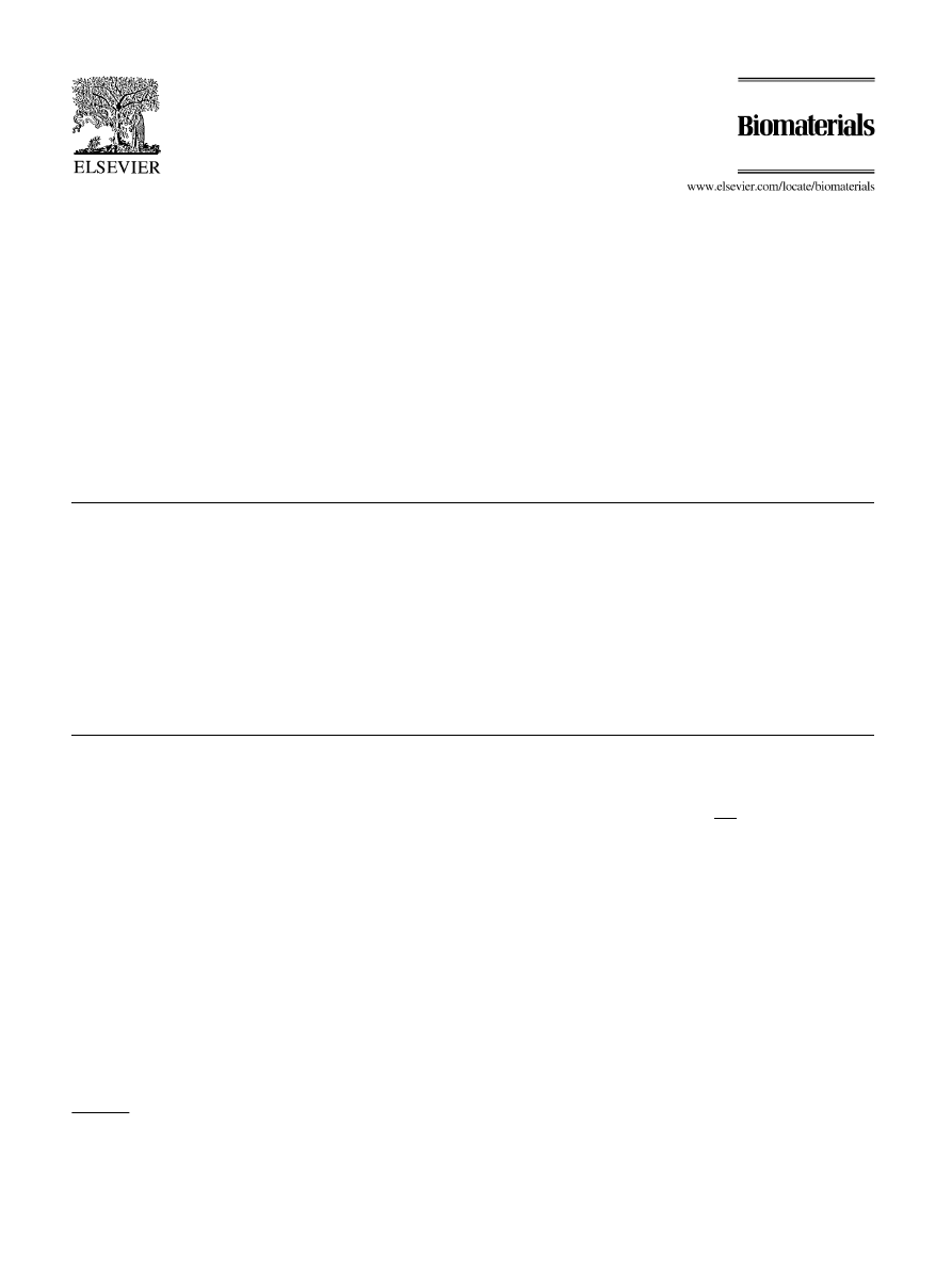
2.2. Wear testing
The wear testing of polymer and polymer composite
disks were performed on a Falex friction and wear test
machine model # ISC450PC. A schematic of this
machine is shown in Fig. 1. A polymer sample was
placed on the rotating plate with a stationary pin as the
counterface subjected to an induced load. The stationary
pin materials were 440 stainless steel balls with a
diameter of 0.635 cm. The wear test conditions used in
each case were: linear speed of 0.10 ms
1
, 10 N load,
radius of 8.0 mm and 200,000 cycles (revolutions). The
total linear distance was approximately 10 km. All tests
were performed at room temperature under dry sliding
conditions. Wear tests for unfilled UHMWPE and the
Al–Cu–Fe/UHMWPE samples were performed in tri-
plicate and the results are reported as the average and
standard deviation of the measurements. The aluminum
oxide/UHMWPE sample was tested one time in order to
establish the abrasive nature of other fillers known to
prevent polymer wear [15]. Volume loss from the
composite samples was determined from profilometery
measurements of the wear tracks. Pin wear was
determined from the mass lost from the stationary 440
stainless steel pin after testing. Wear tracks were
characterized by scanning electron microscopy (Hitachi)
equipped with a secondary electron detector (Hitachi)
and energy dispersive spectrometer (Oxford) for X-ray
analysis. Following the wear experiments, polymer
samples were coated with a thin conductive layer of
gold for analysis by scanning electron microscopy.
2.3. Mechanical testing
Mechanical testing was performed using three-point
bending tests on a dynamic mechanical analyzer (Perkin
Elmer DMA7e). Samples of UHMWPE, alumina/
UHMWPE, and Al–Cu–Fe/UHMWPE were cut into
bars approximately 1.5 mm in height and 3 mm in width.
They were subjected to 55 mN of static force and 50 mN
of oscillatory dynamic force (1 Hz frequency). The
samples were examined over a temperature range of
30–1001C at a scan rate of 51C/min. The storage
modulus (E
0
; Pa) and tan d (E
00
=E
0
or ratio of loss
modulus (E
00
) to storage modulus) were used to compare
the mechanical strength of the materials.
2.4. Cytotoxicity evaluation
Cytotoxicity tests of Al–Cu–Fe/UHMWPE samples
were performed by a direct contact assay outlined by the
American Standards for Tests and Measures [16]. NIH/
3T3 mouse fibroblasts were used as the cell line for these
experiments. Cells were cultured using low-glucose
Dulbecco’s modified eagle medium (DMEM, Sigma)
with 10% fetal bovine serum (FBS, Sigma), 10 mg/ml
insulin (Sigma), 10 units/ml penicillin/streptomycin (Sig-
ma), and 100 mg/ml l-ascorbic acid (Sigma) in a
humidified incubator with 5% CO
2
at 371C. Once the
cells were cultured to confluency, they were transferred
by trypsinization [0.25% trypsin (Sigma) in Hank’s
balanced salt solution (HBSS, Sigma)] into 6-well plates
at a cell density of approximately 300 cells/mm
2
. The
cells were allowed to adhere to the plates and grow for
24 h to near confluency.
Samples of UHMWPE, Al–Cu–Fe/UHMWPE, and
stainless steel disks with a coating of cis-polyisoprene
were sterilized by swabbing with 70% ethanol and
allowing them to dry in aseptic conditions under
ultraviolet light for longer than 6 h. The media was
removed from the well plates containing the cells and the
samples were placed into the wells, leaving one well
empty for comparison. Media was added to the wells
and the plates with samples were placed in an incubator
for another 24 h.
After 24 h, the media was removed and replaced with
Karnovsky’s fixative (2.5% gluteraldehyde, 2.0% para-
formaldehyde, 0.1 m sodium cacodylate). The fixative
was allowed to stand for 24 h before replacing with
crystal violet dye (CVD) in a 20% ethanol solution.
After 24 h, the CVD solution was replaced with 70%
ethanol for 2 h. The ethanol was removed and the
samples were allowed to air dry. The samples were then
inspected for any cytotoxic response at the polymer
interface using light microscopy. UHMWPE served as
the negative control and cis-polyisoprene served as the
positive control.
3. Results and discussion
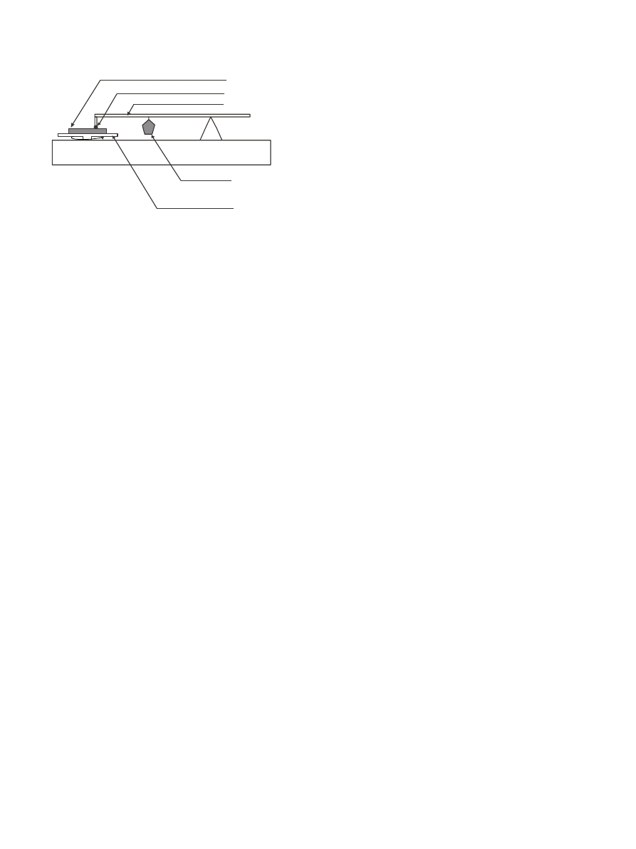
3.1. Wear testing
Results from pin-on-disk wear testing showed that
Al–Cu–Fe filled UHMWPE disks had enhanced wear
resistance to volume loss, as compared to unfilled
UHMWPE and alumina filled UHMWPE, rotating
against a stationary 440 stainless steel pin (Fig. 2). The
ω
Sample
Round Counterface
Lever Arm
Weight
Rotating Plate
Fig. 1. Schematic of pin-on-disk wear testing apparatus.
B.C. Anderson et al. / Biomaterials 23(2002) 1761–1768
1763
volume loss from Al–Cu–Fe/UHMWPE was shown to
be statistically less than the UHMWPE (p
o0:001). The
Al–Cu–Fe/UHMWPE samples showed approximately a
35% decrease in volume loss compared
to the
UHMWPE samples. The wear tracks were further
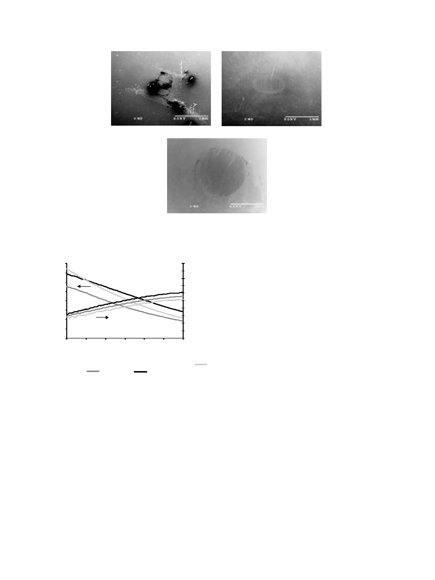
characterized by SEM (Fig. 3). The SEM micrographs
showthe behavior of the substrate after the induced
wear. The UHMWPE samples (Fig. 3A) showed a
smooth wear track surface approximately 900 mm in
width. The Al–Cu–Fe/UHMWPE samples (Fig. 3B)
showed similar wear to the UHMWPE with a slightly
narrower wear track, approximately 850 mm. This is
indicative of less indentation of the spherical counter-
face pin into the sample and lowabrasion of the
counterface. The small, light-colored, circular areas on
the wear track in Fig. 3B are the Al–Cu–Fe quasicrystals
exposed at the wear interface.
The alumina sample (Fig. 3C) showed a much wider
wear track than the UHMWPE and Al–Cu–Fe/
UHMWPE samples, approximately 1500 mm. This is
due to the abrasion of the 440 stainless steel counterface
by the exposed aluminum oxide particles in the
composite at the wear interface. In addition, irregular,
light-colored patterns are observed in the wear track on
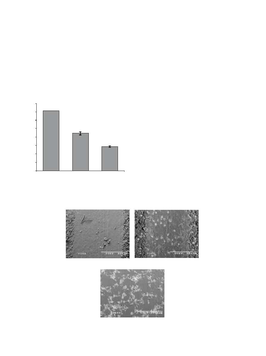
the aluminum oxide/UHMWPE composite (Fig. 3C).
X-ray elemental mapping (Fig. 4) of these areas indicate
the lighter areas are rich in iron, chromium and their
metal oxides. This result confirms that the counterface
stainless steel pin was abraded by the aluminum oxide
particles and the debris was imbedded in the polymer
composite.
The volume loss of the unfilled UHMWPE was due to
both deformation and removal of the polymer during
wear. The improved wear resistance of the Al–Cu–Fe/
UHMWPE composites has been attributed to the high
hardness, high Young modulus, and lowcoefficient of
friction of the Al–Cu–Fe quasicrystalline filler and the
increased strength of the polymer composite in compar-
ison to unfilled UHMWPE. Lowcoefficients of friction
for quasicrystalline alloys were first reported by Dubois
et al. [24]. Other materials, such as hard Cr-steel,
have high hardness and a Young modulus that is
(A)
(B)
(C)
Fig. 3. Wear tracks analysis of (A) UHMWPE (B) Al–Cu–Fe/UHMWPE and (C) alumina/UHMWPE samples by SEM.
0
0.1
0.2
0.3
0.4
0.5
0.6
0.7
0.8
Alumina/UHMWPE
UHMWPE
Al-Cu-Fe/UHMWPE
Volume Loss (mm
3
)
Fig. 2. Volume loss from UHMWPE, Al–Cu–Fe/UHMWPE, and
Al
2
O
3
/UHMWPE samples after wear testing. Error bars represent
standard deviations of the measurements.
B.C. Anderson et al. / Biomaterials 23(2002) 1761–1768
1764
approximately double that of i-Al–Cu–Fe but display
higher coefficients of friction. The lowcoefficients of
friction for quasicrystals were determined to arise from
reduced electronic interactions whereas higher electronic
interactions in materials such as Cr-steel contribute to
sticking effects [24]. As specified in the experimental
section, the Al–Cu–Fe material used in these experi-
ments was a mixture of icosahedral (i-Al–Cu–Fe, 60%)
and b-cubic (b-Al–Cu–Fe, 40%) phases. Coefficients of
friction for i-Al–Cu–Fe and b-Al–Cu–Fe were found to
be very similar [25,26]. However, the presence of b-Al–
Cu–Fe in bulk i-Al–Cu–Fe samples caused rapid
deterioration in wear resistance [25]. In contrast, when
used as a filler in epoxy composites, the mixed phase i,b-
Al–Cu–Fe powder imparted comparable wear resistance
to single phase i-Al–Cu–Fe powders [27].
The wear of the counterface 440 stainless steel ball by
the UHMWPE composites has been summarized in
Fig. 5. Abrasion of the 440 pin from contact with the
Al–Cu–Fe/UHMWPE composite was low and statisti-
cally the same as the unfilled UHMWPE to a 0.05 level.
SEM micrographs of the 440 stainless steel pins used for
wear testing are shown in Fig. 6. By contrast, the
aluminum oxide filled UHMWPE sample was very
abrasive. Aluminum oxide is known for its high
hardness, abrasive nature, and aspherical particle
morphology. During these tests, noticeable amounts of
the counterface 440 stainless steel pin material were
worn away by the aluminum oxide filled composite.
Steel from the pin was embedded in the alumina/
UHMWPE composite and observed by SEM/EDS
(Fig. 4). While aluminum oxide filled polymers typically
offer excellent wear resistance [18–20], the high wear of
the alumina/UHMWPE composite was attributed to the
steel rich wear interface that developed on the aluminum
oxide composite during wear. The wear resistance of
steel filled polymer composites has been shown to be
very poor [14].
3.2. Mechanical testing
Results from the mechanical testing revealed that the
Al–Cu–Fe/UHMWPE shows better mechanical proper-
ties than neat UHMWPE or the alumina/UHMWPE
composite. The storage modulus of the Al–Cu–Fe/
UHMWPE was higher than the UHMWPE for the
Fig. 4. SEM/EDS X-ray elemental mapping of the aluminum oxide/UHMWPE composite. Left to right: secondary electron image, carbon, oxygen;
Rowtwo: aluminum, copper, iron.
0
0.05
0.1
0.15
0.2
0.25
0.3
0.35
0.4
0.45
Alumina/UHMWPE
UHMWPE
Al-Cu-Fe/UHMWPE
Mass Loss (mg)
Fig. 5. Mass loss from 440 stainless steel pin after wear testing on
UHMWPE, Al–Cu–Fe/UHMWPE, and Al
2
O
3
/UHMWPE surfaces.
Error bars represent standard deviations of the measurements.
B.C. Anderson et al. / Biomaterials 23(2002) 1761–1768
1765
entire temperature range of 30–901C (Fig. 7). The
materials were tested at higher temperatures because
these measurements give an indication of long term
polymer behavior due to time-temperature superposi-
tion principles. The higher storage modulus indicates
that a composite material has a higher ‘‘stiffness’’ than
the pure material. This is almost always the case with
filled polymer composites containing a particulate that
has a much higher modulus than that of the polymer
used as the composite matrix. If there is good bonding
between the filler and the polymer matrix the modulus
will increase due to a strengthening of the polymer-filler
heterogeneous interface. The alumina/UHMWPE also
showed a higher modulus than the unfilled material.
However, the strength quickly decayed with tempera-
ture, which was not as much with the Al–Cu–Fe/
UHMWPE. At 371C, the storage modulus of the Al–
Cu–Fe/UHMWPE
was
1.26 10
9
Pa,
whereas
the
UHMWPE was only 1.00 10
9
Pa.
The tan d data showed a trend similar to the storage
modulus. The Al–Cu–Fe/UHMWPE displayed a higher
tan d; meaning that the storage modulus remained
higher than the UHMWPE relative to their loss moduli.
Alumina/UHMWPE displayed a lower tan d value for
the entire temperature range indicating an increased
energy disipation via viscous deformation.
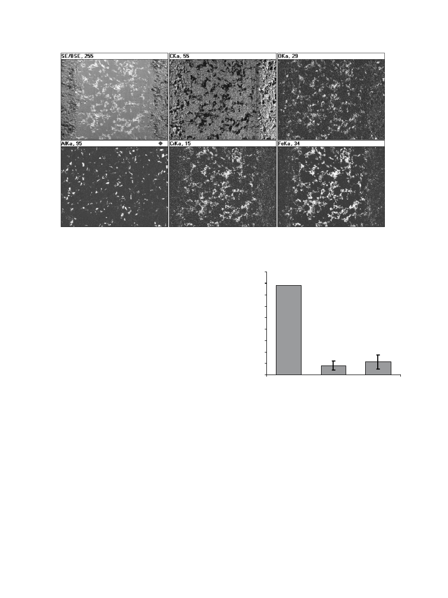
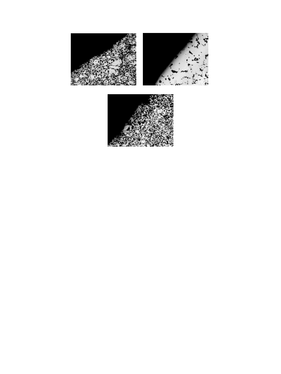
3.3. Cytotoxicity evaluation
The results of the cytotoxicity evaluations indicated
that the Al–Cu–Fe/UHMWPE composites displayed
no cytotoxic behavior. Images of the cell/polymer
interface for the Al–Cu–Fe/UHMWPE, positive control
(cis-polyisoprene), and negative control (UHMWPE)
are shown in Fig. 8. The positive control (Fig. 8B)
elicited a cytotoxic response, as expected. The cell
density near the cis-polyisoprene interface is extremely
lowand the fewcells that are present appear to have
detached from the surface of the well plate. There is also
considerable cellular debris present, likely due to cell
lysis. At a distance >2 mm from the polymer interface,
the cell layer is thick and the cells are confluent. The
negative control (Fig. 8A) elicited no cytotoxic response.
The cell density is constant from the bulk to the
interface. The Al–Cu–Fe/UHMWPE sample (Fig. 8C)
displayed
similar
cytotoxic
characteristics
as
the
negative control. The cell density is constant up to
the sample interface with all the cells fully adhered to the
well plate.
(A)
(B)
(C)
Fig. 6. Analysis of 440 stainless steel pin from wear testing in contact with (A) UHMWPE (B) Al–Cu–Fe/UHMWPE and (C) alumina/UHMWPE
samples by SEM.
0.0E+00
2.0E+08
4.0E+08
6.0E+08
8.0E+08
1.0E+09
1.2E+09
1.4E+09
1.6E+09
30
40
50
60
70
80
90
Temperature (
o
C)
Storage Modulus (Pa)
0
0.05
0.1
0.15
0.2
0.25
0.3
0.35
0.4
0.45
0.5
tangent
δ
Fig. 7. Dynamic mechanical analysis of polymer samples:
alumina/UHMWPE,
UHMWPE,
Al–Cu–Fe/UHMWPE.
B.C. Anderson et al. / Biomaterials 23(2002) 1761–1768
1766
4. Conclusions
It was concluded that Al–Cu–Fe/UHMWPE compo-
sites are potential candidate materials for prosthetic
acetebular cups used in total hip replacement proce-
dures. The wear on Al–Cu–Fe/UHMWPE composites,
as tested by pin-on-disk tribology, was statistically lower
than neat UHMWPE. The wear on the counterface
material in these tests, a 440 stainless steel ball, was
statistically the same for both the Al–Cu–Fe/UHMWPE
and the UHMWPE. The wear on both the substrate and
the counterface for the alumina/UHMWPE sample was
much higher than either the UHMWPE or Al–Cu–Fe/
UHMWPE samples. Dynamic mechanical analysis
determined that the mechanical strength, represented
by the storage modulus and tan d; was improved by the
addition of the Al–Cu–Fe quasicrystals to UHMWPE.
The storage modulus increased by over 25% at 371C
and tan d was lower over the entire temperature range
for
the
Al–Cu–Fe/UHMWPE
relative
to
the
UHMWPE. Direct contact cytotoxicity tests revealed
that the Al–Cu–Fe/UHMWPE composites elicited the
same cytotoxic response as pure UHMWPE that is
routinely used for acetabular cup prosthetics.
Acknowledgements
The authors would like to thank Brandon Vogel,
Daniel Kuster and Ian Kenning for their help with the
cytotoxicity testing and MatthewBesser and Daniel J.
Sordelet for their assistance with the tribology experi-
ments. We would also like to acknowledge the United
States Department of Energy’s Ames Lab Materials
Chemistry Division for funding of this project under
contract number W-7405-ENG-82.
References
[1] Wang A, Lin R, Polineni VK, Essner A, Stark C, Dumbleton JH.
Carbon fiber reinforced polyether ether ketone composite as
a bearing surface for total hip replacement. Tribol Int 1998;31:
661–7.
[2] Bonfield W. Composites for bone replacement. J Biomed Eng
1998;10:522–6.
[3] Roberts JC, Ling FF, Jones Jr WR. Fabrication and wear test of a
continuous fiber/particulate composite total surface hip replace-
ment. ASLE Trans 1983;26:367–75.
[4] Goldsmith AAJ, Dowson D, Isaac GH, Lancaster JG. A
comparative joint simulator study of the wear of metal-on-metal
and alternative material combinations in hip replacement. Proc
Inst Mech Eng 2000;214:39–47.
[5] Amstutz HC, Campbell P, Kossovsky N, Clarke IC. Mechanism
and clinical significance of wear debris-induced osteolysis. Clin
Orthop 1992;276:7–18.
[6] Howie DW, Haynes DR, Rogers SD, McGee MA, Pearcey MJ.
The response to particulate debris. Orthop Clin N Am 1993;
24:571–81.
[7] Maloney WJ, Peters P, Engh CA, Chandler H. Severe osteolysis
of the pelvis in association with acetabular replacement without
cement. J Bone Jt Surg 1993;75A:1627–35.
[8] Dowd JE, Sychterz CJ, Young AM, Engh CA. Characterization
of long-term femoral-head-penetration rates. Association with
(A)
(B)
(C)
Fig. 8. 3T3 fibroblasts after direct contact cytotoxicity testing (100 times). (A) Negative control, UHMWPE, (B) Positive control, cis-polyisoprene,
(C) Al–Cu–Fe/UHMWPE sample.
B.C. Anderson et al. / Biomaterials 23(2002) 1761–1768
1767

and prediction of osteolysis. J Bone Jt Surg Am 2000;
82–A(8):1102–7.
[9] Shechtman D, Blech I, Gratias D, Cahn JW. Metallic phase with
long-range orientational order and no translational symmetry.
Phys Rev Lett 1984;53:1951–3.
[10] Bloom PD, Sheares VV. Quasicrystal-polymer composite materi-
als and methods. PCT Int Appl 2000, WO 0056538, US Patent
(filed 5/99).
[11] Bloom PD, Otaigbe JU, Sheares VV. High-performance quasi-
crystal-reinforced polymer composites. Proc Am Chem Soc Div
Polym Mater: Sci Eng 1999;80:406–7.
[12] Bloom PD, Baikerikar KG, Otaigbe JU, Sheares VV. Develop-
ment of novel polymer/quasicrystal composite materials. J Mater
Sci Eng A 2000;294–296:156–9.
[13] Bloom PD, Baikerikar KG, Anderegg JW, Sheares VV. Devel-
opment of Al–Cu–Fe quasicrystal-poly(p-phenylene sulfide)
composites. Mater Res Soc Symp 2001;643:K16.3.1–K16.3.12.
[14] Durand JM, Vardavoulias M, Jeandin M. Role of reinforcing
ceramic particles in the wear behavior of polymer-based model
composites. Wear 1995;181–183:833–9.
[15] Standard practice for direct contact cell culture evaluation of
materials for medical devices
FDesignation F813-83. In: The 2000
Annual Book of ASTM Standards. Philadelphia: American
Society for Testing and Materials, 2000. p. 240–3.
[16] Ulreich JB, Chavapilin M. In vitro toxicity testing: a quantitative
microassay. In: Brown SA, editor. Cell-culture test methods.
Philadelphia: ASTM, 1983. p. 102–13.
[17] Brotzman RW, Aikens J, Batllo F, Kullberg M, Kritchevsky G.
Nanomaterials and wear resistant polymers. Ceram Ind 2000;
150:39–43.
[18] Kumar K, Kumar K. Sliding wear studies in epoxy containing
alumina powders. High Temp Mater Processes 1998;17:271–4.
[19] Rieu J, Goeuriot P. Ceramic composites for biomedical applica-
tions. Clin Mater 1993;12:211–7.
[20] Wehner BI, Koster U. Microstructural evolution of alumina
layers on an Al–Cu–Fe quasicrystal during high-temperature
oxidation. Oxid Met 2000;54:445–56.
[21] Gil-Gavatz M, Rouxel D, Pigeat P, Weber B, Dubois J-M.
Surface oxidation of the Al
62
Cu
25.5
Fe
12.5
icosahedral quasicrystal.
Philos Mag A 2000;80:2083–97.
[22] Shield JE, Goldman AI, Anderson IE, Ellis TW, McCallum
RW, Sordelet DJ. Method of making quasicrystal alloy
powder, protective coatings and articles. US Patent No.
5,433,978, 1995.
[23] Dubois JM, Kang SS, von Stebut JJ. Quasicrystalline low-friction
coatings. Mater Sci Lett 1991;10:537–41.
[24] Brunet P, Zhang LM, Sordelet DJ, Besser M, Dubois JM.
Comparative study of microstructural and tribological properties
of sintered, bulk icosahedral samples. J Mater Sci Eng A 2000;
294–296:74–8.
[25] Zhang LM, Dong C, Brunet P, Dubois JM. Influence of valence
electron concentration over friction coefficient of B2-based
approximants. J Mater Sci Eng A 2000;294–296:810–2.
[26] Bloom PD, Sheares VW. Fabrication and wear resistance of Al–
Cu–Fe quasicrystal–epoxy composite materials. Chem Matls,
submitted.
[27] Goldman AI, Anderegg JW, Besser MF, Chang S-L, Delaney
DW, Jenks CJ, Kramer MJ, Lograsso TA, Lynch DW, McCallum
RW, Shield JE, Sordelet DJ, Thiel PA. Quasicrystalline materials.
Am Sci 1996;84:230–41.
B.C. Anderson et al. / Biomaterials 23(2002) 1761–1768
1768
Wyszukiwarka
Podobne podstrony:
Al i Cu 2015
al -cu, Metalurgia i odlewnictwo metali nieżelaznych, nież - spr
stopy al cu
Układ równowagi Cu Fe S, Studia
Al i Cu 2015
Układ r Cu Fe S Gaś Głodzik Kwiatkowski v 2
pytania na sprawko, ZUT-Energetyka-inżynier, I Semestr, Materiały konstrukcyjne, Metale, 3. Stopy Cu
Mat8 Cu Al
Mat 8 Cu Al
Utlenianie stopów typu Fe-Cr-Al. W różnych środowiskach korozyjnych, PRACE PISEMNE na studia, dyplom
Fe-cu, Fizyka
Cu,Al,stopy
Cu al
Niedobory mikroelementów Fe, Cu, porażenie poporodowe, tężyczka, dystrofie
~$edobory mikroelementów Fe, Cu, porażenie poporodowe, tężyczka, dystrofie
więcej podobnych podstron