P
rotein crystals can be very difficult to grow. Even protein crystallographers are often unable
to produce crystals of the high quality that is required to determine the molecular structure
of many proteins. As part of NASA’s educational outreach activities, we have prepared a simple
‘recipe’ for growing protein crystals from Brazil nuts. Please let us know how your attempts at
growing these protein crystals turn out. You may contact us at:
microgravitynews@msfc.nasa.gov
For more up-to-date information about NASA’s macromolecular crystallography program, see:
http://crystal.nasa.gov
Introduction
M
any hours of scientific study and investigation are invested in growing protein crystals – crystals
that, when nearly perfect in form, are very highly prized. The information they reveal about a
protein’s molecular structure makes them very sought after and important to science. Proteins, macro-
molecules involved in everyday functions of the body such as transporting oxygen and chemicals in
blood, forming major components of muscle and skin, and fighting disease, come in over 100,000
varieties. Active sites on molecules of proteins, when inappropriately triggered or absent, can cause
disease or an unwanted function. Scientists seek to locate those active sites so drug designers can
understand their function and then, in some cases, work to block them or render them inactive. For
example, the anti-inflammatory drug ibuprofen works on a specific protein, which is involved early in the
signaling process that tells your body that inflammation should occur. Blocking the active site on this
protein prevents or reduces the inflammation.
Educational Brief
A NASA Recipe For Protein Crystallography
Educational Products
Educators
and Students
Grades
9 - 12
EB-2000-10-183-MSFC
National Aeronautics and
Space Administration
1
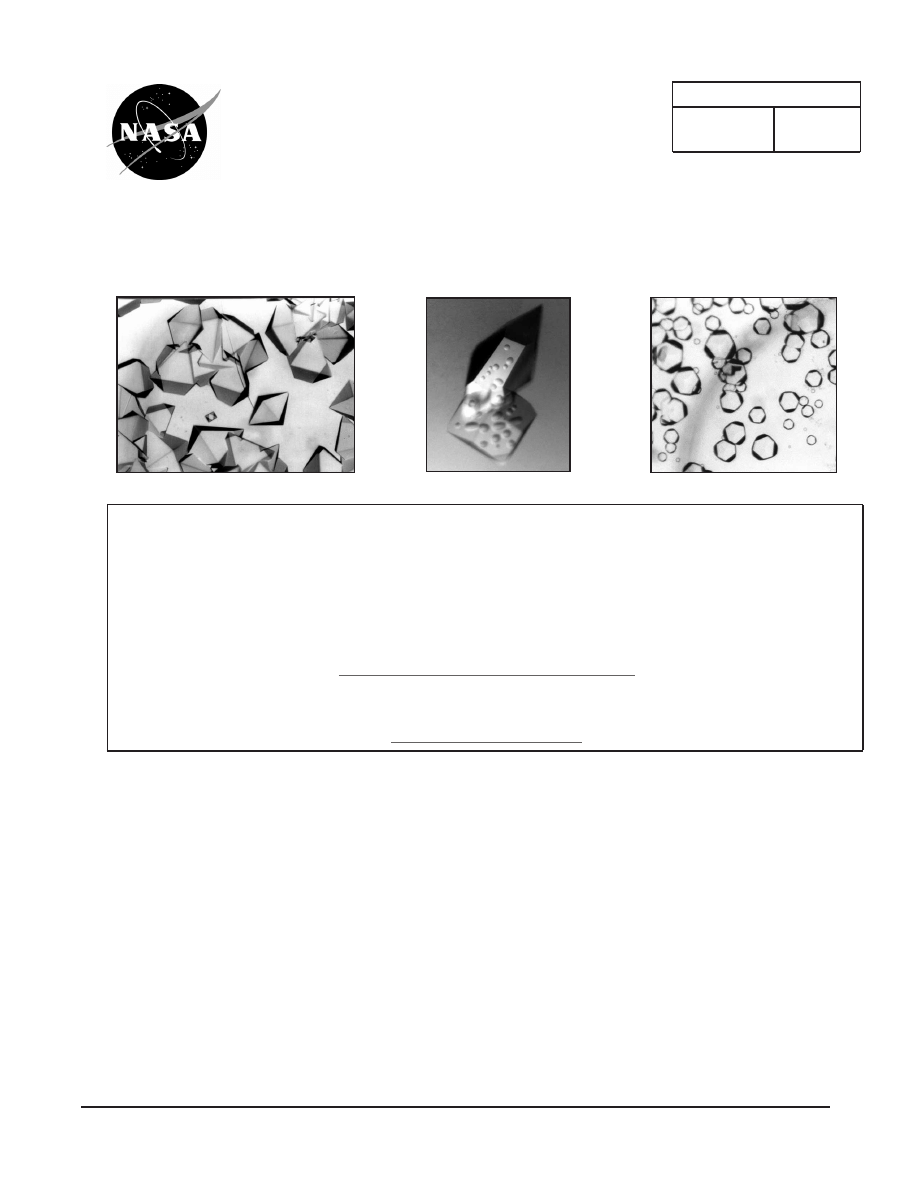
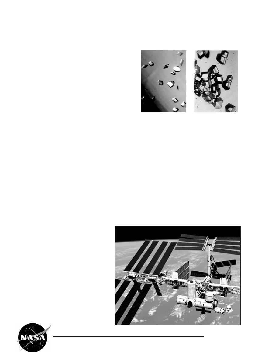
Excelsin Crystals
Excelsin Crystals
Glucose Isomerase Crystals
Glucose Isomerase Crystals
L
L
ysozyme Crystals
ysozyme Crystals
Note: all bold face words are referenced in the glossary in the back of this document.
A
protein crystal is a three-dimen-
sional array of molecules in
which every molecule or specific
group of molecules has the same
orientation and relationship to its
neighbors, as long as the chemical
environment of the solution sur-
rounding the crystal remains the
same. When a protein crystal
forms, protein molecules or groups
of molecules align to produce a
repeating three-dimensional pattern
or array. The effect of bringing these molecules
together in this arrangement is called amplification.
Amplification can be thought of in this way: imag-
ine a football stadium full of people. If only one
person stands up and yells, the sound produced is
not easily heard. But if everybody in the stadium
stands up, faces the same direction and yells at
the same time, that sound can be heard from a
great distance. A very similar thing happens dur-
ing the analysis of a protein crystal. One protein
molecule, by itself, would produce a signal so
weak it would be undetectable. But if all the mole-
cules in the crystal produced the same signal at
exactly the same time, then that signal would be
strong enough to be recorded and decoded. The
more closely oriented or aligned the molecules
making up the protein crystal are, the better the
signal, and the more accurate the molecular infor-
mation.
U
nfortunately, protein molecules
are so small that humans can’t
see them individually, let alone find
a specific site on a molecule or
determine its molecular structure. If
one cell in the human body were the
size of a football stadium, one pro-
tein molecule would be approximate-
ly the size of a can of soda, and
one of the atoms making up that
protein molecule would be about the
size of the fine print on the can.
Comparative sizes of proteins and the atoms
making up proteins
A NASA Recipe For Protein Crystallography
EB-2000-10-183-MSFC
2
A NASA Recipe For Protein Crystallography
EB-2000-10-183-MSFC
3
T
he first known published observation of the
crystallization of a protein was made by F.L.
Hunefeld in 1840 at Leipzig University in Germany.
While working with hemoglobin, Hunefeld obtained
flat, plate-like crystals of this protein when he
pressed the blood of an earthworm between two
slides of glass and allowed the blood to dry very
slowly.
In 1851, Otto Funke, another German
researcher, published a series of articles in which
he described growing hemoglobin crystals by
successively diluting red blood cells with a solvent
such as pure water, alcohol or ether, followed by
slow evaporation of the solvent from the resulting
protein solution.
Early on, scientists grew crystals solely to purify
proteins. Not until the 1930’s did researchers begin
to focus their attention on crystals as a source of
structural information about protein molecules.
They turned to X-ray diffraction, a procedure in
which a pencil-lead-sized X-ray beam is directed
at a crystal. The X-ray beam is scattered by the
crystal, producing a signal that results in tiny pin-
points that can be recorded on film. Data from this
recorded X-ray diffraction pattern has a direct rela-
tionship to the protein’s molecular structure and can
be used to help reveal the structure of a molecule
of the particular protein under investigation. By the
1960’s, scientists were investigating the molecular
structures of an abundance of crystals grown by
biochemists. Further, there was a century-long
backlog of crystals to be investigated. By the
1970’s, however, the
X-ray crystallogra-
phers had become
more selective in
determining which
proteins needed to
be crystallized for
analysis, and produc-
tion of the desired
crystals was no
longer meeting
demand. New
methods for grow-
ing higher-quality
crystals were needed. The inability of scientists to
produce crystals useable for successful analysis of
some proteins had become a bottleneck in the
process of determining the three-dimensional struc-
tures of the molecules of many important proteins.
Typically a protein crystal must be structurally well
ordered and from about 0.2 to 0.5 mm in size. With
a high water content, protein crystals are usually
quite fragile and somewhat difficult to handle.
Protein crystals can be described as "soft" in con-
trast to a "hard" salt crystal.
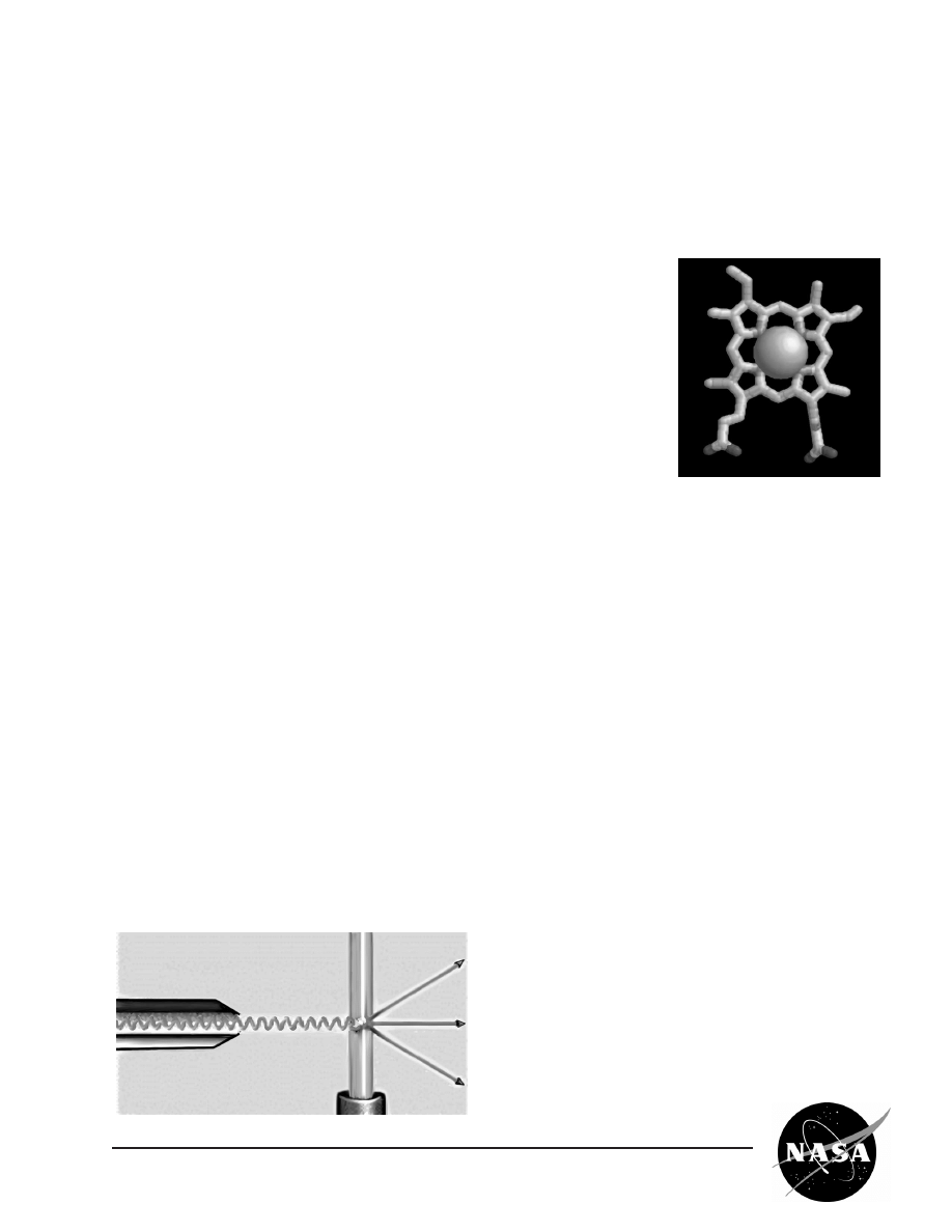
X-ray beam penetrates a
protein crystal
Molecular structure of
the heme ring in the
hemoglobin molecule
History

D
uring the 1970’s, Walter Littke, a space
research pioneer and professor of chemistry at
the University of Freiburg, Germany, was using a
common method of growing protein crystals: placing
a salt solution together with the protein solution.
When two such solutions come in contact, the salt
becomes associated with some of the water mole-
cules in which the protein is dissolved. This causes
the protein molecules to move closer together and
to begin to crystallize. However, many of the crys-
tals produced by this method are fragile, and small
or broken.
By 1980, Littke suspected the culprit in his
unusable crystals to be convection, or fluid flow, in
the solution surrounding the growing protein crys-
tals. This phenomenon occurs in normal gravity,
also known on Earth as one-g. Convection takes
place during crystal growth in one-g as protein mol-
ecules move from the surrounding solution and
assemble in an orderly way to become a part of the
growing crystal lattice. As protein molecules in the
solution move toward a crystal and become a part
of the crystal, the solution bordering the crystal then
contains a lower protein concentration than the
remainder of the solution and, therefore, it has a
lower density. This less-dense solution tends to
rise, and the denser solution sinks under the influ-
ence of gravity, creating fluid flows or convection,
next to the crystal. These convective currents can
have a negative effect on the quality of the crystal
being formed, because they can alter the orienta-
tion and position of the protein molecules as they
become a part of the crystal lattice. This can cause
disorder in the lattice structure of the protein crystal.
These imperfections in the crystal lattice, in turn,
adversely affect X-ray diffraction analysis results, or
the clarity with which a crystallographer can "see"
the precise position each atom occupies in the
three-dimensional structure of the protein molecule.
Another adverse effect of gravity on growing
crystals is sedimentation. Crystals drift to the bot-
tom of a drop of the solution when they have grown
to a mass larger than can be supported by suspen-
sion in the drop. When this happens, partially
formed crystals fall on top of one another and con-
tinue growing into each other. Since X-ray diffrac-
tion analysis requires single crystals, sedimentation
renders potentially high-quality crystals unusable for
data collection.
Scientists began to consider the idea that in a
microgravity environment, with reduced convection
and sedimentation, protein molecules would move
together more slowly, primarily by diffusion. It was
expected that higher quality crystals could be pro-
duced in the microgravity environment, such as that
of the orbiting Space Shuttle.
But just how do scientists grow a protein crystal
in microgravity (or on Earth, for that matter), and
what do they do with it once they have grown it?
Researchers begin with a protein solution that is
supersaturated. A protein solution is prepared by
dissolving the protein, also called the solute, in a
solvent. Proteins have a solubility, which is a
measure of the amount of the protein (solute) that
can be dissolved in a given solvent, under specific
conditions. Growing a crystal, however, requires
supersaturation, which is a less stable condition that
results when somewhat more than maximum
amount of protein that can be dissolved in a solvent
under nominal conditions is, in fact, dissolved in the
solvent.
A NASA Recipe For Protein Crystallography
EB-2000-10-183-MSFC
4
Enter Microgravity
5
M
ost scientists don’t know the precise solubility
of the protein they may be investigating. A
researcher tests many different solution conditions
by varying such parameters as protein concentra-
tion, salt concentration and pH, in order to find the
best conditions required to promote the formation of
usable protein crystals. Once the researcher has
defined an optimum range of solution conditions, he
or she usually uses one of two methods for actually
growing the crystal: vapor diffusion or liquid/liquid
diffusion. Both methods involve changing the con-
ditions of a protein solution to supersaturate the
solution. In vapor diffusion, the crystallographer
places a drop of protein solution in a chamber that
also holds a solution called a precipitant solution,
typically a salt solution. The salt in the precipitant
solution is more concentrated than salt that is in the
protein solution. This causes water vapor to diffuse
through the air from the drop to the precipitant solu-
tion. As water is removed from the drop, the protein
becomes more concentrated, causing protein mole-
cules to move closer together and nucleate. In liq-
uid/liquid diffusion, the researcher diffuses a salt
solution or some other precipitant solution in one
compartment of a two-chamber vial into a protein
solution in the other compartment. As the salt con-
centration increases, some of the water from the
protein solution becomes associated with the salt,
effectively raising the concentration of the protein in
the solution. Under these conditions the protein
molecules come together and begin to crystallize.
To analyze a protein crystal, an X-ray crystallog-
rapher shines an X-ray beam through the crystal.
Unlike a single dental X-ray, which produces a
shadow image of a tooth, these X-ray images have
to be taken many times from different angles to pro-
duce a pattern from the scattered X-ray beam. This
pattern is a map of the intensity of the X-rays after
they diffract through the crystal. The X-rays actually
bounce off the electron clouds that form the outer
structure of each atom. A flawed crystal will yield a
blurred pattern; a well-ordered protein crystal will
yield a series of sharp, more distinct diffraction pat-
terns.
From these patterns, researchers build an
electron density map. With powerful computers
capable of performing complex mathematical cal-
culations, scientists can use the electron density
patterns to determine the structure of the protein
and make computer-generated models of the
three-dimensional structure of a protein molecule.
The models allow researchers to improve
understanding of how the protein functions. In addi-
tion, the models allow scientists to look for receptor
sites and active areas that control a protein’s func-
tion and role in the progression of
diseases.
X-ray diffraction pattern from
a protein crystal
A NASA Recipe For Protein Crystallography
EB-2000-10-183-MSFC
Blueprint for Growing a Crystal
Electron Density Map
A NASA Recipe For Protein Crystallography
EB-2000-10-183-MSFC
6
M
icrogravity has been the chosen environment
for dozens of fundamental science experi-
ments growing macromolecular crystals. Since
NASA’s protein crystal growth program began, prin-
cipal investigators and their research teams have
flown samples of a total of 185 different proteins,
RNA’s, DNA’s and viruses (as of August 1999). The
macromolecules these scientists have studied
range from insulin to lactate dehydrogenase ( a
major enzyme in energy production and an impor-
tant muscle protein in all animals) to thaumatin (a
sweet tasting protein with potential as a sugar sub-
stitute.) Most of NASA’s protein crystal growth
experiments, conducted from 1985 to 1999, have
been flown on Space Shuttle missions. The
remainder were conducted on the Russian space
station
Mir.
Given the great strides NASA’s protein crystal
growth program has made since the first protein
crystal growth experiments were conducted in
microgravity, where is the field headed now? For
many, it is to the International Space Station (ISS).
The ISS will expand the opportunities for growing
crystals in microgravity, enabling continued
advances in understanding the fundamental science
of the crystal growth process. Crystals, which usual-
ly grow more slowly in microgravity, will have more
time to fully develop into usable specimens. Follow-
up experiments, an important and common feature
of all ground-based research, will be more feasible
in space on the ISS.
The International Space Station (ISS)
will provide a platform for long term
microgravity experiments
http://spaceflight.nasa.gov/station
Normal gravity vs. microgravity
grown insulin crystals
Earth (1g) Space (µg)
NASA Macromolecular Crystal Growth Results

7
Abilities Necessary to do Scientific Inquiry
Physical Science
Life Science
Science and Technology
Science in Personal and Social Perspectives
History and Nature of Science
1) Identify questions and concepts that guide
scientific investigations
2) Design and conduct scientific investigations
3) Use technology and mathematics to improve
investigations and communications
4) Formulate and revise scientific explanations
and models using logic and evidence
5) Recognize and analyze alternative explanations
and models
6) Communicate and defend a scientific argument
7) Understandings about scientific inquiry
1) Structure of atoms
2) Structure and properties of matter
3) Chemical reactions
4) Motions and forces
1) The cell
2) Molecular basis of heredity
3) Matter, energy, and organization in living
systems
4) Behavior of organisms
1) Abilities of technological design
2) Understandings about science and technology
1) Personal and community health
2) Science and technology in local, national, and
global challenges
1) Science as human endeavor
2) Nature of scientific knowledge
3) Historical perspectives
A NASA Recipe For Protein Crystallography
EB-2000-10-183-MSFC
Connections to Academic Standards
This NASA Educational Brief supports the following National Academy of Sciences Science Content
Standards:
National Science Education Content Standards (Grades 9-12) supported by the NASA Recipe for
Protein Crystallography.
Isolation, Crystallization & Purification of
Excelsin From Brazil Nuts
Ground Brazil nuts can be extracted with 5% aqueous NaCl at 50˚ to 70˚ C, filtered, then dialyzed
against distilled water to obtain crystals.
This activity takes approximately 60 minutes
(CAUTION: Glass wool should only be handled while wearing gloves.)
Key Words
Aqueous
Dialysis Membrane (or tubing)
Morphology
Clarify
Centrifuge
Pellet
Decant
Opaque
Dialyze
Filtrate
Denature
Supernatant
Materials
Funnel
Styrofoam (for bottom of 500 ml beaker)
Water (distilled preferred)
Table Salt (sodium chloride)
Brazil Nuts (raw, organically grown)
Glass Rod
Centrifuge Tubes
Cheesecloth, Glass Wool, or Filter Paper
Dialysis Membrane (app. 20 cm in length)
Pipette or Medicine Dropper
50 ml graduated cylinder
Thermometer
500 ml and 150 ml beakers
Heating Plate
Small clamp(s)
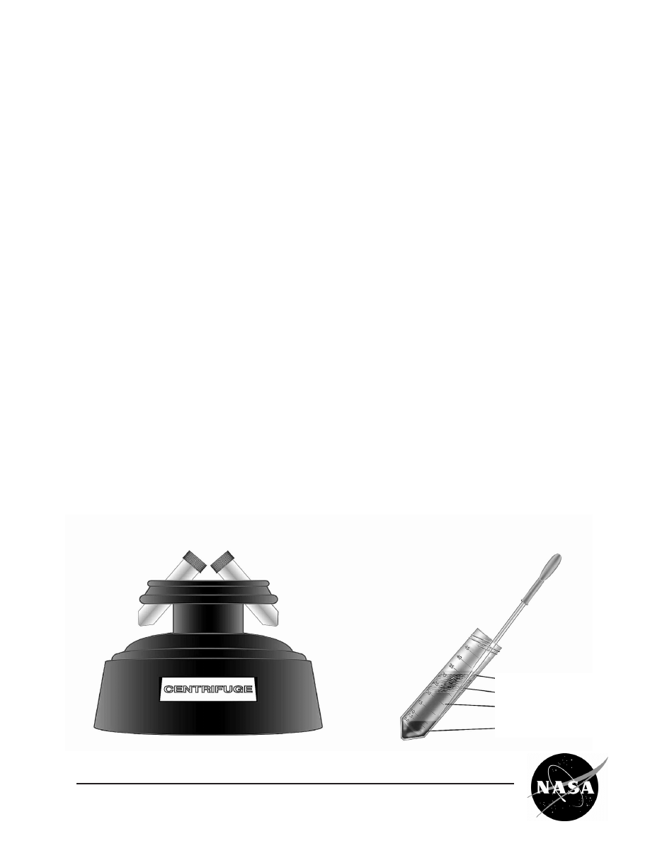
Small Centrifuge (up to 3000rpm)(optional)
Blender or Coffee Grinder
Laboratory Balance
A NASA Recipe For Protein Crystallography
EB-2000-10-183-MSFC
8
9
A NASA Recipe For Protein Crystallography
EB-2000-10-183-MSFC
Methods
Oil
Nut Meats
Aqueous Protein
Solution
Pellet
I. Isolation of Excelsin
1) Record the mass of ~ 8 or 9 raw Brazil nuts
(preferably organically grown). Grind the nuts
to granular size. 1 nut has a mass of approxi-
mately 2.8 grams.
2) Prepare a 5% sodium chloride (NaCl) solution
by dissolving 10 grams of solid NaCl in 200
milliliters of distilled water. Measure 25 ml of
5% NaCl solution into a graduated cylinder
and then pour into a 150 ml beaker. Record
the volume of NaCl solution in the beaker.
(Reserve remaining 5% NaCl solution for mak-
ing 1% NaCl solution in Part II.3.)
3) Create a water bath by placing a flat piece of
styrofoam (can be cut from the bottom of a sty-
rofoam cup) into the bottom of a 500 ml
beaker. Pour approximately 50 ml of water into
the 500 ml beaker. Place the smaller 150 ml
beaker (that contains the NaCl solution) in the
larger 500 ml beaker (that contains the water).
Place the beaker assembly on the heating
plate and heat until the water is at 50˚C.
Measure the final temperature and record.
4) Add the ground nuts to the NaCl solution in the
150 ml beaker and stir with a glass rod to mix.
Continue heating the water bath to maintain
the temperature of the NaCl solution and
ground nut mixture at 50˚C to 70˚C for 20 min-
utes, stirring occasionally. Record the temper-
ature at regular intervals. (Be careful not to stir
vigorously or to heat above the designated
temperature range because this may cause
the protein to denature or break down).
5)
(Caution: Glass wool should only be han-
dled when wearing gloves.)
Place 1-2 layers
of glass wool (or 10 layers of cheesecloth or
filter paper) in a funnel and place over a
beaker. Pour the NaCl/nut mixture through
glass wool. Measure and record the volume
of filtrate. (Note: Filtrate should be opaque.)
6) Option 1 - For classrooms without a cen-
trifuge: Skip steps 7-9 and go directly to the
Crystallization of Excelsin in Section II.

A NASA Recipe For Protein Crystallography
EB-2000-10-183-MSFC
10
7) Option 2 - For classrooms with a centrifuge:
Pour filtrate into two centrifuge tubes and weigh
to make sure tubes are of equal weight.
Record the mass of each centrifuge tube.
8) Centrifuge for 20 minutes at 3000 rpm.
Referring to the diagram from the previous
page, pipette off the oil (top layer) and discard.
Carefully push the pipette through the nut meat
layer into the aqueous layer and decant off the
aqueous portion (located above the pellet and
just below the oil). Discard the pellet and oil.
9) Centrifuge the supernatant to clarify the solu-
tion further. The supernatant should now be a
fairly clear brown or brownish-yellow solution.
Measure and record the volume of the extract-
ed protein solution.
1) Soak the dialysis membrane (small molecular
weight, preferably below 100,000 MWCO
(molecular weight cut off)) in room temperature
water for 5 minutes or longer. Take the mem-
brane out of the beaker and tie a knot in one
end. Refer to page 12 for procedures on the
handling of dialysis tubing.
2) Pour the extracted protein/filtrate solution from
step 5 or 8 above into the dialysis
membrane/tubing and tie the other end of the
membrane.
3) Prepare 500 ml of 1% NaCl solution by combin-
ing 100 ml of 5% NaCl solution and 400 ml of
distilled water. Dialyze the protein solution
against the 500 ml of 1% NaCl solution. The
volume of the 1% NaCl solution should be at
least three times the measured volume of the
extracted solution. Record the volume of pro-
tein filtrate/solution and 1% NaCl solution.
Crystals should appear 4 - 10 hours later (They
will appear as white powder in the bottom of
the bag). If crystals do not appear in the 1%
NaCl solution, lower the NaCl concentration to
0%.
4) When the white powder appears, view it under
a microscope while the solution is still in the
bag.
1) Cut one end of the dialysis membrane bag
and decant the solution off of the crystals.
2) Add just enough 5% NaCl solution (1-3 ml) to
get the crystals to go back into solution. Either
retie the bag or seal the open end with a clamp.
Place the bag into a fresh solution of 1% NaCl
and allow to dialyze overnight, or until crystals
appear. Record information about crystals: size,
number, and morphology.
3) By repeating this dialysis process, a protein
can usually be rendered essentially pure. To
obtain larger crystals, slowly decrease the con-
centration of NaCl in solution. For example,
instead of going directly to 1% NaCl, start with
4.5% and decrease in increments of 0.5%.
III. Recrystalization and Purification of Excelsin
II. Crystallization of Excelsin
Lab Record
Step
Procedure
Record
I(1)
Brazil nut mass (grams)
g
I(2)
Volume of NaCl solution (ml)
ml
I(3)
Temperature of water bath (˚C)
˚C
I(4)
Temperature of NaCl/nut solution (˚C) – 1st recording
˚C
I(4)
Temperature of NaCl/nut solution (˚C) – 2nd recording
˚C
I(4)
Temperature of NaCl/nut solution (˚C) – 3rd recording
˚C
I(4)
Temperature of NaCl/nut solution (˚C) – 4th recording
˚C
I(4)
Temperature of NaCl/nut solution (˚C) – 5th recording
˚C
I(5)
Volume of protein filtrate (ml)
ml
I(6)
Centrifuge tube #1 mass
g
I(6)
Centrifuge tube #2 mass
mg
I(8)
Volume of extracted protein solution (ml)
ml
II(3)
Volume of protein filtrate (ml)
ml
II(3)
Volume of 1% NaCl solution (ml)
ml
II(3)
Volume of 0% NaCl solution (ml) (if required)
ml
III(2)
Volume of 5% NaCl solution added to bag (ml)
ml
III(2)
Volume of 1% NaCl solution added for dialysis (ml)
ml
III(3)
Volume of 5% NaCl solution added to bag (ml)
ml
III(3)
Volume of 1% NaCl solution added for dialysis (ml)
ml
Record the size, number and morphology of the final Excelsin crystals produced.
Crystal
Morphology (check which box applies)
Number
Size
Excelsin
Oily
mm
Spherical
mm
Platelike
mm
Hexagonal
mm
Needles
mm
Other
mm
A NASA Recipe For Protein Crystallography
EB-2000-10-183-MSFC
11
12
A NASA Recipe For Protein Crystallography
EB-2000-10-183-MSFC
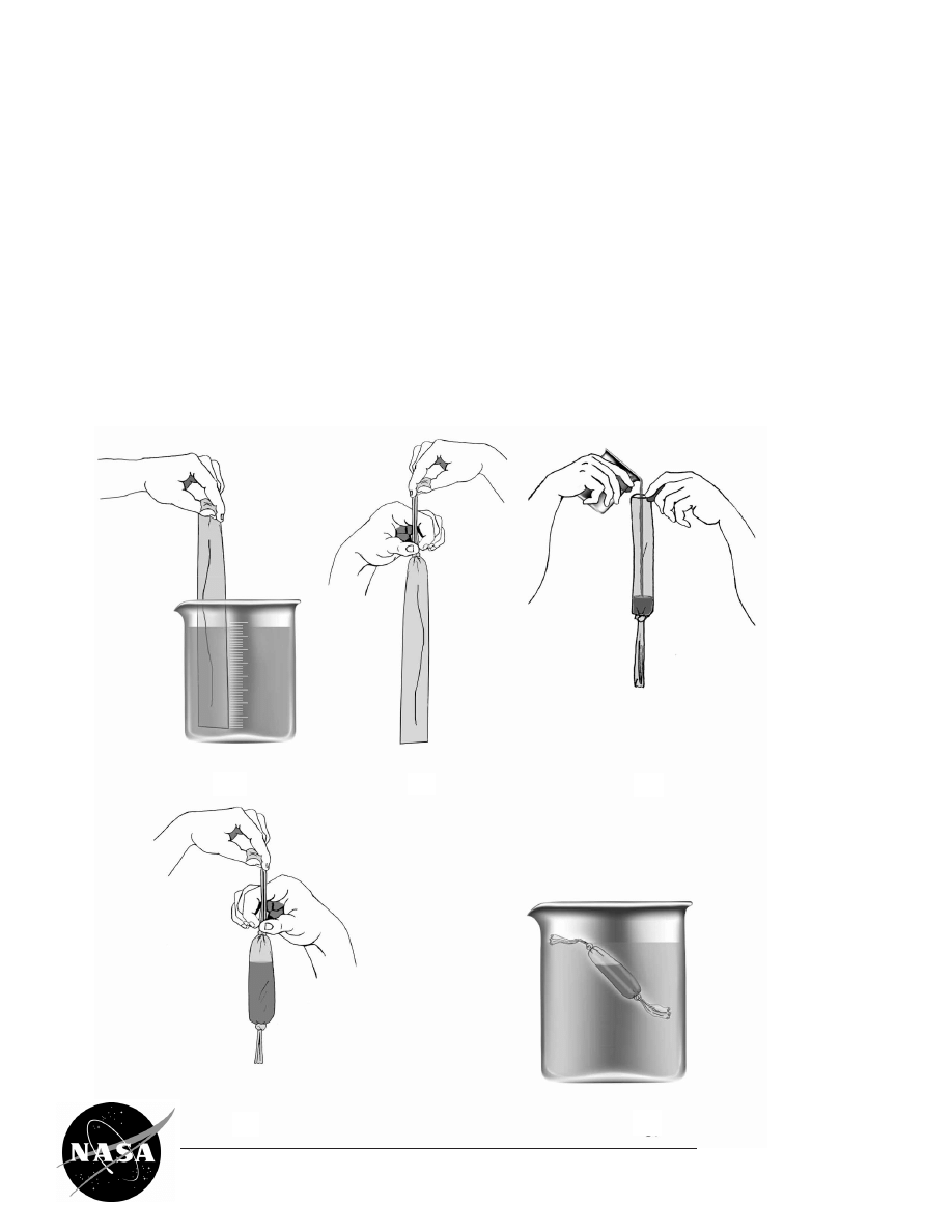
PROCEDURES FOR HANDLING
DIALYSIS TUBING
1) Soak the dialysis tubing in a beaker of distilled
water.
2) Tie the dialysis tubing making sure to tie the
knot from the top end toward the knot, ensuring
that the area to contain the protein solution is
not touched or handled. Touching the tubing
may increase the pore size of the tubing and
result in loss of protein.
3) Pour the protein solution into the dialysis tubing.
4) Tie the remaining open end of the tubing, leav-
ing a small air pocket so the bag will float.
5) Place the dialysis bag in the NaCl solution (see
Lab Method step II.3.)
1
2
3
4
5

13
A NASA Recipe For Protein Crystallography
EB-2000-10-183-MSFC
GLOSSARY
Active site
the portion of a molecule that binds with a substrate molecule
Aqueous
relating to, similar to, containing, or dissolved in water; watery
Clarify
to make clear by removing impurities or solid matter
Concentration
a measurement of the amount of solute that is dissolved in a given quantity of solvent
Convection
heat transfer in a gas or liquid by the circulation of currents from one region to another
Decant
to pour off gently
Denature
to alter the structure of (a protein), as with heat, alkali, or acid, so that some of its
original properties, especially its biological activity, are diminished or eliminated
Density
the ratio of the mass of an object to its volume
Dialysis/Dialyze
the transfer of dissolved solids (solute) across a semipermeable membrane, which
permits or hinders diffusion of molecules according to their size
Dialysis membrane
a semipermeable casing used to dialyze a solution
(tubing)
Diffusion
the spontaneous intermingling of molecules as a result of random thermal motion
Electron density map
a graphical representation of the volume of space that an electron is most likely to
occupy; the smaller the volume of space occupied, the higher the electron density
Filtrate
the liquid passing through the filter during filtration
Gravity
the natural force of attraction of objects to each other due to their masses
Hemoglobin
an iron-containing protein in red blood cells that carries oxygen from the lungs to the
rest of the body
Liquid/liquid diffusion
the diffusion of a precipitant solution into a protein solution across their common
liquid/liquid interface
Macromolecule
a very large molecule, such as a polymer or protein, consisting of many smaller
structural units linked together
Microgravity
an environment in which the apparent weight of a system is small compared to its
actual weight (due to gravity)
Molecular weight
the sum of the weight of all the atoms in a molecule, also called
formula weight

14
A NASA Recipe For Protein Crystallography
EB-2000-10-183-MSFC
Morphology
the shape of a crystal
Nucleate
to gather, as about a nucleus or center
Opaque
impenetrable by light; neither transparent nor translucent
Pellet
a small, solid or densely packed mass
pH
a measure of the acidity or alkalinity of a solution, numerically equal to 7 for neutral
solutions, increasing with increasing alkalinity and decreasing with increasing acidity;
the pH scale commonly in use ranges from 0 to 14
Precipitant solution
a solution which causes the formation of a precipitate
Precipitant
a solid or solid phase separated from a solution
Protein
any of a group of complex organic macromolecules composed of one or more chains
of amino acids; proteins are fundamental components of all living cells and include
many substances, such as enzymes, hormones, and antibodies, that are necessary for
the proper functioning of an organism
Receptor site
a region, often the exposed part of a membrane protein, that binds a substance but
does not catalyze a reaction in the chemical it binds
Sediment
the material that settles to the bottom of a liquid
Sedimentation
the settling of materials to the bottom of a liquid; this settling is due to gravity
Solute
the dissolved substance in a solution; in salt water, salt is the solute
Solution
a homogeneous or uniform mixture of two or more substances
Solvent
a substance used to dissolve a solute to form a solution; in salt water, water is the
solvent
Supernatant
the clear fluid floating above a sediment or precipitate
Supersaturated
the state of a solution when it contains more solute (dissolved substance) than it can
theoretically hold
Vapor diffusion
a process of diffusion in which a drop of protein solution is suspended above a precipi-
tant and sealed from the air, resulting in equilibration through the vapor phase
X-ray diffraction
the scattering of x-ray beams by crystal atoms, producing a diffraction pattern that
yields information about the structure of the crystal
A NASA Recipe For Protein Crystallography
EB-2000-10-183-MSFC
15
If you live in:
Alaska
Arizona
California
Hawaii
Idaho
Montana
Nevada
Oregon
Utah
Washington
Wyoming
Please contact:
NASA Educator Resource Center
Mail Stop 253-2
NASA Ames Research Center
Moffett Field, CA 94035-1000
Phone:
(650) 604-3574
FAX:
(650) 604-3445
http://ccf.arc.nasa.gov/dx/basket/trc/trchome.html
Illinois
Indiana
Michigan
Minnesota
Ohio
Wisconsin
NASA Educator Resource Center
NASA John H. Glenn Research Center at Lewis Field
21000 Brookpark Rd., MS 8-1
Cleveland, OH 44135
Phone:
(216) 433-2017
FAX:
(216) 433-3601
http://www.grc.nasa.gov/WWW/PAO/html/edteacher.htm
NASA Educator Resource Centers
N
ASA's Central Operation of Resources for Educators (CORE)
was established for the national
and international distribution of NASA-produced educational materials in audiovisual format.
Educators can obtain a catalogue and an order form by one of the following methods:
NASA CORE
Lorain County Joint Vocational School
15181 State Route 58
Oberlin, OH 44074-9799
Phone: (440) 775-1400
Fax: (440) 775-1460
E-mail: nasaco@leeca.org
Home Page: http://core.nasa.gov
Educator Resource Center Network
(ERCN)
To make additional information available
to the education community, NASA has created
the NASA Educator Resource Center (ERC) net-
work. Educators may preview, copy, or receive
NASA materials at these sites. Phone calls are
welcome if you are unable to visit the area. A list
of the centers and the regions they serve follows.

A NASA Recipe For Protein Crystallography
EB-2000-10-183-MSFC
16
Colorado
Kansas
Nebraska
New Mexico
North Dakota
Oklahoma
South Dakota
Texas
Space Center Houston
NASA Educator Resource Center for Johnson Space Center
1601 NASA Road One
Houston, TX 77058
Phone:
(281) 244-2129
FAX:
(281) 483-9638
http://www.jsc.nasa.gov
Florida
Georgia
Puerto Rico
Virgin Islands
NASA Educator Resource Center
Mail Code ERC
NASA Kennedy Space Center
J.F. Kennedy Space Center, FL 32899
Phone:
(321) 867-4090
FAX:
(321) 867-7242
http://www/ksc.nasa.gov
Kentucky
North Carolina
South Carolina
Virginia
West Virginia
Virginia Air and Space Center
Educator Resource Center for NASA Langley Research Center
600 Settlers Landing Road
Hampton, VA 23669-4033
Phone:
(757) 727-0990 Ext. 75
FAX:
(757) 727-0898
http://www.vasc.org/erc/
NASA Educator Resource Laboratory
Mail Code 130.3
NASA Goddard Space Flight Center
Greenbelt, MD 20771-0001
Phone:
(301) 286-8570
FAX:
(301) 286-1781
http://pao.gsfc.nasa.gov/gsfc/educ/trl/welcome.html
Connecticut
Delaware
District of Columbia
Maine, Maryland
Massachusetts
Pennsylvania
New Hampshire
New Jersey
New York
Rhode Island
Vermont
Alabama
Arkansas
Louisiana
Missouri
Tennessee
Iowa
U.S. Space & Rocket Center
Educator Resource Center for NASA Marshall Space Flight Center
One Tranquility Base
Huntsville, AL 35758
Phone:
(256) 544-5812
FAX:
(256) 544-5820
http://www.msfc.nasa.gov/education/erc

A NASA Recipe For Protein Crystallography
EB-2000-10-183-MSFC
17
GSFC/Wallops Flight Facility
NASA Educator Resource Center
Building J-17
Wallops Island, VA 23337
Phone:
(757) 824-2298
FAX:
(757) 824-1776
http://WFF.nasa.gov
NASA Educator Resource Center for
NASA Dryden Flight Research Center
45108 N. 3rd Street East
Lancaster, CA 93535
Phone:
(661) 948-7347
Fax:
(661) 948-7068
http://www.dfrc.nasa.gov/trc
/
NASA JPL Educator Resource Center
Village at Indian Hill
1460 East Holt Avenue, Suite 20
NASA Jet Propulsion Laboratory
Pomona, CA 91767
Phone:
(909) 397-4420
Fax:
(909) 397-4470
http://learn.jpl.nasa.gov
Arizona and
Southern California
Virginia and
Maryland’s Eastern
Shores
Mississippi
NASA Educator Resource Center
NASA Stennis Space Center
Building 1200
Stennis Space Center, MS 39529-6000
Phone:
(228) 688-3220
FAX:
(228) 688-2824
http://education.scc.nasa.gov/htmls/trc/trc.htm
R
egional Educator Resource Centers
offer more educators access to NASA educational materials. N A S A
has formed partnerships with universities, museums, and other educational institutions to serve as
regional ERCs in many states. A complete list of regional ERCs is available through CORE, or elec-
tronically via NASA Spacelink at
http://spacelink.nasa.gov/ercn/
N
ASA's Education Home Page
serves as a cyber-gateway to information regarding educational
programs and services offered by NASA for the American education community. This high-level
directory of information provides specific details and points of contact for all of NASA's educational
efforts, Field Center offices, and points of presence within each state. Visit this resource at the follow-
ing address:
http://education.nasa.gov
The Jet Propulsion
Laboratory (JPL)
serves inquiries related
to space and planetary
exploration and other
JPL activities.

A NASA Recipe For Protein Crystallography
EB-2000-10-183-MSFC
18
N
ASA Spacelink
is one of NASA's electronic
resources specifically developed for the edu-
cational community. Spacelink is a "virtual library"
in which local files and hundreds of NASA World
Wide Web links are arranged in a manner familiar
to educators. Using the Spacelink search engine,
educators can search this virtual library to find
information regardless of its location within NASA.
Special events, missions, and intriguing NASA
websites are featured in Spacelink's "Hot Topics"
and "Cool Picks" areas. Spacelink may be
accessed at: http://spacelink.nasa.gov
NASA Spacelink is the official home to electronic
versions of NASA's Educational Products. A
complete listing of NASA Educational Products
can be found at the following address:
http://spacelink.nasa.gov/products
N
ASA Television (NTV)
features Space
Shuttle mission coverage, live special events,
interactive educational live shows, electronic field
trips, aviation and space news, and historical
NASA footage. Programming has a 3-hour block
— Video (News) File, NASA Gallery, and
Education File — beginning at noon Eastern and
repeated five more times throughout the day. Live
feeds preempt regularly scheduled programming.
Check the Internet for program listings at:
http://www.nasa.gov/ntv
For more information on NTV, contact:
NASA TV
NASA Headquarters
Code P-2
Washington, DC 20546-0001
Phone (202) 358-3572
NTV Weekday Programming Schedules
(Eastern Times)
Video File NASA Gallery Education File
12-1 p.m. 1-2 p.m. 2-3 p.m.
3-4 p.m. 4-5 p.m. 5-6 p.m.
6-7 p.m. 7-8 p.m. 8-9 p.m.
9-10 p.m. 10-11 p.m. 11-12 p.m.
How to Access Information on NASA's
Education Program, Materials, and Services
EP-1999-06-345-HQ.
This brochure serves as a guide to accessing a
variety of NASA materials and services for educa-
tors. Copies are available through the ERC net-
work, or electronically via NASA Spacelink.
Spacelink & NASA Television
Online Evaluation
Please take a moment to evaluate this product at
http://ehb2.gsfc.nasa.gov/edcats/educational_brief
Your evaluation and suggestions are vital to continually
improving NASA educational materials.
Wyszukiwarka
Podobne podstrony:
1 DIETA PROTEINOWA
FABP ang. fatty acids binding proteins
Dieta proteinowa dr.dukana, Zdrowie
DIETA PROTEINOWA
Arakawa et al 2011 Protein Science
Kulki Proteinowe
Methods in Enzymology 463 2009 Quantitation of Protein
DIETA PROTEINOWA
Fluorescent proteins as a toolkit for in vivo imaging 2005 Trends in Biotechnology
Dieta Proteinowa przepisy na 101 deserów
Beta barrel proteins form bacte Nieznany
Producing proteins in transgenic plants and animals
Dieta proteinowa(1)
Making recombinant proteins in animals
(kulki proteinowe 17uniwersalnych tonących)id 1350
17 uniwersalnych tonących kulek proteinowych
więcej podobnych podstron