
REVIEWS
Renal failure in atherosclerotic renovascular
disease: pathogenesis, diagnosis, and intervention
R G Woolfson
Atherosclerotic renovascular disease (ARVD)
is increasingly recognised as an important
cause of both chronic and end stage renal fail-
ure. These patients tend to do badly on
dialysis, which reflects their systemic athero-
sclerotic burden. In an e
Vort to delay and per-
haps prevent their need for renal replacement
therapy, some patients are subjected to a
variety of medical, radiological and surgical
interventions, although evidence for each is
sparse. The purpose of this review is to
describe the epidemiology and pathophysiol-
ogy of renal failure in ARVD, discuss the avail-
able diagnostic techniques, consider the evi-
dence for benefit from intervention in the
context of pathogenesis and finally, identify
those gaps in our knowledge which impede the
practice of evidence based medicine.
Epidemiology of ARVD
The prevalence of ARVD in patients with
chronic renal failure is not known but dialysis
registry data provide some epidemiological
information about ARVD among patients who
develop end stage renal failure (ESRF). Over a
20 year period in an American haemodialysis
unit, Mailloux et al reported a 16% incidence
of ARVD among new patients with a median
age of 70 years (range 37–86 years).
1
Similarly,
in an 18 month retrospective study in a UK
haemodialysis unit, Scoble et al reported a
14% incidence of ARVD among patients over
the age of 50 years.
2
ESRF patients with
ARVD generally present with substantial
comorbidities and have a poor prognosis on
dialysis.
1
Given their poor prospects, many
patients may not be o
Vered dialysis and there-
fore these figures are likely to underestimate
the true incidence of the disease. These stud-
ies do identify age as a risk factor for ARVD
and a cause for ESRF. Consistent with this, in
a series of 133 hypertensive patients with
chronic renal failure (mean (SD) creatinine
clearance 51(26) ml/min) not due to glomeru-
lonephritis or polycystic kidney disease, the
incidence of atherosclerotic renal artery steno-
sis (ARAS) was shown to rise progressively
with age (see fig 1).
3
The prevalence of ARAS in patients who are
undergoing investigation for atherosclerosis is
in proportion to their burden of extrarenal dis-
ease (see fig 2). Therefore it is much higher in
patients with aortoiliac disease than in patients
undergoing coronary angiography, in whom
cardiac pain may be the result of a relatively
small burden of atherosclerosis but with a par-
ticularly critical distribution. This relationship
is confirmed by studies that have reported the
prevalence of extrarenal vascular disease in
patients with proved high grade ARAS.
4–6
Louie et al investigated the prevalence of
carotid and peripheral vascular disease in 60
patients with ARAS graded as greater than or
less than 60%.
6
In the less severe group, 25%
and 50% of patients were a
Vected by carotid
and peripheral vascular disease respectively
increasing to 46% and 73% for those with
ARAS exceeding 60%. Given that diabetes
mellitus is a risk factor for systemic atheroscle-
rosis, it is not surprising that it is also associated
with an increased prevalence of ARAS.
7 8
The true incidence of ARVD may also be
underestimated as a result of its varied presen-
tation which includes the patient with recur-
rent “flash” pulmonary oedema. The diagnosis
may be suspected in an elderly uraemic
arteriopath with a normal urinary sediment
and absent proteinuria, however, the presence
of proteinuria, even up to nephrotic range, with
or without evidence of glomerular bleeding,
does not exclude the diagnosis.
9 10
Progression of ARAS
Reported rates of progression of ARAS vary
between 18% and 53% over mean follow up
periods which range from 24 to 52 months.
11–15
The risk of progression appears to be deter-
mined by the severity of disease at the time of
diagnosis. Zierler et al used serial duplex Dop-
pler scans to show that 8% of normal arteries
developed a stenosis exceeding 60% at three
years whereas 48% of those with a significant
but not critical (that is, <60%) stenosis at
baseline progressed.
14
The reported incidence
of complete occlusion ranges from 7% to
16%
11 12 14
and this tends to a
Vect kidneys with
baseline stenoses exceeding 60% in patients
with bilateral disease.
Aside from baseline severity, the identifica-
tion of other risk factors for progression of
ARAS remains contentious and does not
explain why only some stenoses progress. Some
studies report no correlation with blood
Figure 1
Incidence of ARAS increases with age.
3
100
75
50
0
25
Age (years)
No stenosis
% Stenosis
50–59
60–69
> 70
< 50% stenosis
> 50% stenosis
Postgrad Med J 2001;77:68–74
68
Department of
Nephrology, Middlesex
Hospital, UCLH Trust,
Mortimer Street,
London W1N 8AA, UK
Correspondence to:
Dr Woolfson
r.woolfson@ucl.ac.uk
Submitted 27 March 2000
Accepted 22 June 2000
www.postgradmedj.com
pressure, smoking, diabetes mellitus, hyperlipi-
daemia, or the presence of coronary or periph-
eral vascular disease.
11 12 16
In contrast, Crowley
et al reported that age, female gender, hyper-
tension, severity of coronary disease, and
ARVD at baseline were independent variables
for progression of ARAS in 1214 patients with
a mean follow up of 2.59 years.
15
Data which
suggest that the rate of stenosis progression is
falling, perhaps secondary to better control of
hyperlipidaemia or hypertension, are not con-
vincing.
Development of renal atrophy
The important adverse outcome in ARAS is
the development of renal atrophy and dysfunc-
tion which may result directly from the
stenosis, as occurs in patients with fibromuscu-
lar dysplasia (FMD).
11
In 85 patients with
ARAS who underwent repeated angiography,
Schreiber et al noted progression of stenoses in
44% of kidneys, although renal atrophy (reduc-
tion in renal length
>1.5 cm) aVected 70%.
11
When patients without progressive stenoses
(n=48) were considered separately from those
with progressive stenoses (n=37), renal atrophy
and increased creatinine were significantly
more likely in the progressive group but even
so, approximately one quarter of the non-
progressive group also demonstrated worse
function and renal atrophy. This study provides
strong evidence that progressive parenchymal
injury and renal dysfunction reflects not just
progress of the underlying stenosis but also
another pathological process.
Two subsequent studies have confirmed the
relationship between severity of ARAS and risk
of renal atrophy. Guzman et al performed
repeated duplex Doppler at six monthly inter-
vals in 54 patients with ARAS over a mean fol-
low up period of 44 months.
17
Renal atrophy
(exceeding 1 cm) was not observed in kidneys
with baseline ARAS less than 60%, but by 12
months had a
Vected 26% of kidneys with
baseline ARAS exceeding 60%. When patients
were subdivided into those with unilateral or
bilateral ARAS, then the 12 month risk of atro-
phy was 13% and 43% respectively. Similarly,
Caps et al reported a 24 month cumulative
incidence of renal atrophy (exceeding 1 cm) of
5.5% in those with no baseline ARAS, 11.7%
in those with stenoses less than 60% and
20.8% in those with stenoses exceeding 60%.
18
The development of renal atrophy in non-
progressive ARAS could be due to vascular
dysfunction in the intrarenal microcirculation
distal to the stenosis rather than hypoperfusion
secondary to a critical stenosis. Lerman et al
used electron beam computer tomography to
measure whole kidney, cortical and medullary
blood flow in 42 patients with ARAS, FMD, or
essential hypertension who had previously
undergone
renal
angiography
and
were
matched for blood pressure and baseline
creatinine.
19
Even when corrected for renal vol-
ume, whole kidney perfusion and cortical per-
fusion were significantly less in the ARAS
group compared with the groups with FMD or
essential
hypertension
(p<0.05),
although
medullary blood flow was conserved. Consist-
ent with this evidence of abnormal cortical
perfusion in ARAS, whole kidney and cortical
blood flow correlated significantly with the
degree of renal artery stenosis in the FMD
group but not in the patients with ARAS. Simi-
lar results were reported by Tullis et al who
used duplex Doppler to show bilateral abnor-
mal renal haemodynamics in patients with uni-
lateral ARAS exceeding 60%.
20
Farmer and colleagues have explored the
relationship between ARAS and renal func-
tion.
21
Seventy four patients with angiographi-
cally proved ARAS underwent simultaneous
estimation of isotopic glomerular filtration rate
(GFR) and DMSA scintigraphy to accurately
calculate individual kidney function. A signifi-
cant correlation (p=0.016) was demonstrated
between the degree of stenosis and the GFR of
the a
Vected kidney, but there was no significant
di
Verence in GFR between paired kidneys
when only one was stenosed. Ostensibly, this
study provides further evidence to support a
relationship between renal function and degree
of stenoses. However, given that renal function
is similarly reduced in both stenosed and non-
stenosed kidneys, it could also be concluded
that there is an underlying systemic process
which a
Vects parenchymal function of both
kidneys and which is also responsible for the
ARAS. This conclusion is consistent with a
recent report that the severity of renal dysfunc-
tion did not correlate with the severity of
stenosis in 63 patients with ARAS.
22
CONCLUSION
The presence of progressive ARAS is an
important risk factor for the development of
renal atrophy and dysfunction. However, evi-
dence that renal atrophy and dysfunction can
develop in the absence of progressive stenosis is
also compelling. It is essential that diagnostic
techniques and therapeutic strategies should
recognise both these processes.
Figure 2
Prevalence of ARAS exceeding 50% found in patients under investigation for
atherosclerotic disease elsewhere (MI = myocardial infarction). References for the figure are
listed at the end of the paper.
50
30
20
0
10
Aortic
disease
Peripheral
vascular disease
Coronary
angiography
MI
Heart
failure
%
40
Brewster
et al
1975
Olin
et al
1990
V
alentine
et al
1993
Choudhri
et al
1990
Wilms
et al
1990
Salmon
et al
1990
Swartbol
et al
1992
Missouris
et al
1994
V
etrovec
et al
1989
Harding
et al
1992
Jean
et al
1994
Crowley
et al
1996
Uzu
et al
1997
MacDowall
et al
1998
Renal failure in atherosclerotic renovascular disease
69
www.postgradmedj.com

Investigation of the patient with ARAS
A variety of techniques are available to
diagnose ARAS. These include renal arteriog-
raphy, ultrasound with duplex Doppler, mag-
netic resonance angiography, and captopril
scintigraphy.
Renal arteriography has long been considered
the gold standard investigation despite the risks
of radiocontrast nephropathy and the precipi-
tation of cholesterol emboli syndrome.
23 24
Sig-
nificant morbidity and mortality make this
investigation a relatively unattractive screening
test, especially in an aging patient group with
increasing comorbidities and a high prevalence
of non-insulin dependent diabetes mellitus.
Ultrasound can measure renal length to
provide evidence of renal asymmetry or aortic
atherosclerosis, which may suggest the possi-
bility of ARAS. Some departments routinely
undertake duplex Doppler, although the tech-
nique is di
Ycult and time consuming. Repro-
ducibility varies significantly from centre to
centre and even in expert hands, may be
unsuccessful
in
up
to
20%
of
patients.
Common pitfalls include obscuration of vessels
by overlying bowel gas and shadows, poor con-
trol of angle of beam, insensitivity to stenoses
less than 50%, inability to di
Verentiate between
severe stenosis and occlusion, and failure to
detect accessory vessels. The development of
intravascular ultrasound may usefully charac-
terise atherosclerotic plaques, but the tech-
nique is likely to be associated with the same
complications as any other intervention.
Magnetic resonance angiography is fast be-
coming the new gold standard investigation,
especially with dynamic non-nephrotoxic con-
trast medium infusion (guadolinium) which
has reduced signal loss due to saturation and
turbulence. Recent technical developments
have significantly improved the speed of data
acquisition, quality of images and diagnostic
sensitivity, but examinations remain lengthy
and claustrophobic for patients. In contrast to
angiography, radiographers can complete the
investigation, although the computer recon-
struction is highly skilled.
Captopril scintigraphy is commonly used in
non-uraemic patients with renovascular hyper-
tension and is of proved e
Ycacy in both
diagnosis and also prediction of blood pressure
lowering outcome after intervention.
25
How-
ever, careful patient preparation is critical:
angiotensin converting enzyme (ACE) inhibi-
tors, angiotensin II blockers, and diuretics
should be discontinued (which may be danger-
ous in a patient with heart failure), and the
patient should be fasted but adequately hy-
drated. The safety of administration of a single
dose of captopril (25 mg or 50 mg) in patients
with high grade ARAS is unclear, although the
risk of acute renal failure from therapeutic
ACE inhibition is well recognised. A variety of
isotopic tracers are available of which the tech-
netium labels, and in particular
99
Tc-MAG3,
give the best images in patients with renal
impairment. A variety of diagnostic criteria are
used and include changes in divided function,
the time activity curve, and residual cortical
activity. In general, the inclusion of more crite-
ria (and performance of scans before and after
captopril) increases diagnostic sensitivity but
there is still marked observer variability. False
negatives may occur in patients with single kid-
neys, segmental stenoses, or bilateral disease.
Although widely reported in non-uraemic
patients
with
renovascular
hypertension,
25
there are few data regarding the use of captopril
scintigraphy to diagnose ARAS in uraemia.
Datseris et al undertook captopril MAG3
renography in 41 patients with a GFR less than
41 ml/min/1.73m
2
.
26
Seven patients were cat-
egorised as being at high risk of significant
ARAS and this was confirmed in five of these
patients who subsequently underwent angio-
graphy. The authors noted that scintigraphy
findings tended to be non-specific when the
GFR was less than 10 ml/min/1.73m
2
or if the
divided function was less than 10%.
A few studies have compared these di
Verent
investigations in patients with ARAS and mild
renal failure. Kaplan-Pavlovcic and Nadja
compared duplex Doppler with captopril scin-
tigraphy in 28 patients with a mean blood pres-
sure of 175/106 mm Hg of whom 36% had a
creatinine greater than 120 µmol/l.
27
Using
angiography as gold standard, they reported no
di
Verence in sensitivity, specificity, positive
predictive value, or negative predictive value
between these tests. In another study of 89
patients (mean blood pressure 169/96 mm Hg,
creatinine range 60–800 µmol/l) with angio-
graphically proved ARAS exceeding 60%, the
sensitivity and negative predictive value of
magnetic resonance angiography (97% and
98%, respectively) exceeded that of duplex
Doppler (81% and 88%, respectively).
28
CONCLUSION
The lack of comparative data regarding these
di
Verent diagnostic techniques in patients with
renal failure is disappointing. But it seems
likely that, although currently limited by avail-
ability, magnetic resonance angiography is des-
tined to replace both contrast angiography and
captopril scintigraphy as the investigation of
choice in the patient with ARVD. What nuclear
medicine might be best positioned to o
Ver is a
scan which can discriminate renal dysfunction
secondary to critical stenosis from dysfunction
due
to
obliterative
microvascular
disease,
perhaps similar to those developed for hiber-
nating myocardium. Until then, measurement
of renal size, individual kidney GFR, and renal
biopsy are the only ways to ascertain that renal
tissue is viable and that the consideration of
revascularisation is worthwhile.
Revascularisation in patients with ARAS
Current aims of revascularisation include
recovery or preservation of renal function and
the treatment of resistant hypertension. But
given
the
expanding
indications
for
and
benefits from ACE inhibition (and angiotensin
II receptor blockade) in cardiovascular disease,
the demand for interventions which reverse
angiotensin II dependent renal dysfunction is
set to increase. Interventions commonly under-
taken to treat patients with ARAS include a
variety of surgical procedures, angioplasty
70
Woolfson
www.postgradmedj.com

(PTRA) and PTRA with stent deployment
(PTRAS). There are few randomised prospec-
tive data comparing any of these interventions
with one another or even with “best” medical
management; the history of intervention in this
disease has largely been driven by technical
development.
Current criteria for intervention in a sten-
osed kidney include renal size, with bipolar
length less than 8 cm frequently used as a cut
o
V for revascularisation. In some centres, renal
biopsy is used to di
Verentiate ischaemic but
recoverable renal parenchyme from irreparably
damaged tissue. Measurement of the single
kidney GFR allows the clinician to quantify the
functional contribution from a stenosed kidney
and by comparison with the contralateral
kidney may help di
Verentiate between dysfunc-
tion due to the ARAS as opposed to more gen-
eralised microvascular obliteration. Anecdotal
reports of the rescue of patients with progres-
sive ARAS from dialysis by revascularisation
suggest that these criteria are best used in
combination.
SURGERY
A full discussion of surgical procedures is
beyond the scope of this review but anecdotal
reports suggest that surgery which bypasses the
grossly diseased aorta may o
Ver better results,
perhaps by reduction of atheroembolic events.
Renal outcomes after surgical revascularisation
have been reported by several groups over the
last 20 years.
29
These show improved renal
function in 50% (range 22%–77%) of patients,
stable function in 30% (range 12%–53%),
worse function in 20% (11%–44%), and an
overall surgical mortality up to 17%. Patient
selection is critical with analyses indicating
much greater risk for elderly patients, with dif-
ferent determinants of survival at 30 days
(ischaemic heart disease, congestive cardiac
failure, and cerebral vascular disease) com-
pared with 90 days (preoperative renal func-
tion, age, and presence of an abdominal aortic
aneurysm).
30
ANGIOPLASTY
(
PTRA
)
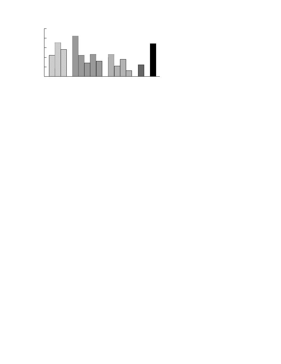
After PTRA, studies report that about 40% of
patients have improved renal function with the
remainder split equally between stable or worse
function.
31
One very informative study re-
ported that renal function outcome at three
and 12 months after PTRA depended on the
mean creatinine before intervention.
32
As with
surgery, results were not at all encouraging in
those patients with poor baseline function
(mean creatinine 461 µmol/l) of whom over
50% had either worse function or required
dialysis at 12 months (see fig 3).
PTRA PLUS STENT DEPLOYMENT
(
PTRAS
)
The development of the stent has allowed
interventional radiologists to attempt treat-
ment of more severe atherosclerotic lesions,
particularly those extending from the renal
ostia. Perhaps as a result of this di
Verent case
selection, results appear less satisfactory and
only 27% (range 15%–36%) of patients have
improved renal function, 52% (range 29%–
91%) stable function, and the remaining 21%
(range 0%–45%) worse function.
33 34
PTRAS is
associated with a vigorous inflammatory reac-
tion and average restenosis rates of 13% (range
9%–25%) have been reported during follow up
(range 12–24 months).
33 34
CONCLUSION
These studies do not provide overwhelming
encouragement to intervene in ARAS but
ostensibly indicate that surgical reconstruction
is the best option; however, this is likely to
reflect case selection and the use of stents in
patients with ostial stenoses which are usually a
marker of more advanced atherosclerotic dis-
ease. A recently published comparison between
PTRA and PTRAS has shown improved
patency rates in stented arteries, although this
was not associated with functional benefit.
35
This mismatch between technical success and
functional outcome has previously been re-
ported,
36 37
and provides evidence of on-going
parenchymal injury, presumably due to con-
tinuing uncontrolled atheroembolic disease
from
proximal
unstable
atherosclerotic
plaques; such reports question the contribution
of the stenosis per se to renal dysfunction in
ARAS. Even if function is improved, there is
only very limited evidence to suggest that renal
atrophy which is due to a proximal stenosis can
be reversed by intervention.
38
Most studies
report improved blood pressure and reduced
dosage of antihypertensive medication after
intervention, however, the need for medication
is rarely abolished and any benefit is not usually
sustained.
34 39 40
Medical interventions
A recent retrospective study from the Mayo
Clinic has suggested that long term e
Vective
management of hypertension and renal func-
tion can be achieved in patients with unilateral
ARAS.
41
In contrast, overall mortality and kid-
ney function were worse in medically managed
patients with bilateral ARAS or ARAS in a sin-
gle kidney,
41
which is consistent with a previous
report of 38% two year mortality in patients
with bilateral ARAS managed medically.
42
Although specific evidence of benefit is absent,
medical interventions routinely applied to
patients with ARAS include the prescription of
aspirin and active management of conventional
risk factors such as hypertension, dyslipidae-
mia,
diabetes
mellitus,
and
cessation
of
Figure 3
Renal function at three months and 12 months
after PTRA is determined by baseline function.
32
Mean
baseline creatinine: group A, 190 µmol/l; group B: 217
µmol/l; and group C, 461 µmol/l.
100
75
50
0
25
Effect of PTRA on groups
Dialysis
%
A
3 months
B
C
A
12 months
B
C
Worse
Stable
Improved
Renal failure in atherosclerotic renovascular disease
71
www.postgradmedj.com
smoking. There are also no data regarding spe-
cific risk factors in uraemia, such as hyperho-
mocysteinaemia, increased oxidative stress, or
endogenous inhibitors of nitric oxide synthase.
In short, “best medical treatment” in patients
with ARAS remains unknown.
HYPERTENSION
Although excessive blood pressure lowering in
patients with bilateral ARAS can lead to
progressive elevation of plasma creatinine,
43
data regarding optimal blood pressure levels
may be inferred from large studies. Tight blood
pressure control was associated with improved
outcomes in both the Modification of Diet in
Renal Disease study and UK Prospective
Diabetes Study Group studies,
44 45
both of
which likely included a significant proportion
of patients with ARAS. Two other studies have
shown specific renoprotection from ACE inhi-
bition in uraemic patients of whom a pro-
portion will have had ARAS.
46 47
With regard to
their safe use in patients, clinicians should be
reassured that no excess of adverse renal events
has been reported in large multicentre studies
which have investigated ACE inhibition in the
treatment of patients with heart failure, a
significant proportion of whom will have had
underlying ARAS. Furthermore, two recent
studies have shown that control of hyper-
tension by ACE inhibition is safe and e
Vective
in patients with ARAS and is not associated
with an increased risk of renal atrophy.
18 48
Nevertheless, these drugs should be introduced
at the lowest dose with renal function checked
after three to five days.
DYSLIPIDAEMIA
Dyslipidaemia is a risk factor for atherosclero-
sis, although curiously several studies demon-
strated no relationship between cholesterol
concentrations and progression of ARAS.
11 12 16
However, subtle lipid abnormalities may be
characteristic.
49
There are no data that report
benefit from cholesterol lowering in ARVD.
PLAQUE INSTABILITY
Recent data reports increased cardiovascular
morbidity and mortality in patients with
irregular
as
opposed
to
smooth
carotid
plaques.
50
The increased risk, which must
reflect a systemic predisposition to unstable
atherosclerotic plaques, did not correlate with
conventional risk factors and this suggests
additional as yet unrecognised factor(s) for
plaque progression. Similarly, the very high
incidence of recurrent disease after coronary
angioplasty in haemodialysis patients suggests
that plaques behave di
Verently in uraemia.
51
Unstable atherosclerotic plaques may embolise
cholesterol crystals and other debris that lodge
in the dependent circulation, even down to the
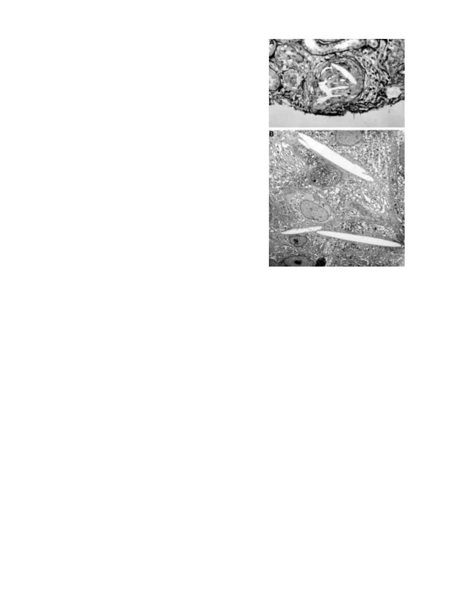
capillary level. In the kidney, cholesterol
embolisation can lead to progressive microvas-
cular obliteration, chronic inflammation and
worsening renal failure, with the diagnosis
clinched by the characteristic appearance of
intravascular cholesterol clefts on renal biopsy
(see fig 4).
The recent association between raised acute
phase
proteins,
unstable
atherosclerotic
plaques and increased risk of myocardial
infarction suggests that systemic inflammation
predisposes
to
plaque
instability.
The
importance of this observation is supported by
recent data which show that reduction in C
reactive protein is associated with reduction in
coronary risk.
52
A similar association between
raised acute phase proteins and atherosclerosis
has also been shown in dialysis and renal failure
patients,
53 54
although there are no data from
intervention studies.
Statins can cause atherosclerotic plaque
regression and may also have a specific role in
the management of the unstable plaque. As
well as inhibition of hepatocyte synthesis of
cholesterol, statins decrease macrophage chol-
esterol synthesis; increase macrophage low
density lipoprotein degradation; inhibit platelet
derived growth factor induced proliferation of
vascular smooth muscle cells and fibroblasts;
inhibit thrombosis; improve endothelial func-
tion; and
suppress
inflammation.
55
These
properties may promote plaque stability and
explain the reduction in levels of acute phase
proteins observed in patients treated with a
statin in the Cholesterol and Recurrent Events
study.
52
Consistent with this role, we recently
reported the successful treatment of a patient
with spontaneous cholesterol emboli syndrome
Figure 4
(A) Characteristic histological appearance on
light microscopy of a cholesterol cleft in a small artery with
evidence of intimal thickening, concentric hypertrophy, and
interstitial inflammation. Embolised cholesterol crystals
dissolve during the fixation process. (B) Electron
micrograph demonstrating a cholesterol cleft in an a
Verent
glomerular arteriole. The destination vessel depends on
crystal size and this variability may determine the clinical
presentation.
72
Woolfson
www.postgradmedj.com

a
Vecting the renal and pedal circulations.
56
Treatment with simvastatin lead to resolution
of ischaemic symptoms, recovery of renal func-
tion, and normalisation of acute phase pro-
teins. Of course anecdotes are inadequate and a
definitive prospective trial of statin therapy in
patients with ARVD is long overdue.
Conclusion
Epidemiological studies clearly indicate those
patient groups at increased risk of developing
ARVD, although its varied presentation means
the clinician must remain vigilant. Recent
studies have emphasised that renal dysfunction
and atrophy result from both the stenosis and
downstream vascular disease. Although the
contribution of each process to an individual
patient’s ARVD varies, it is tempting to
consider both as di
Verent manifestations of a
primary
disease
against
which
treatment
should be directed.
Developments in magnetic resonance tech-
nology and its increased availability means that
there is now a useful diagnostic technique for
ARAS on the horizon for all. However, the
ideal diagnostic test would di
Verentiate renal
injury secondary to proximal stenosis from
irreversible microvascular disease. But as yet,
only renal biopsy can define the reason for dys-
function and the potential for recovery with
certainty. Improved diagnostic sensitivity will
hopefully inform the debate over management
of ARAS. Given the dual pathology, treatment
of the stenosis alone is not likely to be adequate
and the development of novel aggressive anti-
atherogenic therapy will probably make these
invasive interventions redundant in the future.
At present, optimum medical treatment re-
mains undefined and it is not known whether
the treatment of conventional cardiovascular
risk factors (blood pressure lowering, choles-
terol lowering, and aspirin) matches the benefit
from intervention. These continuing uncer-
tainties highlight the problem which can arise
when clinical management is driven by techno-
logical development rather that clinical benefit.
The need for randomised prospective studies
of available interventions has never been
greater.
1 Mailloux LU, Napolitano B, Bellucci AG, et al. Renal vascu-
lar disease causing end-stage renal disease, incidence, clini-
cal correlates, and outcomes: a 20-year clinical experience.
Am J Kidney Dis 1994;24:622–9.
2 Scoble JE, Maher ER, Hamilton G, et al. Atherosclerotic
renovascular disease causing renal impairment—a case for
treatment. Clin Nephrol 1989;31:119–22.
3 Coen G, Manni M, Giannoni MF, et al. Ischemic nephropa-
thy in an elderly nephrologic and hypertensive population.
Am J Nephrol 1998;18:221–7.
4 Missouris CG, Papavassiliou MB, Khaw K, et al. High
prevalence of carotid artery disease in patients with athero-
matous renal artery stenosis. Nephrol Dial Transplant
1998;13:945–8.
5 Zierler RE, Bergelin RO, Polissar NL, et al. Carotid and
lower extremity arterial disease in patients with renal artery
atherosclerosis. Arch Intern Med 1998;158:761–7.
6 Louie J, Isaacson JA, Zierler RE, et al. Prevalence of carotid
and lower extremity arterial disease in patients with renal
artery stenosis. Am J Hypertens 1994;7:436–9.
7 Olin JW, Melia M, Young JR, et al. Prevalence of atheroscle-
rotic renal artery stenosis in patients with atherosclerosis
elsewhere. Am J Med 1990;88:46N–51N.
8 Sawicki PT, Kaiser S, Heinemann L, et al. Prevalence of
renal artery stenosis in diabetes mellitus—an autopsy study.
J Intern Med 1991;229:489–92.
9 Thadhani R, Pascual M, Nickeleit V, et al. Preliminary
description of focal segmental glomerulosclerosis in patients
with renovascular disease. Lancet 1996;347:231–3.
10 Greenberg A, Bastacky SI, Iqbal A, et al. Focal segmental
glomerulosclerosis associated with nephrotic syndrome in
cholesterol atheroembolism: clinicopathological correla-
tions. Am J Kidney Dis 1997;29:334–44.
11 Schreiber MJ, Pohl MA, Novick AC. The natural history of
atherosclerotic and fibrous renal artery disease. Urol Clin
North Am 1984;11:383–92.
12 Tollefson DF, Ernst CB. Natural history of atherosclerotic
renal artery stenosis associated with aortic disease. J Vasc
Surg 1991;14:327–31.
13 Zierler RE, Bergelin RO, Isaacson JA, et al. Natural history
of atherosclerotic renal artery stenosis: a prospective study
with duplex ultrasonography. J Vasc Surg 1994;19:250–7.
14 Zierler RE, Bergelin RO, Davidson RC, et al. A prospective
study of disease progression in patients with atherosclerotic
renal artery stenosis. Am J Hypertens 1996;9:1055–61.
15 Crowley JJ, Conlon PJ, Santos RM, et al. Incidence and pro-
gression of renal artery atherosclerosis in patients undergo-
ing cardiac catheterization. Circulation 1996;94:I–299.
16 Dean RH, Kie
Ver RW, Smith BM, et al. Renovascular
hypertension: anatomic and renal function changes during
drug therapy. Arch Surg 1981;116:1408–15.
17 Guzman RP, Zierler RE, Isaacson JA, et al. Renal atrophy
and arterial stenosis. A prospective study with duplex ultra-
sound. Hypertension 1994;23:346–50.
18 Caps MT, Zierler RE, Polissar NL, et al. Risk of atrophy in
kidneys with atherosclerotic renal artery stenosis. Kidney Int
1998;53:735–42.
19 Lerman LO, Taler SJ, Textor SC, et al. Computed
tomography-derived intrarenal blood flow in renovascular
and essential hypertension. Kidney Int 1996;49:846–54.
20 Tullis MJ, Zierler RE, Caps MT, et al. Clinical evidence of
contralateral renal parenchymal injury in patients with uni-
lateral atherosclerotic renal artery stenosis. Ann Vasc Surg
1998;12:122–7.
21 Farmer CKY, Cook GJR, Blake GM, et al. Individual kidney
function in atherosclerotic nephropathy is not related to the
presence of renal artery stenosis. Nephrol Dial Transplant
1999;14:2880–4.
22 Suresh M, Laboi P, Mamtora H, et al. Relationship of renal
dysfunction to proximal arterial disease severity in athero-
sclerotic renovascular disease. Nephrol Dial Transplant 2000;
15:631–6.
23 Thadhani RI, Camargo CA Jr, Xavier RJ, et al. Atheroem-
bolic renal failure after invasive procedures. Natural history
based on 52 histologically proven cases. Medicine (Balti-
more) 1995;74:350–8.
24 Belenfant X, Meyrier A, Jacquot C. Supportive treatment
improves survival in multivisceral cholesterol crystal embo-
lism. Am J Kidney Dis 1999;33:840–50.
Box 1: Risk factors for development of
ARVD
x Age
x Female gender
x Extrarenal atherosclerosis
x Diabetes mellitus
x Hypertension
x Smoking
x Hypercholesterolaemia
Box 2: Clinical presentations of ARVD
x Hypertension
x Deterioration in renal function or acute
renal failure after introduction of ACE
inhibition or angiotensin II receptor
blockade
x Chronic renal failure
x Variable proteinuria, ranging to
nephrotic syndrome
x Recurrent “flash pulmonary oedema”
Box 3: Pathogenesis of renal injury in
ARVD
x Hypertensive nephrosclerosis
x Hypoperfusion due to critical stenosis
x Atheroembolic renal disease
Renal failure in atherosclerotic renovascular disease
73
www.postgradmedj.com

25 Woolfson RG, Neild GH. The true clinical significance of
renography in nephro-urology. Eur J Nucl Med 1997;24:
557–70.
26 Datseris IE, Bomanji JB, Brown EA, et al. Captopril renal
scintigraphy in patients with hypertension and chronic renal
failure. J Nucl Med 1994;35:251–4.
27 Kaplan-Pavlovcic S, Nadja C. Captopril renography and
duplex Doppler sonography in the diagnosis of renovascular
hypertension. Nephrol Dial Transplant 1998;13:313–7.
28 Leung DA, Ho
Vmann U, Pfammatter T, et al. Magnetic
resonance angiography versus duplex sonography for
diagnosing renovascular disease. Hypertension 1999;33:726–
31.
29 Greco BA, Breyer-Lewis J. Atheromatous renovascular dis-
ease. In: Johnson R, Feehally J, eds. Principles of nephrology.
London: Mosby International, 2000: 13.65.1–14.
30 Hallett JW Jr, Textor SC, Kos PB, et al. Advanced renovas-
cular hypertension and renal insu
Yciency: trends in medical
comorbidity and surgical approach from 1970 to 1993. J
Vasc Surg 1995;21:750–9.
31 Greco BA, Breyer JA. Atherosclerotic ischemic renal
disease. Am J Kidney Dis 1997;29:167–87.
32 Connolly JO, Higgins RM, Walters HL, et al. Presentation,
clinical features and outcome in di
Verent patterns of athero-
sclerotic renovascular disease. Q J Med 1994;87:413–21.
33 Tuttle KR, Raabe RD. Endovascular stents for renal artery
revascularization. Curr Opin Nephrol Hypertens 1998;7:695–
701.
34 Isles CG, Robertson S, Hill D. Management of renovascular
disease: a review of renal artery stenting in ten studies. Q J
Med 1999;92:159–67.
35 van de Ven PJ, Kaatee R, Beutler JJ, et al. Arterial stenting
and balloon angioplasty in ostial atherosclerotic renovascu-
lar disease: a randomised trial. Lancet 1999;353:282–6.
36 Mikhail A, Cook GJ, Reidy J, et al. Progressive renal
dysfunction despite successful renal artery angioplasty in a
single kidney. Lancet 1997;349:926.
37 Tuttle KR, Chouinard RF, Webber JT, et al. Treatment of
atherosclerotic ostial renal artery stenosis with the intravas-
cular stent. Am J Kidney Dis 1998;32:611–22.
38 Sos TA, Pickering TG, Sniderman K, et al. Percutaneous
transluminal renal angioplasty in renovascular hypertension
due to atheroma or fibromuscular dysplasia. N Engl J Med
1983;309:274–9.
39 Plouin PF, Chatellier G, Darne B, et al. Blood pressure out-
come of angioplasty in atherosclerotic renal artery stenosis:
a randomized trial. Essai Multicentrique Medicaments vs
Angioplastie (EMMA) Study Group. Hypertension 1998;31:
823–9.
40 Webster J, Marshall F, Abdalla M, et al. Randomised
comparison of percutaneous angioplasty vs continued
medical therapy for hypertensive patients with atheroma-
tous renal artery stenosis. Scottish and Newcastle Renal
Artery Stenosis Collaborative Group. J Hum Hypertens
1998;12:329–35.
41 Chabova V, Schirger A, Stanson AW, et al. Outcomes of
atherosclerotic renal artery stenosis managed without revas-
cularization. Mayo Clin Proc 2000;75:437–44.
42 Baboolal K, Evans C, Moore RH. Incidence of end-stage
renal disease in medically treated patients with severe bilat-
eral atherosclerotic renovascular disease. Am J Kidney Dis
1998;31:971–7.
43 Textor SC, Novick AC, Tarazi RC, et al. Critical perfusion
pressure for renal function in patients with bilateral athero-
sclerotic renal vascular disease. Ann Intern Med 1985;102:
308–14.
44 Klahr S. Role of dietary protein and blood pressure in the
progression of renal disease. Kidney Int 1996;49:1783–6.
45 UK Prospective Diabetes Study Group.Tight blood pres-
sure control and risk of macrovascular and microvascular
complications in type 2 diabetes: UKPDS 38. BMJ
1998;317:703–13.
46 Maschio G, Alberti D, Janin G, et al. E
Vect of the
angiotensin-converting-enzyme inhibitor benazepril on the
progression of chronic renal insu
Yciency. N Engl J Med
1996;334:939–4.
47 Ruggenenti P, Perna A, Gherardi G, et al. renoprotective
properties of ACE-inhibition in non-diabetic bephropathies
with non-nephrotic proteinuria. Lancet 1999;354:359–64.
48 Tullis MJ, Caps MT, Zierler RE, et al. Blood pressure, anti-
hypertensive medication, and atherosclerotic renal artery
stenosis. Am J Kidney Dis 1999;33:675–81.
49 Scoble JE, de Takats D, Ostermann ME, et al. Lipid profiles
in patients with atherosclerotic renal artery stenosis.
Nephron 1999;83:117–21.
50 Rothwell PM, Villagra R, Gibson R, et al. Evidence of a
chronic systemic cause of instability of atherosclerotic
plaques. Lancet 2000;355:19–24.
51 Rinehart AL, Herzog CA, Collins AJ, et al. A comparison of
coronary angioplasty and coronary artery bypass grafting
outcomes in chronic dialysis patients. Am J Kidney Dis
1995;25:281–90.
52 Ridker PM, Rifai N, Pfe
Ver MA, et al. Long-term eVects of
pravastatin on plasma concentration of C-reactive protein.
The Cholesterol and Recurrent Events (CARE) Investiga-
tors. Circulation 1999;100:230–5.
53 Zimmermann J, Herrlinger S, Pruy A, et al. Inflammation
enhances cardiovascular risk and mortality in hemodialysis
patients. Kidney Int 1999;55:648–58.
54 Stenvinkel P, Heimburger O, Paultre F, et al. Strong associ-
ation between malnutrition, inflammation, and atheroscle-
rosis in chronic renal failure. Kidney Int 1999;55:1899–911.
55 Rosenson RS, Tangney CC. Antiatherothrombotic proper-
ties of statins: implications for cardiovascular event reduc-
tion. JAMA 1998;279:1643–50.
56 Woolfson RG, Lachmann H. Improvement in renal choles-
terol emboli syndrome after simvastatin. Lancet 1998;351:
1331–2.
References for figure 2
Brewster DC, Retana A, Waltman AC, et al. Angiography in the
management of aneurysms of the abdominal aorta. Its value
and safety. N Engl J Med 1975;292:822–5.
Olin JW, Melia M, Young JR, et al. Prevalence of atherosclerotic
renal artery stenosis in patients with atherosclerosis else-
where. Am J Med 1990;88:46N–51N.
Valentine RJ, Myers SI, Miller GL, et al. Detection of
unsuspected renal artery stenoses in patients with abdominal
aortic aneurysms: refined indications for preoperative aortog-
raphy. Ann Vasc Surg 1993;7:220–4.
Choudhri AH, Cleland JG, Rowlands PC, et al. Unsuspected
renal artery stenosis in peripheral vascular disease. BMJ
1990;301:1197–8.
Wilms G, Marchal G, Peene P, et al. The angiographic incidence
of renal artery stenosis in the arteriosclerotic population. Eur
J Radiol 1990;10:195–7.
Salmon P, Brown MA. Renal artery stenosis and peripheral vas-
cular disease: implications for ACE inhibitor therapy. Lancet
1990;336:321.
Swartbol P, Thorvinger BO, Parsson H, et al. Renal artery ste-
nosis in patients with peripheral vascular disease and its cor-
relation to hypertension. A retrospective study. Int Angiol
1992;11:195–9.
Missouris CG, Buckenham T, Cappuccio FP, et al. Renal artery
stenosis: a common and important problem in patients with
peripheral vascular disease. Am J Med 1994;96:10–4.
Vetrovec GW, Landwehr DM, Edwards VL. Incidence of renal
artery stenosis in hypertensive patients undergoing coronary
angiography. J Intervent Cardiol 1989;2:69–76.
Harding MB, Smith LR, Himmelstein SI, et al. Renal artery ste-
nosis: prevalence and associated risk factors in patients
undergoing routine cardiac catheterization. J Am Soc Nephrol
1992;2:1608–16.
Jean WJ, al-Bitar I, Zwicke DL, et al. High incidence of renal
artery stenosis in patients with coronary artery disease. Cathet
Cardiovasc Diagn 1994;32:8–10.
Crowley JJ, Conlon PJ, Santos RM, et al. Incidence and
progression of renal artery atherosclerosis in patients
undergoing cardiac catheterization. Circulation 1996;94:I–
299.
Uzu T, Inoue T, Fujii T, et al. Prevalence and predictors of renal
artery stenosis in patients with myocardial infarction. Am J
Kidney Dis 1997;29:733–8.
MacDowall P, Kalra PA, O’Donoghue DJ, et al. Risk of morbid-
ity from renovascular disease in elderly patients with conges-
tive cardiac failure. Lancet 1998;352:13–6.
74
Woolfson
www.postgradmedj.com

doi: 10.1136/pmj.77.904.68
2001 77: 68-74
Postgrad Med J
R G Woolfson
intervention
disease: pathogenesis, diagnosis, and
Renal failure in atherosclerotic renovascular
http://pmj.bmj.com/content/77/904/68.full.html
Updated information and services can be found at:
These include:
References
http://pmj.bmj.com/content/77/904/68.full.html#related-urls
Article cited in:
http://pmj.bmj.com/content/77/904/68.full.html#ref-list-1
This article cites 53 articles, 13 of which can be accessed free at:
service
Email alerting
box at the top right corner of the online article.
Receive free email alerts when new articles cite this article. Sign up in the
Notes
http://group.bmj.com/group/rights-licensing/permissions
To request permissions go to:
http://journals.bmj.com/cgi/reprintform
To order reprints go to:
http://group.bmj.com/subscribe/
To subscribe to BMJ go to:
Wyszukiwarka
Podobne podstrony:
68 74 ROZ w spr bezpieczenst Nieznany (2)
68 307 POL ED02 2001
kpk, ART 74 KPK, 2001
kpk, ART 74 KPK, 2001
74 75 307 POL ED02 2001
68 69 306 pol ed02 2001
74 306 pol ed02 2001
Med Czyn Rat1 Ostre zatrucia Materialy
FARMAKOLOGIA WYKŁAD III RAT MED ST
Med Czyn Rat6 Gospodarka wodno elektrolitowa Materialy
2001 08 28
bph pbk raport roczny 2001
2001 11 29
74 Nw 11 Obwody drukowane
więcej podobnych podstron