
Facial Chemical Peels
Facial Chemical Peels
Jean Paul Font, MD
Jean Paul Font, MD
David C. Teller, MD
David C. Teller, MD
Grand Rounds Presentation
Grand Rounds Presentation
Department of Otolaryngology
Department of Otolaryngology
University of Texas Medical Branch at
University of Texas Medical Branch at
Galveston
Galveston
March 18, 2007
March 18, 2007

History
History
Egypt - first evidence
Egypt - first evidence
of exfoliants use
of exfoliants use
–
Sun-damaged skin was
Sun-damaged skin was
a sign of lower rank in
a sign of lower rank in
society
society
–
Sour milk- contain lactic
Sour milk- contain lactic
acid, an alpha-hydroxy
acid, an alpha-hydroxy
acid commonly used
acid commonly used
today
today
Turks - use fire to
Turks - use fire to
produce a thermal
produce a thermal
exfoliation
exfoliation

History
History
1882 P.G Unna, German dermatologist
1882 P.G Unna, German dermatologist
described resorcinol, salicylic acid, phenol,
described resorcinol, salicylic acid, phenol,
trichloroacetic acid
trichloroacetic acid
1903 Mackee began using phenol for acne
1903 Mackee began using phenol for acne
scarring (Chairman of dermatology at NYU)
scarring (Chairman of dermatology at NYU)
1961 Baker and Gordon presented a peel
1961 Baker and Gordon presented a peel
formula with one patient with a 3 month
formula with one patient with a 3 month
follow up, became the standard formula
follow up, became the standard formula
1966 Baker published results in 250
1966 Baker published results in 250
patients
patients

Aging
Aging
Define as the process of
Define as the process of
system's deterioration
system's deterioration
(
(
Hanbook of the Biology of Aging
Hanbook of the Biology of Aging
2006)
2006)
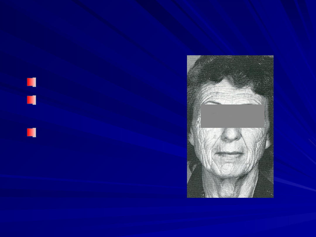
Facial skin changes is one
Facial skin changes is one
of the most apparent
of the most apparent
examples of aging
examples of aging

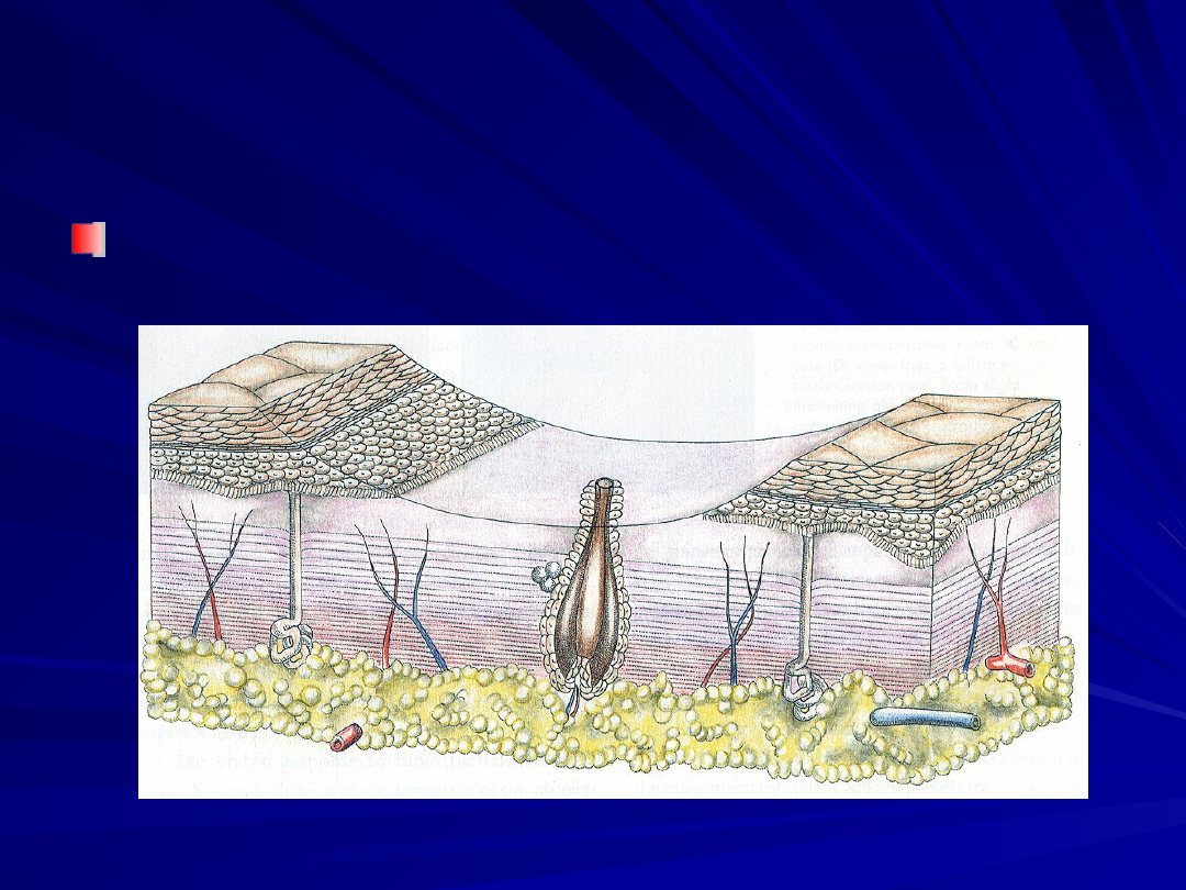
Histology
Histology
Actinic changes - photochemical effects of
Actinic changes - photochemical effects of
solar radiation exposure
solar radiation exposure
–
Disorderly arrangement of epidermis
Disorderly arrangement of epidermis
–
Degeneration of the elastic network
Degeneration of the elastic network
–
Mottled pigmentation
Mottled pigmentation
–
Lymphocytic infiltration
Lymphocytic infiltration
–
Decrease in collagen
Decrease in collagen
–
Flattening of the dermal-epidermal junction
Flattening of the dermal-epidermal junction
–
Epidermal cell atypia
Epidermal cell atypia
–
Increased melanocytes, but they were
Increased melanocytes, but they were
unevenly distributed and contained variable
unevenly distributed and contained variable
amounts of melanin
amounts of melanin
Peel Skin Histology
Peel Skin Histology
Chemical burn of the epidermis and the outer
Chemical burn of the epidermis and the outer
dermis
dermis
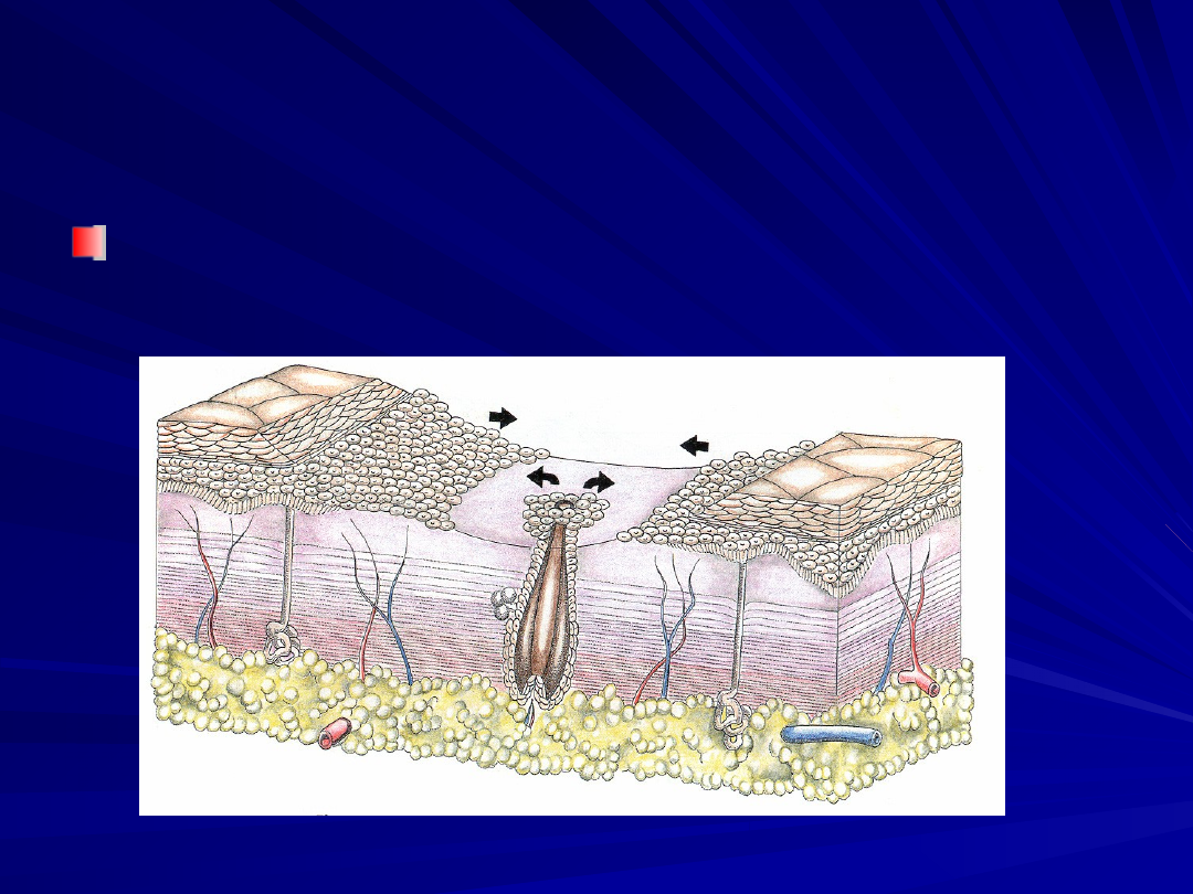
Peel Skin Histology
Peel Skin Histology
First 2 to 5 days - Regenerates from
First 2 to 5 days - Regenerates from
follicular and eccrine duct epithelium
follicular and eccrine duct epithelium
Peel Skin Histology
Peel Skin Histology
Fresh, orderly, organized epidermis
Fresh, orderly, organized epidermis
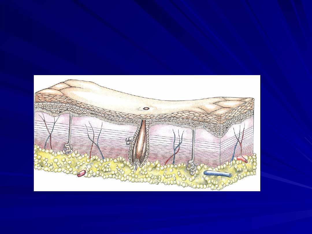
Peel Skin Histology
Peel Skin Histology
At 2 weeks - new
At 2 weeks - new
collagen formation
collagen formation
begins and may
begins and may
continue up to 1 year
continue up to 1 year
–
New bands of dermis
New bands of dermis
2- to 3-mm-thick
2- to 3-mm-thick
–
Thin, compact, parallel
Thin, compact, parallel
collagen bundles
collagen bundles
arranged horizontally
arranged horizontally
along the epidermal-
along the epidermal-
dermal matrix
dermal matrix

Peel Skin Histology
Peel Skin Histology
Other changes
Other changes
–
Melanocytes contain fine, evenly
Melanocytes contain fine, evenly
distributed melanin granules
distributed melanin granules
–
Impaired melanin synthesis with a
Impaired melanin synthesis with a
generalized bleaching effect
generalized bleaching effect
–
Decrease lymphocytic infiltration
Decrease lymphocytic infiltration
Treat cutaneous lesions
Treat cutaneous lesions
Replace atypical
Replace atypical
keratinocytes with normal
keratinocytes with normal
epidermal cells
epidermal cells
Kligman concluded that
Kligman concluded that
chemical peel reduced
chemical peel reduced
the development of new
the development of new
neoplasms
neoplasms
Litton decreased the rate
Litton decreased the rate
of appearance of
of appearance of
precancerous and early
precancerous and early
cancerous lesions after a
cancerous lesions after a
phenol chemical peel
phenol chemical peel

Patient Selection
Patient Selection
"The ideal patient is a thin-skinned
"The ideal patient is a thin-skinned
female with fair complexion and fine
female with fair complexion and fine
rhytids."
rhytids."
Skin type and the amount of
Skin type and the amount of
photodamage present
photodamage present
Fitzpatrick classified the skin types
Fitzpatrick classified the skin types
–
Color and acute solar radiation response
Color and acute solar radiation response
The Glogau classification based on the
The Glogau classification based on the
degree of photoaging
degree of photoaging

Fitzpatrick Classification
Fitzpatrick Classification
Fitzpatrick skin type I and type II are good candidates
Fitzpatrick skin type I and type II are good candidates
Type III and greater - increased risk pigment
Type III and greater - increased risk pigment
complications
complications
Type
Type
Color
Color
Tanning response
Tanning response
I
I
White
White
Always burns, never tans
Always burns, never tans
II
II
White
White
Usually burns, tans less than average
Usually burns, tans less than average
III
III
White
White
Sometimes burns mildly, tans about average
Sometimes burns mildly, tans about average
IV
IV
Brown
Brown
Rarely burns, tans more than average and with ease
Rarely burns, tans more than average and with ease
V
V
Dark brown
Dark brown
Very rarely burns, tans very easily
Very rarely burns, tans very easily
VI
VI
Black
Black
Never burns, tans very easily
Never burns, tans very easily

Glogau classification
Glogau classification
Group
Group
Classificati
Classificati
on
on
Skin characteristics
Skin characteristics
Peel
Peel
I
I
Mild
Mild
Little wrinkling or scarring and
Little wrinkling or scarring and
no keratoses
no keratoses
Superficial
Superficial
II
II
Moderate
Moderate
Early wrinkling, mild scarring,
Early wrinkling, mild scarring,
and sallow color with early
and sallow color with early
actinic keratoses
actinic keratoses
Medium
Medium
III
III
Advanced
Advanced
Persistent wrinkling,
Persistent wrinkling,
discoloration with
discoloration with
telangectasias and actinic
telangectasias and actinic
keratoses
keratoses
Medium
Medium
IV
IV
Severe
Severe
Wrinkling—superficial to deep
Wrinkling—superficial to deep
actinic keratoses ± skin
actinic keratoses ± skin
cancer
cancer
Medium to
Medium to
Deep
Deep
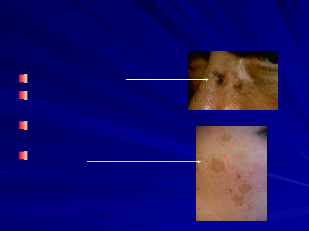
Aesthetic Indications
Aesthetic Indications
Rhytids
Rhytids
Spotty
Spotty
hyperpigmentation
hyperpigmentation
Superficial acne
Superficial acne
scarring
scarring
Therapeutic Indications
Therapeutic Indications
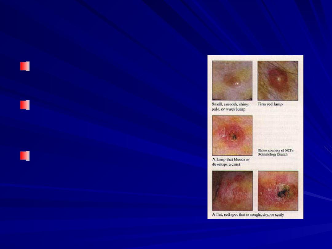
Actinic keratoses
Actinic keratoses
Superficial basal cell
Superficial basal cell
carcinomas
carcinomas
Lentigo maligna
Lentigo maligna
lentigines
lentigines
Melasma
Melasma
(discoloration of
(discoloration of
skin caused by
skin caused by
pregnancy)
pregnancy)

Contraindications
Contraindications
Relative
Relative
Contraindications
Contraindications
–
Darker skin type
Darker skin type
(Fitzpatrick IV-VI)
(Fitzpatrick IV-VI)
–
History Keloid
History Keloid
–
History of herpes
History of herpes
infections
infections
–
Cardiac abnormalities
Cardiac abnormalities
–
A history of diabetes
A history of diabetes
mellitus or previous facial
mellitus or previous facial
irradiation
irradiation
–
Unrealistic patient
Unrealistic patient
expectations
expectations
–
Telangiectasias
Telangiectasias
–
Anticipation of inadequate
Anticipation of inadequate
photo protection
photo protection
Absolute
Absolute
Contraindications
Contraindications
–
Significant hepatorenal
Significant hepatorenal
disease
disease
–
HIV-positive patient
HIV-positive patient
–
Significant
Significant
immunosuppression
immunosuppression
–
Emotional instability or
Emotional instability or
mental illness
mental illness
–
Ehlers-Danlos syndrome
Ehlers-Danlos syndrome
–
Scleroderma or collagen
Scleroderma or collagen
vascular diseases
vascular diseases
–
Accutane treatment
Accutane treatment
(within 6–12 months
(within 6–12 months
before)
before)

Patient Preparation
Patient Preparation
History of herpes infections
History of herpes infections
–
Prophylaxis with Valtrex or Acyclovir for 2 wks
Prophylaxis with Valtrex or Acyclovir for 2 wks
Skin preparation
Skin preparation
–
Vitamin A derivative therapy 4 weeks before
Vitamin A derivative therapy 4 weeks before
the procedure
the procedure
Speeds epidermal healing
Speeds epidermal healing
Thins stratum corneum
Thins stratum corneum
Increases the depth of a chemical peel
Increases the depth of a chemical peel
–
Stop sun exposure - 2 months before the
Stop sun exposure - 2 months before the
procedure
procedure
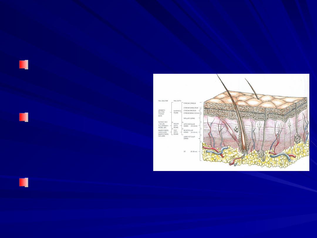
Chemical Peel Depths
Chemical Peel Depths
Superficial
Superficial
–
Epidermal loss
Epidermal loss
Medium
Medium
–
Injury to superficial
Injury to superficial
dermis
dermis
Deep
Deep
–
Mid-dermal injury
Mid-dermal injury
Chemical Peel
Chemical Peel
Frosting - keratin
Frosting - keratin
protein denaturation
protein denaturation
–
Level I - erythema with
Level I - erythema with
streaky surface
streaky surface
whitening
whitening
–
Level II - white-coated
Level II - white-coated
frosting with erythema
frosting with erythema
showing through
showing through
–
level III - solid white
level III - solid white
enamel frosting with
enamel frosting with
little or no background of
little or no background of
erythema (penetration
erythema (penetration
through the papillary
through the papillary
dermis)
dermis)

Superficial Peels
Superficial Peels
Necrosis of the epidermis
Necrosis of the epidermis
Healing time from 1 to 4 days
Healing time from 1 to 4 days
Improve pigmentary irregularities
Improve pigmentary irregularities
Improve minor surface changes
Improve minor surface changes
Fresher appearance to facial skin
Fresher appearance to facial skin
Superficial Peels
Superficial Peels
Different Solutions
Different Solutions
–
10% to 20% Trichloracetic
10% to 20% Trichloracetic
acid (TCA)
acid (TCA)
–
Jessner's solution
Jessner's solution
(resorcinol, 14 g; salicylic acid, 14 g;
(resorcinol, 14 g; salicylic acid, 14 g;
lactic acid, 14 mL; ethanol, 100 mL)
lactic acid, 14 mL; ethanol, 100 mL)
–
Glycolic acid (50% to 70%)
Glycolic acid (50% to 70%)
Level I frosting
Level I frosting
Postoperative
Postoperative
–
Mild cleanser, moisturizers
Mild cleanser, moisturizers
and sunscreens
and sunscreens
Glycolic acid can be
Glycolic acid can be
used to peel skin of all
used to peel skin of all
skin types with minimal
skin types with minimal
risk
risk

Medium Peel
Medium Peel
Necrosis of the epidermis & inflammation
Necrosis of the epidermis & inflammation
within the papillary dermis
within the papillary dermis
Improvement of skin texture in moderate
Improvement of skin texture in moderate
photodamaged skin (grade II Glogau)
photodamaged skin (grade II Glogau)
Removes of epidermal or superficial lesions
Removes of epidermal or superficial lesions
–
Actinic keratoses
Actinic keratoses
–
Repair mild rhytides
Repair mild rhytides
–
Improve pigmentary dyschromias
Improve pigmentary dyschromias
–
Improve depressed scars
Improve depressed scars

Trichloracetic acid (TCA)
Trichloracetic acid (TCA)
TCA approaching 50% or higher were used
TCA approaching 50% or higher were used
to achieve injury to the superficial dermis
to achieve injury to the superficial dermis
At this concentration TCA is unreliable and
At this concentration TCA is unreliable and
associated with a higher incidence of
associated with a higher incidence of
complications (
complications (
pigmentary dyschromia, textural
pigmentary dyschromia, textural
change, and even scarring
change, and even scarring
)
)
Combination of products improves the
Combination of products improves the
absorption of the lower concentration of
absorption of the lower concentration of
TCA without the associated complications
TCA without the associated complications
–
Solid CO2 freezing with trichloracetic acid 35%
Solid CO2 freezing with trichloracetic acid 35%
–
Jessner's solution + 35% TCA
Jessner's solution + 35% TCA
–
Glycolic acid 70% plus 35% TCA
Glycolic acid 70% plus 35% TCA

Medium Peel
Medium Peel
Brody
Brody
–
First developed solid CO2 applied with acetone to the skin
First developed solid CO2 applied with acetone to the skin
–
Freezing technique break the epidermal barrier for a more
Freezing technique break the epidermal barrier for a more
even and complete penetration
even and complete penetration
Monheit
Monheit
–
Jessner's solution destroyed the epidermal barrier by
Jessner's solution destroyed the epidermal barrier by
breaking up individual epidermal cells
breaking up individual epidermal cells
Coleman
Coleman
–
70% glycolic acid before the application of 35% TCA.
70% glycolic acid before the application of 35% TCA.
–
Results similar to that of Jessner's solution
Results similar to that of Jessner's solution
Deeper penetration of the 35% TCA and a more
Deeper penetration of the 35% TCA and a more
even application of the peeling solution
even application of the peeling solution
Phenol 88% by itself will give a medium-depth peel
Phenol 88% by itself will give a medium-depth peel

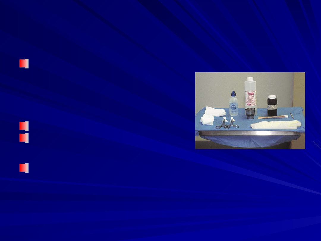
Patient Preparation
Patient Preparation
Vigorous cleaning and degreasing are
Vigorous cleaning and degreasing are
necessary for even penetration
necessary for even penetration
–
Septisol and acetone
Septisol and acetone
–
Debrided of stratum corneum and
Debrided of stratum corneum and
excessive scale
excessive scale
A splotchy peel is usually the result of
A splotchy peel is usually the result of
uneven penetration of peel solution
uneven penetration of peel solution
because of residual oil or stratum
because of residual oil or stratum
corneum
corneum
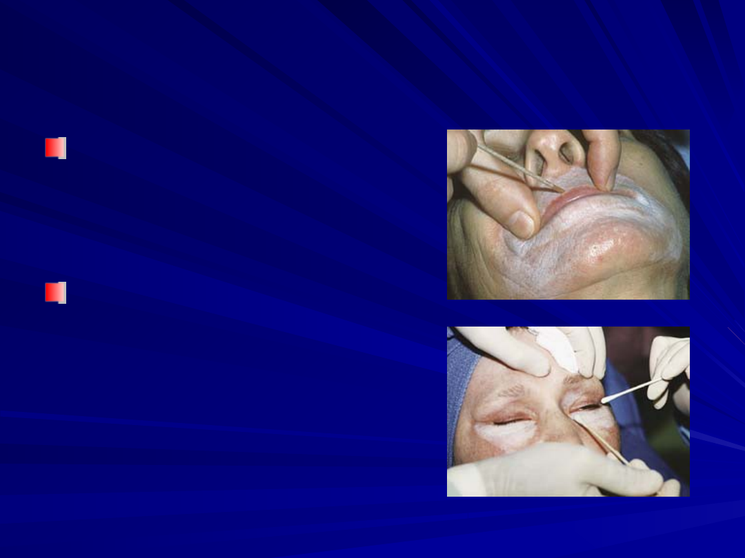
Medium Peel
Medium Peel
TCA is painted evenly
TCA is painted evenly
–
Forehead to temple to
Forehead to temple to
cheeks and finally to the
cheeks and finally to the
lips and eyelids
lips and eyelids
–
Eyelids within 1 to 2 mm
Eyelids within 1 to 2 mm
of the lower eyelid margin
of the lower eyelid margin
Amount of TCA
Amount of TCA
delivered is dependent
delivered is dependent
on:
on:
–
Number of applications
Number of applications
–
Degree of saturation
Degree of saturation
–
Pressure applied to the
Pressure applied to the
skin
skin
–
Contact time
Contact time
Medium Peel
Medium Peel
White frost appears complete
White frost appears complete
on the treated area within 30
on the treated area within 30
seconds to 2 minutes
seconds to 2 minutes
Before re-treating an area
Before re-treating an area
one should wait at least 3 to
one should wait at least 3 to
4 minutes before
4 minutes before
determining for asymmetry
determining for asymmetry
Eyelid skin and bony
Eyelid skin and bony
prominences have a high
prominences have a high
propensity for scarring
propensity for scarring
(limited to a level II frosting)
(limited to a level II frosting)
An assistant standby with
An assistant standby with
sterile eye wash in case
sterile eye wash in case
agent spills into the eye
agent spills into the eye
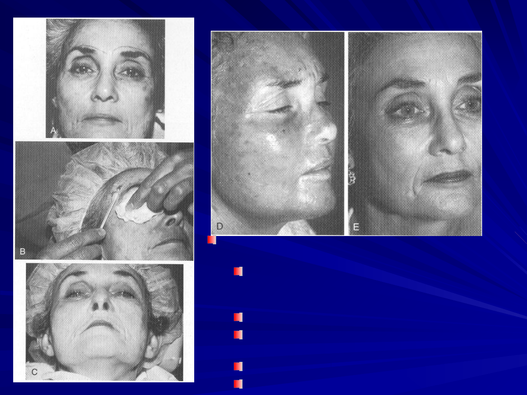
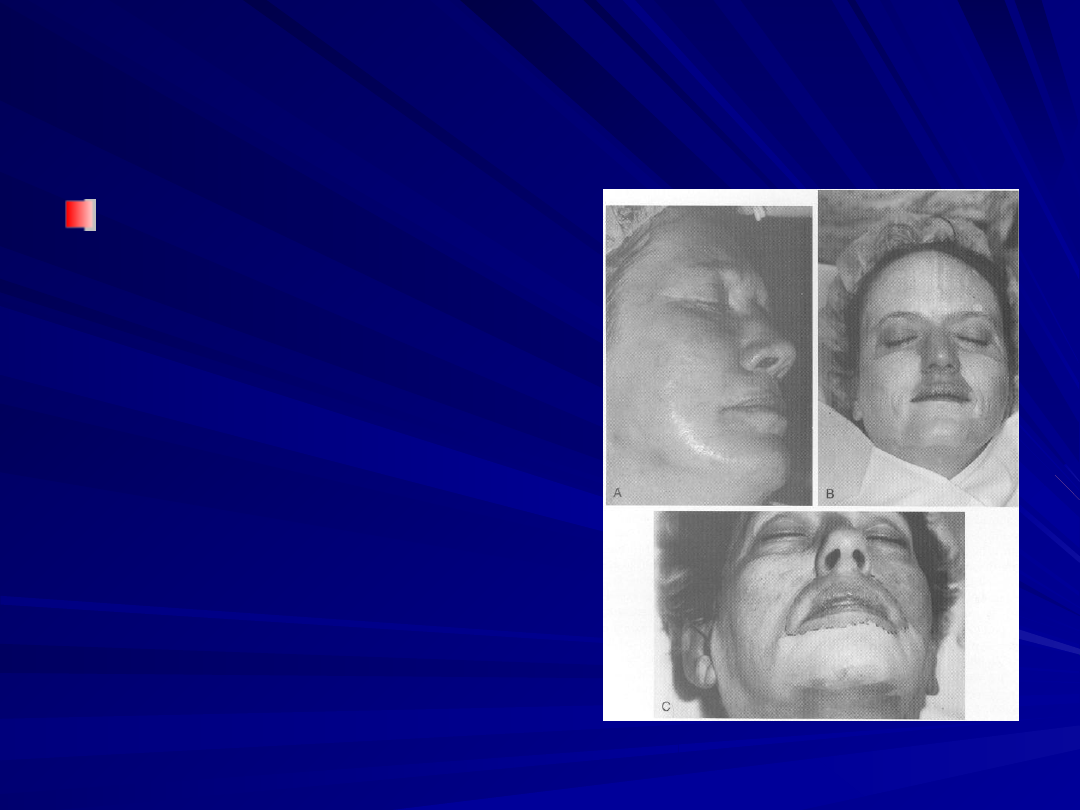
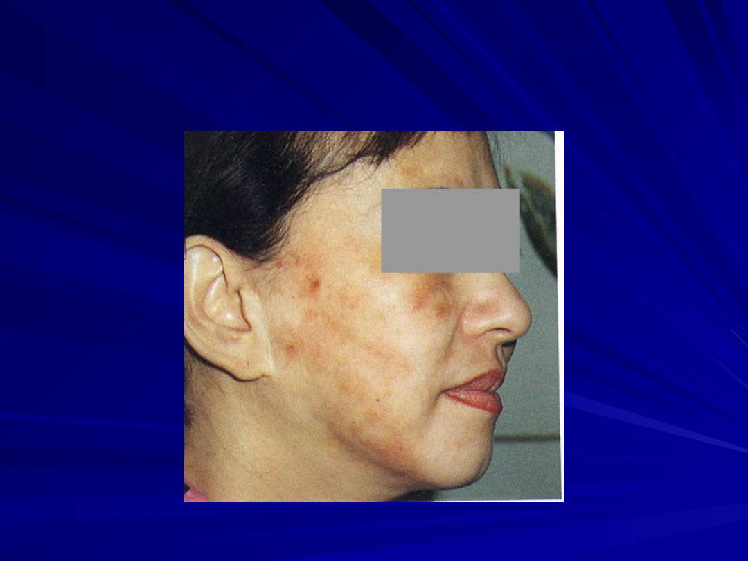
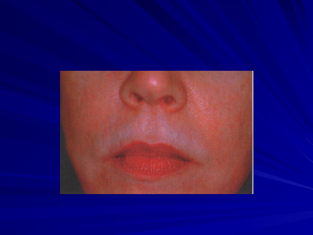
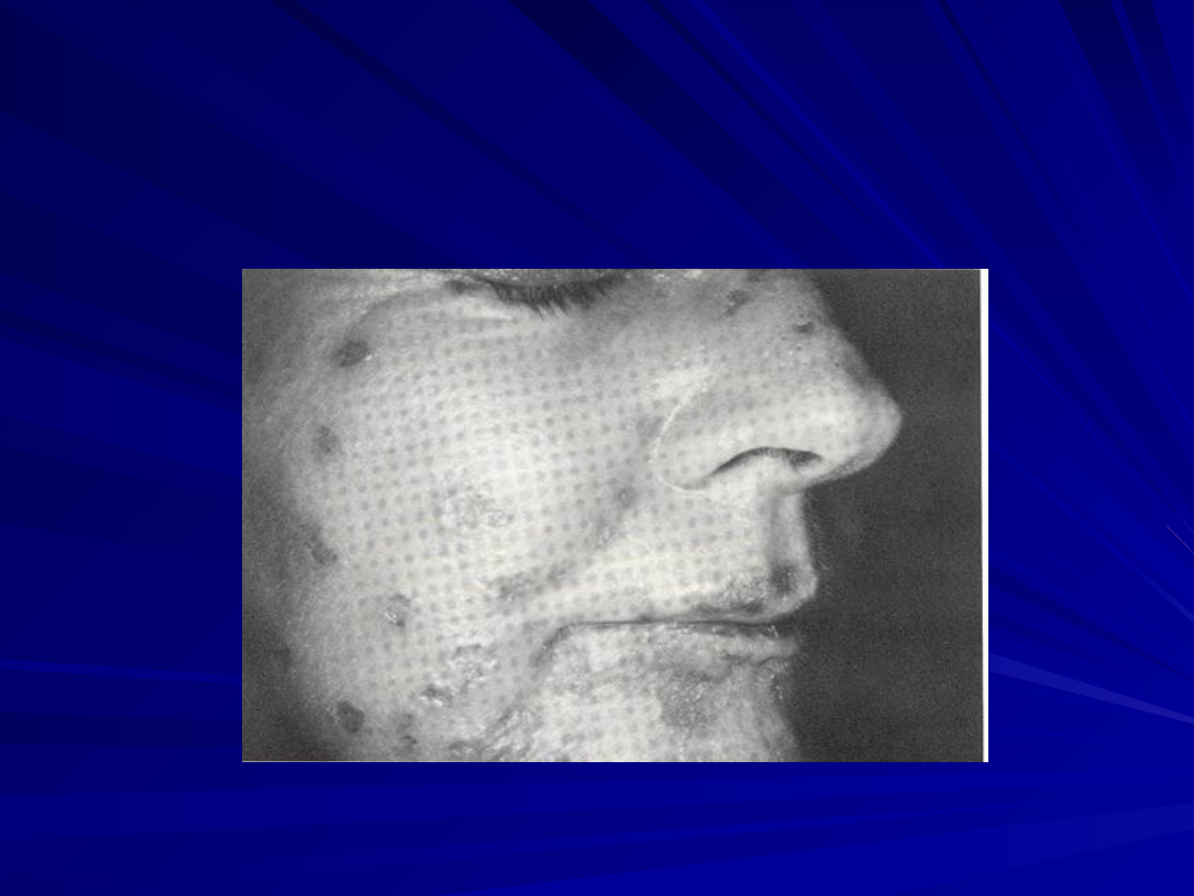
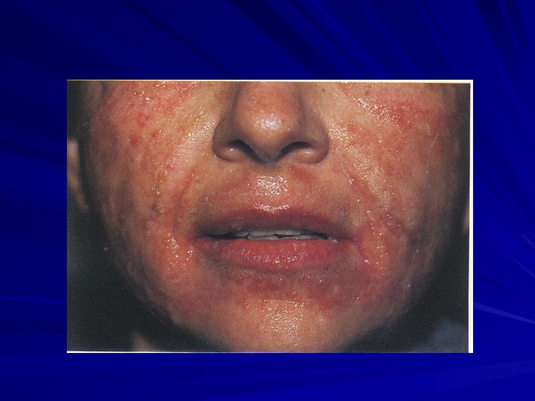
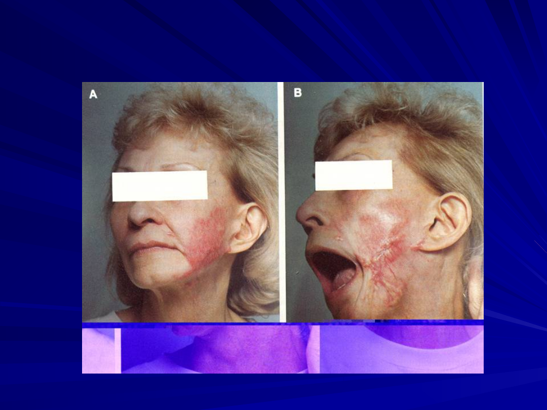
Jessner's TCA peel for moderate
photoaging skin, Glogau level II.
A, Preoperative view demonstrating
rhytides, lentigenes, keratoses, and
sallow skin.
B, Jessner's solution applied to face.
C, Full application 35% TCA with a level
III frosting.
D. Four days after chemical peel.
E, Six months after chemical peel

Medium Peel
Medium Peel
Dark crusts peels off on day 5 to 7
Dark crusts peels off on day 5 to 7
then erythema appears and soon fade
then erythema appears and soon fade
Repeat medium-depth chemical peel
Repeat medium-depth chemical peel
should not be performed for at least 1
should not be performed for at least 1
year
year
There is improvement of collagen
There is improvement of collagen
thickness progressing over a 6- to 13-
thickness progressing over a 6- to 13-
month period
month period

Deep Chemical Peel
Deep Chemical Peel
Glogau III and IV photoaging skin
Glogau III and IV photoaging skin
–
Deeper grooves and wrinkles
Deeper grooves and wrinkles
Deep peels are usually performed using
Deep peels are usually performed using
the Baker-Gordon solution
the Baker-Gordon solution
–
Phenol 88% 3 mL, Septisol 8 drops, Croton oil 3
Phenol 88% 3 mL, Septisol 8 drops, Croton oil 3
drops, Distilled water 2 mL
drops, Distilled water 2 mL
Septisol acts as a surfactant which results
Septisol acts as a surfactant which results
in more even penetration
in more even penetration
Croton oil is epidermolytic enhancing the
Croton oil is epidermolytic enhancing the
absorption of phenol
absorption of phenol

Deep Chemical Peel
Deep Chemical Peel
Phenol >80%
Phenol >80%
–
Keratin protein binds to the phenol
Keratin protein binds to the phenol
creating large molecules preventing
creating large molecules preventing
further penetration of the peel solution
further penetration of the peel solution
Phenol <50%
Phenol <50%
–
produce deeper penetration and more
produce deeper penetration and more
destruction than desired
destruction than desired
Tape Occlusion
Tape Occlusion
Occlusion of the
Occlusion of the
peeling solution
peeling solution
with tape increases
with tape increases
its penetration
its penetration
creating injury to
creating injury to
the mid-reticular
the mid-reticular
dermis
dermis
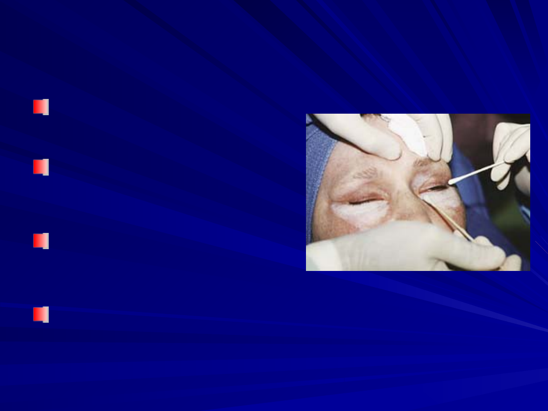
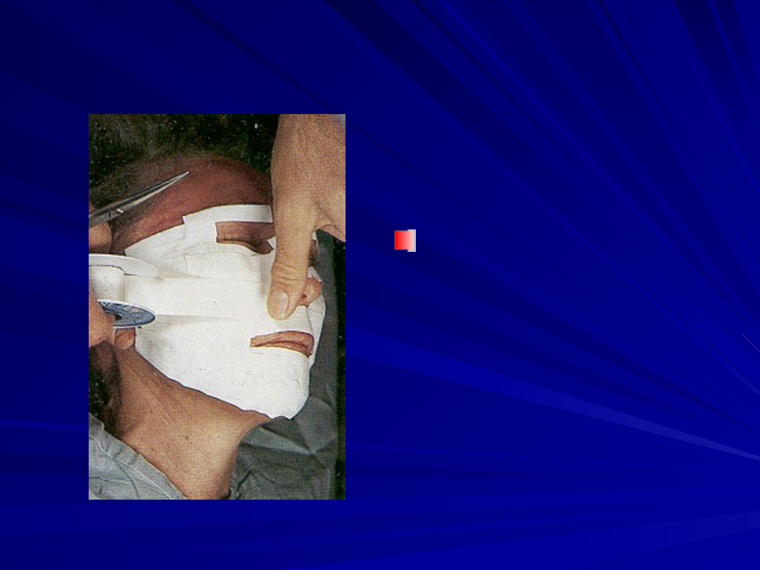
Deep Chemical Peel
Deep Chemical Peel
Face is divided into six
Face is divided into six
aesthetic subunits
aesthetic subunits
–
Forehead, perioral region,
Forehead, perioral region,
bilateral cheeks, nose, and
bilateral cheeks, nose, and
periorbital region
periorbital region
–
15-minute time interval
15-minute time interval
between units
between units
White frost that is carried 2 to 3
White frost that is carried 2 to 3
mm across the vermilion border
mm across the vermilion border
Lower eyelids need to be
Lower eyelids need to be
treated to within 1 to 2 mm of
treated to within 1 to 2 mm of
the ciliary margin
the ciliary margin
Upper eyelid above supratarsal
Upper eyelid above supratarsal
fold
fold

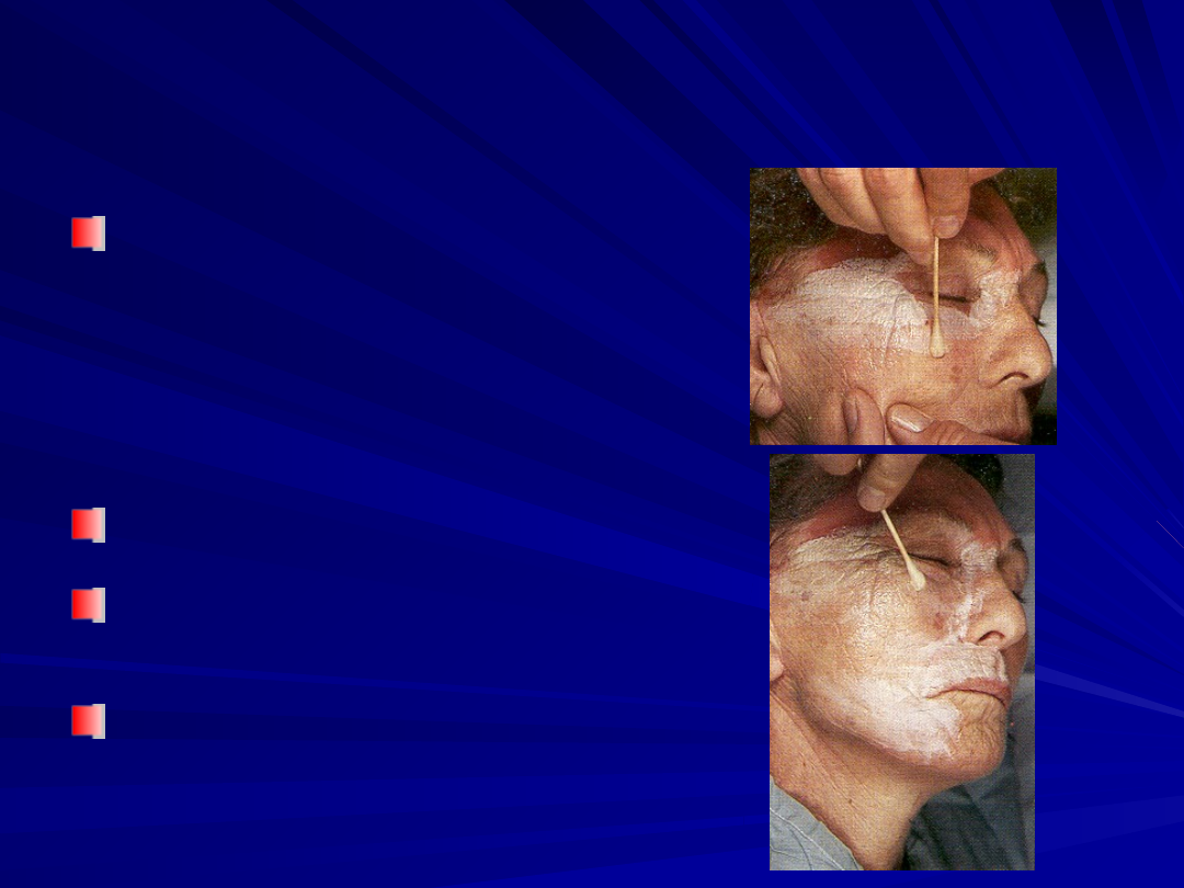
Deep Chemical Peel
Deep Chemical Peel
Erythema may take months to resolve
Erythema may take months to resolve
Evaluated in 3 to 4 days to observe the
Evaluated in 3 to 4 days to observe the
amount of wound healing and residual
amount of wound healing and residual
crusting
crusting
Sun avoidance 6 weeks and minimize sun
Sun avoidance 6 weeks and minimize sun
exposure for up to 6 months (Sunscreen
exposure for up to 6 months (Sunscreen
with an SPF of 3)
with an SPF of 3)
Splotchy hyperpigmentation (2 – 6 weeks)
Splotchy hyperpigmentation (2 – 6 weeks)
–
Retin A, hydroquinone and triamcinolone may
Retin A, hydroquinone and triamcinolone may
provide an improvement
provide an improvement
Deep Chemical Peel
Deep Chemical Peel

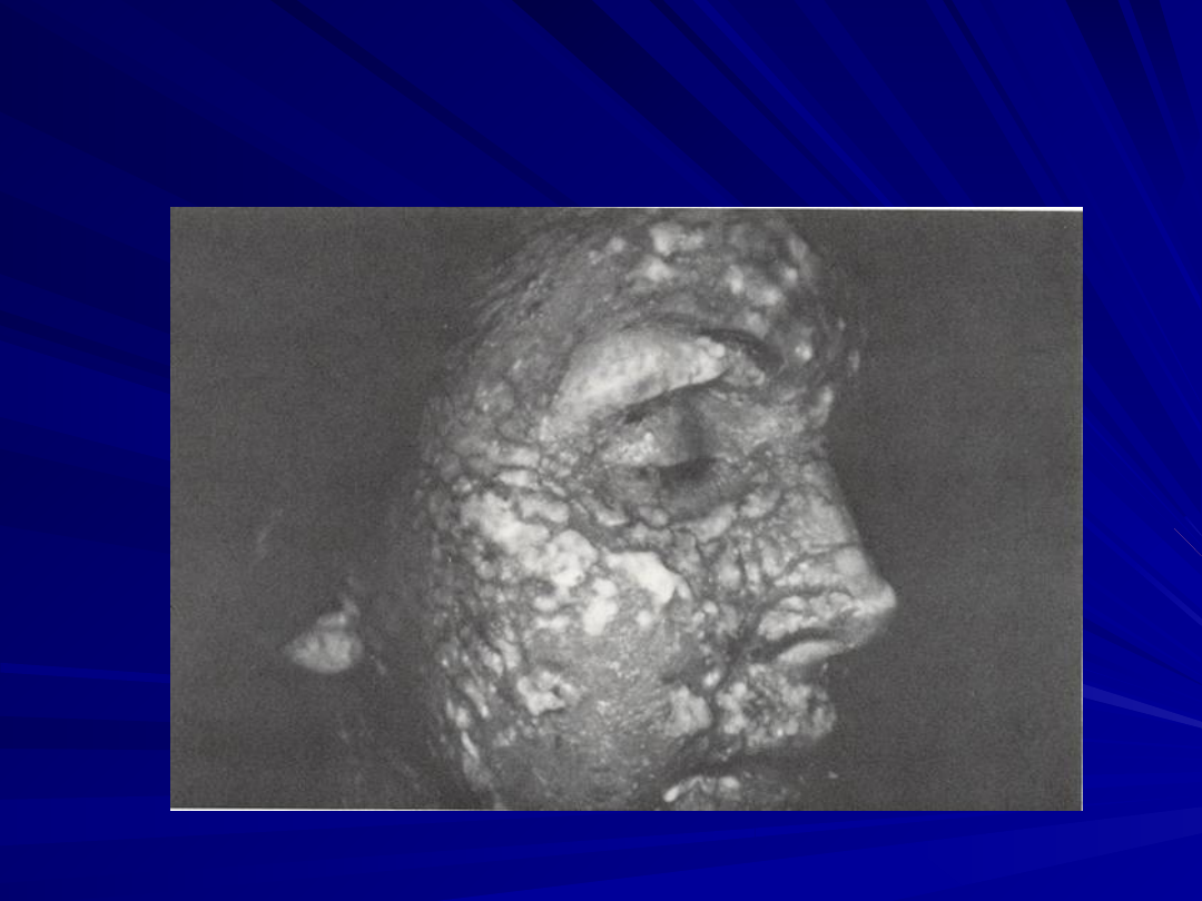
Phenol Toxicity
Phenol Toxicity
Cardiotoxic & eliminated hepatic and renal
Cardiotoxic & eliminated hepatic and renal
Monitored setting
Monitored setting
–
Cardiac status, pulse-oximetry, and blood pressure
Cardiac status, pulse-oximetry, and blood pressure
Volume loading with intravenous fluids before,
Volume loading with intravenous fluids before,
during, and after phenol peeling
during, and after phenol peeling
Botta advocates force diuresis (furosemide given
Botta advocates force diuresis (furosemide given
10 min before phenol)
10 min before phenol)
Waiting as much as 20 to 30 minutes between unit
Waiting as much as 20 to 30 minutes between unit
Recognize
Recognize
–
First - CNS stimulation,
First - CNS stimulation,
Tremors, hyperreflexia, and hypertension.
Tremors, hyperreflexia, and hypertension.
–
Later - CNS depression, respiratory failure, hypotension,
Later - CNS depression, respiratory failure, hypotension,
and cardiac arrhythmias ensuing rapidly.
and cardiac arrhythmias ensuing rapidly.

Sequelae
Sequelae
–
Pigmentary changes
Pigmentary changes
–
Persistence of rhytids
Persistence of rhytids
–
Prolonged erythema
Prolonged erythema
–
Hypertrophic
Hypertrophic
subepidermal healing
subepidermal healing
–
Milia
Milia
–
Skin pore prominence
Skin pore prominence
–
Increased prominence
Increased prominence
of telangiectasias
of telangiectasias
–
Darkening and growth
Darkening and growth
of preexisting nevi
of preexisting nevi
Complications
Complications
–
Skin infection
Skin infection
Herpes simplex virus
Herpes simplex virus
Pseudomonas
Pseudomonas
organisms
organisms
Staphylococcus/Strepto
Staphylococcus/Strepto
coccus organisms
coccus organisms
Candida organisms
Candida organisms
–
Ectropion
Ectropion
–
Cardiac arrhythmias
Cardiac arrhythmias
–
Renal failure
Renal failure
–
Facial scarring
Facial scarring
Hyperpigmentation
Hyperpigmentation
Hypopigmentation
Hypopigmentation
Herpes outbreak
Herpes outbreak
Candida infection
Candida infection
Pseudomonal infection
Pseudomonal infection
Scarring
Scarring

Conclusion
Conclusion
Chemical peeling is an technique that removes
Chemical peeling is an technique that removes
superficial lesions and improves the texture of skin
superficial lesions and improves the texture of skin
Careful patient selection and education are crucial
Careful patient selection and education are crucial
to both the patient's final result and his or her
to both the patient's final result and his or her
satisfaction
satisfaction
Learning the technique is a small part of the
Learning the technique is a small part of the
process; postoperative care and close patient
process; postoperative care and close patient
follow-up are equally important
follow-up are equally important
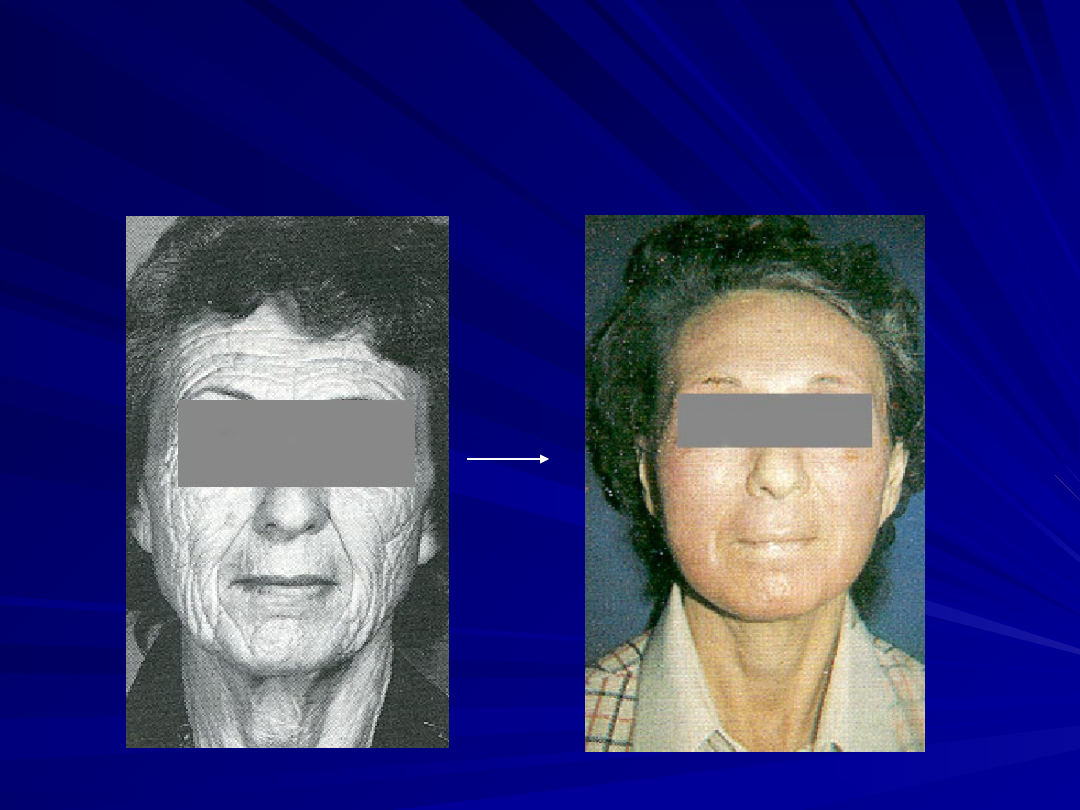
Clinical and histological changes are long-lasting
Clinical and histological changes are long-lasting
(15 to 20 years) and may be permanent for some
(15 to 20 years) and may be permanent for some
patients
patients
A complication can also be permanent!
A complication can also be permanent!

References
References
Deborshi R. AblativeFacial Resurfacing Dermatologic Clinics. 23(3), July 2005
Deborshi R. AblativeFacial Resurfacing Dermatologic Clinics. 23(3), July 2005
Gary D. M. MEDIUM-DEPTH CHEMICAL PEELS. Dermatologic Clinics. 19(3), July 2001
Gary D. M. MEDIUM-DEPTH CHEMICAL PEELS. Dermatologic Clinics. 19(3), July 2001
Langsdon, P. Comparison of the Laser and Phenol Chemical Peel in Facial Skin
Langsdon, P. Comparison of the Laser and Phenol Chemical Peel in Facial Skin
Resurfacing.
Resurfacing.
Brody HJ. Chemical Peeling. St Louis, Mo: Mosby-Year Book; 1992:1-5
Brody HJ. Chemical Peeling. St Louis, Mo: Mosby-Year Book; 1992:1-5
Brody HJ: Chemical Peeling and Resurfacing. St. Louis, Mosby, 1997, pp 109–110
Brody HJ: Chemical Peeling and Resurfacing. St. Louis, Mosby, 1997, pp 109–110
Monheit GD: Advances in chemical peeling. Facial Plast Surg Clin North Am 2:5–9, 1994
Monheit GD: Advances in chemical peeling. Facial Plast Surg Clin North Am 2:5–9, 1994
Monheit GD: The Jessner's-TCA peel. Facial Plast Surg Clin North Am 2:21–22, 1994
Monheit GD: The Jessner's-TCA peel. Facial Plast Surg Clin North Am 2:21–22, 1994
Monheit GD, Zeitouni NC: Skin resurfacing for photoaging: Laser resurfacing versus
Monheit GD, Zeitouni NC: Skin resurfacing for photoaging: Laser resurfacing versus
chemical peeling. Cosmet Dermatol 10:11–22, 1997
chemical peeling. Cosmet Dermatol 10:11–22, 1997
Rubin M: Manual of Chemical Peels. Philadelphia, Lippincott, 1995, pp 120–121
Rubin M: Manual of Chemical Peels. Philadelphia, Lippincott, 1995, pp 120–121
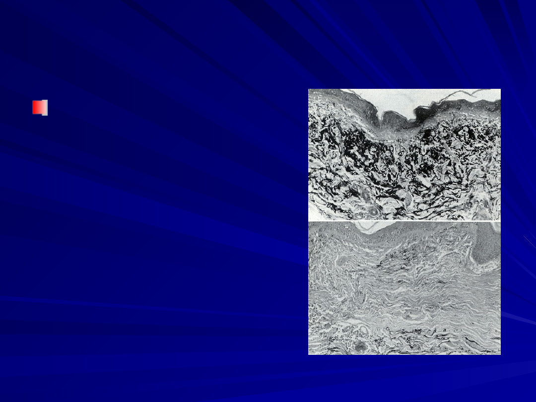
Stegman SJ: A comparative histologic study of the effects of three peeling agents and
Stegman SJ: A comparative histologic study of the effects of three peeling agents and
dermabrasion on normal and sundamaged skin. Aesthetic Plast Surg 6:123–135, 1982
dermabrasion on normal and sundamaged skin. Aesthetic Plast Surg 6:123–135, 1982
Cummings: MANAGEMENT OF AGING SKIN
Cummings: MANAGEMENT OF AGING SKIN
.
.
Otolaryngology: Head & Neck Surgery, 4th
Otolaryngology: Head & Neck Surgery, 4th
ed
ed
,
,
2005. Chapter 29
2005. Chapter 29
Tse Y, Ostad A, Lee HS, et al. A Clinical and histologic evaluation of two medium-depth
Tse Y, Ostad A, Lee HS, et al. A Clinical and histologic evaluation of two medium-depth
peels: glycolic acid versus Jessner's trichloroacetic acid. Dermatol Surg. 1996;22:781-786
peels: glycolic acid versus Jessner's trichloroacetic acid. Dermatol Surg. 1996;22:781-786
Kligman A.M. Long-term histologic follow-up of phenol face peel.
Kligman A.M. Long-term histologic follow-up of phenol face peel.
Plast Reconstr Surg
Plast Reconstr Surg
(1985) 75 : pp 652-659
(1985) 75 : pp 652-659
Halaas YP Medium Depth Peels, Facial Plastic Surgery Clinics of North America,
Halaas YP Medium Depth Peels, Facial Plastic Surgery Clinics of North America,
12(3):297-304, 2004
12(3):297-304, 2004
Document Outline
- Facial Chemical Peels
- History
- Slide 3
- Aging
- Histology
- Peel Skin Histology
- Slide 7
- Slide 8
- Slide 9
- Slide 10
- Treat cutaneous lesions
- Patient Selection
- Fitzpatrick Classification
- Glogau classification
- Aesthetic Indications
- Therapeutic Indications
- Contraindications
- Patient Preparation
- Chemical Peel Depths
- Chemical Peel
- Superficial Peels
- Superficial Peels
- Medium Peel
- Trichloracetic acid (TCA)
- Medium Peel
- Slide 26
- Slide 27
- Slide 28
- Slide 29
- Slide 30
- Deep Chemical Peel
- Slide 32
- Tape Occlusion
- Slide 34
- Slide 35
- Slide 36
- Phenol Toxicity
- Slide 38
- Hyperpigmentation
- Hypopigmentation
- Herpes outbreak
- Candida infection
- Pseudomonal infection
- Scarring
- Conclusion
- References
Wyszukiwarka
Podobne podstrony:
Positron emission tomography slides
face painting lesson 3 id 16748 Nieznany
chem wykład 11
c3 stal po ob ciep-chem, Politechnika Poznańska, Edukacja Techniczno Informatyczna, Semestr II, Mate
20094048eko-chem, zarzadzanie kryzysowe
chem fiz L Dok1
Sprawozdanie nr 8 chem
chem
BAT wielkotonazowe chem org
laboratorium chem inz proc
chem wykład 13
chem bud wyklad I JM
Podst poj i pr chem id 366070 Nieznany
Chem kolokwium odp, Studia, I Semestr, Chemia Budowlana
zjawisko fiz a reakcja chem, dydaktyka, konspekty
Wyniki egzamin IIIrok-inż.chem., III rok semestr letni, inżynieria chemiczna
chem fiz 14 11 zad id 111352 Nieznany
anali srod chem anali4 id 59578 Nieznany (2)
więcej podobnych podstron