
Muscle mass gain observed in patients with short bowel syndrome
subjected to resistance training
Ellen Cristini Freitas Araújo
a
, Vivian Marques Miguel Suen
b,
⁎
,
Julio Sergio Marchini
b
, Helio Vannucchi
c
a
Food Science and Experimental Nutrition, Division of Clinical Nutrition, Department of Internal Medicine, Faculty of Medicine of Ribeirão Preto,
University of São Paulo, São Paulo 14 049-900, Brazil
b
Division of Clinical Nutrition, Department of Internal Medicine, Faculty of Medicine of Ribeirão Preto,
University of São Paulo, São Paulo 14 049-900, Brazil
c
Department of Internal Medicine, Faculty of Medicine of Ribeirão Preto, University of São Paulo, São Paulo 14 049-900, Brazil
Received 11 May 2007; revised 30 November 2007; accepted 2 December 2007
Abstract
Few studies are available about the evaluation of resistance training in patients with protein-
energy malnutrition. To assess the effects of resistance training on the recovery of nutritional status
of patients with short bowel syndrome, with a small bowel remnant of less than 100 cm, 9 patients of
both sexes with protein-energy malnutrition after extensive resection of the small bowel were
submitted to resistance training of progressive intensity consisting of concentric and eccentric work
exercises for the upper limbs, trunk, and lower limbs, with the individuality and limitations of each
patients being respected. Food consumption was monitored by 24-hour food recall performed during
the initial phase of the study, before and 7 and 14 weeks after physical training, and by a dietary
record for a period of 3 days of oral feeding. The nutrients administered by the enteral and parenteral
route were recorded. A significant increase in total arm area (P
≤ .01) and fat-free mass (P ≤ .01)
was observed as determined by computed tomography. An increase in total energy ingestion and
carbohydrate consumption (P
≤ .01) was also observed. In addition, the activity of the enzyme
carnosinase was increased after resistance training (P
≤ .01). The present results show that resistance
training in patients with short bowel syndrome and protein-energy malnutrition can be considered to
be a part of the nonmedicamentous treatment of these patients, leading to better nutrient use and to a
gain of lean mass.
© 2008 Elsevier Inc. All rights reserved.
Keywords:
Human; Protein-energy malnutrition; Short bowel syndrome; Physical activity; Resistance training
Abbreviations:
E.C., Escherichia Coli; KV, kilovolt; Mg/creat/24 h, milligram of creatinine in 24 hours;
μmol/ml/h, micromole
per milliliter per hour; USP, University of São Paulo.
1. Introduction
Short bowel syndrome occurs after extensive resection of
the small bowel. Among the causes of mesenteric ischemia
are emboli and infarction of the superior mesenteric artery
. Because of the loss of an extensive portion of the small
bowel, the patients develop severe protein-energy malnutri-
tion, requiring parenteral nutrition during the immediate and
late postoperative period, a fundamental procedure for in-
creased survival
.
The nutritional status expresses the extent to which the
physiologic nutrient requirements are being met to maintain
adequate composition and function
. Malnutrition predis-
poses to a series of severe complications including a
Available online at www.sciencedirect.com
Nutrition Research 28 (2008) 78
–82
www.elsevier.com/locate/nutres
⁎ Corresponding author. Department of Internal Medicine, Ribeirao
Preto School of Medicine, São Paulo University, Ribeirão Preto, São Paulo
14 049-900, Brazil. Tel.: +55 16 3602 3375; fax: 55 16 3633 6695.
E-mail address:
(V.M.M. Suen).
0271-5317/$
– see front matter © 2008 Elsevier Inc. All rights reserved.
doi:

tendency to infection, deficient wound healing, respiratory
failure, cardiac insufficiency, reduced protein synthesis at the
hepatic level with production of abnormal metabolites, and
reduced glomerular filtration and production of gastric juice
. Physical inactivity causes muscle weakening, drastically
reducing the capacity to generate muscle work, affecting the
ability to
“live independently”
. Resistance or strength
physical training has been pointed out as the cause of
positive hypertrophic adaptation of skeletal muscle
.
Training with resistance or strength exercises can help
reverse the malnutrition commonly occurring among
patients with renal failure. This type of training is
characterized by weight lifting, which results in increased
muscle mass, improving physical function and attenuating
progressive muscle loss
.
In a study in which a low-protein diet potentially inducing
malnutrition was administered to male Wistar rats to
determine the physiologic and metabolic changes because
of malnutrition in a control and in an exercised group, Neiva
et al
concluded that malnutrition associated with
sedentarism causes important alterations in patterns con-
sidered to be normal, with physical exercise potentiating the
results obtained and aiding nutritional recovery.
To our knowledge, few data are available about resisted
physical exercise applied to patients who underwent
enterectomy. Thus, there is an urgent need to transmit
information about the importance of resistance training as
part of treatment to the professionals involved in the
recovery of patients with protein-energy malnutrition. On
the basis of the information, we believe that resisted
physical exercise is associated with improved nutritional
status in patients who underwent enterectomy, aiding their
nutritional recovery.
2. Methods and materials
2.1. Patients
A total of 9 patients with short bowel syndrome, 4 women
and 5 men older than 30 years followed at the Metabolic Unit
of the University Hospital, Faculty of Medicine of Ribeirão
Preto, University of São Paulo (São Paulo, Brazil),
participated in the study. The study was approved by the
research ethics committee of the University Hospital, Faculty
of Medicine of Ribeirão Preto, University of São Paulo, and
all patients gave written informed consent to participate.
2.2. Experimental design
The patients were submitted to evaluation of nutritional
status before and after 14 weeks of resistance physical
training, with each individual acting as his own control. The
evaluation consisted of anthropometry, evaluation of food
intake by 2 types of dietary survey, 24-hour diet recall and
3-day food record, and measurement of energy expenditure
by indirect calorimetry. Computed tomography was used as
the imaging method. The patients were submitted to
resistance training twice a week for a period of 14 weeks.
The inclusion criterion was not to have participated in any
type of regular physical exercise in the last 12 months. The
evaluation of nutritional status was repeated after the period
of physical training. All evaluations were performed before
and after the resistance training. Each individual served as
his own control. Each evaluation method and the respective
references are described below.
2.3. Anthropometry
The anthropometric measurements performed were
weight, height, skin folds, arm circumference, and calcula-
tion of arm muscle circumference
, and the results were
defined as mild, moderate, and severe malnutrition
.
2.4. Laboratory data
Venous blood samples were collected and used to determine
total proteins, albumin, and carnosinase (Escherichia coli:
3.4.13.20)
; 24-hour urine samples were also obtained.
Urinary creatinine level was determined by reaction with a
picrate solution in alkaline medium, forming a red complex
that was measured photometrically. The determination was
performed using a Labtest kit (Lagoa Santa, Minas Gerais,
Brazil) and a Beckman DU640 spectrophotomer (Corona, CA)
at 510 nm.
2.5. Evaluation of food intake
Food intake was determined by the sum and the mean of
the results obtained with the 24-hour diet recall, with the
3-day diet record
and with enteral and parenteral
nutrition. The data were analyzed before and after physical
training. Food intake was calculated with the aid of a
computer program (Programa de Apoio à Nutrição [Nutrition
Support Program] version 2.5, licensed by Escola Paulista de
Medicina
—Nutritional therapy, São Paulo, Brazil).
2.6. Measurement of resting energy expenditure
Resting energy expenditure was determined before and after
the end of physical training using a Sensor Medics calorimeter
(Sensor Medics Corporation, Yorba Linda, Calif)
.
2.7. Computed tomography
Images of the midpoint of the nondominant arm were
obtained (Tomoscan LX-C, Matrix 512 and 320; Eastlake,
OH) at a speed of 9.5 seconds at 30 kV. The axis was oriented
at 90° using a 256 × 256 matrix. Readings of total area and of
muscle and bone areas were obtained with a Mini-Moop
digitizing board (Eching, Bavaria, Germany) plus an
associated computation program using a digital pen to circle
the figure exposed on photographic paper to measure the
different areas (total area, muscle area, and bone area). To
determine the muscle area, bone area was measured and
subtracted from muscle area, and to determine the adipose
area, the muscle and bone areas were summed and the value
was subtracted from the total area
79
E.C.F. Araújo et al. / Nutrition Research 28 (2008) 78
–82

2.8. Program of physical training with weights
The subjects were submitted to resistance training of
progressive intensity, with exercises of concentric and
eccentric work for upper limbs, trunk, and lower limbs,
with the individualities and limitations of each patient
being respected. All exercise sessions were monitored for
patient compliance. The training program lasted 14 weeks,
with 2 sessions per week lasting approximately 60 minutes.
During the first 2 weeks, 4 exercise sessions were held to
permit the individuals to familiarize themselves with the
equipment (Athletics 2001 mechanotherapy station, Albar-
reja, Fuenlabrada, Madrid), and with the exercise techni-
ques. Eight different types of exercise, pectoral, back,
shoulders, biceps, triceps, thigh, calf, and abdomen, were
used. The subjects first executed the exercises for large
muscle groups and then the remaining ones. They also
performed general warm-up exercises for 3 minutes. Three
series of 8 repetitions were executed (maximum load for
8 repetitions), corresponding to an approximate intensity of
80% of the maximum load for the muscle groups in
general, except for the calf and abdomen, with abdominal
exercises being performed when possible. Three series of
10 to 12 repetitions were executed for the calf and
abdominal exercises. When an individual increased his
strength to the point of being able to perform the exercises
with ease, a new load was added. A resting period of
±1 minute was allowed between exercise series
.
2.9. Statistical analysis
Data regarding total energy, carbohydrate, protein and
lipid intake; anthropometric measurements; adipose area and
muscle area obtained by computed tomography; and resting
energy expenditure were analyzed statistically by nonpara-
metric analysis of variance using the GraphPad software,
version 3.00 for Windows 95, San Diego, Calif. The
differences detected in the variable between the pre- and
postexercise period were determined by the nonparametric
Wilcoxon test
. The level of significance was set at P
≤
.05 in all analyses and values are presented as means ± SD.
3. Results and discussion
At the beginning of the study all patients presented some
degree of malnutrition regarding the different variables
studied. Analysis of the data presented in
shows that
before resistance training, all patients presented a mild,
moderate, or severe weight loss as indicated by the
measurement of tricipital skin fold and arm fat index, in
addition to loss of body muscle mass of a mild or moderate
degree, as indicated by estimated arm muscle circumference.
After physical training, arm muscle circumference was
the anthropometric measurement showing a statistically
significant difference (P
b .05). In addition, the resting
metabolic rate measured (
), the carnosinase enzyme
(
), and total arm area and fat-free mass (
demonstrated a statistically significant difference (P
≤ .01).
No significant difference was observed regarding weight,
body mass index, tricipital skin fold, bicipital skin fold,
subscapular skin fold, or suprailiac skin fold (
After the training period, there was a statistically
significant increase in total caloric and carbohydrate intake
(
).
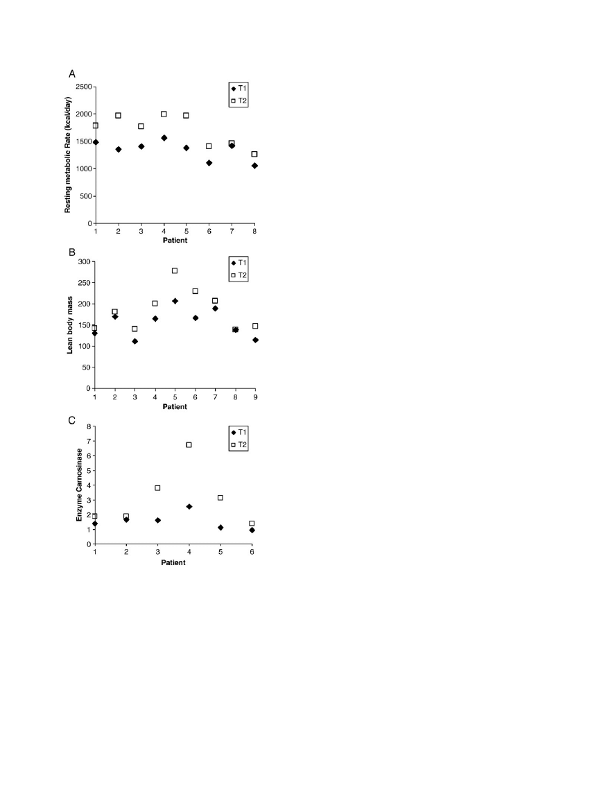
(A, B, and C) presents the data for lean body
Table 1
Anthropometric measurements, resting energy expenditure, body
composition obtained by computed tomography, and serum carnosinase
levels before and after physical training in subjects
Before physical
training
After physical
training
Age
50.7 ± 4.5
-
Height
1.63 ± 0.08
-
Weight (kg)
50.7 ± 5.4
51.9 ± 5.4
Body mass index (kg/m
2
)
19.1 ± 1.8
19.5 ± 1.8
Triceps skin fold (mm)
8.2 ± 3.6
7.4 ± 3.0
Arm fat index
0.76 ± 0.31
-
Arm muscle circumference (cm)
21.7 ± 1.8
22.5 ± 2.1 ⁎
Urinary creatinine (mg per
creatinine per 24 h) (n = 6)
0.86 ± 0.37
1.01 ± 0.32
Resting metabolic rate (kcal/d)
5622 ± 744
7106 ± 1187 ⁎⁎
Carnosinase (
μmol/mL per hour)
(n = 6)
1.53 ± 0.57
3.11 ± 1.97 ⁎⁎
⁎ Significantly different values (means ± SD) after resisted training at
P
≤ .01 as determined by Wilcoxon test.
⁎⁎ Significantly different values (means ± SD) after resisted training at
P
≤ .05 as determined by Wilcoxon test.
Fig. 1. Total arm area results in subjects at T1 and T2 during the study. TAA
indicates total arm area at P
≤ .01; fat-free mass (FFM) at *P ≤ .01 as
determined by the Wilcoxon test. FM indicates fat mass.
Table 2
Daily intake of total energy, protein, fat, and carbohydrates, before and after
the resisted physical activity in subjects
Before
After
Total calories (kJ/d)
7081 ± 1221
8439 ± 1852 ⁎
Protein (kJ)
1467 ± 268
1584 ± 355
Fat (kJ)
1902 ± 815
2487 ± 1584
Carbohydrates (kJ)
3708 ± 915
4368 ± 798
⁎ Significantly different values (means ± SD) after resisted training at
P
≤ .01 as determined by the Wilcoxon test.
80
E.C.F. Araújo et al. / Nutrition Research 28 (2008) 78
–82
mass, resting metabolic rate, and carnosinase activity of each
patient (variables related to increased muscle mass).
An increase in resting metabolic rate (P
b .01) was
observed in the present study before and after resistance
training. This result suggests that the intensity and volume of
physical exercise performed were sufficient to induce
changes in energy expenditure. The significant increase in
total energy intake (kcal/d) after resistance training and also
the increased carbohydrate intake during the same period
were probably because of the modification of energy
expenditure that increased the energy requirements and the
daily energy consumption because of resistance training.
Carbohydrates are recognized as the main source of energy
during physical training, thus sparing the consumption of
proteins as an energy substrate
Other authors
have also shown this increase in
elderly patients, inducing an increase of approximately 15%
in total energy intake. Pratley et al
, in a study of the
effect of resistance physical exercise on elderly men, also
observed an increase in fat-free mass. However, they did not
observe changes in food intake during resistance training.
In the present study, the patients were similar to elderly
individuals, with evolution of the disease, surgeries,
compromised absorption levels, large weight loss, sedentary
life style, and consequent loss of fat-free mass, factors that
contributed to a loss of quality of life. However, with
resistance training, there was an increase in resting metabolic
rate and in fat-free mass, with improved quality of life for the
patients, who became able to perform the exercises
independently without direct assistance.
During periods of inanition, the organism remains in a
catabolic state, with depletion of lean mass because of a
reduction of body reserves
. However, in the present
study, there was a significant increase in body muscle mass
and in arm muscle circumference after physical training. It is
possible that the stimulus was efficient in changing the
direction of the process from catabolism to anabolism, a fact
that was not observed by Nelson et al
in elderly women
after progressive resistance training. Urinary creatinine
excretion was not correlated with muscle mass among the
patients studied. Urinary creatinine excretion depends on
factors such as physical activity and metabolic status and is
lower in malnourished individuals
A significant increase in serum carnosinase activity was
observed after resistance training. Serum carnosinase is an
enzyme that hydrolyzes its substrate, carnosine, into its
constituent amino acids alanine and histidine. Carnosine
(
β-alanyl-1-histidine) is a dipeptide abundantly distributed
throughout the body in organs such as muscles and kidneys
. According to Dubin et al
, histidine is one of the
substrates necessary for protein synthesis. Reduced urinary
excretion of 3-methyl-histidine (a histidine derivative) and
low carnosinase activity are phenomena described for
patients with reduced muscle mass formation such as uremic
patients and patients with progressive muscular dystrophy
. Thus, we conclude that the increase in carnosinase
activity was related to the increase in body muscle mass.
Tomography data revealed an increase in total arm area
and fat-free mass after physical training. The present study
involved 28 training sessions with 24 repetitions per session
at 80% maximum load and muscle evaluation by tomo-
graphy that showed that physical exercise performed under
the conditions described promoted an increase in muscle area
and in total arm area. Similar results were reported by other
authors in a 52-week study conducted on elderly males
Fig. 2. Individual data points for resting metabolic rate (RMR), lean body
mass (LBM), and carnosinase enzyme activity before (T1) and after (T2)
resistance physical training for each patient. Resting metabolic rate,
LBM, and carnosinase enzyme activity at *P
≤ .01 as determined by the
Wilcoxon test.
81
E.C.F. Araújo et al. / Nutrition Research 28 (2008) 78
–82

In contrast to these results, Moritani et al
found no
muscle changes after the measurement of skin folds and arm
circumference. The study involved 24 training sessions with
20 repetitions per session at 66% maximum load per
exercise. These divergent results may be probably explained
by differences in the intensity and duration of training and in
the techniques used for evaluation. Frontera et al
and
Pyka et al
, in a study of the increase in muscle fiber area
measured by subcutaneous muscle biopsy specimens and
tomography after resistance training in elderly subjects,
observed a significant increase in type I and II fibers. These
results suggest that the ability to increase muscle mass is
preserved in debilitated individuals.
We conclude that the proposed resistance training led to
metabolic modifications in the patients studied, indicating
better nutrient assimilation, helping nutritional recovery, and
indicating that this is an important part of the nonmedica-
mentous treatment of patients with short bowel syndrome.
References
[1] Brandt L. Vascular disorders of the intestine. In: Goldman L, Ausiello
D, editors. Cecil textbook of medicine. 22th ed. Philadelphia:
Saunders; 2004. p. 872-8.
[2] Nonino CB, Borges RM, Pasquali LP, Marchini JS. Oral dietetic
therapy in patients with short bowel syndrome. Rev Nutr Campinas
2001;14:201-5.
[3] Bistrian BR. Nutritional assessment. In: Goldman L, Ausiello D,
editors. Cecil textbook of medicine. Philadelphia: Saunders; 2004.
p. 1312-5.
[4] Torun B, Chew F. Protein-energy malnutrition. In: Shills M, Olson J,
Shike M, Ross AC, editors. Modern nutrition in health and disease.
Baltimore: Lippincott Williams and Wilkins; 1998. p. 963-87.
[5] Lopes J, Russell DM, Whitwell J, Jeejeebhoy KN. Skeletal muscle
function in malnutrition. Am J Clin Nutr 1982;36:602-10.
[6] Evans WJ, Campbell WW. Sarcopenia and age-related changes in body
composition and functional capacity. J Nutr 1993;123:465-8.
[7] Evans WJ. Exercise training guidelines for the elderly. Med Sci Sports
Exerc 1999;31:12-7.
[8] Castaneda C, Grossi L, Dwyer J. Potential benefits of resistance
exercise training on nutritional status in renal failure. J Ren Nutr
1998;8:2-10.
[9] Neiva CM, Guerino MR, Mello MAR. Análise dos efeitos da
desnutrição próteico-calórica sobre as respostas ao exercício agudo
(single section)
—parâmetros metabólicos. Motriz 1995;1:32-43.
[10] Marchini JS, Unamuno MRDL, Fonseca RMHR, Rodrigues MMP,
Dutra de Oliveira JE. Métodos antropométricos para avaliação
do estado nutricional de adultos. Rev Nutr PUCCAMP 1992;5:
121-42.
[11] Frisancho AR. New standards of weight and body composition by
frame size and height for assessment of nutritional status of adults and
the elderly. Am J Clin Nutr 1984;40:808-19.
[12] Bando K, Shimotsuji T, Toyoshima H, Hayashi C, Miyai K.
Fluorometric assay of human serum carnosinase activity in normal
children, adults and patients with myopathy. Ann Clin Biochem
1984;21:510-4.
[13] Dywer J, Picciano MF, Raiten DJ, Members of the Steering
Committee. Estimation of usual intakes: what we eat in America
—
NHANES. J Nutr 2003;133:609S-23S.
[14] Ferrannini E. The theoretical bases of indirect calorimetry: a review.
Metabolism 1988;37:287-301.
[15] Heymsfield SB, Olafson RP, Kutner MH, Nixon DW. A radiographic
method of quantifying protein-calorie undernutrition. Am J Clin Nutr
1979;32:693-702.
[16] Lukaski HC. Methods for the assessment of human body composition:
traditional and new. Am J Clin Nutr 1987;46:537-56.
[17] Fleck SJ, Kraemer WJ. Fundamentos do treinamento de força
muscular. 2nd ed. Porto Alegre: Artmed; 1999. p. 20-183.
[18] Daniels WW. Biostatistics: a foundation for analysis in the health
sciences. 7th ed. New York: John Wiley & Sons; 1998. p. 669-74.
[19] Achten J, Halson SL, Moseley L, Rayson MP, Casey A, Jeukendrup
AE. Higher dietary carbohydrate content during intensified running
training results in better maintenance of performance and mood states.
J Appl Physiol 2004;60:167-75.
[20] Campbell WW, Crim MC, Young VR, Evans WJ. Increase energy-
requirement and changes in body-composition with resistance training
in older adults. Am J Clin Nutr 1994;60:167-75.
[21] Pratley R, Nicklas B, Rubin M, Miller J, Smith A, Smith M, et al.
Strength training increase resting metabolic rate and norepinephrine
levels in healthy 50- to 65-yr-old men. J Appl Physiol 1994;76:133-7.
[22] Nelson ME, Fiatorarone MA, Layne JE, Trice I, Economos CD,
Fielding RA. Analysis of body-composition techniques and models for
detecting change in soft tissue with strength training. Am J Clin Nutr
1996;63:678-86.
[23] Wang ZM, Gallagher D, Nelson ME, Matthews DE, Heymsfield SB.
Total-body skeletal muscle mass: evaluation of 24-h urinary creatinine
excretion by computed axial tomography. Am J Clin Nutr 1996;63:
863-9.
[24] Quinn PJ, Boldyrevt AA, Formazuyk VE. Carnosine: its properties,
functions and potential therapeutic applications. Mol Aspects Med
1992;13:379-444.
[25] Dubin S, McKee K, Battish S. Essential amino acid reference profile
affects the evaluation of enteral feeding products. J Am Diet Assoc
1994;8:884-7.
[26] Moritani T, DeVries HA. Potential for gross muscle hypertrophy in
older men. J Gerontol 1980;35:672-82.
[27] Frontera WR, Meredith CN, O'Reilly KP, Knuttgen HG, Evans WJ.
Strength conditioning in older men skeletal muscle hypertrophy and
improved function. J Appl Physiol 1988;64:1038-44.
[28] Pyka G, Lindenberg E, Charette S, Marcus R. Muscle strength and
fiber adaptations to a year-long resistance training program in elderly
men and women. J Gerontol 1994;49:22-7.
82
E.C.F. Araújo et al. / Nutrition Research 28 (2008) 78
–82
Document Outline
- Muscle mass gain observed in patients with short bowel syndrome subjected to resistance trainin.....
Wyszukiwarka
Podobne podstrony:
Effects of Clopidogrel?ded to Aspirin in Patients with Recent Lacunar Stroke
Difficult airway management in a patient with traumatic asphyxia
Impaired Sexual Function in Patients with BPD is Determined by History of Sexual Abuse
Konstatinos A Land versus water exercise in patients with coronary
A Ser49Cys Variant in the Ataxia Telangiectasia, Mutated, Gene that Is More Common in Patients with
Difficult airway management in a patient with traumatic asphyxia
Proton Magnetic Resonance Spectroscopy of the Medial Prefrontal Cortex in Patients With Deficit Schi
Serum cytokine levels in patients with chronic low back pain due to herniated disc
The Effects of Probiotic Supplementation on Markers of Blood Lipids, and Blood Pressure in Patients
The relationship of Lumbar Flexion to disability in patients with low back pain
High Choline Concentrations in the Caudate Nucleus in Antipsychotic Naive Patients With Schizophreni
Breast and other cancers in 1445 blood relatives of 75 Nordic patients with ataxia telangiectasia
Continuous mechanical chest compression during in hospital cardiopulmonary resuscitation of patients
A Proton MRSI Study of Brain N Acetylaspartate Level After 12 Weeks of Citalopram Treatment in Drug
Trace Element Levels in Hashimoto Thyro Patients with Subclinical Hypothyroidism
Glutamate and Glutamine Measured With 4 0 T Proton MRS in Never Treated Patients With Schizophrenia
więcej podobnych podstron