
Resuscitation 82 (2011) 155–159
Contents lists available at
Resuscitation
j o u r n a l h o m e p a g e :
w w w . e l s e v i e r . c o m / l o c a t e / r e s u s c i t a t i o n
Clinical paper
Continuous mechanical chest compression during in-hospital cardiopulmonary
resuscitation of patients with pulseless electrical activity
夽
Hendrik Bonnemeier
, Gregor Simonis
, Göran Olivecrona
, Britta Weidtmann
,
Matthias Götberg
, Gunther Weitz
, Ivana Gerling
, Ruth Strasser
, Norbert Frey
a
Klinik für Innere Medizin III, Kardiologie und Angiologie, Universitätsklinikum Schleswig-Holstein, Campus Kiel, Arnold-Heller-Str. 3, Kiel 24105, Germany
b
Medizinische Klinik/Kardiologie und Intensivmedizin, Herzzentrum Dresden, Dresden, Germany
c
Department of Cardiology, Heart and Lung Division, Lund University Hospital, Lund, Sweden
d
Institut für Rechtsmedizin, Universitätsklinikum Schleswig-Holstein Campus Lübeck, Lübeck, Germany
e
Medizinische Klinik I, Universitätsklinikum Schleswig-Holstein, Campus Lübeck, Lübeck, Germany
a r t i c l e i n f o
Article history:
Received 30 August 2007
Received in revised form 30 August 2010
Accepted 29 October 2010
Keywords:
Cardiopulmonary resuscitation
Mechanical chest compression device
Pulseless electrical activity
a b s t r a c t
Survival after in-hospital pulseless electrical activity (PEA) cardiac arrest is poor and has not changed
during the last 10 years. Effective chest compressions may improve survival after PEA. We investigated
whether a mechanical device (LUCAS
TM
-CPR) can ensure chest compressions during cardiac arrest accord-
ing to guidelines and without interruption during transport, diagnostic procedures and in the catheter
laboratory.
Methods: We studied mechanical chest compression in 28 patients with PEA (pulmonary embolism
(PE) n = 14; cardiogenic shock/acute myocardial infarction; n = 9; severe hyperkalemia; n = 2; sustained
ventricular arrhythmias/electrical storm; n = 3) in a university hospital setting.
Results: During or immediately after CPR, 21 patients underwent coronary angiography and or pul-
monary angiography. Successful return of a spontaneous circulation (ROSC) was achieved in 27 out of
the 28 patients. Ten patients died within the first hour and three patients died within 24 h after CPR.
A total of 14 patients survived and were discharged from hospital (13 without significant neurological
deficit). Interestingly, six patients with PE did not have thrombolytic therapy due to contraindications.
CT-angiography findings in these patients showed fragmentation of the thrombus suggesting throm-
bus breakdown as an additional effect of mechanical chest compressions. No patients exhibited any
life-threatening device-related complications.
Conclusion: Continuous chest compression with an automatic mechanical device is feasible, safe, and
might improve outcomes after in-hospital-resuscitation of PEA. Patients with PE may benefit from effec-
tive continuous chest compression, probably due to thrombus fragmentation and increased pulmonary
artery blood flow.
© 2010 Elsevier Ireland Ltd. All rights reserved.
1. Introduction
The incidence of pulseless electrical activity (PEA) after in-
hospital cardiac arrest (IHCA) is unchanged for the last 10 years
(29–37%), and similar to the incidence of asystole (30–39%). Both
PEA and asystole have similar rates of survival to hospital discharge
(about 10%).
Ventricular fibrillation (VF) accounts for 23–40%
of IHCAs and has higher rates of survival (30–40% to hospital
夽 A Spanish translated version of the abstract of this article appears as Appendix
in the final online version at
doi:10.1016/j.resuscitation.2010.10.019
∗ Corresponding author. Tel.: +49 431 597 1441; fax: +49 431 597 1470.
E-mail addresses:
,
(H. Bonnemeier).
discharge) due to effective treatment with defibrillation. Survival
from PEA and asystole depends on treating the underlying cause
of cardiac arrest and this often requires a longer period of chest
compressions (CC). Studies show that high quality CC is difficult
to achieve on manikins and real patients during long periods of
resuscitation even when performed by hospital staff.
Pulseless electrical activity is often seen after pulmonary
embolism (PE) or coronary artery thrombosis (e.g., main-stem
occlusion) and is associated with poor survival.
treatment during CPR for PE induced cardiac arrest has been shown
to have good survival in small case series but larger case series have
not shown this.
Chest compressions are important for the defibrillation suc-
cess and survival from VF, both in humans
We aimed to evaluate if effective continuous chest compression
0300-9572/$ – see front matter © 2010 Elsevier Ireland Ltd. All rights reserved.
doi:

156
H. Bonnemeier et al. / Resuscitation 82 (2011) 155–159
Table 1
Consecutive patients with PEA undergoing CPR with LUCAS for IHCA.
Gender
Age
Underlying diagnosis for PEA
LUCAS compression (min)
Outcome
1.
Female
47yo
Fulminant pulmonary embolism
50
ROSC
2.
Male
60yo
STEMI/main stem thrombosis
85
ROSC
3.
Male
68yo
Fulminant pulmonary embolism
100
Survival
4.
Female
74yo
Fulminant pulmonary embolism
20
Survival
5.
Female
81yo
Fulminant pulmonary embolism
35
Survival
6.
Male
66yo
Fulminant pulmonary embolism
25
Survival
7.
Female
60yo
Fulminant pulmonary embolism
10
Survival
8.
Male
64yo
STEMI/stent-thrombosis prox. LCx
15
Survival
9.
Male
72yo
STEMI/thromb. occlusion prox. LAD
120
Survival
10.
Male
54yo
Fulminant pulmonary embolism
20
Survival
11.
Female
60yo
Fulminant pulmonary embolism
10
Survival
12.
Female
35yo
Fulminant pulmonary embolism
180
ROSC
13.
Male
69yo
Severe hyperkalemia/cardiomyopathy
40
Survival
14.
Male
64yo
ICD-testing during CRT-ICD-implantation
60
ROSC
15.
Male
70yo
STEMI/stent-thrombosis prox. LAD
30
Survival/N
16.
Female
34yo
Fulminant pulmonary embolism
70
ROSC
17.
Male
82yo
PAVR/ during balloon-occlusion
45
ROSC
18.
Male
66yo
Amiodarone during incess. VT
60
Survival
19.
Female
78yo
Fulminant pulmonary embolism
20
ROSC
20.
Male
71yo
Cardiogenic shock/AMI
45
ROSC
21
Male
59yo
STEMI/left main-stem occlusion
75
Survival
22.
Male
71yo
NSTEMI/thrombolytic CABG-occlusion
20
ROSC
23.
Male
80yo
Fulminant pulmonary embolism
25
Deceased
24.
Female
59yo
Electric storm/ICD patient with DCM
40
ROSC
25.
Male
77yo
Fulminant pulmonary embolism
30
ROSC
26.
Female
51yo
Fulminant pulmonary embolism
60
ROSC
27.
Male
62yo
Severe hyperkalemia/patient on dialysis
10
Survival
28.
Male
69yo
Cardiogenic shock/AMI
25
ROSC
Survival = hospital discharge without significant neurological deficits (CRC 1 + 2).
Survival/N = hospital discharge with significant neurological deficits (CRC
≥ 3).
AMI = acute myocardial infarction. CABG = coronary artery bypass grafting. DCM = dilative cardiomyopathy. ICD = implantable cardioverter defibrillator. PAVR = percutaneous
aortic valve replacement. NSTEMI = non-ST-segment elevation myocardial infarction. STEMI = ST-segment elevation myocardial infarction.
(rate of 100 min
−1
, compression depth of 50 mm, 50/50 duty cycle
and adequate recoil) using a mechanical chest compression device
(LUCAS
TM
) is safe and feasible during treatment of patients with
PEA cardiac arrest.
2. Methods
Patients were enrolled from August 2006 to August 2008 in three
European university hospitals (Lübeck and Dresden, Germany and
Lund, Sweden). Resuscitation events were studied among patients
that experienced cardiac arrest, defined by the documented loss of
a pulse and respirations as well as the delivery of (initially man-
ual) chest compressions. Out of hospital cardiac arrest (OHCA)
cases were not included. Only patients with PEA as the under-
lying rhythm were investigated. Other cardiac arrest treatments
included were: diagnostic imaging using coronary angiography,
pulmonary angiography and CT-angiography during mechanical
chest compressions. Imaging was followed by treatment with per-
cutaneous coronary intervention (PCI) and thrombolysis when
indicated. Following return of spontaneous circulation (ROSC)
comatose patients were treated with hypothermia in the intensive
care unit according to local protocols.
Continuous chest compressions were delivered mechanically
using LUCAS
TM
CPR (Jolife, Sweden). This device can be used to
deliver chest compression according to the current guidelines
without interruptions during prolonged resuscitation, patient
transport, acute diagnostic procedures, and during coronary
angiography.
The use of LUCAS
TM
for IHCA was left to the
discretion of the resuscitation team, however, in all three centres
the use of the LUCAS
TM
device was already established for patients
with ongoing CPR on the wards, the coronary care units, the inten-
sive care units, and the catheterization laboratory for more than
12 months before the initiation of the present study. Following
the intervention, all patients were intensively screened for life-
threatening device-related complications and some of the deceased
patients underwent a forensic necropsy.
The predefined endpoints were: ROSC, 24 hour survival, hospital
discharge with Cerebral Performance Category (CPC) 1 or 2 and
device-related complications.
3. Results
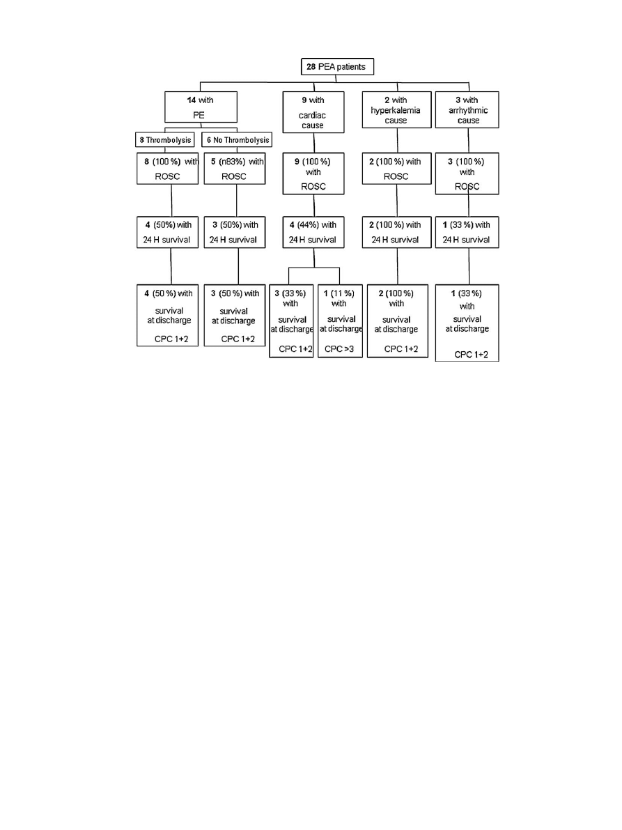
A total of 28 patients with PEA were included in the study. Most
patients were enrolled in the university hospital Lübeck where 21
consecutive patients with PEA cardiac arrests on internal medicine
wards were enrolled. Within the enrolment period there were 215
in-hospital resuscitations in the university hospital Lübeck includ-
ing 52 patients with PEA. Between January 2008 and August 2008 a
total of 4 non-consecutive patients were enrolled in the heart cen-
ter Dresden, and 3 patients in the department of cardiology of the
Lund university.
The 28 patients with PEA in the study included 10 were
females, 18 males and the mean age was 64.4
± 12 (mean ± SD)
years (range 34–82 years). The underlying cause of PEA was:
PE (n = 14), cardiogenic shock/acute myocardial infarction (n = 9),
severe hyperkalemia (n = 2) and sustained ventricular arrhyth-
mias/electric storm (n = 3). LUCAS
TM
CC were performed for a
median duration of 37.5 min (range 10 and 180 min) (
During or directly after CPR, 21 patients underwent coronary
angiography/pulmonary angiography. Initial ROSC was achieved in
27 out of 28 patients. Ten patients died within the first hour, another
three patients died within 24 h after CPR. A total of 14 patients sur-
vived and were discharged from hospital (13 without significant
neurological deficits – CPC 1 and 2). Six of the 14 patients with PE
did not undergo thrombolytic therapy because they had contraindi-
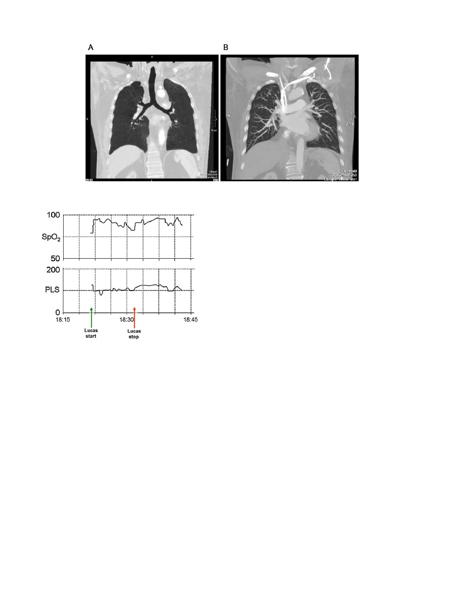
cations. CT-angiography in these patients showed fragmentation of
the thrombus even though thrombolytic therapy was not was given
(
). None of the patients exhibited significant or serious injuries
H. Bonnemeier et al. / Resuscitation 82 (2011) 155–159
157
Fig. 1. Flow chart of treatment and outcome data for all 28 PEA patients studied, according to the predefined endpoints: ROSC, 24 h survival, hospital discharge with good
Cerebral Performance Category (CPC 1 + 2).
associated with LUCAS
TM
CC. The deceased patients that under-
went forensic necropsy showed no evidence of a device related
injury.
4. Discussion
We report a case series of good outcomes after continuous chest
compression using LUCAS
TM
and early imaging and intervention for
IHCA due to PEA. We found that almost 50% of the patients survived
to discharge to their homes with good or moderate neurological
function (CPC 1 and 2).
Neurologically intact survival rates have not improved in
more than a decade, and overall survival rates of in-hospital-
cardiopulmonary resuscitation are still alarmingly low for patients
with PEA. Cardiopulmonary resuscitation of IHCA has been shown
to be inconsistent and often does not meet guideline recommen-
dations, even when performed by well-trained hospital staff.
Studies show that even experienced rescuers produce shallower
and slower compressions over time, without being aware. In an
effort to improve manual CPR, several mechanical devices are
available and mainly in use by out-of-hospital-emergency medi-
cal services. The setting and environment, the response time, the
medical and diagnostic equipment, and the patient population are
all different for CPR after IHCA, compared to OHCA. Resuscitation
from IHCA would be expected to be more successful, but even
with experienced hospital staff and CPR training programs, resus-
citation skills deteriorate over time. Furthermore, translation from
training to actual cardiac arrest settings and rescuer fatigue during
CPR limit IHCA CPR quality.
23
Mechanical CPR devices offer new
opportunities for IHCA resuscitation as they help to sustain circu-
lation with consistent compressions according to the guidelines
during prolonged resuscitation efforts, transportation, and during
interventional procedures such as PCI. There is clinical evidence
that mechanical CPR devices provide chest compressions more
reliably at a set rate and depth and thus generate better hemo-
dynamic characteristics than manual chest compressions.
Furthermore, using mechanical CPR it is possible to “buy time” in
an effective manner ensuring adequate circulation and allowing
interventional procedures treatments – i.e., primary angioplasty
or computed tomography. In addition to these practical benefits,
experimental data show significantly increased flow and ROSC
levels with mechanical CPR devices compared to manual chest
compression.
Another significant benefit of mechanical chest compression
for the clinical management of IHCA is becoming clearer: in the
catheter laboratory, one of the pivotal points of IHCA, interventions
are not possible without interrupting manual chest compressions.
Usually, CPR is difficult in the catheter laboratory because effec-
tive manual chest compressions are difficult due to the gantry
around the patient’s chest and the height of the table. Furthermore,
interventions are hindered during manual compressions there is
significant radiation exposure to the staff performing CPR. Our
experience from several IHCA cases treated with the LUCAS device
in the catheter laboratory supports previous observations that this
device is feasible, safe and highly effective in this setting. Mechani-
cal chest compressions are also useful during emergency computer
tomography.
Besides the significant advantages of continuous CPR, effec-
tive external chest compression may also provide additional
therapeutic effects in patients with PEA due to PE. After long-
term LUCAS-compression we found considerable CT evidence of
mechanical thrombus fragmentation as a surrogate marker of
increased pulmonary artery flow (
Thus, from our point of view, the integration of an automatic
mechanical compression device into the in-hospital chain of sur-
vival, significantly improves IHCA resuscitation management and
infrastructure, and, above all, seems also to increase clinical out-
come (compared to data from IHCA registries).
158
H. Bonnemeier et al. / Resuscitation 82 (2011) 155–159
Fig. 2. Frontal reconstructed CT images in lung-window (A) and pulmonary angiography (B) setting demonstrating no injuries of thoracic and abdominal organs after
long-term LUCAS-compression.
Fig. 3. The upper panel shows the increase in SpO
2
on pulse oximetry during
LUCAS
TM
chest compression rising from approximately 55% to approximately 90%.
The lower panel shows the pulse rate during LUCAS
TM
chest compressions (stable
around 100/min). The green arrow shows were LUCAS
TM
chest compressions starts
and the red arrow shows were the patient regains circulation (ROSC).
Our study has a number of weaknesses. We have presented a
small number of cardiac arrests that represent only a small propor-
tion of all cardiac arrests occurring over the time period. Most of the
cases came from one centre (Lübeck) and selection bias will have
contributed to the good outcomes. We do not report the overall
outcomes for all cardiac arrest patients in the study centres during
the time of the study. There is no formal control group to make a
comparison with standard CPR. We cannot say for certain which
aspect of care resulted in the good outcomes we report.
Ongoing multi-centre randomized controlled studies will pro-
vide more evidence about the role of compression devices in CPR.
Our findings do however suggest that CPR for IHCA with a mechan-
ical device is safe and feasible, and can help improve the care of
IHCA patients.
5. Conclusion
Continuous chest compression with an automatic mechanical
device seems to be feasible, safe, and might improve outcomes after
in-hospital-resuscitation of PEA cardiac arrest. Patients with PE
may benefit from effective continuous chest compression, proba-
bly due to thrombus fragmentation and increased pulmonary artery
blood flow.
Conflict of Interest statement
There are no potential conflicts of interest to disclose.
Acknowledgements
We are indebted to all the patients and hospital staff partic-
ipating in this study. The photographer Dagmar Angermann of
the Institut für Rechtsmedizin, Universitätsklinikum Schleswig-
Holstein, Campus Lübeck is gratefully acknowledged for the precise
pictures.
References
1. Meaney PA, Nadkarni VM, Kern KB, Indik JH, Halperin HR, Berg RA. Rhythms and
outcomes of adult in-hospital cardiac arrest. Crit Care Med 2009.
2. Peberdy MA, Kaye W, Ornato JP, et al. Cardiopulmonary resuscitation of adults
in the hospital: a report of 14720 cardiac arrests from the national registry of
cardiopulmonary resuscitation. Resuscitation 2003;58:297–308.
3. Peberdy MA, Ornato JP, Reynolds P, Thacker LR, Weil MH. The first documented
cardiac arrest rhythm in hospitalized patients with heart failure. Resuscitation
2009;80:1346–50.
4. Skogvoll E, Nordseth T. The early minutes of in-hospital cardiac arrest: shock or
CPR? A population based prospective study. Scand J Trauma Resusc Emerg Med
2008;16:11.
5. Ferguson RP, Phelan T, Haddad T, Hinduja A, Dubin NH. Survival after in-hospital
cardiopulmonary resuscitation. South Med J 2008;101:1007–11.
6. Jantti H, Silfvast T, Turpeinen A, Kiviniemi V, Uusaro A. Quality of cardiopul-
monary resuscitation on manikins: on the floor and in the bed. Acta Anaesthesiol
Scand 2009;53:1131–7.
7. Peberdy MA, Silver A, Ornato JP. Effect of caregiver gender, age, and feedback
prompts on chest compression rate and depth. Resuscitation 2009;80:1169–74.
8. Perkins GD, Benny R, Giles S, Gao F, Tweed MJ. Do different mattresses affect the
quality of cardiopulmonary resuscitation? Intensive Care Med 2003;29:2330–5.
9. Sugerman NT, Edelson DP, Leary M, et al. Rescuer fatigue during actual
in-hospital cardiopulmonary resuscitation with audiovisual feedback: a
prospective multicenter study. Resuscitation 2009;80:981–4.
10. Gallerani M, Manfredini R, Ricci L, et al. Sudden death from pulmonary throm-
boembolism: chronobiological aspects. Eur Heart J 1992;13:661–5.
11. Bailen MR, Cuadra JA, Aguayo De Hoyos E. Thrombolysis during cardiopul-
monary resuscitation in fulminant pulmonary embolism: a review. Crit Care
Med 2001;29:2211–9.
12. Bonnemeier H, Olivecrona G, Simonis G, et al. Automated continuous chest
compression for in-hospital cardiopulmonary resuscitation of patients with
pulseless electrical activity: a report of five cases. Int J Cardiol 2008.
13. Paradis NA, Martin GB, Rivers EP, et al. Coronary perfusion pressure and the
return of spontaneous circulation in human cardiopulmonary resuscitation.
JAMA 1990;263:1106–13.
14. Christenson J, Andrusiek D, Everson-Stewart S, et al. Chest compression frac-
tion determines survival in patients with out-of-hospital ventricular fibrillation.
Circulation 2009;120:1241–7.
15. Steen S, Liao Q, Pierre L, Paskevicius A, Sjoberg T. The critical importance of
minimal delay between chest compressions and subsequent defibrillation: a
haemodynamic explanation. Resuscitation 2003;58:249–58.

H. Bonnemeier et al. / Resuscitation 82 (2011) 155–159
159
16. 2005 American heart association guidelines for cardiopulmonary resuscitation
and emergency cardiovascular care. Circulation 2005;112:IV1–203.
17. Handley AJ, Koster R, Monsieurs K, Perkins GD, Davies S, Bossaert L. Euro-
pean resuscitation council guidelines for resuscitation 2005. Section 2. Adult
basic life support and use of automated external defibrillators. Resuscitation
2005;67:S7–23.
18. Wagner H, Van der Pals J, Olsson HR, Gotberg M, Harnek J, Olivecrona G. Mechan-
ical chest compression devices can save lives in the cath lab. Resuscitation
2008;77:S12.
19. Larsen AI, Hjornevik AS, Ellingsen CL, Nilsen DW. Cardiac arrest with continu-
ous mechanical chest compression during percutaneous coronary intervention.
A report on the use of the LUCAS device. Resuscitation 2007;75:454–
9.
20. Friberg H, Rundgren M. Submersion, accidental hypothermia and cardiac arrest,
mechanical chest compressions as a bridge to final treatment: a case report.
Scand J Trauma Resusc Emerg Med 2009;17:7.
21. Nielsen N, Sandhall L, Schersten F, Friberg H, Olsson SE. Successful resuscitation
with mechanical CPR, therapeutic hypothermia and coronary interven-
tion during manual CPR after out-of-hospital cardiac arrest. Resuscitation
2005;65:111–3.
22. Wirth S, Korner M, Treitl M, et al. Computed tomography during cardiopul-
monary resuscitation using automated chest compression devices – an initial
study. Eur Radiol 2009;19:1857–66.
23. Abella BS, Alvarado JP, Myklebust H, et al. Quality of cardiopulmonary resusci-
tation during in-hospital cardiac arrest. JAMA 2005;293:305–10.
24. Abella BS, Sandbo N, Vassilatos P, et al. Chest compression rate during CPR are
sub-optimal: a prospective study during in-hospital cardiac arrest. Circulation
2005;111:428–34.
25. Wik L. Automatic and manual mechanical external chest compression devices
for cardiopulmonary resuscitation. Resuscitation 2000;47:7–25.
26. Timerman S, Cardoso LF, Ramires JA, Halperin H. Improved hemodynamic per-
formance with a novel chest compression device during treatment of in-hospital
cardiac arrest. Resuscitation 2004;61:273–80.
27. Steen S, Liao Q, Pierre L, Paskevicius A, Sjöberg T. Evaluation of LUCAS, a new
device for automatic mechanical compression and active decompression resus-
citation. Resuscitation 2002;55:285–99.
28. Rubertsson S, Karlsten R. Increased cortical cerebral blood flow with LUCAS, a
new device for mechanical chest compressions compared to standard external
compressions during experimental cardiopulmonary resuscitation. Resuscita-
tion 2005;65:357–63.
Document Outline
Wyszukiwarka
Podobne podstrony:
Spectrum of ATM Gene Mutations in a Hospital based Series of Unselected Breast Cancer Patients
Resuscitation- The use of intraosseous devices during cardiopulmonary resuscitation, MEDYCYNA, RATOW
Impact of resuscitation system errors on survival from in hospital cardiac arrest
C07 Lect09 Continuum Mechanics3 MC
In hospital cardiac arrest Is it time for an in hospital chain of prevention
Pharmacokinetics of intraosseous and central venous drug delivery during cardiopulmonary resuscitati
C07 Lect08 Continuum Mechanics2 MC
LearningExpress Reading Comprehension Success in 20 Minutes a Day 3rd
C07 Lect11 Continuum Mechanics5 MC
C07 Lect12 Continuum Mechanics6 MC
C07 Lect10 Continuum Mechanics4 MC
Impact of resuscitation system errors on survival from in-hospital cardiac arrest, MEDYCYNA, RATOWNI
#0207 – Giving Birth in a Hospital
Describe the role of the dental nurse in minimising the risk of cross infection during and after the
19 Mechanisms of Change in Grammaticization The Role of Frequency
Unit 1 Principles SAFE, HYGIENIC AND SECURE WORKING ENVIRONMENTS IN HOSPITALITY
więcej podobnych podstron