
Biomaterials 23 (2002) 2939–2944
Soft tissue findings above submerged titanium implants
F
a histological and spectroscopic study
K.A. Schlegel*, S. Eppeneder
1
, J. Wiltfang
Maxillofacial Surgery Department, Friedrich Alexander University, Erlangen-Numberg, Gluckstrasse 11, 91054 Erlangen, Germany
Received 1 March 2001; accepted 6 December 2001
Abstract
The aim of this study was to check the titanium level within the muco-periosteal flaps covering submerged titanium implants. The
investigated material included 38 biopsies taken after 2.4–18 months (mean: 5.9) after implant insertion. Due to the evident time
delay between implantation and taking the biopsy any influence of the implantation trauma itself was excluded. The implants came
from the following producers: HaTi (Matthys, Switzerland), ITI (Straumann, Switzerland) and Branemark (Nobelbiocare, Sweden).
The surface areas of these implants differ in size and structure. A comparison between the titanium impregnation of the investigated
biopsies did not demonstrate any remarkable influence of the surface differences. This can be explained by the fact that only the top
diameter and not the implant surface as a whole was the contact area with the excised tissue. Titanium in the biopsies was analysed
in terms of its effect histologically and regarding the titanium quantity by spectrophotometry. Even the highest titanium
contamination was without a negative effect on the muco-periosteal cover flaps. A correlation between time delay between
implantation and biopsy or of the titanium amount and tissue reactions was not demonstrable. In summary, the results again
highlighted the biological acceptance of titanium. r 2002 Elsevier Science Ltd. All rights reserved.
Keywords: Endosseous implants; Titanium; Soft tissue reaction; Implant surface
1. Introduction
Clinically used biomaterials should be non-toxic and
neither carcinogenic nor allergenic or radioactive. In
cases of endosseous oral implants high mechanical
stability is also essential [1]. Therefore, ceramic implants
are now more or less outdated and titanium implants of
different designs dominate [2]. Even Cobalt, chromium,
molybdenum and tantalum are no longer important
implant materials due to the high biological acceptance
and clinical practicability of titanium implants. The bio-
response to endosseous implants was classified by
Osborn [3] into three categories: Bio-tolerant, bio-inert
and bio-reactive. These groups are characterized by
distanceosteogenesis (bio-tolerant), contactosteogenesis
(bio-inert) or a physico-chemical linkage between the
implant and the surrounding bone (bioreactive). These
groups of a histologically observed implant incorpora-
tion are not only influenced by the surgical procedures
but also by the implant material [4,1]. Nowadays, we
know that the post-insertion healing time is of greater
importance than the material itself [5]. Without this
unloaded healing, even the most bioacceptable materials
will be separated from the bone by a fibrous membrane
of varying thickness. Titanium in contact with oxygen is
immediately covered by a titaniumoxide layer (a-case),
starting as a titanium monoxide and ending up as rutile
surface, a titanium dioxide [6,2]. Rutile is described as
‘‘a stable crystalline form similar to porcelain in its
bioreactive behaviour’’ [2]. Due to the rutile surface,
there is little degradation of titanium and titanium
implants should not cause metallosis. Nevertheless,
there are some publications which report titanium not
only in the bone around enosseous implants but also in
regional lymphatic nodules as well as in liver, kidney
and spleen [7–9].
To classify the biocompatability of implant materials,
histological tissue analysis is a common procedure
[10–17]. In addition, X-ray scanning spectrography is
*Corresponding author.
E-mail address:
schlegel@mkg.imed.uni-erlangen.de
(K.A. Schlegel).
1
Private dental office, Germany.
0142-9612/02/$ - see front matter r 2002 Elsevier Science Ltd. All rights reserved.
PII: S 0 1 4 2 - 9 6 1 2 ( 0 1 ) 0 0 4 2 3 - 9
used and enzyme tests are also introduced [15,18]. All
these methods try to establish the influence of implanted
material on living tissue around these foreign bodies.
Sometimes, there are also correlations between the
quantity of titanium in tissues and the evident cellular
reactions studied [13,16,18]. In general, the clinical
relevance of such findings is interpreted as undramatic.
Currently, all studies concentrate on the bone-implant
contact areas, but there has been no previous attempt to
provide information on the titanium in the muco-
periosteal flap above submerged implants.
2. Materials and methods
Thirty-eight implants from different manufacturers
were inserted into 34 patients and covered by a muco-
periosteal flap over a time period of 2.4–18 months
(mean: 5.9 months) to achieve unloaded healing. The
time necessary to osseointegrate implants is set at 3
months in the mandible and 6 months in the maxilla
[19]. After this time, the soft tissue above the implants
was removed by a trephine burr for bioptic analysis. The
material was placed in 10% puffered formalin solution
for fixation. After the common dehydration procedure,
these biopsies were embedded in paraffin and microtome
sections of 3 mm thickness were made. From all sections
three cuts remained unstained for further investigations.
All the other ones were stained with HE, van Gieson
and Berlin Blue (Table 1). To avoid artefacts resulting
from the fixation process, additionally we used the
Kardasewitsch reaction to exclude formalin pigments in
six cases. To remove such artefacts, the unstained
cuttings are put into a solution of NH
4
OH 1–5% in
70% ethylene alcohol. After a period of 5 min to 4 h all
previously existing artefacts disappeared without com-
promising the specific staining later on [20].
The remaining material which was not necessary for
histology was used for induced coupled plasma (ICP)-
emissionspectroscopy to expose the content of titanium
in the material. In this way, all biopsy material was as
well analysed histologically as for ICP-emissionspectro-
scopy. The ICP technique is valued as a very effective
method [18]. Since the technique is based on a fluid
material, the specimens are first ashed under pressure in
an oxygen atmosphere within a closed system. Then the
ash is dissolved and injected into the hot core of argon
plasma burning in a concentric tube of silicon (‘‘torch’’)
which is energized by a high-frequency generator via an
induction roll. To spray in the ash solution, we used a
concentric pneumatic technique with Pt/Ir-Capillaries
(Jobin-Yvon) or ultrasound (Mod.UNSP-1, Plasma
Therm Inc.) in combination with a peristatic suction
pump working at 0.9 ml/min. Using this procedure, the
investigated aerosol is exsiccated and the atomized
particles expose not only the quality but also the
Fig. 1. Iron incorporation
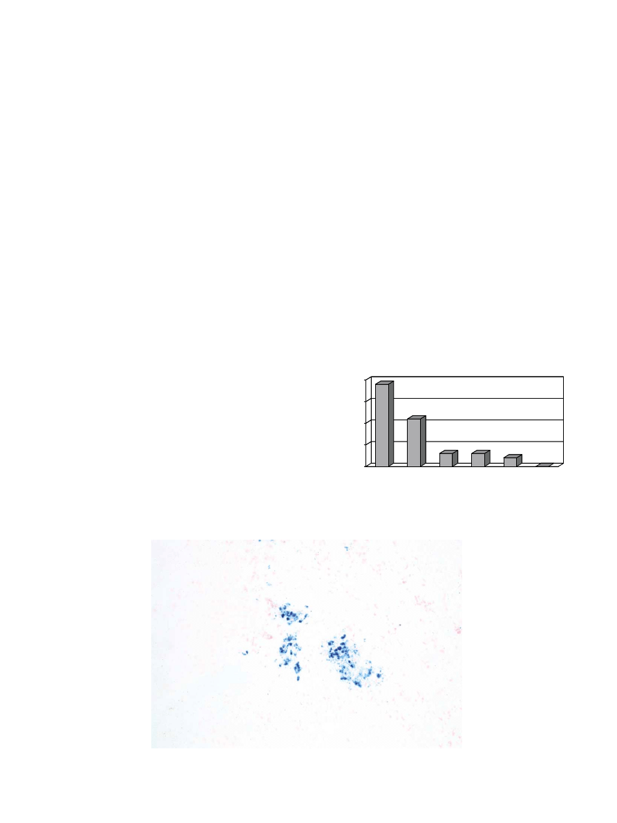
Fdemonstrated by Berlin Blue reaction.
19
11
3
3
2
0
0
5
10
15
20
0
I
I-II
II
II-III
III
Grad
No.Cases
19
11
3
3
2
0
0
5
10
15
20
0
I
I-II
II
II-III
III
Grad
No.Cases
Fig. 2. (Graph). Distribution of the observed cases and their grade of
Fe
+
containing.
K.A. Schlegel et al. / Biomaterials 23 (2002) 2939–2944
2940

quantity of the titanium via beam emission to the
spectrometer. The ICP-spectrometer JY38P used was
produced by Instruments S.A., Jobin-Yvon. The stimu-
lating unit came from Plasma-Therm. The maximum
power of the high-frequency generator was 1.5 kW,
frequency 27.12 MHz. The spectrometer is thermostati-
sized and combined with a Czerny Turner model with
1 m beam focus and a holographic net of 2400 lines/mm.
The spectral analysis potential is 0.02 mm. The mono-
chromatic analysis was carried out with a PDP-11/03
calculator system.
3. Results
In all cases, iron-containing intracellular pigments
were evident by staining with the Berlin Blue reactions
(Figs. 1 and 2). There were also black particles of
varying sizes (Fig. 3). The lack of a repulse reaction in
the tissue around these foreign bodies was obvious.
Signs of inflammation characterized by macrophages,
lymphocytes and plasma cells around these irritants
were mostly mild [21,4,22] (Figs. 4–6). In the epithelium,
an orthokeratotic reaction was the norm (Fig. 7). There
were also keratohyaline granules and at the surface
there were keratinous cells without nuclei. Some cases
were with isolated epitheliae islets. A possible correla-
tion was sought between implant types, delay after
insertion and the level of inflammation (Figs. 8–10). The
inflammation was graded based on the cellular elements:
Grade I=none, Grade II=low grade infiltration, Grade
III=medium grade infiltration and Grade IV=high
amount of cell infiltration.
4. Discussion
The high biocompatability of titanium is well known
[19,11]. While titanium alloys such as Ti6A14V are used
in USA [14], Europeans prefer pure titanium which was
the material used in our study [2]. Corrosion products
from metallic elements are often an irritating factor after
implantation [23]. Due to the ceramic-like layer (rutile)
on pure titanium surfaces, corrosion-related problems
have not been published regarding such pure titanium
implants. Since our samples were collected at least 2.4
months after implantation, no primary wound-healing
reactions are still to be expected. Instead, we find only
material-related typical reactions. In general, a correla-
tion between levels of inflammation, titanium concen-
tration and insertion time did not exist. This illustrates
the good bio-acceptance of pure titanium. Also, Perren
[24–26] did not see such a correlation, when he counted
the number of macrophages, leucocytes, neutrophilic
granulocytes and the granuloma like chronical inflam-
matory reactions. The levels of inflammation observed
by us and related to the different implant products are
nearly equal to those of lTI (Straumann, Switzerland)
Fig. 3. Foreign particles grade II–III in the covering soft tissue excision (magn. 4 ).
1
5
7
8
10
7
0
0
5
10
0
0-I
I
I-II
II
II-III
III
Grade
No.cases
Fig. 4. (Graph). Distribution of the found chronical inflammations of
the periimplant soft tissues according to grades.
K.A. Schlegel et al. / Biomaterials 23 (2002) 2939–2944
2941
1.7, HaTi (Matthys, Switzerland) 1.8 and Branemark
(Nobelbiocare, Sweden) 1.3 despite the impressive
surface differences. The soft tissue flap above the
implants may not have covered the implant diameter
completely over the observation time.
Small leaks are often not clinical evident but
can cause a mild chronic infection. Regarding the
epithelial reaction of the flaps, there were mostly
orthokeratotic cell structures but also a few medium-
sized epithelial hyperplasias which could be linked
to such leaks. The titanium concentration in the
biopsies varied widely (Figs. 9 and 10). The mean
value in mg/kg biopsy material depended on the
type of implant inserted. The concentration of titanium
in tissues surrounding titanium implants is found in
bone with
o2100 by Ducheyne 1984, in soft tissues with
56–3700 by Agius 1988 (cit. 4). In rabbit tissue a
titanium concentration of 17.4 plus/11.4 minus is
normal [13].
Fig. 5. Inflammatory reaction grade I–III, zones of granulation tissue rich in capillaries and fibroblasts (magn. 40 ).
Fig. 6. Fibrosis grade III as a result of chronic inflammatory reaction (magn. 40 ).
13
1
1
0
0
5
10
15
I
II
II-III
III
Grade
No.cases
Fig. 7. (Graph). Orthoceratotic reactions of the periimplant tissues.
K.A. Schlegel et al. / Biomaterials 23 (2002) 2939–2944
2942

0
1
2
3
0
2
4
6
8
10
12
14
16
18
HaTi
Branemark
ITI
grades of imflammation (1-3)
t since insertion in months
Fig. 8. (Graph). Correlation between implant types, time since insertion and level of inflammation found in the soft tissues.
0
200
400
600
800
1000
1200
1400
1600
1800
0
2
4
6
8
10
12
14
16
18
20
HaTi
Branemark
ITI
titanium concentration in mg / kg biopsy
time since insertion
Fig. 9. (Graph). Correlation between implant types, time since insertion and titanium concentration found in the surrounding soft tissues.
0
1
2
3
0
200
400
600
800
1000
1200
1400
1600
1800
2000
HaTi
Branemark
ITI
grades of inflammation (1-3)
titanium concentration in mg/kg material
Fig. 10. (Graph). Correlation between implant types, time since insertion and titanium concentration found in the surrounding soft tissues.
K.A. Schlegel et al. / Biomaterials 23 (2002) 2939–2944
2943

In HaTi (15 cases) the mean finding was 322.19 mg, in
ITI material (4 cases) 127.28 mg and in Branemark (19
cases) a mean of 290.11 mg was measured (Fig. 9). Since
the ITI surface is improved nearly 10-fold due to the
plasma flame spraying, it is difficult to explain this series
of average titanium content. But we must take into
consideration the fact that only the surface diameter was
in contact with the biopsied areas.
Variations in titanium content could be a result of
mechanical irritations inflicted upon implants by steel
instruments during insertion [27], an explanation also
offered in another context by Fischer-Brandies et al. [8]
and Schliephake et al. [9].
We must assume that the implant-covering flap was
never absolutely immobile and the existing small move-
ment may have led to an eraser-like effect which
impregnated the tissue continuously with titanium.
Since the titanium transfer to the muco-periosteal flap
during insertion is only a minor possibility due to the
standardized surgical technique, the ‘‘eraser’’ effect may
be the only remaining explanation.
References
[1] Williams DF. Biocompatibility of clinical implant materials. New
York: CRC Press, 1971.
[2] Steinemann S. Werkstoff Titan. In: Schroeder A, Sutter F,
Krekeler G, editors. Orale Implantologie. Stuttgart: Thieme, 1988.
[3] Osborn JF, Kovacs E, Kallenberger A. Hydroxylapatitkeramik-
Entwicklung eines neuen Biowerkstoffes und erste tierexperimen-
telle Ergebnisse. Dtsch Zahn
.arztl Z 1980;35:54–6.
[4] Anderson JM, Miller KM. Biomaterial, biocompatibility and the
macrophage. Biomaterials 1984;5:5–10.
[5] Donath K, Kirsch, A. Welche Bedeutung hat die prim
.are
Stabilisation von Implantaten f
.ur die oss.are Integration w.a
hrend der Einheilphase? Dtsch Z Zahn
.arztl Implantol II 1986:
11–17.
[6] Kasemo B. Biocompatibility of titanium implants: surface science
aspects. J Prosthet Dent 1983;49:832–7.
[7] Ferguson AB, Akahoshi Y, Lang P, Hodge ES. Trace metal ion
concentration in liver, kidney, spleen and lung of normal rabbits.
J Bone Jt Surg 1962;44:317–22.
[8] Fischer–Brandies E, Zeintl W, Schramel P, Brenner K-U. Zur
Frage der Gewebebelastung mit Titan nach Schraubenosteo-
synthese. Dtsch Z Mund-Kiefer-Gesichts-Chir 1993;17:93–4.
[9] Schliephake H, Reiss J, Urban R, Neukam FW, Guenay H.
Freisetzung von Titan aus Schraubenimplantaten. Dtsch Z
Zahn
.arztl Implantol 1991;7:6–10.
[10] Bagnall RD. An approach to the soft tissue/synthetic material
interface. J Biomed Mater Res 1977;11:939–46.
[11] Donath K. Klinische und histopathologische Befunde im
Implantatlagergewebe bei Titan Implantaten. ZWR 1987;96:
14–6.
[12] Ferguson AB, Laing P, Hodge ES. The ionization of metal
implants in living tissues. J Bone Jt Surg 1960;42:77–90.
[13] Laing PG, Ferguson AB, Hodge ES. Tissue reaction in rabbit
muscle exposed to metallic implants. J Biomed Mater Res 1967;1:
135–49.
[14] Parr GR, Gardner LK, Toth RW. Titanium: the mystery metal of
implant dentistry dental material aspects. J Prosthet Dent
1985;54:410–4.
[15] Rae T. A study on the effects of particulate metals of orthopaedic
interest on murine macrophages in vitro. J Bone Jt Surg 1975;
57:444–50.
[16] Rae T. The biological response to titanium and titanium–
aluminium-vanadium
alloy
particles.
Biomaterials
1986;7:
30–6.
[17] Riede UN, Ruedi T, Limacher F. Quantitative und morpholo-
gische Erfassung der Gewebereaktion auf Metallimplantate
(Osteosynthesematerial). Arch Orthop Unfall–Chir 1974;79:
205–15.
[18] Schramel P, Klose B-J, Hasse S. Die Leistungsf
.ahigkeit der ICP-
Emissionsspektroskopie zur Bestimmung von Spurenelementen in
biologisch-medizinischen und in Umweltproben. Fresenius Z
Anal Chem 1982;310:209–16.
[19] Branemark, et al. Osseointegrated implants in the treatment of the
edentulous jaw: experience from a year period. Stockholm:
Almquist u. Wiksell, 1977.
[20] Gotfredsen K, Budtz–Joergensen E, Nimb Jensen L. A method
for preparing and staining histological sections containing
titanium implants for light microscopy. Stain Technol 1989;
64:121–7.
[21] Adams DO. The granulomatous inflammatory response. Am J
Pathol 1976;84:164–91.
[22] Coleman D, King RN, Andrade JD. The foreign body reaction: a
chronic inflammatory response. J Biomed Mater Res 1974;8:
199–211.
[23] Barbosa MA. Corrosion of Metallic Implants. In: Handbook of
Biomaterial Properties. London, Weinheim, New York, Tokyo,
Melbourne, Madras: Chapman & Hall, 1998.
[24] Perren SM. A dynamic compression plate. Acta Orthop Scand
1969;125:7–16.
[25] Donath K, Laass M, Guenzl H-J. The histopathology of different
foreign-body reactions in oral soft tissue and bone tissue.
Virchows Arch A 1992;420:131–7.
[26] Strassl H. Experimentelle Studie
.uber das Verhalten von
titanbeschichteten Werkstoffen hinsichtlich der Gewebskom-
patibilit
.at im Vergleich zu anderen Metallimplantaten. .Ost Z
Stomat 1978;75:82–98.
[27] Weber H, Sauer K-H, Geis-Gerstorfer J, Kratzenstein B. Zur
Metallaufnahme
durch
implantologische
und
prothetische
Ma
nahmen. Z Zahn
.arztl Implantol 1986;2:6–67.
K.A. Schlegel et al. / Biomaterials 23 (2002) 2939–2944
2944
Document Outline
Wyszukiwarka
Podobne podstrony:
soft tissue2
Tissues
sprawko soft start
szko IO soft ppt
Topic 13 AHL Plants IB III Lecture 2 Plant Tissues and Organs
Plant Tissues3
mFAQ 2 13 Instalacja polskiej wersji językowej oprogramowania LOGO! Soft Comfort
TISSUE REPAIR-Podstawowe fazy procesu gojenia tkanek miękkich, biomechanika(2)
Nye Soft power id 325343 Nieznany
Burroughs The Soft Machine
skład chemia MULTI-SURFACE SOFT CLEANSER
tpl soft manual
więcej podobnych podstron