
Chemico-Biological Interactions 184 (2010) 212–217
Contents lists available at
Chemico-Biological Interactions
j o u r n a l h o m e p a g e :
w w w . e l s e v i e r . c o m / l o c a t e / c h e m b i o i n t
Role of hydroquinone–thiol conjugates in benzene-mediated toxicity
Serrine S. Lau
, Christopher L. Kuhlman
, Shawn B. Bratton
, Terrence J. Monks
a
Southwest Environmental Health Sciences Center, Department of Pharmacology and Toxicology, The University of Arizona Health Sciences Center, Tucson,
AZ 85721, United States
b
Division of Pharmacology & Toxicology, College of Pharmacy, The University of Texas at Austin, Austin, TX 78712, United States
c
Institute for Cellular and Molecular Biology, The University of Texas at Austin, Austin, TX 78712, United States
a r t i c l e i n f o
Article history:
Available online 23 December 2009
Keywords:
Hydroquinone–thiol conjugates
Hematotoxicity
Myleotoxicity
Protein modifications
Covalent adduction
a b s t r a c t
Hydroquinone (HQ) is a metabolite of benzene, and in combination with phenol (PHE), reproduces ben-
zene myelotoxicity. HQ readily oxidizes to 1,4-benzoquinone (1,4-BQ) followed by the reductive addition
of glutathione (GSH). Subsequent cycles of oxidation and GSH addition give rise to a variety of mono-, and
multi-GSH substituted conjugates. Following administration of PHE/HQ (1.1 mmol/kg/0.9 mmol/kg, ip)
to male Sprague–Dawley (SD) rats, 2-(glutathion-S-yl)HQ [GS-HQ], 2,5-bis-(glutathion-S-yl)HQ [2,5-GS-
HQ], 2,6-bis-(glutathion-S-yl)HQ [2,6-GS-HQ], and 2,3,5-tris-(glutathion-S-yl)HQ [2,3,5-GS-HQ] were all
identified in bone marrow. 2-(Cystein-S-ylglycine)HQ [2-(CysGly)HQ], 2-(cystein-S-yl)HQ [2-(Cys)HQ],
and 2-(N-acetylcystein-S-yl)HQ [2-(NACys)HQ] were also found in the bone marrow of PHE/HQ and ben-
zene treated rats and mice, indicating the presence of an active mercapturic acid pathway within bone
marrow. Moreover, 2,6-GS-HQ and 2,3,5-GS-HQ were hematotoxic when administered to rats. All of
the HQ–GSH conjugates retain the ability to redox cycle and generate reactive oxygen species (ROS),
and to arylate target proteins. Recent in vitro and in vivo studies in our laboratory revealed lysine and
arginine residues as primary targets of 1,4-BQ, GS-HQ and 2-(NACys)HQ adduction. In contrast 1,4-BQ-
adduction of cysteine residues may be a transient interaction, where physiological conditions dictate
adduct stability. The generation of ROS and alkylation of proteins may both contribute to benzene-
mediated myelotoxicity, and the two processes may be inter-dependent. However, the precise molecular
mechanism by which benzene and HQ–GSH conjugates induce hematotoxicity remains to be determined.
Within 18 h of administration of PHE/HQ to SD rats a significant decrease in blood lymphocyte count was
observed. At this early time point, erythrocyte counts and hemoglobin concentrations remained within
the normal range. Concomitant with the decrease in lymphocyte count, western blot analysis of bone
marrow lysate, using HQ–GSH and 4-hydroxy-2-nonenal (4HNE) specific antibodies, revealed the pres-
ence of HQ–GSH- and 4HNE-derived protein adducts. Identification of these adducts is required before
the functional significance of such protein modifications can be determined.
© 2009 Elsevier Ireland Ltd. All rights reserved.
Abbreviations: 2-BrHQ-NAC, 2-bromo-6-(N-acetylcystein-S-yl)hydroquinone;
1,4-BQ, 1,4-benzoquinone; ECL, enhanced chemiluminesence; EPO, erythropoi-
etin;
␥-GT, ␥-glutamyl transpeptidase; GS-HQ, 2-(glutathion-S-yl)HQ; 2,5-GS-HQ,
2,5-bis-(glutathion-S-yl)HQ; 2,6-GS-HQ, 2,6-bis-(glutathion-S-yl)HQ; 2,3,5-GS-
HQ,
2,3,5-tris-(glutathion-S-yl)HQ;
2-(CysGly)HQ,
2-(cystein-S-ylglycine)HQ;
2-(Cys)HQ,
2-(cystein-S-yl)HQ;
2-(NACys)HQ,
2-(N-acetylcystein-S-yl)-
hydroquinone; 4HNE, 4-hydroxy-2-nonenal; HQ, hydroquinone; PHE, phenol;
QT, quinol–thioether; ROS, reactive oxygen species.
∗ Corresponding author at: Southwest Environmental Health Sciences Center,
Department of Pharmacology and Toxicology, College of Pharmacy, University of
Arizona, P.O. Box 210207, 1703 E. Mabel Street, Tucson, AZ 85721, United States.
Tel.: +1 520 626 460; fax: +1 520 626 6944.
E-mail address:
(S.S. Lau).
1. Introduction
Benzene, a major industrial chemical and environmental pollu-
tant, causes a variety of hematological disorders in man, including
aplastic anemia, myelodysplastic syndrome, and acute myeloge-
nous leukemia. While benzene must be metabolized to yield its
hematotoxic and leukemogenic effects, no single metabolite of ben-
zene reproduces these effects in vivo. Co-administration of PHE and
HQ, however, does lead to myelotoxicity in rodents
. A phar-
macokinetic interaction between these two metabolites results
in increased concentrations of both metabolites in bone mar-
row
. Peroxidase and/or phenoxy-radical mediated oxidation
of HQ then theoretically initiates redox cycling and formation of
the reactive electrophile, 1,4-BQ, which is considered to be one
of the ultimate hematotoxic metabolites of benzene
. 1,4-
BQ is an electrophile, and covalent interactions of quinones with
0009-2797/$ – see front matter © 2009 Elsevier Ireland Ltd. All rights reserved.
doi:

S.S. Lau et al. / Chemico-Biological Interactions 184 (2010) 212–217
213
nucleophilic sites within cellular macromolecules may contribute
to the toxic effects of benzene
. Indeed, the combined
treatment of PHE and [
14
C]-HQ increases myelotoxicity with con-
comitant increases in covalently bound radiolabel in blood and
bone marrow
. Moreover, elevated levels of benzene oxide
and HQ-derived (1,4-BQ?) adducts of hemoglobin and albumin
have been observed in workers subjected to benzene exposure
Focusing on cysteine-targeted protein adducts, McDonald et al.
reported that 1,4-BQ protein binding was favored over benzene
oxide in mouse bone marrow. Although modification on selective
target proteins occurs in bone marrow of mice following treatment
with [
14
C]-benzene
, the exact nature of the adducted metabo-
lite(s) and/or the specific site of adduction on target proteins are not
known. Of particular relevance to the ability of benzene to induce
aneuploidy, and other forms of chromosomal aberrations, histones
were identified as potential targets of unknown reactive benzene
metabolites
. 1,4-BQ readily conjugates with glutathione (GSH)
to give 2-GS-HQ, 2,3-GS-HQ, 2,5-GS-HQ, 2,6-GS-HQ and 2,3,5-GS-
HQ
. Moreover, HQ–GSH conjugates are present in the bone
marrow of rats and mice following co-administration of PHE/HQ
and metabolized to more reactive cysteinylgylcine and cys-
teine conjugates via the mercapturic acid pathway in bone marrow.
Because HQ–thioether metabolites have an enhanced capacity to
both redox cycle [Monks et al. (this issue)] and arylate tissue
macromolecules
, we suggest that they play an important
role in benzene-mediated toxicity via a mechanism involving the
production of ROS and/or macromolecular arylation. Interestingly,
lysine residues appear to be a preferred target of quinone–thiother
adduction
. ROS produced as a result of HQ–thioether redox
cycling are also capable of oxidatively modifying both proteins and
DNA thereby producing toxicity. Herein we report the presence of
HQ–thioether and 4HNE-derived protein adducts following in vivo
administration of PHE/HQ to rats. These two inter-dependent path-
ways of protein modification may contribute to benzene induced
myleotoxicity.
2. Materials and methods
2.1. Materials
HQ and PHE were purchased from Sigma–Aldrich (St. Louis, MO).
Cell Lysis Buffer (10
×) was purchased from Cell Signaling Technol-
ogy (Danvers, MA). Complete Protease Inhibitor Cocktail Tablets
were purchased from Roche (Madison, WI). Antibody sources were
as follows: rabbit anti-2-Br-6-(N-acetylcystein-S-yl)hydroquinone
(anti-2-BrHQ-NAC) in-house
; anti-4HNE antibody was a gen-
erous gift from Dr. Dennis R. Petersen (University of Colorado
Health Sciences Center) produced and characterized as previously
described
; peroxidase labeled goat anti-rabbit IgG, Vec-
tor Laboratories (Burlingame, CA). Enhanced chemi-luminescent
reagent (ECL) and Hyperfilm ECL were purchased from Amersham
Life Science (Arlington Heights, IL). Acivicin was obtained from
Sigma.
2.2. Animals
Male and female SD rats (n = 2–4@ 2–5 months) were pur-
chased from Harlan Sprague–Dawley (Houston, TX) and used for
all experiments. All animals were housed on a 12 h light/dark cycle
and allowed food and water ad libitum. Blood samples were col-
lected in EDTA coated micro-cuvettes via the retro-orbital sinus
and cardiocentesis. Consistent with humane practices, animals
were anesthetized via pentobarbital injection prior to blood col-
lection. Blood samples were submitted to University Animal Care
Pathology Services (Tucson, AZ) for complete blood count analy-
sis.
2.3. Treatment protocols and isolation of bone marrow
Male
and
female
SD
rats
were
co-administered
PHE
(1.1 mmol/kg) and HQ (0.9 mmol/kg) and dissolved in a vehi-
cle consisting of 0.85% phosphate-buffered saline (PBS):ethanol
(60:40). Protective clothing was used and proper ventilation
ensured to limit exposure to HQ, a potential carcinogen. After 18 h
animals were euthanized by pentobarbital overdose, and each
femur was quickly removed, cleansed of muscle, and placed on
ice. The epiphyseal plates of each femur were then removed, and
the marrow was flushed with 5 mL of ice cold 0.85% PBS, pelleted
under centrifugation (Eppendorf, Model 5804R) for 10 min at
13,500 rpm, and suspended in Cell Lysis Buffer with protease
inhibitor. The cell suspensions were quickly probe sonicated for
30 s on ice, freeze–thawed three times, and centrifuged (Eppendorf,
Model 5415R) for 10 min at 13,500 rpm. The supernatant was then
removed and stored at
−80
◦
C.
␥-Glutamyl transpeptidase (␥-GT)
activity and total protein levels were determined as previously
described
. One unit of
␥-GT activity is defined as 1 mol of
p-nitroanilide formed/min at 37
◦
C.
2.4. 1D western analysis
Proteins (40
g) and protein standards were loaded at 120 V
(constant) through a 3% (w/v) acrylamide stacking gel and resolved
at 140 V through the 10% (w/v) acrylamide resolving gel. Proteins
were transferred to PVDF electrophoretically
and duplicate
gels were stained with 0.05% (w/v) Imperial Blue (Pierce, Rockford,
IL). Blots were incubated overnight at 4
◦
C with either affinity-
purified rabbit anti-BrHQ-NAC antibodies diluted 1:20 in TBST
or affinity purified rabbit anti-4HNE antibodies diluted 1:5000 in
TBST. We have demonstrated previously that anti-2-BrHQ-NAC
antibodies detect in vivo covalent protein adducts of 2-BrHQ, HQ,
and their corresponding GSH conjugates
. Additionally the anti-
4HNE antibodies were characterized in Dr. Petersen’s laboratory
. Blots were incubated with goat anti-rabbit IgG (HRP-labeled)
diluted 1:3000 in TBST for 1 h at room temperature, washed and
then incubated for 1 min in ECL solution. Finally blots were exposed
to Hyperfilm ECL for 1–5 min, and the stained duplicate gels were
aligned with the western blot. The immunopositive bands from
the parallel Imperial Blue stained gel will be subjected to in-gel
digestion followed by LC–MS/MS for protein identification using
established protocol
2.5. Cell lines and culture conditions
HL-60 cells were maintained in RPMI 1640 medium (Gibco BRL,
Grand Island, NY) containing 20% fetal bovine serum (FBS) in a 37
◦
C,
5% CO
2
regulated incubator. Cells were routinely cultured at a den-
sity of 1.0
× 10
6
cells/mL. Immediately prior to all experiments, cells
were washed and resuspended in RPMI 1640 containing 25 mM
HEPES and 10% FBS.
2.6. Serum erythropoietin determination
The RIA kit for erythropoietin (EPO) determination (DSL Inc.,
Webster, TX) was a competitive binding assay using a radiolabeled
ligand. Standards and samples (100
L) were processed accord-
ing to the manufacturer’s suggested protocol and analyzed on a
␥-counter (1282 CompuGamma, LKB Wallac). EPO concentrations
were determined by comparing the DPM of a given sample with
those of known standards.
214
S.S. Lau et al. / Chemico-Biological Interactions 184 (2010) 212–217
Table 1
Effect of phenol/hydroquinone on blood lymphocyte counts.
Lymphocyte count (K/
L)
% decrease
Phenol/HQ treated
Rat 1
0.51
86
Rat 2
1.57
58
Rat 3
0.42
89
Rat 4
1.53
59
Control
Rat 1
4.18
Rat 2
3.22
a
Normal range of rat lymphocytes: 3.8–15.3 (K/
L).
3. Results
3.1. Effect of blood counts after phenol/hydroquinone
administration
Complete blood counts of rats 18 h following co-administration
of PHE/HQ (1.1/0.9 mmol/kg, ip) to rats revealed significant reduc-
tion (58–89%) of lymphocyte counts when compared to control
values (
). All other leukocyte and erythrocyte counts were
within their normal ranges following treatment.
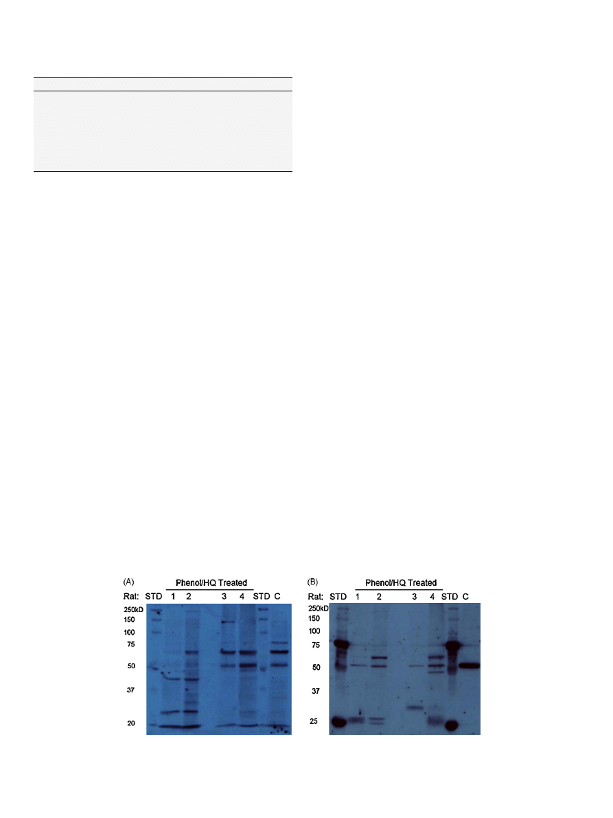
3.2. 1D western detection of HQ–GSH/4HNE-protein adducts in
bone marrow
It
should
be
noted
that
immediately
following
co-
administration of phenol/hydroquinone, a transient (15–20 min)
neurological side-effect is observed. Interestingly the intensity of
this side-effect correlates with the intensity of protein immuno-
staining in the western blot analyses. Such correlations indicate
that
inter-individual
differences
in
absorption/distribution
(revealed in the neurological response “bioassay”) correlate with
inter-individual differences in covalent binding and toxicity. It is
therefore essential to present data from each individual animal in
. Unique GS-HQ-immunopositive bands (26, 43, and 140 kD)
were detected in bone marrow protein of rats co-administered
HQ/PHE. GS-HQ-immunopositive bands, at 50 and 65 kD, were
detected in bone marrow protein from both treated and untreated
control male rats (
A). 4HNE immunopositive bands (25, 27,
43 and 59 kD) were detected in bone marrow protein of treated
rats (
B). The 50 kD protein band that was immunopositive
for HQ–thioether adduction in both control and treated rats
also appeared to be immunopositive for 4HNE. Interestingly, the
50 kD 4HNE immunopositive band is of much stronger inten-
sity in untreated bone marrow protein relative to the similar
protein from rats co-administered PHE/HQ (
B). The immuno-
reactive bands from control rat represent endogenously modified
protein(s), possibly derived from dietary sources.
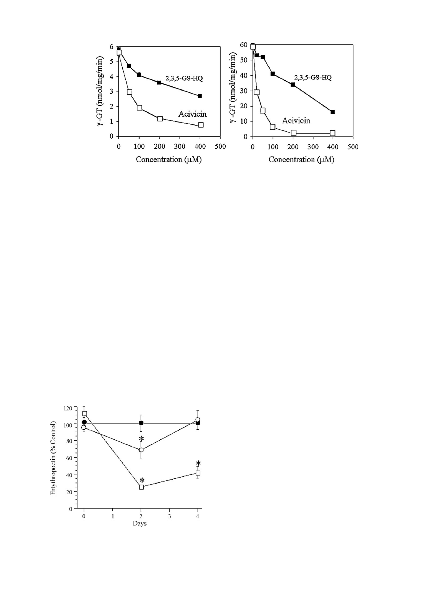
3.3. Inhibition of
-GT
A key enzyme in the GSH cycle,
␥-GT is involved in salvaging
cysteine. Since cysteine availability is rate limiting in the for-
mation of GSH, inhibition of
␥-GT enhances the susceptibility of
cells to oxidative damage. 2,3,5-GS-HQ inhibits
␥-GT in freshly
isolated bone marrow homogenates and HL-60 cell lysates, in a
concentration-dependent manner (
). Acivicin was used as a
positive control and Triton X-100 treated homogenates as a neg-
ative control. Acivicin, a potent inhibitor of
␥-GT, also induces
hematotoxicity in humans and rats
. Thus, inhibition of
␥-
GT in hematopoietic tissue dramatically reduces intracellular GSH
levels
, and TGHQ depletes GSH in HL-60 cells and induces
apoptosis in an ROS-dependent manner
. HQ–thioethers might
therefore adversely affect cellular GSH stores thereby exacerbating
the effects of ROS generation in bone marrow.
3.4. Reactivity of HQ–thioethers: effect on plasma erythropoietin
Erythrotoxicity of several of these conjugates was determined
in rats using the erythrocyte
59
Fe incorporation assay
istration of 2,3,5-GS-HQ (17
mol/kg, iv), 2,6-GS-HQ (50 mol/kg,
iv), and benzene (11 mmol/kg, sc) significantly decreased
59
Fe
incorporation into reticulocytes to 45
± 6%, 28 ± 3%, and 20 ± 9% of
control values, respectively. Although the doses of 2,3,5-GS-HQ and
2,6-GS-HQ represented only 0.2% and 0.4% of the dose of benzene,
both conjugates reduced
59
Fe incorporation to the same degree
as benzene. These data suggest that HQ–GSH conjugates are ery-
throtoxic and may contribute to benzene-mediated hematotoxicity
. Because HQ–GSH conjugates are capable of damaging cells
within the proximal tubules of the kidney
, the major site of
EPO production, we questioned whether 2,3,5-GS-HQ and 2,6-GS-
HQ might also be capable of indirectly inducing anemia by reducing
serum EPO levels. Consistent with this reasoning, both 2,3,5-GS-HQ
and 2,6-GS-HQ significantly reduced circulating EPO levels with
levels returning to control values at 4 days in animals treated with
2,3,5-GS-HQ (
). Two possibilities exist for the recovery of EPO
levels with continued dosing. Firstly the % of reduction is modest
(30% of control), and stimulation of the repair response in the kid-
ney may be proceeding between days 2 and 4. Alternatively, the
remaining healthy cells may well be compensating to the transient
decrease by increasing EPO synthesis. The liver might also produce
EPO if the kidneys are compromised. However when the damage to
Fig. 1. 1D Western blotting of protein adducts in bone marrow. (A) HQ–GSH adducts and (B) 4-HNE adducts in rat bone marrow following co-administration of phenol
(1.1 mmol/kg) and hydroquinone (0.9 mmol/kg). Lane 1,2 and 3,4 are phenol/HQ treated female and male SD rats, respectively; STD, ladder; C, vehicle treated control male
rat.
S.S. Lau et al. / Chemico-Biological Interactions 184 (2010) 212–217
215
Fig. 2. The effect of 2,3,5-GS-HQ on
␥-GT activity in bone marrow (left) and HL-60 cell (right) lysates. Bone marrow was flushed from the femurs of male Sprague–Dawley
rats with 1 ml of Tris/KCl (20 mM/1.15%) containing 1% Triton X-100. The marrow was homogenized, incubated on ice for 30 min, and centrifuged at 800
× g for 10 min. The
supernatant was used as the enzyme source. (
) 2,3,5-GS-HQ; () acivicin.
kidney is too severe, as in the case following 2,6-GS-HQ administra-
tion the recovery in EPO was minimal, indicating that the kidneys
are the primary organ for EPO production.
4. Discussion
HQ–thioethers are present in the bone marrow of rats follow-
ing co-administration of PHE/HQ, the majority of which appear
to be generated in situ and further metabolized via the mercap-
turic acid pathway
. This pathway is important in modulating
the reactivity of HQ–GSH conjugate. Based on the (re)activity of
HQ–GSH conjugates, we speculated that some of the hematotoxic
effects attributed to HQ (or 1,4-BQ) may, in fact, be mediated by
their thiol conjugates. Indeed, as shown by Monks et al (this issue),
HQ–GSH conjugates are far more efficient generators of superox-
ide anion than the HQ/1,4-BQ redox couple. Moreover, HQ–GSH
conjugates are toxic to developing erythrocytes in vivo
Whether this is a direct effect of these conjugates in bone marrow,
or whether additional tissues/organs contribute to the observed
erythrotoxicity is not known. The hematopoietic microenviron-
ment is regulated by stromal cells which secrete a variety of
cytokines, including interleukin-1 (IL-1), tumor necrosis factor-
␣, granulocyte-macrophage colony-stimulating factor (GM-CSF),
granulocyte colony-stimulating factor (G-CSF), and macrophage
Fig. 3. Effect of HQ–GSH conjugates on rat serum erythropoietin levels. Rats were
given daily doses of either (
䊉) saline; () 2,3,5-GS-HQ (10 mol/kg, i.v.) or; () 2,6-
GS-HQ (50
mol/kg, i.v.). Each point represents the mean ± SEM (n = 5). One-way
ANOVA; SNK.
colony-stimulating factor (M-CSF). In addition, kidney peritubu-
lar cells produce and secrete the hormone, erythropoietin (EPO)
. These growth factors interact with various hematopoietic
stem/progenitor cells to control cellular proliferation and differen-
tiation. Because HQ–GSH conjugates are capable of damaging cells
within the proximal tubules of the kidney
, the major site of
EPO production, we hypothesized that 2,3,5-GS-HQ and 2,6-GS-HQ
might also be capable of indirectly inducing anemia by reducing
serum EPO levels. Consistent with this reasoning, both 2,3,5-GS-HQ
and 2,6-GS-HQ caused reductions in circulating EPO levels (
We have previously shown that quinone–thioethers are “erythro-
toxic” based on a reduction in iron uptake
. However, EPO
drives RBC production and consequently the incorporation of iron.
It is therefore possible that the reduced iron uptake involved both
decreased production of EPO as well as direct toxicity to exist-
ing RBCs. In this report we have shown that quinone–thioethers
induced a reduction in circulating lymphocytes, consistent with
bone marrow toxicity, as previously reported
. However a pos-
sible redistribution of lymphocytes from the peripheral blood into
tissues cannot be ruled out.
Adduction of proteins by reactive electrophiles is not a ran-
dom event, but rather specific proteins appear to be targeted.
These protein targets may differ between different electrophiles,
as illustrated with HQ–GSH and 4HNE modified proteins (
but may also exhibit overlap (e.g. the 50 kD proteins). Chemical
structure, reactivity, and ability to localize within the various intra-
cellular compartments are several characteristics that likely govern
which proteins are targeted by a given electrophile. We have pro-
vided evidence in support of the existence of “electrophile binding
motifs” (EBMs) within proteins in the kidney that are selectively
adducted by reactive electrophiles following GS-HQ administration
. Whether similar EBMs exists within the bone marrow adduc-
tome deserves attention. With respect to the reactive metabolites
derived from GS-HQ, they appear to selectively target (i) proteins
with a high lysine (basic amino acid) content, and specifically (ii)
proteins that contain lysine residues either flanking a potentially
nucleophilic amino acid [KXK], or containing two lysine residues
preceded or followed by a nucleophilic amino acid [XKK or KKX]
. Although we have detected GS-HQ-adducted proteins in bone
marrow of PHE/HQ treated rats (
) the identity of these pro-
teins, and the site(s) of adduction, remain to be determined. One
such target may be
␥-GT. Indeed, HQ–GSH conjugates are sub-
strates for, and inhibit, renal
␥-GT
. Consistent with this view,
2,3,5-GS-HQ inhibits
␥-GT in bone marrow cell lysates and in HL-60
cells (

216
S.S. Lau et al. / Chemico-Biological Interactions 184 (2010) 212–217
Both “free” and protein-bound HQ–GSH conjugates (and
metabolites thereof) retain the ability to redox cycle and generate
ROS. One consequence of which is the initiation of lipid peroxida-
tion. Cysteine thiols are common targets of reactive electrophilic
metabolites, and of endogenous electrophiles, including lipid-
derived
␣,-unsaturated aldehydes, such as 4-hydroxynonenal
(4HNE) and 4-oxononenal (4ONE), and ROS. In addition to the
formation of HQ–GSH-derived protein adducts, PHE/HQ adminis-
tration to rats also results in the formation of 4HNE-protein adducts
(
). Although in most cases, the toxicological significance of
protein adducts remains uncertain, physiological concentrations of
either 4HNE or 4ONE cause the cross-linking of bovine brain tubu-
lin, and an inability of tubulin to polymerize
. 4HNE also reduces
ERK-1/2 phosphorylation causing a loss of activity and of nuclear
localization. Interestingly, the loss of ERK activity is caused by a
single 4HNE modification, on histidine 178. While the precise tox-
icological implication of proteins modified by 4HNE is not known,
recent evidence suggests a role in the pathogenesis of several dis-
eases
Conflict of interest
None.
Acknowledgements
This work was supported by RO1 GM070890 (SSL). The authors
also acknowledge the support of the P30 ES006694 Southwest
Environmental Health Sciences Center, in particular the Arizona
Proteomics Consortium (APC) and Dr. George Tsaprailis, Director
of the APC. The generous gift of anti-4HNE antibody from Dr. Den-
nis R. Petersen at the University of Colorado Health Sciences Center
is greatly appreciated.
References
[1] D.A. Eastmond, M.T. Smith, R.D. Irons, An interaction of benzene metabolites
reproduces the myelotoxicity observed with benzene exposure, Toxicol. Appl.
Pharmacol. 91 (1) (1987) 85–95.
[2] A. Legathe, B.A. Hoener, T.N. Tozer, Pharmacokinetic interaction between ben-
zene metabolites, phenol and hydroquinone, in B6C3F1 mice, Toxicol. Appl.
Pharmacol. 124 (1) (1994) 131–138.
[3] S.J. Pirozzi, M.J. Schlosser, G.F. Kalf, Prevention of benzene-induced myelo-
toxicity and prostaglandin synthesis in bone marrow of mice by inhibitors of
prostaglandin H synthase, Immunopharmacology 18 (1) (1989) 39–55.
[4] D.G. Schattenberg, W.S. Stillman, J.J. Gruntmeir, K.M. Helm, R.D. Irons, D. Ross,
Peroxidase activity in murine and human hematopoietic progenitor cells:
potential relevance to benzene-induced toxicity, Mol. Pharmacol. 46 (2) (1994)
346–351.
[5] R.C. Smart, V.G. Zannoni, DT-diaphorase and peroxidase influence the covalent
binding of the metabolites of phenol, the major metabolite of benzene, Mol.
Pharmacol. 26 (1) (1984) 105–111.
[6] C.J. Smith, Y. Zhang, C.M. Koboldt, J. Muhammad, B.S. Zweifel, A. Shaffer,
J.J. Talley, J.L. Masferrer, K. Seibert, P.C. Isakson, Pharmacological analysis of
cyclooxygenase-1 in inflammation, Proc. Natl. Acad. Sci. U.S.A. 95 (22) (1998)
13313–13318.
[7] D.J. Thomas, A. Sadler, V.V. Subrahmanyam, D. Siegel, M.J. Reasor, D. Wierda, D.
Ross, Bone marrow stromal cell bioactivation and detoxification of the benzene
metabolite hydroquinone: comparison of macrophages and fibroblastoid cells,
Mol. Pharmacol. 37 (2) (1990) 255–262.
[8] R.D. Irons, Quinones as toxic metabolites of benzene, J. Toxicol. Environ. Health
16 (5) (1985) 673–678.
[9] M.J. Schlosser, R.D. Shurina, G.F. Kalf, Metabolism of phenol and hydroquinone
to reactive products by macrophage peroxidase or purified prostaglandin H
synthase, Environ. Health Perspect. 82 (1989) 229–237.
[10] V.V. Subrahmanyam, P. Doane-Setzer, K.L. Steinmetz, D. Ross, M.T. Smith,
Phenol-induced stimulation of hydroquinone bioactivation in mouse bone
marrow in vivo: possible implications in benzene myelotoxicity, Toxicology
62 (1) (1990) 107–116.
[11] Renal toxicology, in: I.G. Sipes, C.A. Maqueen, A.J. Gandolfi (Eds.), Comprehen-
sive Toxicology, Elsevier Science Pub Co., 1997.
[12] V.V. Subrahmanyam, P. Kolachana, M.T. Smith, Metabolism of hydroquinone
by human myeloperoxidase: mechanisms of stimulation by other phenolic
compounds, Arch. Biochem. Biophys. 286 (1) (1991) 76–84.
[13] V.V. Subrahmanyam, D. Ross, D.A. Eastmond, M.T. Smith, Potential role of free
radicals in benzene-induced myelotoxicity and leukemia, Free Radic. Biol. Med.
11 (5) (1991) 495–515.
[14] W.F. Greenlee, J.D. Sun, J.S. Bus, A proposed mechanism of benzene toxicity: for-
mation of reactive intermediates from polyphenol metabolites, Toxicol. Appl.
Pharmacol. 59 (2) (1981) 187–195.
[15] M.T. Smith, J.W. Yager, K.L. Steinmetz, D.A. Eastmond, Peroxidase-dependent
metabolism of benzene’s phenolic metabolites and its potential role in
benzene toxicity and carcinogenicity, Environ. Health Perspect. 82 (1989)
23–29.
[16] R.D. Irons, W.F. Greenlee, D. Wierda, J.S. Bus, Relationship between benzene
metabolism and toxicity. A proposed mechanism for the formation of reactive
intermediates from poly-phenol metabolites, Adv. Exp. Med. Biol. 136A (1982)
229.
[17] K. Yeowell-O’Connell, N. Rothman, M.T. Smith, R.B. Hayes, G. Li, S. Waidyanatha,
M. Dosemeci, L. Zhang, S. Yin, N. Titenko-Holland, S.M. Rappaport, Hemoglobin
and albumin adducts of benzene oxide among workers exposed to high levels
of benzene, Carcinogenesis 19 (9) (1998) 1565–1571.
[18] K. Yeowell-O’Connell, N. Rothman, S. Waidyanatha, M.T. Smith, R.B. Hayes,
G. Li, W.E. Bechtold, M. Dosemeci, L. Zhang, S. Yin, S.M. Rappaport, Protein
adducts of 1,4-benzoquinone and benzene oxide among smokers and non-
smokers exposed to benzene in China, Cancer Epidemiol. Biomarkers Prev. 10
(8) (2001) 831–838.
[19] T.A. McDonald, K. Yeowell-O’Connell, S.M. Rappaport, Comparison of protein
adducts of benzene oxide and benzoquinone in the blood and bone marrow
of rats and mice exposed to [14C/13C6]benzene, Cancer Res. 54 (18) (1994)
4907–4914.
[20] K.E. Williams, T.A. Carver, J.J. Miranda, A. Kautiainen, J.S. Vogel, K. Dingley, M.A.
Baldwin, K.W. Turteltaub, A.L. Burlingame, Attomole detection of in vivo protein
targets of benzene in mice: evidence for a highly reactive metabolite, Mol. Cell.
Proteomics 1 (11) (2002) 885–895.
[21] S.S. Lau, B.A. Hill, R.J. Highet, T.J. Monks, Sequential oxidation and glutathione
addition to 1,4-benzoquinone: correlation of toxicity with increased glu-
tathione substitution, Mol. Pharmacol. 34 (6) (1988) 829–836.
[22] S.B. Bratton, S.S. Lau, T.J. Monks, Identification of quinol thioethers in bone
marrow of hydroquinone/phenol-treated rats and mice and their potential
role in benzene-mediated hematotoxicity, Chem. Res. Toxicol. 10 (8) (1997)
859–865.
[23] H.E. Kleiner, T.W. Jones, T.J. Monks, S.S. Lau, Immunochemical analysis of
quinol–thioether-derived covalent protein adducts in rodent species sensitive
and resistant to quinol–thioether-mediated nephrotoxicity, Chem. Res. Toxicol.
11 (11) (1998) 1291–1300.
[24] M.T. Labenski, A.A. Fisher, H.H. Lo, T.J. Monks, S.S. Lau, Protein electrophile-
binding motifs: lysine-rich proteins are preferential targets of quinones, Drug
Metab. Dispos. 37 (6) (2009) 1211–1218.
[25] A.A. Fisher, M.T. Labenski, S. Malladi, V. Gokhale, M.E. Bowen, R.S. Milleron, S.B.
Bratton, T.J. Monks, S.S. Lau, Quinone electrophiles selectively adduct “elec-
trophile binding motifs” within cytochrome c, Biochemistry 46 (39) (2007)
11090–11100.
[26] H.E. Kleiner, M.I. Rivera, N.R. Pumford, T.J. Monks, S.S. Lau, Immunochemical
detection of quinol–thioether-derived protein adducts, Chem. Res. Toxicol. 11
(11) (1998) 1283–1290.
[27] J.R. Roede, B.J. Stewart, D.R. Petersen, Decreased expression of peroxiredoxin 6
in a mouse model of ethanol consumption, Free Radic. Biol. Med. 45 (11) (2008)
1551–1558.
[28] S.S. Lau, T.J. Monks, J.I. Everitt, E. Kleymenova, C.L. Walker, Carcinogenicity
of a nephrotoxic metabolite of the “nongenotoxic” carcinogen hydroquinone,
Chem. Res. Toxicol. 14 (1) (2001) 25–33.
[29] H. Towbin, T. Staehelin, J. Gordon, Electrophoretic transfer of proteins from
polyacrylamide gels to nitrocellulose sheets: procedure and some applications,
Proc. Natl. Acad. Sci. U.S.A. 76 (9) (1979) 4350–4354.
[30] S. Baruchel, M. Bernstein, V.M. Whitehead, S. Devine, B. Bell, R. Dubowy, H. Grier,
C. Kretschmar, A.M. Langevin, T. Vietti, A phase I study of acivicin in refractory
pediatric solid tumors. A Pediatric Oncology Group study, Invest New Drugs 13
(3) (1995) 211–216.
[31] R.H. Earhart, J.D. Khandekar, D. Faraggi, R.A. Schinella, T.E. Davis, Phase II trial
of continuous drug infusions in advanced ovarian carcinoma: acivicin versus
vinblastine, Invest. New Drugs 7 (2/3) (1989) 255–260.
[32] J.A. Maroun, D.J. Stewart, S. Verma, W.K. Evans, E. Eisenhauer, Phase I study of
acivicin and cisplatin in non-small-cell lung cancer. A National Cancer Institute
of Canada study, Am. J. Clin. Oncol. 13 (5) (1990) 401–404.
[33] J.K. Dethmers, A. Meister, Glutathione export by human lymphoid cells:
depletion of glutathione by inhibition of its synthesis decreases export and
increases sensitivity to irradiation, Proc. Natl. Acad. Sci. U.S.A. 78 (12) (1981)
7492–7496.
[34] D.J. van den Dobblesteen, C.S.I. Nobel, A.F. Slater, S. Orrenius, Regulation and
mechanisms of apoptosis in T lymphocytes, Arch. Toxicol. (Suppl.) 19 (1996)
77–85.
[35] S.B. Bratton, S.S. Lau, T.J. Monks, The putative benzene metabolite 2, 3,
5-tris(glutathion-S-yl)hydroquinone depletes glutathione, stimulates sphin-
gomyelin turnover, and induces apoptosis in HL-60 cells, Chem. Res. Toxicol.
13 (7) (2000) 550–556.
[36] K.L. Blanchard, A.M. Acquaviva, D.L. Galson, H.F. Bunn, Hypoxic induction of the
human erythropoietin gene: cooperation between the promoter and enhancer,
each of which contains steroid receptor response elements, Mol. Cell Biol. 12
(12) (1992) 5373–5385.

S.S. Lau et al. / Chemico-Biological Interactions 184 (2010) 212–217
217
[37] J.E. Groopman, J.M. Molina, D.T. Scadden, Hematopoietic growth factors. Biol-
ogy and clinical applications, N. Engl. J. Med. 321 (21) (1989) 1449–1459.
[38] B.A. Hill, H.H. Lo, T.J. Monks, S.S. Lau, The role of gamma-glutamyl transpep-
tidase in hydroquinone–glutathione conjugate mediated nephrotoxicity, Adv.
Exp. Med. Biol. 283 (1991) 749–751.
[39] B.J. Stewart, J.A. Doorn, D.R. Petersen, Residue-specific adduction of tubulin by
4-hydroxynonenal and 4-oxononenal causes cross-linking and inhibits poly-
merization, Chem. Res. Toxicol. 20 (8) (2007) 1111–1119.
[40] D.R. Petersen, J.A. Doorn, Reactions of 4-hydroxynonenal with proteins and
cellular targets, Free Radic. Biol. Med. 37 (7) (2004) 937–945.
Document Outline
- Role of hydroquinone–thiol conjugates in benzene-mediated toxicity
Wyszukiwarka
Podobne podstrony:
1 s2 0 S000925099800520X main
1 s2 0 S0009250907002394 main
1 s2 0 S0009254115002983 mainid Nieznany
1 s2 0 S0020025512001946 main
1 s2 0 S0378382002000085 main
1 s2 0 S0304397599001000 main
1 s2 0 S0006291X05021595 main
1 s2 0 S0040603111000104 main 2
1 s2 0 S0944501312001358 main
1 s2 0 S0166218X96000583 main
1 s2 0 S0005273614000303 main
1 s2 0 S0304397502001342 main
1 s2 0 S0377221798003622 main (1)
1 s2 0 S0022169496031423 main
1 s2 0 S1046592814002101 main
więcej podobnych podstron