
1
IAEA
International Atomic Energy Agency
This set of 91 slides is based on Chapter 8 authored by
W. Strydom, W. Parker, and M. Olivares
of the IAEA publication
(ISBN 92-0-107304-6):
Radiation Oncology Physics:
A Handbook for Teachers and Students
Objective:
To familiarize students with basic physical and clinical principles of
radiotherapy with megavoltage electron beams.
Chapter 8
Electron Beams: Physical and Clinical Aspects
Slide set prepared in 2006 (updated Aug2007)
by E.B. Podgorsak (McGill University, Montreal)
Comments to S. Vatnitsky:
dosimetry@iaea.org
IAEA
Radiation Oncology Physics: A Handbook for Teachers and Students - 8.1 Slide 1 (2/91)
CHAPTER 8.
TABLE OF CONTENTS
8.1.
Central axis depth dose distributions in water
8.2.
Dosimetric parameters of electron beams
8.3.
Clinical considerations in electron beam therapy

2
IAEA
Radiation Oncology Physics: A Handbook for Teachers and Students - 8.1 Slide 1 (3/91)
8.1 CENTRAL AXIS DEPTH DOSE DISTRIBUTIONS
Megavoltage electron beams
represent an important
treatment modality in modern radiotherapy, often
providing a unique option in the treatment of super-
ficial tumours.
•
Electrons have been used in radiotherapy since the early
1950s.
•
Modern high-energy linacs typically provide, in addition to
two photon energies, several electron beam energies in the
range from 4 MeV to 25 MeV.
IAEA
Radiation Oncology Physics: A Handbook for Teachers and Students - 8.1.1 Slide 1 (4/91)
8.1 CENTRAL AXIS DEPTH DOSE DISTRIBUTIONS
8.1.1 General shape of the depth dose curve
The general shape of the
central axis depth dose curve
for electron beams differs from that of photon beams.

3
IAEA
Radiation Oncology Physics: A Handbook for Teachers and Students - 8.1.1 Slide 2 (5/91)
8.1 CENTRAL AXIS DEPTH DOSE DISTRIBUTIONS
8.1.1 General shape of the depth dose curve
•
The
surface dose
is relatively
high (of the order of 80 - 100%).
•
Maximum dose
occurs at a depth
referred to as the
depth of dose
maximum z
max
.
•
Beyond z
max
the dose drops off
rapidly and levels off at a small
low level dose called the
brems-
strahlung tail
(of the order of a
few per cent).
The
electron beam central axis percentage depth dose
curve
exhibits the following characteristics:
IAEA
Radiation Oncology Physics: A Handbook for Teachers and Students - 8.1.1 Slide 3 (6/91)
8.1 CENTRAL AXIS DEPTH DOSE DISTRIBUTIONS
8.1.1 General shape of the depth dose curve
Electron beams are almost
monoenergetic
as they leave
the linac accelerating waveguide.
In moving toward the patient through:
•
Waveguide exit window
•
Scattering foils
•
Transmission ionization chamber
•
Air
and interacting with photon collimators, electron cones
(applicators) and the patient,
bremsstrahlung radiation is
produced
. This radiation constitutes the bremsstrahlung
tail of the electron beam PDD curve.

4
IAEA
Radiation Oncology Physics: A Handbook for Teachers and Students - 8.1.2 Slide 1 (7/91)
8.1 CENTRAL AXIS DEPTH DOSE DISTRIBUTIONS
8.1.2 Electron interactions with absorbing medium
As the electrons propagate through an absorbing
medium, they interact with atoms of the absorbing
medium by a variety of elastic or inelastic
Coulomb
force interactions
.
These
Coulomb interactions
are classified as follows:
•
Inelastic collisions with orbital electrons of the absorber atoms.
•
Inelastic collisions with nuclei of the absorber atoms.
•
Elastic collisions with orbital electrons of the absorber atoms.
•
Elastic collisions with nuclei of the absorber atoms.
IAEA
Radiation Oncology Physics: A Handbook for Teachers and Students - 8.1.2 Slide 2 (8/91)
8.1 CENTRAL AXIS DEPTH DOSE DISTRIBUTIONS
8.1.2 Electron interactions with absorbing medium
Inelastic collisions between the incident electron and
orbital electrons of absorber atoms
result in loss of
incident electron’s kinetic energy through ionization and
excitation of absorber atoms (
collision
or
ionization loss
).
Absorber atoms can be ionized through two types of
ionization collision:
•
Hard collision
in which the ejected orbital electron gains enough
energy to be able to ionize atoms on its own (these electrons are
called delta rays).
•
Soft collision
in which the ejected orbital electron gains an
insufficient amount of energy to be able to ionize matter on its
own.

5
IAEA
Radiation Oncology Physics: A Handbook for Teachers and Students - 8.1.2 Slide 3 (9/91)
8.1 CENTRAL AXIS DEPTH DOSE DISTRIBUTIONS
8.1.2 Electron interactions with absorbing medium
Elastic collisions between the incident electron and nuclei
of the absorber atoms
result in:
•
Change in direction of motion of the incident electron (elastic
scattering).
•
A very small energy loss by the incident electron in individual
interaction, just sufficient to produce a deflection of electron’s path.
Incident electron loses kinetic energy through cumulative
action of multiple scattering events, each scattering event
characterized by a small energy loss.
IAEA
Radiation Oncology Physics: A Handbook for Teachers and Students - 8.1.2 Slide 4 (10/91)
8.1 CENTRAL AXIS DEPTH DOSE DISTRIBUTIONS
8.1.2 Electron interactions with absorbing medium
Electrons traversing an absorber lose their kinetic energy
through
ionization collisions
and
radiation collisions
.
The rate of energy loss per gram and per cm
2
is called the
mass stopping power and it is a sum of two components:
•
Mass collision stopping power
•
Mass radiation stopping power
The
rate of energy loss
for a therapy electron beam in
water and water-like tissues, averaged over the electron’s
range,
is about 2 MeV/cm
.

6
IAEA
Radiation Oncology Physics: A Handbook for Teachers and Students - 8.1.3 Slide 1 (11/91)
8.1 CENTRAL AXIS DEPTH DOSE DISTRIBUTIONS
8.1.3 Inverse square law (virtual source position)
In contrast to a photon beam,
which has a distinct focus
located at the accelerator x ray
target, an
electron beam
appears to originate from a point
in space
that does not coincide
with the scattering foil or the
accelerator exit window.
The term “
virtual source
position
” was introduced to
indicate the virtual location of
the electron source.
IAEA
Radiation Oncology Physics: A Handbook for Teachers and Students - 8.1.3 Slide 2 (12/91)
8.1 CENTRAL AXIS DEPTH DOSE DISTRIBUTIONS
8.1.3 Inverse square law (virtual source position)
Effective source-surface distance SSD
eff
is defined as
the distance from the virtual source position to the
edge of the electron cone applicator.
The inverse square law may be used for small SSD
differences from the nominal SSD to make cor-
rections to absorbed dose rate at z
max
in the patient for
variations in air gaps g between the actual patient
surface and the nominal SSD.

7
IAEA
Radiation Oncology Physics: A Handbook for Teachers and Students - 8.1.3 Slide 3 (13/91)
8.1 CENTRAL AXIS DEPTH DOSE DISTRIBUTIONS
8.1.3 Inverse square law (virtual source position)
A common
method for determining SSD
eff
consists of
measuring the dose rate at z
max
in phantom for various
air gaps g starting with at the electron cone.
• The following inverse square law relationship holds:
• The measured slope of the linear plot is:
• The effective SSD is then calculated from:
max
(
0)
D
g
=
2
max
eff
max
eff
max
max
(
0)
SSD
SSD
( )
D
g
z
g
z
D
g
=
+
+
=
+
k
=
1
SSD
eff
+
z
max
SSD
eff
=
1
k
+
z
max
IAEA
Radiation Oncology Physics: A Handbook for Teachers and Students - 8.1.3 Slide 4 (14/91)
8.1 CENTRAL AXIS DEPTH DOSE DISTRIBUTIONS
8.1.3 Inverse square law (virtual source position)
Typical example of data measured in determination of
virtual source position SSD
eff
normalized to the edge of
the electron applicator (cone).
SSD
eff
=
1
k
+
z
max

8
IAEA
Radiation Oncology Physics: A Handbook for Teachers and Students - 8.1.3 Slide 5 (15/91)
8.1 CENTRAL AXIS DEPTH DOSE DISTRIBUTIONS
8.1.3 Inverse square law (virtual source position)
For practical reasons the
nominal SSD
is usually a fixed
distance (e.g., 5 cm) from the distal edge of the electron
cone (applicator) and coincides with the linac isocentre.
Although the
effective SSD
(i.e., the virtual electron
source position) is determined from measurements at
z
max
in phantom, its value does not change with change
in the depth of measurement.
The
effective SSD
depends on electron beam energy
and must be measured for all energies available in the
clinic.
IAEA
Radiation Oncology Physics: A Handbook for Teachers and Students - 8.1.4 Slide 1 (16/91)
8.1 CENTRAL AXIS DEPTH DOSE DISTRIBUTIONS
8.1.4 Range concept
By virtue of being surrounded by a Coulomb force field,
charged particles, as they penetrate into an absorber
encounter numerous Coulomb interactions with orbital
electrons and nuclei of the absorber atoms.
•
Eventually, a charged particle will lose all of its kinetic energy
and come to rest at a certain depth in the absorbing medium
called the
particle range
.
•
Since the stopping of particles in an absorber is a statistical
process several definitions of the range are possible.

9
IAEA
Radiation Oncology Physics: A Handbook for Teachers and Students - 8.1.4 Slide 2 (17/91)
8.1 CENTRAL AXIS DEPTH DOSE DISTRIBUTIONS
8.1.4 Range concept
Definitions of particle range: (1)
CSDA range
•
In most encounters between the charged particle and absorber
atoms the energy loss by the charged particle is minute so that it
is convenient to think of the charged particle as losing its kinetic
energy gradually and continuously in a process referred to as the
continuous slowing down approximation
(CSDA - Berger and
Seltzer).
•
The CSDA range or the mean path length of an electron of initial
kinetic energy E
o
can be found by integrating the reciprocal of the
total mass stopping power over the energy from E
o
to 0:
R
CSDA
=
S(E)
0
E
o
1
dE
IAEA
Radiation Oncology Physics: A Handbook for Teachers and Students - 8.1.4 Slide 3 (18/91)
8.1 CENTRAL AXIS DEPTH DOSE DISTRIBUTIONS
8.1.4 Range concept
3.052
3.545
4.030
4.506
4.975
9.320
13.170
3.255
3.756
4.246
4.724
5.192
9.447
13.150
6
7
8
9
10
20
30
CSDA
range
in water
(g/cm
2
)
CSDA
range
in air
(g/cm
2
)
Electron
energy
(MeV)
•
CSDA range is a calculated
quantity that represents the
mean path length along the
electron’s trajectory.
•
CSDA range is not the depth
of penetration along a
defined direction.
CSDA range for electrons in air and water

10
IAEA
Radiation Oncology Physics: A Handbook for Teachers and Students - 8.1.4 Slide 4 (19/91)
8.1 CENTRAL AXIS DEPTH DOSE DISTRIBUTIONS
8.1.4 Range concept
Several other range definitions are in use for electron beams:
•
Maximum range R
max
•
Practical range R
p
•
Therapeutic range R
90
•
Therapeutic range R
80
•
Depth R
50
•
Depth R
q
IAEA
Radiation Oncology Physics: A Handbook for Teachers and Students - 8.1.4 Slide 5 (20/91)
8.1 CENTRAL AXIS DEPTH DOSE DISTRIBUTIONS
8.1.4 Range concept
Maximum range R
max
is defined as the depth at
which the extrapolation of
the tail of the central axis
depth dose curve meets the
bremsstrahlung background.
R
max
is the largest
penetration depth of
electrons in absorbing
medium.

11
IAEA
Radiation Oncology Physics: A Handbook for Teachers and Students - 8.1.4 Slide 6 (21/91)
8.1 CENTRAL AXIS DEPTH DOSE DISTRIBUTIONS
8.1.4 Range concept
Practical range R
p
is defined as the depth
at which the tangent
plotted through the
steepest section
of the electron depth
dose curve intersects
with the extrapolation
line of the bremsstrahlung
tail.
IAEA
Radiation Oncology Physics: A Handbook for Teachers and Students - 8.1.4 Slide 7 (22/91)
8.1 CENTRAL AXIS DEPTH DOSE DISTRIBUTIONS
8.1.4 Range concept
Depths R
90
, R
80
,
and
R
50
are defined
as depths on the
electron PDD curve
at which the PDDs
beyond the depth
of dose maximum z
max
attain values of 90%,
80%, and 50%,
respectively.

12
IAEA
Radiation Oncology Physics: A Handbook for Teachers and Students - 8.1.4 Slide 8 (23/91)
8.1 CENTRAL AXIS DEPTH DOSE DISTRIBUTIONS
8.1.4 Range concept
Depth R
q
is defined
as that depth where
the tangent through
the dose inflection
point intersects the
maximum dose level.
IAEA
Radiation Oncology Physics: A Handbook for Teachers and Students - 8.1.5 Slide 1 (24/91)
8.1 CENTRAL AXIS DEPTH DOSE DISTRIBUTIONS
8.1.5 Buildup region
The
buildup region
for electron
beams, like for photon beams,
is the depth region between the
phantom surface and the depth
of dose maximum z
max
.
The
surface dose
for megavoltage
electron beams is relatively large
(typically between 75% and 95%)
in contrast to the surface dose for
megavoltage photon beams which
is of the order of 10% to 25%.

13
IAEA
Radiation Oncology Physics: A Handbook for Teachers and Students - 8.1.5 Slide 2 (25/91)
8.1 CENTRAL AXIS DEPTH DOSE DISTRIBUTIONS
8.1.5 Buildup region
Unlike in photon beams,
the
percentage surface
dose
in electron beams
increases with
increasing energy.
In contrast to photon
beams, z
max
in electron
beams does not follow
a specific trend with
electron beam energy;
it is a result of machine
design and accessories
used.
IAEA
Radiation Oncology Physics: A Handbook for Teachers and Students - 8.1.6 Slide 1 (26/91)
8.1 CENTRAL AXIS DEPTH DOSE DISTRIBUTIONS
8.1.6 Dose distribution beyond z
max
The
dose beyond z
max
, especially at relatively low
megavoltage electron beam energies, drops off sharply
as a result of the scattering and continuous energy
loss by the incident electrons.
As a result of bremsstrahlung energy loss by the
incident electrons in the head of the linac, air and the
patient, the
depth dose curve beyond the range of
electrons is attributed to the bremsstrahlung photons
.

14
IAEA
Radiation Oncology Physics: A Handbook for Teachers and Students - 8.1.6 Slide 2 (27/91)
8.1 CENTRAL AXIS DEPTH DOSE DISTRIBUTIONS
8.1.6 Dose distribution beyond z
max
The
bremsstrahlung contamination
of electron beams
depends on electron beam energy and is typically:
•
Less than 1% for
4 MeV electron beams.
•
Less than 2.5% for
10 MeV electron beams.
•
Less than 4% for
20 MeV electron beams.
IAEA
Radiation Oncology Physics: A Handbook for Teachers and Students - 8.1.6 Slide 3 (28/91)
8.1 CENTRAL AXIS DEPTH DOSE DISTRIBUTIONS
8.1.6 Dose distribution beyond z
max
Electron dose gradient
G
is defined as follows:
The dose gradient G
for lower electron beam
energies is steeper than
that for higher electron
energies.
G
=
R
p
R
p
R
q

15
IAEA
Radiation Oncology Physics: A Handbook for Teachers and Students - 8.2.1 Slide 1 (29/91)
8.2 DOSIMETRIC PARAMETERS OF ELECTRON BEAMS
8.2.1 Electron beam energy specification
The
spectrum of the electron beam
is very complex and
is influenced by the medium the beam traverses.
•
Just before exiting the waveguide through the beryllium exit
window the electron beam is almost monoenergetic.
•
The electron energy is degraded randomly when electrons pass
through the exit window, scattering foil, transmission ionization
chamber and air. This results in a relatively broad spectrum of
electron energies on the patient surface.
•
As the electrons penetrate into tissue, their spectrum is
broadened and degraded further in energy.
IAEA
Radiation Oncology Physics: A Handbook for Teachers and Students - 8.2.1 Slide 2 (30/91)
8.2 DOSIMETRIC PARAMETERS OF ELECTRON BEAMS
8.2.1 Electron beam energy specification
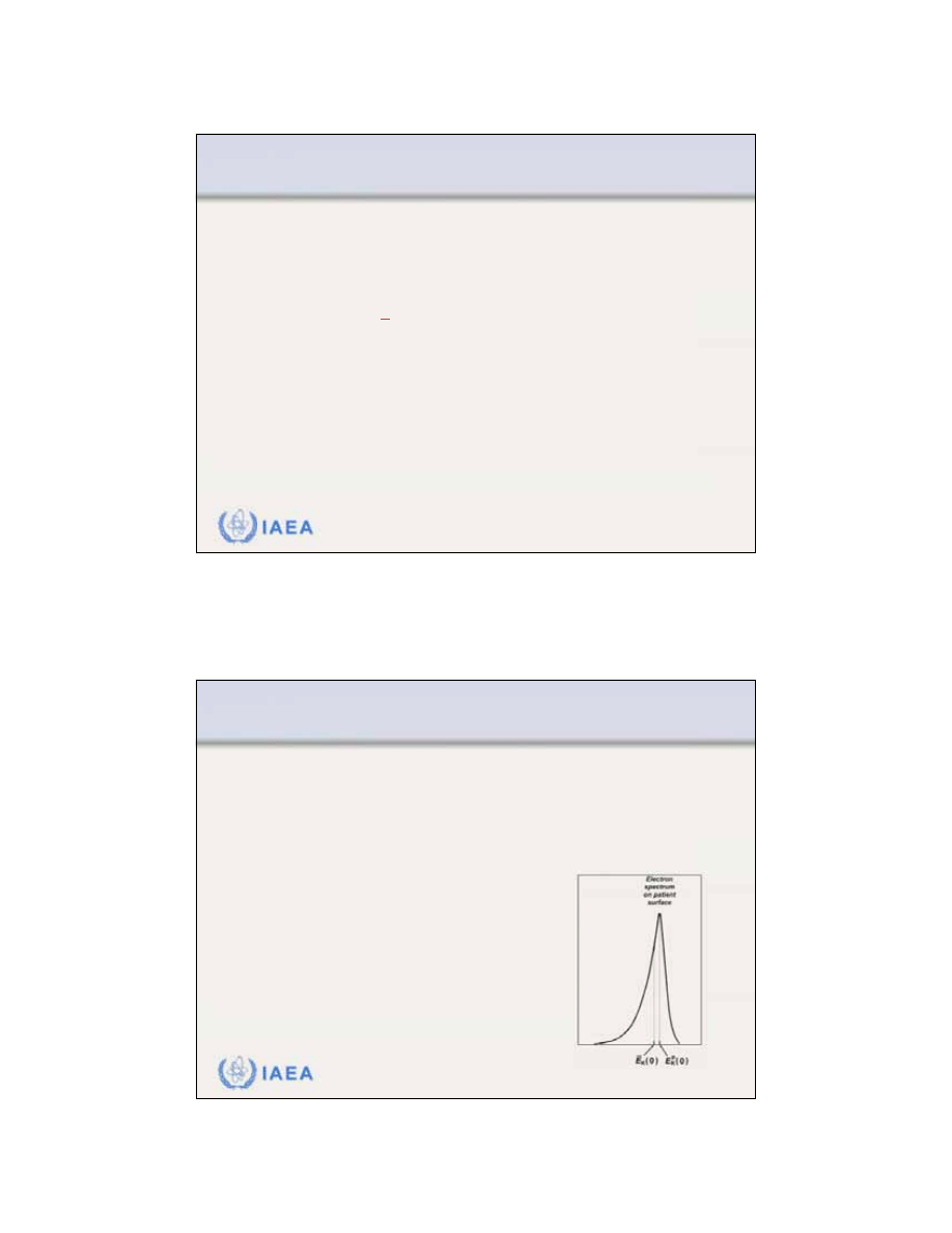
The
spectrum of the electron beam
depends on the point
of measurement in the beam.
16
IAEA
Radiation Oncology Physics: A Handbook for Teachers and Students - 8.2.1 Slide 3 (31/91)
8.2 DOSIMETRIC PARAMETERS OF ELECTRON BEAMS
8.2.1 Electron beam energy specification
Several parameters are used for describing the beam
quality of an electron beam
:
•
Most probable energy
of the electron beam on phantom
surface.
•
Mean energy
of electron beam on the phantom surface.
•
Half-value depth R
50
on the percentage depth dose curve of the
electron beam.
•
Practical range R
p
of the electron beam.
K
(0)
E
p
K
(0)
E
IAEA
Radiation Oncology Physics: A Handbook for Teachers and Students - 8.2.1 Slide 4 (32/91)
8.2 DOSIMETRIC PARAMETERS OF ELECTRON BEAMS
8.2.1 Electron beam energy specification
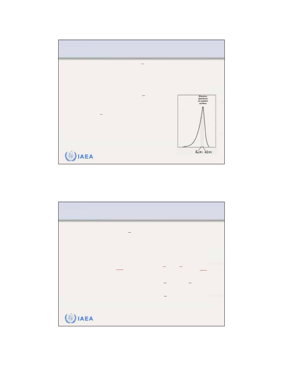
The
most probable energy
on the phantom surface
is defined by the position of the spectral peak.
the most probable of the electrons, is related to the
practical range R
p
(in cm) of the electron beam through
the following polynomial equation:
For water:
E
K
p
(0)
=
C
1
+
C
2
R
p
+
C
3
R
p
2
C
1
=
0.22 MeV
C
2
=
1.98 MeV/cm
C
3
=
0.0025 MeV/cm
2
E
K
p
(0)
E
K
p
(0),
17
IAEA
Radiation Oncology Physics: A Handbook for Teachers and Students - 8.2.1 Slide 5 (33/91)
8.2 DOSIMETRIC PARAMETERS OF ELECTRON BEAMS
8.2.1 Electron beam energy specification
The
mean electron energy
of the electron beam on
the phantom surface is slightly smaller than the most
probable energy on the phantom surface as a result
of an asymmetrical shape of the electron spectrum.
The mean electron energy is
related to the half-value depth R
50
as:
Constant C for water is 2.33 MeV/cm.
E
K
(0)
E
K
(0)
E
K
(0)
=
CR
50
E
K
p
(0)
IAEA
Radiation Oncology Physics: A Handbook for Teachers and Students - 8.2.1 Slide 6 (34/91)
8.2 DOSIMETRIC PARAMETERS OF ELECTRON BEAMS
8.2.1 Electron beam energy specification
Harder
has shown that the most probable energy
and the mean energy of electron beam at a depth z
in the phantom or patient decrease linearly with z.
Harder’s relationships are expressed as follows:
and
Note:
p
p
K
K
p
( )
(0) 1
z
E z
E
R
=
E
K
p
(z)
p
( )
(0) 1
z
E z
E
R
E(z)
E
K
p
(z
=
0)
=
E
K
p
(0)
E
K
p
(z
=
R
p
)
=
0
E(z
=
0)
=
E(0)
E(z
=
R
p
)
=
0
18
IAEA
Radiation Oncology Physics: A Handbook for Teachers and Students - 8.2.2 Slide 1 (35/91)
8.2 DOSIMETRIC PARAMETERS OF ELECTRON BEAMS
8.2.2 Typical depth dose parameters as a function of energy
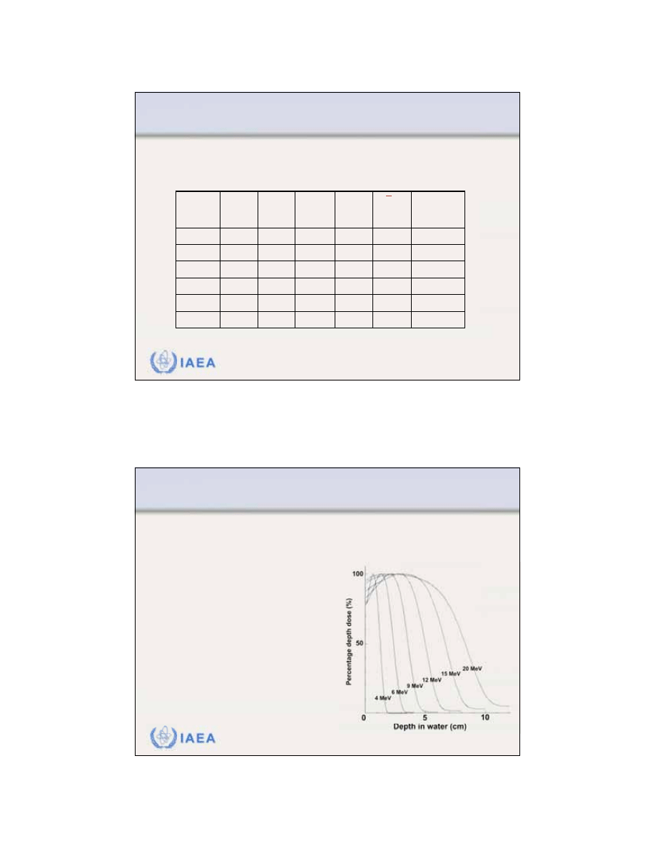
Typical
electron beam depth dose parameters
that
should be measured for each clinical electron beam.
96
17.4
9.1
7.3
5.9
5.5
18
92
14.0
7.5
6.1
5.2
4.7
15
90
11.3
6.0
4.8
4.1
3.7
12
86
9.2
4.8
3.9
3.3
3.1
10
83
7.2
4.0
3.0
2.6
2.4
8
81
5.6
2.9
2.2
1.8
1.7
6
Surface
dose %
(MeV)
R
p
(cm)
R
50
(cm)
R
80
(cm)
R
90
(cm)
Energy
(MeV)
E(0)
IAEA
Radiation Oncology Physics: A Handbook for Teachers and Students - 8.2.3 Slide 1 (36/91)
8.2 DOSIMETRIC PARAMETERS OF ELECTRON BEAMS
8.2.3 Percentage depth dose
Similarly to PDDs for photon beams, the
PDDs for
electron beams
, at a given source-surface distance
SSD, depend upon:
•
Depth z in phantom (patient).
•
Electron beam kinetic energy
E
K
(0) on phantom surface.
•
Field size A on phantom
surface.

19
IAEA
Radiation Oncology Physics: A Handbook for Teachers and Students - 8.2.3 Slide 2 (37/91)
8.2 DOSIMETRIC PARAMETERS OF ELECTRON BEAMS
8.2.3 Percentage depth dose
The
PDDs of electron beams
are measured with:
•
Cylindrical, small-volume ionization chamber in water phantom.
•
Diode detector in water phantom.
•
Parallel-plate ionization chamber in water phantom.
•
Radiographic or radiochromic film in solid water phantom.
IAEA
Radiation Oncology Physics: A Handbook for Teachers and Students - 8.2.3 Slide 3 (38/91)
8.2 DOSIMETRIC PARAMETERS OF ELECTRON BEAMS
8.2.3 Percentage depth dose
Measurement of electron beam PDDs:
•
If an
ionization chamber
is used, the measured depth ionization
distribution must be converted into a depth dose distribution by
using the appropriate stopping power ratios, water to air, at
depths in phantom.
•
If
diode
is used, the diode ionization signal represents the dose
directly, because the stopping power ratio, water to silicon, is
essentially independent of electron energy and hence depth.
•
If
film
is used, the characteristic curve (H and D curve) for the
given film should be used to determine the dose against the film
density.
20
IAEA
Radiation Oncology Physics: A Handbook for Teachers and Students - 8.2.3 Slide 4 (39/91)
8.2 DOSIMETRIC PARAMETERS OF ELECTRON BEAMS
8.2.3 Percentage depth dose
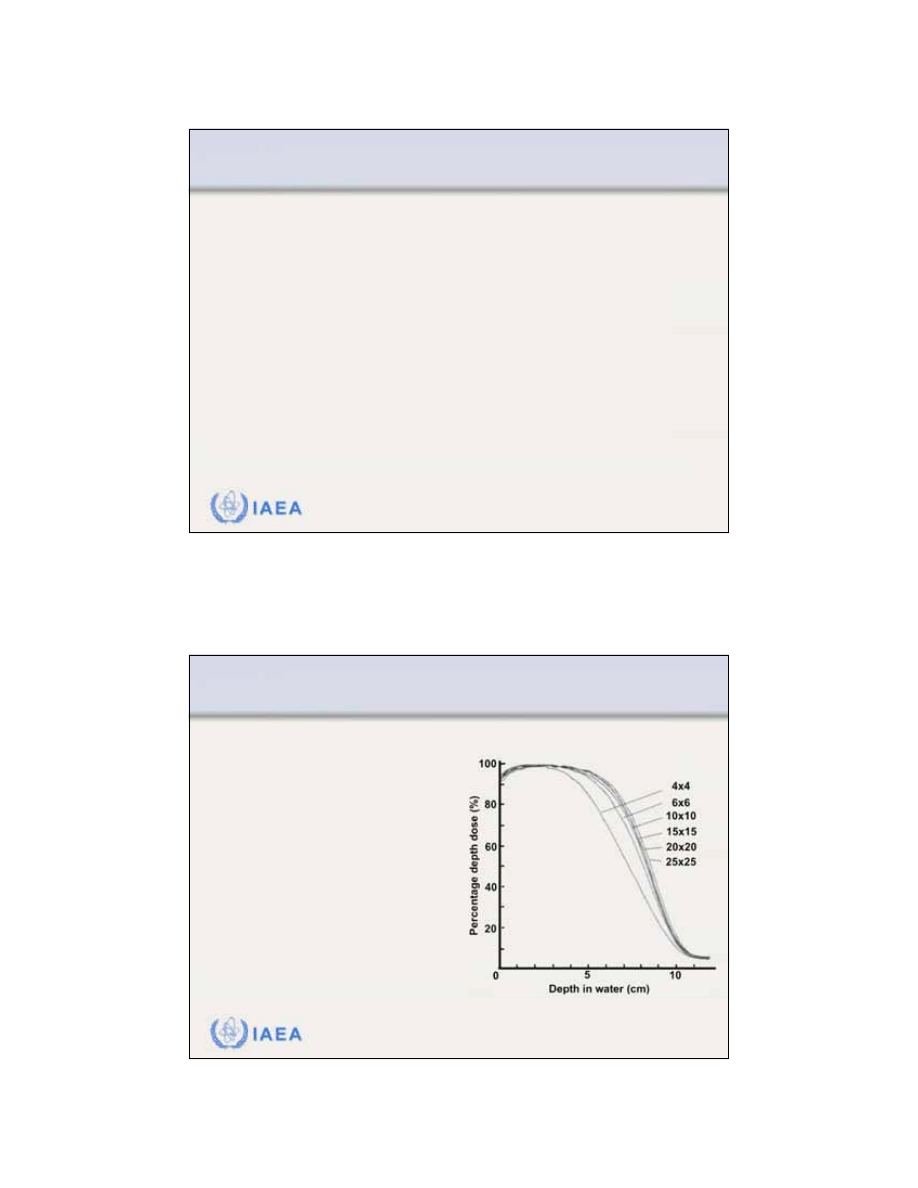
Dependence of PDDs on electron beam field size.
For relatively large field sizes the PDD distribution at a
given electron beam energy is essentially independent
of field size.
When the side of the electron field is smaller than the
practical range R
p
, lateral electronic equilibrium will not
exist on the beam central axis and both the PDDs as
well as the output factors exhibit a significant depen-
dence on field size.
IAEA
Radiation Oncology Physics: A Handbook for Teachers and Students - 8.2.3 Slide 5 (40/91)
8.2 DOSIMETRIC PARAMETERS OF ELECTRON BEAMS
8.2.3 Percentage depth dose
PDDs for small electron fields
For a decreasing field size,
when the side of the field
decreases to below the R
p
value for a given electron
energy:
•
Depth of dose maximum
decreases.
•
Surface dose increases.
•
R
p
remains essentially
constant, except when the field
size becomes very small.
21
IAEA
Radiation Oncology Physics: A Handbook for Teachers and Students - 8.2.3 Slide 6 (41/91)
8.2 DOSIMETRIC PARAMETERS OF ELECTRON BEAMS
8.2.3 Percentage depth dose
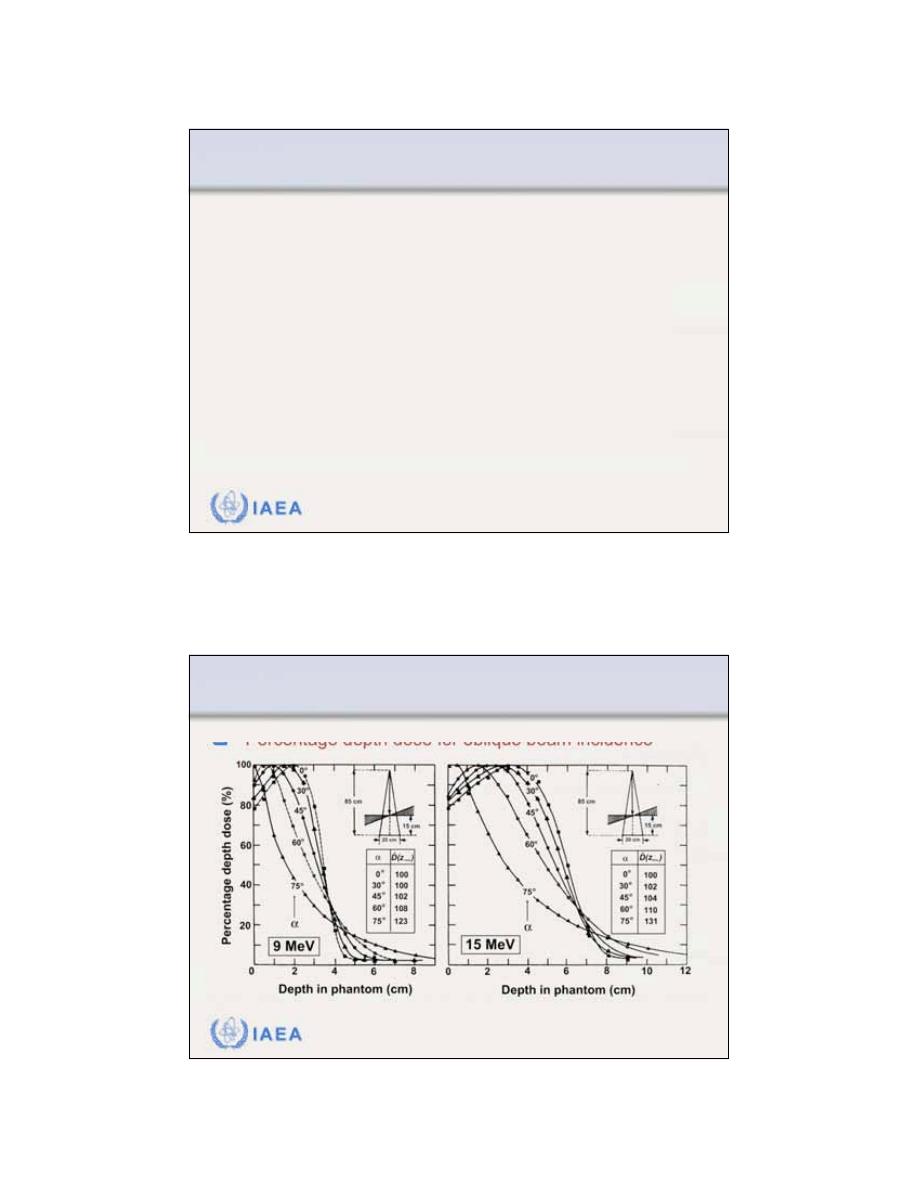
PDDs for oblique incidence.
The
angle of obliquity
is defined as the angle between
the electron beam central axis and the normal to the
phantom or patient surface. Angle corresponds to
normal beam incidence.
For oblique beam incidences, especially at large angles
the PDD characteristics of electron beams deviate
significantly from those for normal beam incidence.
=
0
IAEA
Radiation Oncology Physics: A Handbook for Teachers and Students - 8.2.3 Slide 7 (42/91)
8.2 DOSIMETRIC PARAMETERS OF ELECTRON BEAMS
8.2.3 Percentage depth dose
Percentage depth dose for oblique beam incidence

22
IAEA
Radiation Oncology Physics: A Handbook for Teachers and Students - 8.2.3 Slide 8 (43/91)
8.2 DOSIMETRIC PARAMETERS OF ELECTRON BEAMS
8.2.3 Percentage depth dose
Depth dose for oblique beam incidence
The obliquity effect becomes significant for angles of
incidence exceeding 45
o
.
The obliquity factor accounts for the change in
depth dose at a given depth z in phantom and is
normalized to 1.00 at z
max
at normal incidence .
The obliquity factor at z
max
is larger than 1 (see insets on
previous slide).
OF(
,z)
=
0
IAEA
Radiation Oncology Physics: A Handbook for Teachers and Students - 8.2.4 Slide 1 (44/91)
8.2 DOSIMETRIC PARAMETERS OF ELECTRON BEAMS
8.2.4 Output factors
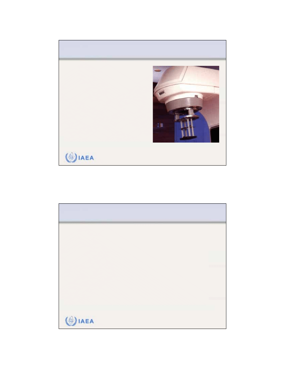
The
output factor
•
For a given electron energy and
•
For a given field size (delineated by applicator or cone)
is defined as the ratio of the dose for the specific field
size (applicator) to the dose for a 10x10 cm
2
reference field size (applicator), both measured:
•
At depth z
max
on the beam central axis in phantom
•
At a nominal SSD of 100 cm.
23
IAEA
Radiation Oncology Physics: A Handbook for Teachers and Students - 8.2.4 Slide 2 (45/91)
8.2 DOSIMETRIC PARAMETERS OF ELECTRON BEAMS
8.2.4 Output factors
When using electron beams
from a linac, the photon
collimator must be opened to
the appropriate setting for a
given electron applicator.
Typical
electron applicator
sizes
at nominal SSD are:
•
Circular with diameter: 5 cm
•
Square: 10x10 cm
2
; 15x15 cm
2
;
20x20 cm
2
; and 25x25 cm
2
.
IAEA
Radiation Oncology Physics: A Handbook for Teachers and Students - 8.2.4 Slide 3 (46/91)
8.2 DOSIMETRIC PARAMETERS OF ELECTRON BEAMS
8.2.4 Output factors
Often collimating blocks made of lead or a low melting
point alloy (e.g., cerrobend) are used for field shaping.
These blocks are attached to the end of the electron
cone (applicator) and produce the required irregular
field.
Output factors
, normalized to the standard 10x10 cm
2
electron cone, must be measured for all custom-made
irregular fields.

24
IAEA
Radiation Oncology Physics: A Handbook for Teachers and Students - 8.2.4 Slide 4 (47/91)
8.2 DOSIMETRIC PARAMETERS OF ELECTRON BEAMS
8.2.4 Output factors
For small irregular field sizes the extra shielding affects
not only the output factors but also the PDD distribution
because of the lack of lateral scatter.
For custom-made small fields, in addition to output
factors, the full electron beam PDD distribution should
be measured.
IAEA
Radiation Oncology Physics: A Handbook for Teachers and Students - 8.2.5 Slide 1 (48/91)
8.2 DOSIMETRIC PARAMETERS OF ELECTRON BEAMS
8.2.5 Therapeutic range
The depth of the 90% dose level on the beam central
axis (
R
90
) beyond z
max
is defined as the
therapeutic
range
for electron beam therapy.
•
R
90
is approximately equal to E
K
/4 in cm of water, where E
K
is
the nominal kinetic energy in MeV of the electron beam.
•
R
80
, the depth that corresponds to the 80% PDD beyond z
max
,
may also be used as the therapeutic range and is approximated
by E
K
/3 in cm of water.
25
IAEA
Radiation Oncology Physics: A Handbook for Teachers and Students - 8.2.6 Slide 1 (49/51)
8.2 DOSIMETRIC PARAMETERS OF ELECTRON BEAMS
8.2.6 Profiles and off-axis ratio
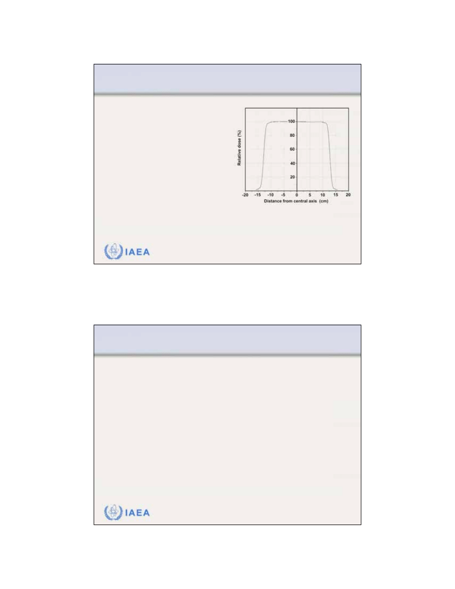
A
dose profile
represents a
plot of dose at a given
depth in phantom against
the distance from the
beam central axis.
The profile is measured in
a plane perpendicular to
the beam central axis at a
given depth z in phantom.
Dose profile measured at a depth
of dose maximum z
max
in water
for a 12 MeV electron beam and
25x25 cm
2
applicator cone.
IAEA
Radiation Oncology Physics: A Handbook for Teachers and Students - 8.2.6 Slide 2 (50/91)
8.2 DOSIMETRIC PARAMETERS OF ELECTRON BEAMS
8.2.6 Profiles and off-axis ratio
Two
different normalizations
are used for beam profiles:
•
Profile data for a given depth in phantom may be normalized to
the dose at z
max
on the central axis (point P). The dose value on
the beam central axis for then represents the central axis
PDD value.
•
Profile data for a given depth in phantom may also be normalized
to the value on the beam central axis (point Q). The values off
the central axis for are then referred to as the off-axis
ratios (OARs).
max
z
z
max
z
z
26
IAEA
Radiation Oncology Physics: A Handbook for Teachers and Students - 8.2.7 Slide 1 (51/91)
8.2 DOSIMETRIC PARAMETERS OF ELECTRON BEAMS
8.2.7 Flatness and symmetry
According to the International
Electrotechnical Commission (IEC)
the
specification for beam flatness
of
electron beams is given for z
max
under two conditions:
•
Distance between the 90% dose
level and the geometrical beam
edge should not exceed 10 mm
along major field axes and 20 mm
along diagonals.
•
Maximum value of the absorbed
dose anywhere within the region
bounded by the 90% isodose
contour should not exceed 1.05
times the absorbed dose on the
axis of the beam at the same depth.
IAEA
Radiation Oncology Physics: A Handbook for Teachers and Students - 8.2.7 Slide 2 (52/91)
8.2 DOSIMETRIC PARAMETERS OF ELECTRON BEAMS
8.2.7 Flatness and symmetry
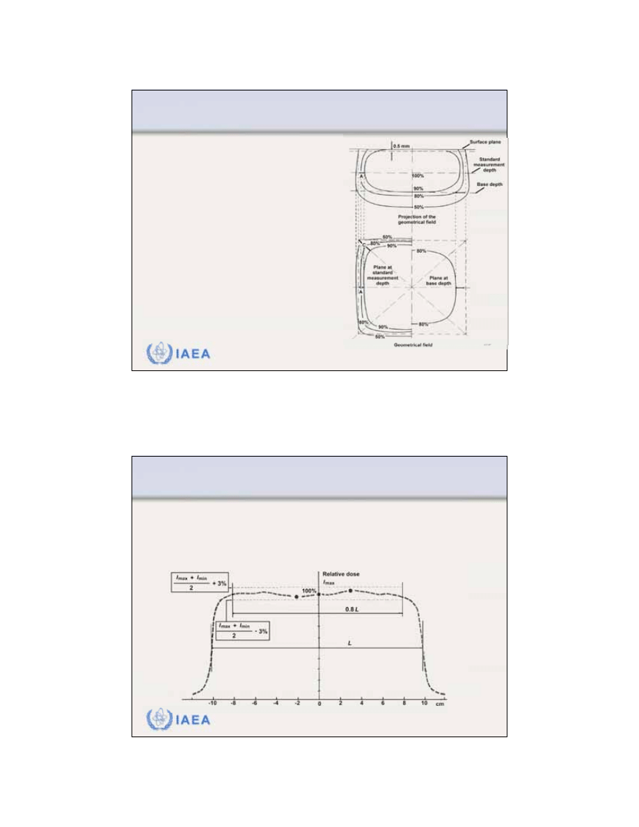
According to the International Electrotechnical Commission (IEC)
the
specification for symmetry
of electron beams requires that the
cross-beam profile measured at depth z
max
should not differ by
more than 3% for any pair of symmetric points with respect to the
central ray.

27
IAEA
Radiation Oncology Physics: A Handbook for Teachers and Students - 8.3.1 Slide 1 (53/91)
8.3 CLINICAL CONSIDERATIONS
8.3.1 Dose specification and reporting
Electron beam therapy
is usually applied in treatment of
superficial or subcutaneous disease.
Electron beam treatment is usually delivered with a single
direct electron field at a nominal SSD of 100 cm.
The dose in electron beam therapy is usually prescribed
at a depth that lies at, or beyond, the distal margin of the
target.
IAEA
Radiation Oncology Physics: A Handbook for Teachers and Students - 8.3.1 Slide 2 (54/91)
8.3 CLINICAL CONSIDERATIONS
8.3.1 Dose specification and reporting
To maximize
healthy tissue sparing
beyond the tumour
and to provide relatively homogeneous target coverage
treatments are usually prescribed at z
max
, R
90
, or R
80
.
•
If the treatment dose is specified at R
80
or R
90
, the skin dose may
exceed the prescription dose.
•
Since the maximum dose in the target may exceed the
prescribed dose by up to 20%, the maximum dose should be
reported for all electron beam treatments.
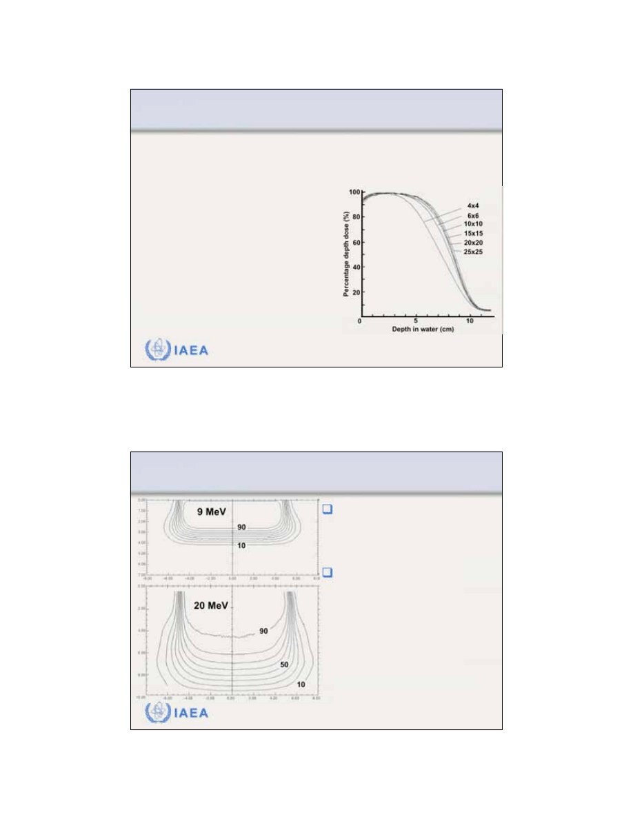
28
IAEA
Radiation Oncology Physics: A Handbook for Teachers and Students - 8.3.2 Slide 1 (55/91)
8.3 CLINICAL CONSIDERATIONS
8.3.2 Small field sizes
The
PDD curves for electron beams
do not depend on field
size, except for small fields where the side of the field is
smaller than the practical range of the electron beam.
When lateral scatter equilibrium
is not reached at small electron
fields, in comparison to
a 10x10 cm
2
field:
•
Dose rate at z
max
decreases.
•
Depth of maximum dose, z
max
,
moves closer to the surface.
•
PDD curve becomes less steep.
IAEA
Radiation Oncology Physics: A Handbook for Teachers and Students - 8.3.3 Slide 1 (56/91)
8.3 CLINICAL CONSIDERATIONS
8.3.3 Isodose distributions
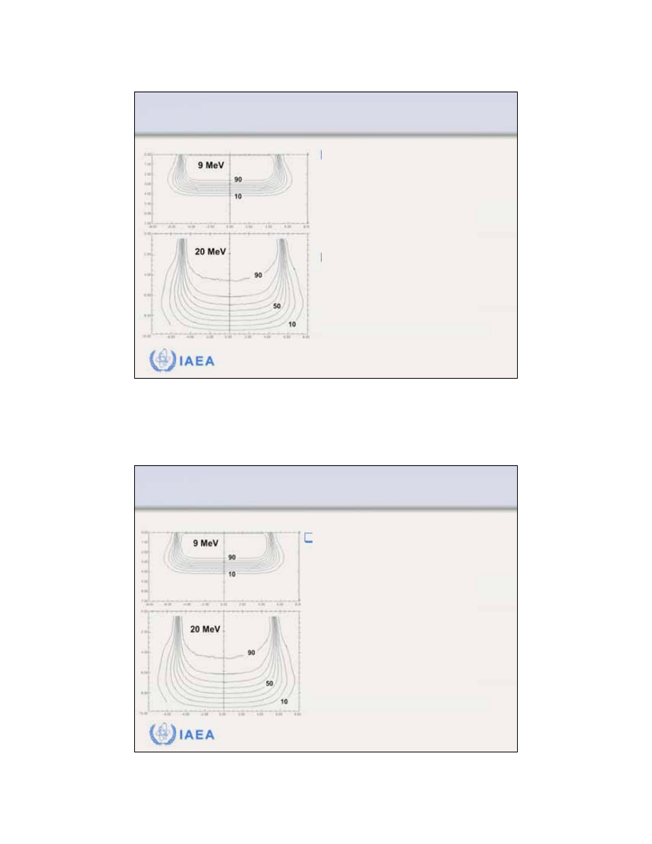
Isodose curves
are lines
connecting points of equal
dose in the irradiated
medium.
Isodose curves are usually
drawn at regular intervals
of absorbed dose and are
expressed as a percentage
of the dose at a reference
point, which is usually
taken as the z
max
point on
the beam central axis.
29
IAEA
Radiation Oncology Physics: A Handbook for Teachers and Students - 8.3.3 Slide 2 (57/91)
8.3 CLINICAL CONSIDERATIONS
8.3.3 Isodose distributions
As an electron beam
penetrates a medium
(absorber), the beam
expands rapidly below the
surface because of electron
scattering on absorber
atoms.
The spread of the isodose
curves varies depending on:
•
Isodose level
•
Energy of the beam
•
Field size
•
Beam collimation
IAEA
Radiation Oncology Physics: A Handbook for Teachers and Students - 8.3.3 Slide 3 (58/91)
8.3 CLINICAL CONSIDERATIONS
8.3.3 Isodose distributions
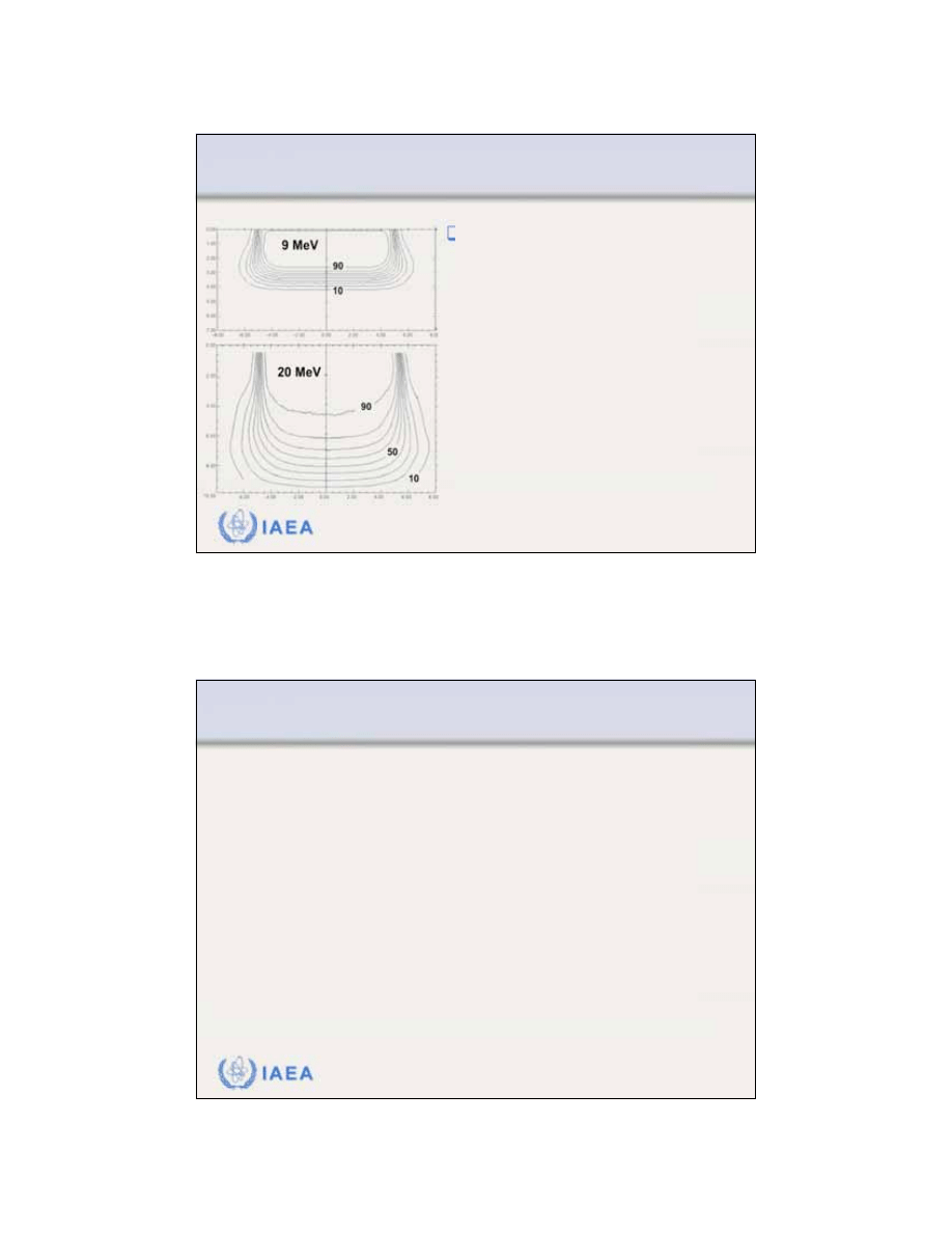
A particular characteristic of
electron beam isodose curves
is the
bulging out
of the low
value isodose curves (<20%)
as a direct result of the
increase in electron scattering
angle with decreasing electron
energy.
30
IAEA
Radiation Oncology Physics: A Handbook for Teachers and Students - 8.3.3 Slide 4 (59/91)
8.3 CLINICAL CONSIDERATIONS
8.3.3 Isodose distributions
At energies above 15 MeV
electron beams exhibit a
lateral
constriction
of the higher value
isodose curves (>80%). The
higher is the electron beam
energy, the more pronounced
is the effect.
IAEA
Radiation Oncology Physics: A Handbook for Teachers and Students - 8.3.3 Slide 5 (60/91)
8.3 CLINICAL CONSIDERATIONS
8.3.3 Isodose distributions
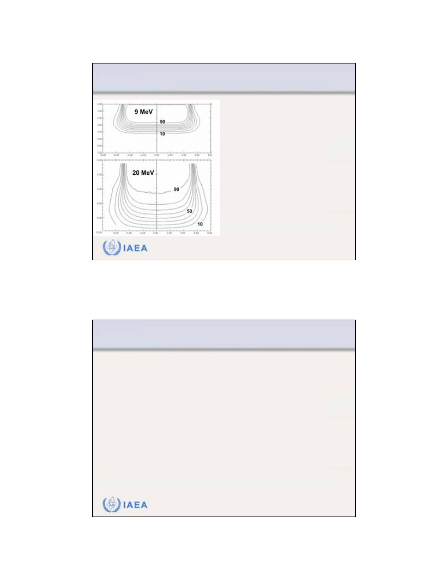
The term
penumbra
generally defines the region at the
edge of the radiation beam over which the dose rate
changes rapidly as a function of distance from the beam
central axis.
The
physical penumbra
of an electron beam may be
defined as the distance between two specified isodose
curves at a specified depth in phantom.
31
IAEA
Radiation Oncology Physics: A Handbook for Teachers and Students - 8.3.3 Slide 6 (61/91)
8.3 CLINICAL CONSIDERATIONS
8.3.3 Isodose distributions
In determination of the
physical penumbra
of an
electron beam the ICRU
recommends that:
•
The 80% and 20% isodose
curves be used.
•
The specified depth of
measurement be R
85
/2, where
R
85
is the depth of the 85%
dose level beyond z
max
on the
electron beam central ray.
IAEA
Radiation Oncology Physics: A Handbook for Teachers and Students - 8.3.3 Slide 7 (62/91)
8.3 CLINICAL CONSIDERATIONS
8.3.3 Isodose distributions
In electron beam therapy, the
air gap
is defined as the
separation between the patient and the end of the
applicator cone. The standard air gap is 5 cm.
With increasing air gap:
•
The low value isodose curves diverge.
•
The high value isodose curves converge toward the central
axis of the beam.
•
The physical penumbra increases.

32
IAEA
Radiation Oncology Physics: A Handbook for Teachers and Students - 8.3.4 Slide 1 (63/91)
8.3 CLINICAL CONSIDERATIONS
8.3.4 Field shaping
To achieve a more customized electron field shape, a
lead or metal alloy cut-out
may be constructed and
placed on the applicator as close to the patient as
possible.
Field shapes may be determined from conventional or
virtual simulation, but are most often prescribed
clinically by a physician prior to the first treatment.
As a rule of thumb, divide the practical range R
p
by 10 to
obtain the approximate thickness of lead required for
shielding (<5%).
IAEA
Radiation Oncology Physics: A Handbook for Teachers and Students - 8.3.4 Slide 2 (64/91)
8.3 CLINICAL CONSIDERATIONS
8.3.4 Field shaping
For certain treatments, such as treatments of the lip,
buccal mucosa, eyelids or ear lobes, it may be
advantageous to use an
internal shield
to protect the
normal structures beyond the target volume.
Internal shields are usually coated with low atomic
number materials to minimize the electron back-
scattering into healthy tissue above the shield.

33
IAEA
Radiation Oncology Physics: A Handbook for Teachers and Students - 8.3.4 Slide 3 (65/91)
8.3 CLINICAL CONSIDERATIONS
8.3.4 Field shaping
Extended SSDs
have various effects on electron beam
parameters and are generally not advisable.
In comparison with treatment at nominal SSD of 100 cm
at extended SSD:
•
Output is significantly lower
•
Beam penumbra is larger
•
PDD distribution changes minimally.
An effective SSD based on the virtual source position is
used when applying the inverse square law to correct
the beam output at z
max
for extended SSD.
IAEA
Radiation Oncology Physics: A Handbook for Teachers and Students - 8.3.5 Slide 1 (66/91)
8.3 CLINICAL CONSIDERATIONS
8.3.5 Irregular surface correction
Uneven air gaps
as a result of curved patient surfaces are
often present in clinical use of electron beam therapy.
Inverse square law corrections can be made to the dose
distribution to account for the sloping surface.
From F.M. Khan:
“The Physics of
Radiation Therapy”
g = air gap
z = depth below surface
SSD
eff
= distance between the
virtual source and surface
eff
2
eff
o
eff
eff
(SSD
, )
SSD
(SSD , )
SSD
)
D
g z
z
D
z
g
z
+
=
+
=
+ +

34
IAEA
Radiation Oncology Physics: A Handbook for Teachers and Students - 8.3.5 Slide 2 (67/91)
8.3 CLINICAL CONSIDERATIONS
8.3.5 Irregular surface correction
The inverse square correction alone does not account for
changes in side scatter as a result of
beam obliquity
which:
•
Increases side scatter at the depth of maximum dose, z
max
•
Shifts z
max
toward the surface
•
Decreases the therapeutic depths R
90
and R
80
.
From F.M. Khan:
“The Physics of
Radiation Therapy”
= obliquity factor
which
accounts for the change in depth
dose at a point in phantom at depth z
for a given angle of obliquity but
same SSD
eff
as for
eff
2
eff
o
eff
eff
(SSD
, )
SSD
(SSD , )
OF( , )
SSD
)
D
g z
z
D
z
z
g
z
+
=
+
=
+ +
=
0
OF(z,
)
IAEA
Radiation Oncology Physics: A Handbook for Teachers and Students - 8.3.6 Slide 1 (68/91)
8.3 CLINICAL CONSIDERATIONS
8.3.6 Bolus
Bolus
made of tissue equivalent material, such as wax,
is often used in electron beam therapy:
•
To increase the surface dose.
•
To shorten the range of a given electron beam in the patient.
•
To flatten out irregular surfaces.
•
To reduce the electron beam penetration in some parts of the
treatment field.
Although labour intensive, the use of bolus in electron
beam therapy is very practical, since treatment plan-
ning software for electron beams is limited and empirical
data are normally collected only for standard beam
geometries.

35
IAEA
Radiation Oncology Physics: A Handbook for Teachers and Students - 8.3.6 Slide 2 (69/91)
8.3 CLINICAL CONSIDERATIONS
8.3.6 Bolus
The use of computed tomography (CT) for treatment
planning enables
accurate determination of tumour
shape
and
patient contour
.
If a wax bolus is constructed such that the total distance
from the bolus surface to the required treatment depth is
constant along the length of
the tumour, then the shape
of the resulting isodose
curves will approximate
the shape of the tumour
as determined with
CT scanning.
IAEA
Radiation Oncology Physics: A Handbook for Teachers and Students - 8.3.7 Slide 1 (70/91)
8.3 CLINICAL CONSIDERATIONS
8.3.7 Inhomogeneity corrections
The presence of
tissue inhomogeneities (
also referred to
as
heterogeneities)
such as lung or bone can greatly affect
the dose distributions for electron beams .
The dose inside an inhomogeneity is difficult to calculate or
measure, but the effect of an inhomogeneity on the dose
beyond the inhomogeneity is relatively simple to measure
and quantify.

36
IAEA
Radiation Oncology Physics: A Handbook for Teachers and Students - 8.3.7 Slide 2 (71/91)
8.3 CLINICAL CONSIDERATIONS
8.3.7 Inhomogeneity corrections
The simplest correction for for a tissue inhomogeneity
involves the scaling of the inhomogeneity thickness by
its electron density relative to that of water and the
determination of the
coefficient of equivalent thickness
(CET).
The electron density of an inhomogeneity is essentially
equivalent to the mass density of the inhomogeneity.
IAEA
Radiation Oncology Physics: A Handbook for Teachers and Students - 8.3.7 Slide 3 (72/91)
8.3 CLINICAL CONSIDERATIONS
8.3.7 Inhomogeneity corrections
CET is used to determine the effective depth in water
equivalent tissue z
eff
through the following expression:
For example:
•
Lung has approximate density of 0.25 g/cm
3
and a CET of 0.25.
•
A thickness of 1 cm of lung is equivalent to 0.25 cm of tissue.
•
Solid bone has approximate density of 1.6 g/cm
3
and a CET of 1.6.
•
A thickness of 1 cm of bone is equivalent to 1.6 cm of tissue.
z
eff
=
z
t(1 CET)
z = actual depth of the point of
interest in the patient
t = thickness of the inhomogeneity
37
IAEA
Radiation Oncology Physics: A Handbook for Teachers and Students - 8.3.7 Slide 4 (73/91)
8.3 CLINICAL CONSIDERATIONS
8.3.7 Inhomogeneity corrections
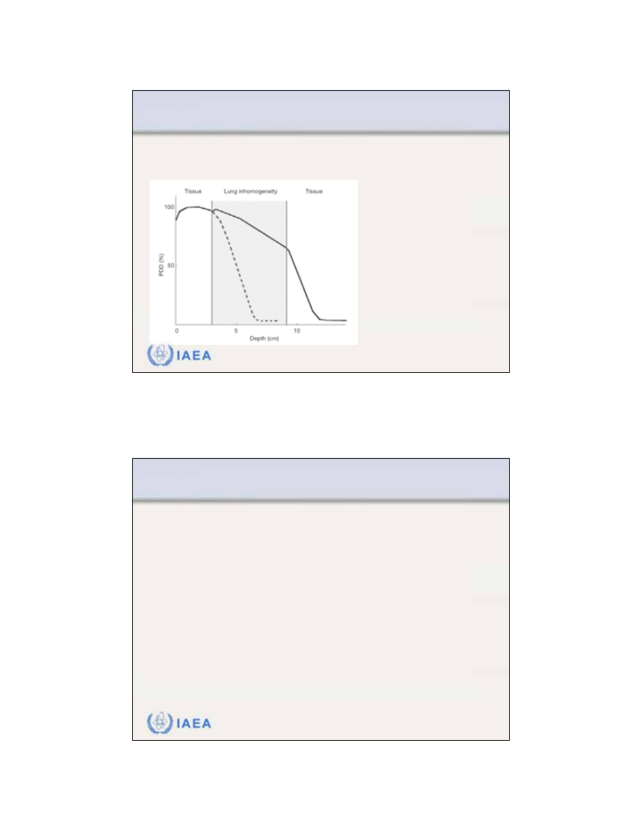
The effect of lung inhomogeneity on the PDD distribution
of an electron beam (energy: 15 MeV, field: 10x10 cm
2
).
Thickness t of lung
inhomogeneity: 6 cm
Tissue equivalent thickness:
z
eff
= 1.5 cm
IAEA
Radiation Oncology Physics: A Handbook for Teachers and Students - 8.3.7 Slide 5 (74/91)
8.3 CLINICAL CONSIDERATIONS
8.3.7 Inhomogeneity corrections
If an electron beam strikes the interface between two
materials either tangentially or at a large oblique angle,
the resulting
scatter perturbation
will affect the dose
distribution at the interface.
The lower density material will receive a higher dose,
due to the increased scattering of electrons from the
higher density side.

38
IAEA
Radiation Oncology Physics: A Handbook for Teachers and Students - 8.3.7 Slide 6 (75/91)
8.3 CLINICAL CONSIDERATIONS
8.3.7 Inhomogeneity corrections
Edge effects
need to be considered in the following
situations:
•
Inside a patient, at the interfaces between internal
structures of different density.
•
On the surface of a patient, in regions of sharp surface
irregularity.
•
On the interface between lead shielding and the surface of
the patient, if the shielding is placed superficially on the
patient or if it is internal shielding.
IAEA
Radiation Oncology Physics: A Handbook for Teachers and Students - 8.3.8 Slide 1 (76/91)
8.3 CLINICAL CONSIDERATIONS
8.3.8 Electron beam combinations
Occasionally, the need arises to abut electron fields.
When
abutting two electron fields
, it is important to
take into consideration the dosimetric characteristics
of electron beams at depth in the patient.
The large penumbra and bulging isodose lines
produce hot spots and cold spots inside the target
volume.

39
IAEA
Radiation Oncology Physics: A Handbook for Teachers and Students - 8.3.8 Slide 2 (77/91)
8.3 CLINICAL CONSIDERATIONS
8.3.8 Electron beam combinations
In general, it is best to avoid using adjacent electron fields.
If the use of abutting fields is absolutely necessary, the
following conditions apply:
•
Contiguous electron beams should be parallel to one another in
order to avoid significant overlapping of the high value isodose
curves at depth in the patient.
•
Some basic film dosimetry should be carried out at the junction of
the fields to ensure that no significant hot or cold spots in dose
occur.
IAEA
Radiation Oncology Physics: A Handbook for Teachers and Students - 8.3.8 Slide 3 (78/91)
8.3 CLINICAL CONSIDERATIONS
8.3.8 Electron beam combinations
Electron - photon field matching
is easier than
electron -
electron field matching.
•
A distribution for photon fields is readily available from a
treatment planning system (TPS) and the location of the
electron beam treatment field as well as the associated hot and
cold spots can be determined relative to the photon field
treatment plan.
•
The matching of electron and photon fields on the skin will
produce a hot spot on the photon side of the treatment.

40
IAEA
Radiation Oncology Physics: A Handbook for Teachers and Students - 8.3.9 Slide 1 (79/91)
8.3 CLINICAL CONSIDERATIONS
8.3.9 Electron arc therapy
Electron arc therapy
is a special radiotherapeutic
treatment technique in which a rotational electron beam
is used to treat superficial tumour volumes that follow
curved surfaces.
While its usefulness in treatment of certain large super-
ficial tumours is well recognized, the technique is not
widely used because it is relatively complicated and
cumbersome, and its physical characteristics are poorly
understood.
IAEA
Radiation Oncology Physics: A Handbook for Teachers and Students - 8.3.9 Slide 2 (80/91)
8.3 CLINICAL CONSIDERATIONS
8.3.9 Electron arc therapy
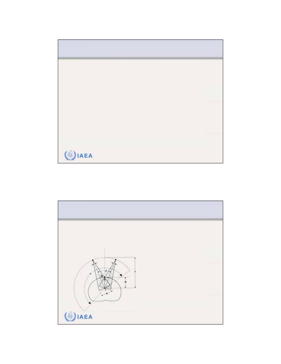
The
dose distribution
in the target volume for electron
arc therapy depends in a complicated fashion on:
•
Electron beam energy
•
Field width w
•
Depth of the isocentre d
i
•
Source-axis distance f
•
Patient curvature
•
Tertiary collimation
•
Field shape as defined by the secondary collimator
41
IAEA
Radiation Oncology Physics: A Handbook for Teachers and Students - 8.3.9 Slide 3 (81/91)
8.3 CLINICAL CONSIDERATIONS
8.3.9 Electron arc therapy
Two approaches to electron arc therapy have been
developed:
•
Electron pseudo-arc
based on a series of overlapping stationary
electron fields.
•
Continuous electron arc
using a continuous rotating electron
beam.
The calculation of dose distributions in electron arc
therapy is a complicated procedure that generally can-
not be performed reliably with the algorithms used for
standard electron beam treatment planning.
IAEA
Radiation Oncology Physics: A Handbook for Teachers and Students - 8.3.9 Slide 4 (82/91)
8.3 CLINICAL CONSIDERATIONS
8.3.9 Electron arc therapy
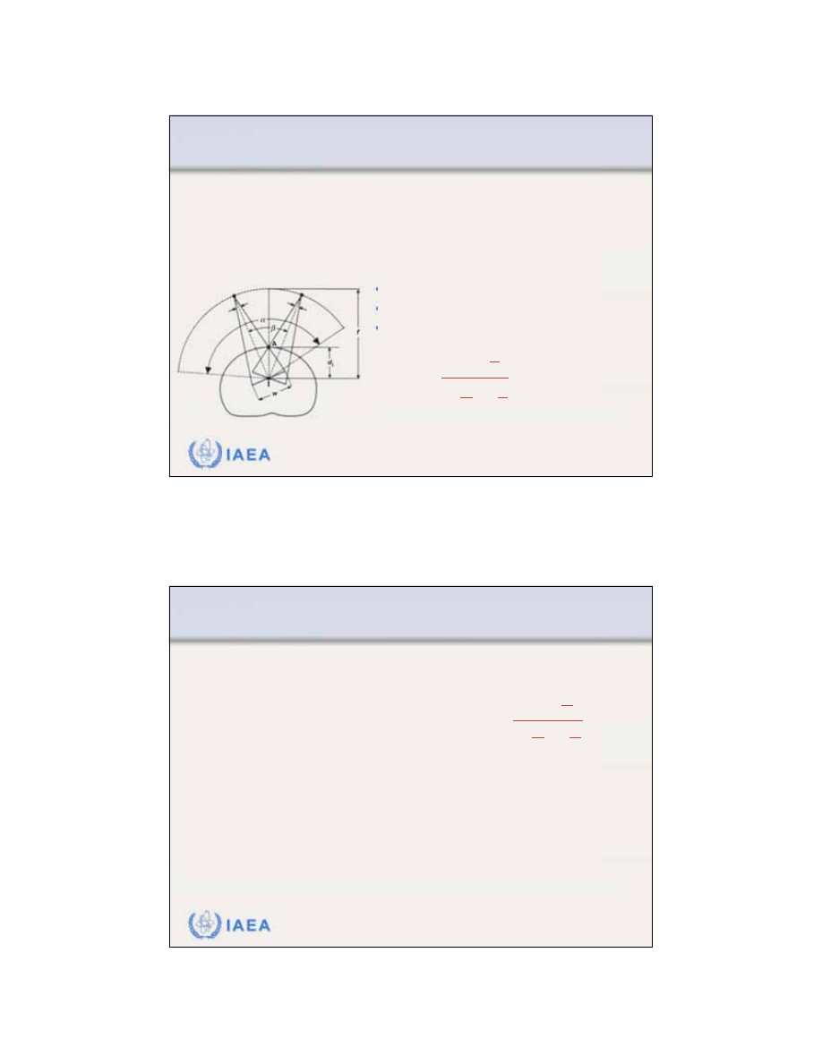
The characteristic angle
concept represents a semi-
empirical technique for treatment planning in electron
arc therapy.
The
characteristic angle
for an
arbitrary point A on the patient
surface is measured between
the central axes of two rotational
electron beams positioned in
such a way that at point A the
frontal edge of one beam
crosses the trailing edge of the
other beam.
42
IAEA
Radiation Oncology Physics: A Handbook for Teachers and Students - 8.3.9 Slide 5 (83/91)
8.3 CLINICAL CONSIDERATIONS
8.3.9 Electron arc therapy
•
w is the nominal field size.
•
f is the virtual source isocentre distance.
•
d
i
is the isocentre depth.
The
characteristic angle
represents a continuous rotation
in which a surface point A receives a contribution from all ray
lines of the electron beam starting with the frontal edge and
finishing with the trailing edge of the rotating electron beam.
w
=
2d
i
sin
2
1
d
i
f
cos
2
IAEA
Radiation Oncology Physics: A Handbook for Teachers and Students - 8.3.9 Slide 6 (84/91)
8.3 CLINICAL CONSIDERATIONS
8.3.9 Electron arc therapy
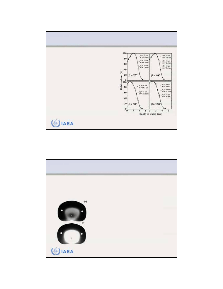
The
characteristic angle
is uniquely determined by
three treatment parameters
•
Source-axis distance f
•
Depth of isocentre d
i
•
Field width w
Electron beams with combinations of d
i
and w that give
the same characteristic angle exhibit very similar radial
percentage depth dose distributions even though they
may differ considerably in individual d
i
and w.
=
i
i
2 sin
2
1
cos
f
2
d
w
d
43
IAEA
Radiation Oncology Physics: A Handbook for Teachers and Students - 8.3.9 Slide 7 (85/91)
8.3 CLINICAL CONSIDERATIONS
8.3.9 Electron arc therapy
The
PDDs for rotational electron
beams
depend only on:
•
Electron beam energy
•
Characteristic angle
•
When a certain PDD is required for
patient treatment one may choose
a that will give the required beam
characteristics.
•
Since d
i
is fixed by the patient
contour, the required is obtained
by choosing the appropriate w.
IAEA
Radiation Oncology Physics: A Handbook for Teachers and Students - 8.3.9 Slide 8 (86/91)
8.3 CLINICAL CONSIDERATIONS
8.3.9 Electron arc therapy
Photon contamination
of the electron beam is of concern
in electron arc therapy, since the photon contribution from
all beams is added at the isocentre and the isocentre may
be at a critical structure.
Comparison between two dose distributions
measured with film in a humanoid phantom:
(a) Small of 10
o
(small field width) exhibiting a
large photon contamination at the isocentre
(b) Large of 100
o
exhibiting a relatively small
photon contamination at the isocentre.
In electron arc therapy the bremsstrahlung dose
at the isocentre is inversely proportional to the
characteristic angle
.

44
IAEA
Radiation Oncology Physics: A Handbook for Teachers and Students - 8.3.9 Slide 9 (87/91)
8.3 CLINICAL CONSIDERATIONS
8.3.9 Electron arc therapy
The
shape of secondary collimator
defining the field
width w in electron arc therapy is usually rectangular and
the resulting treatment volume geometry is cylindrical,
such as for example in the treatment of the chest wall.
When sites that can only be approximated with spherical
geometry, such as lesions of the scalp, are treated, a
custom built secondary collimator defining a non-
rectangular field of appropriate shape must be used to
provide a homogeneous dose in the target volume.
IAEA
Radiation Oncology Physics: A Handbook for Teachers and Students - 8.3.10 Slide 1 (88/91)
8.3 CLINICAL CONSIDERATIONS
8.3.10 Electron therapy treatment planning
The complexity of electron-tissue interactions makes
treatment planning for electron beam therapy
difficult
and look up table type algorithms do not predict well the
dose distribution for oblique incidence and tissue
inhomogeneities.
Early methods in electron beam treatment planning
were empirical and based on water phantom
measurements of PDDs and beam profiles for various
field sizes, similarly to the Milan-Bentley method
developed for use in photon beams.

45
IAEA
Radiation Oncology Physics: A Handbook for Teachers and Students - 8.3.10 Slide 2 (89/91)
8.3 CLINICAL CONSIDERATIONS
8.3.10 Electron therapy treatment planning
The
early
methods in electron treatment planning
accounted for tissue inhomogeneities by scaling the
percentage depth doses using the CET approximation
which provides useful parametrization of the electron
depth dose curve but has nothing to do with the physics
of electron transport.
The
Fermi-Eyges multiple scattering theory
considers a
broad electron beam as being made up of many indivi-
dual pencil beams that spread out laterally in tissue
following a Gaussian function.
IAEA
Radiation Oncology Physics: A Handbook for Teachers and Students - 8.3.10 Slide 3 (90/91)
8.3 CLINICAL CONSIDERATIONS
8.3.10 Electron therapy treatment planning
The
pencil beam algorithm
can account for tissue
inhomogeneities, patient curvature and irregular field
shape.
•
Rudimentary pencil beam algorithms deal with lateral
dispersion but ignore angular dispersion and backscat-tering
from tissue interfaces.
•
Despite applying both the stopping powers and the
scattering powers, the modern refined pencil beam, multiple
scattering algorithms generally fail to provide accurate dose
distributions for most general clinical conditions.

46
IAEA
Radiation Oncology Physics: A Handbook for Teachers and Students - 8.3.10 Slide 5 (91/91)
8.3 CLINICAL CONSIDERATIONS
8.3.10 Electron therapy treatment planning
The most accurate and reliable means to calculate
electron beam dose distributions is through
Monte
Carlo techniques
.
•
The main drawback of the current Monte Carlo approach
to treatment planning is the relatively long computation
time.
•
With increased computing speed and decreasing hard-
ware cost, it is expected that Monte Carlo based electron
dose calculation algorithms will soon become available for
routine electron beam treatment planning.
Wyszukiwarka
Podobne podstrony:
Clinical and diagnostic aspects ofencephalopathy associated with autoimmunethyroid disease (or Hashi
Clinical aspects and molecular of skin ageing
Physical and chemical character Nieznany
Heisenberg, Werner Physics and philosophy
NUCLEAR PHYSICS AND REACTOR THEORY vol2
The Physical and?onomic Geography Of?nada
Drug and Clinical Treatments for Bipolar Disorder
13 Clinical Aspect of Interpret Nieznany
Dell'Osso Epidemiologic and clinical updates on impulse control disorder
Resuscitation Hands on?fibrillation, Theoretical and practical aspects of patient and rescuer safet
Clinical Aspect of Interpretation of Blood Gas Analysis
NUCLEAR PHYSICS AND REACTOR THEORY vol1
15 Clinical Aspect of Interpretation of Blood Gas Analysis PL
Microwave Drying of Parsley Modelling, Kinetics, and Energy Aspects
więcej podobnych podstron