skanuj0004 (505)

Neuroanatomy
6. Brainstem
Oariomotor Interpeduncular Cerebral
«ve(CNIII) fossa pedunde
Brachium of inferior coiliculus
Olive
Pyramid of
medulla
oblongata
inerve CS XII)
Anterior median fissure
■ccessory nerve (CN XI)
Cl spinał nerve, Decussation ventral root of pyramids
^uadrigeminal piąte, superior coiliculus
Pineal
Brachium of superior coiliculus
Quadrigeminal piąte, inferior coiliculus
Superior medullary velum
Superior
cerebellar
pedunde
Rhomboid
fossa
Inferior
cerebellar
pedunde
Vestibular
area

Trochlear nerve
Trigeminal
nerve
Medial
eminence
Middle
cerebellar
pedunde
Facial
coiliculus
Trigone of hypoglossal nerve
Trigone of vagus nerve
Tuberde of nucleus cuneatus
Tuberde of nucleus gracilis
Striae
medullaris
Taenia
cinerea
Cerebral Brachium of
pedunde inferior coiliculus
Superior coiliculus

Accessory
nerve
Posterolateral
sulcus
ryngeal
nerve
fpoglossal nerve
Olive
Cl spinał nerve, ventral root
Anterolateral
sulcus
Inferior coiliculus
Trochlear nerve
Superior cerebellar pedunde
Middle cerebellar pedunde
Inferior cerebellar pedunde
Lateral aperture
Vagus nerve
E Brainstem
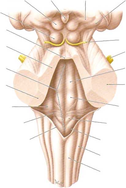
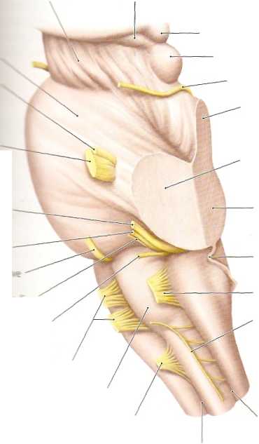
a Anterlorview. The sites ofentryand emergence oftheten pairs oftrue cranial nerves (III—XII) are particularly well displayed in this view. Notę: Cranial nerve II (optic nerve) is a derivative of the diencepha-lon. Notę also the site belowthe pyramids where the pyramidal fibers cross over the midline from each side (decussation of the pyramids). Most of the axons of the large motor pathway for the trunk and limbs cross to the opposite side at this level. b Posterior view. Since the cerebellum has been removed, we can see the rhomboid fossa, which forms the floor of the fourth ventricle. The surface of the fossa is raised by several cranial nerve nuclei, which bulge into the fourth ventride. The cerebellum is connected to the brainstem by three cerebellar peduncles on each side:
• Superior cerebellar pedunde
• Middle cerebellar pedunde
• Inferior cerebellar pedunde
The superior and inferior cerebellar peduncles border portions of the rhomboid fossa and thus contribute to the boundaries of the fourth ventride.
c Left lateral view. In addition to the cerebellar peduncles, this view displays the superior and inferior colliculi. Together with their coun-terparts on the right side, the colliculi form the quadrigeminal piąte (see b), which is a prominent structure of the mesencephalon. The two superior colliculi are part of the visual pathway, while the inferior colliculi are part of the auditory pathway. The trochlear nerve (CN IV) runs forward below the inferior coiliculus, and is the only cranial nerve that emerges from the dorsal side of the brainstem. The olive appears as a prominence on the side of the medulla oblongata. The nuclei within the olive function as a relay station for the motor system (see p. 342).
Wyszukiwarka
Podobne podstrony:
skanuj0004 (505) Neuroanatomy6. Brainstem Oariomotor
skanuj0009 (376) Neuroanatomy 9. Spinał Cord.13 Spinał Cord, Topography A Spinał cord and spinał ner
skanuj0015 (279) Neuroanatomy 10. Sectional Anatomy of the Brain -landtudinal Mrafcsure ■PHlfillilll
skanuj0016 (266) Neuroanatomy 10. Sectional Anatomy of the Brain10.4 Coronal Sections: VII and VIII
skanuj0032 (103) Neuroanatomy 9. Spinał Cord9.2 Spinał Cord, Organization of Spinał Cord Segments Ne
skanuj0005 (488) Neuroanatomy 7. Cerebellum7.3 Cerebellar Pedundes and Tracts Neuroanatomy 7. Cerebe
skanuj0005 (505) o)t [dni] Rys. 2.1. Przyrost wytrzymałości betonu w czasie: a - przy różnych cement
skanuj0009 (376) Neuroanatomy 9. Spinał Cord.13 Spinał Cord, Topography A Spinał cord and spinał ner
skanuj0002 (505) Jak rozpoznać, czy dziecko sięga po narkotyki Naśladowanie ich zachowania rozpoczyn
skanuj0016 (266) Neuroanatomy 10. Sectional Anatomy of the Brain10.4 Coronal Sections: VII and VIII
skanuj0003 (522) Neuroanatomy5.
skanuj0005 (488) Neuroanatomy 7. Cerebellum7.3 Cerebellar Pedundes and Tracts Neuroanatomy 7. Cerebe
81898 skanuj0003 (505) Teoretycznym źródłem wyboru kryteriów oceny jakości systemów kształcenia osób
55037 skanuj0032 (103) Neuroanatomy 9. Spinał Cord9.2 Spinał Cord, Organization of Spinał Cord Segme
84692 skanuj0006 (505) mIII Interwencja USA w Iraku Poziom jednostki: w o Saddam Husajn był złym prz
więcej podobnych podstron