P1130001
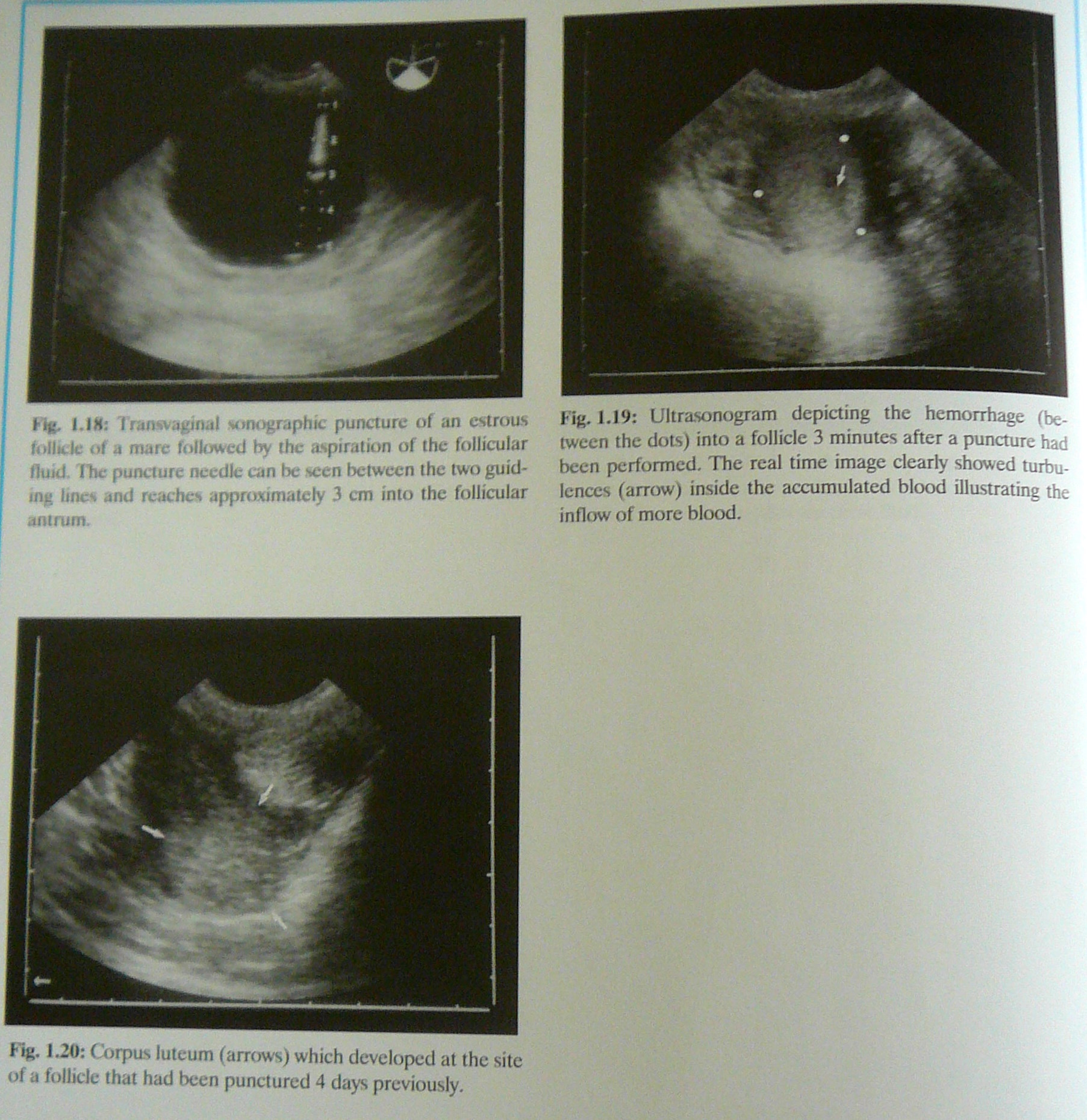
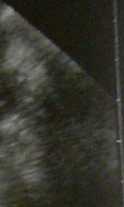
Fig. l.lfl: Transvagjnal sonographic puncture of an estrous foUichs of a fitiiffc foflowcd by the aipiration of the follicular fluid. The puncture needle can be ncen between the two guid-mg iinei and reache* approxtmatcly 3 cm into the follicular ant rum.
Fig. 1.19: Ultrasonogram depicting the hemorrhage (between the dots) into a follicle 3 minutes after a puncture had been perfonmed. The real time image clearly showed turbu-lenccs (arrow) inside the accumulated blood illustrating the inflow of morę blood.
Fig. 1.20: Corpus luteum (arrows) which developed at the site of a follicle that had been punctured 4 days previous!y.
Wyszukiwarka
Podobne podstrony:
43843 P1130002 1.2.1.3 Transvaginal sonographic puncture of fołlicles Transvaginal follide punctnres
20503 P1130038 Fig. 1.81: Transvagina! puncture of an embryonic ycsiclc on Day 29 of gcstution. The
P1130054 Fig- 1.105: Transverse section through the uterine hom (arrows) of a marę with chronię endo
P1130004 1-2.2 Corpora lutea1-22.1 Sonographic images of corpora lutea Useful uhraaomc features tn i
P1130039 Ejcperiences with transvaginal punctures of concep-tuses in the horse are stiłl limitcd. In
P1130005 Fig. 1.24: Intense echogenicily (arrows) at the site of the est-rous follicle one day after
Slajd39 (6) Fig. 10 An allergic reaction to red tattoo pigment is evident (A,B) by the raised scaly
45738 P1130048 Fig. 1.95: Oasct of embryonic mortality on Day 17 of gesta-tion. Signs of abnormality
więcej podobnych podstron