20503 P1130038
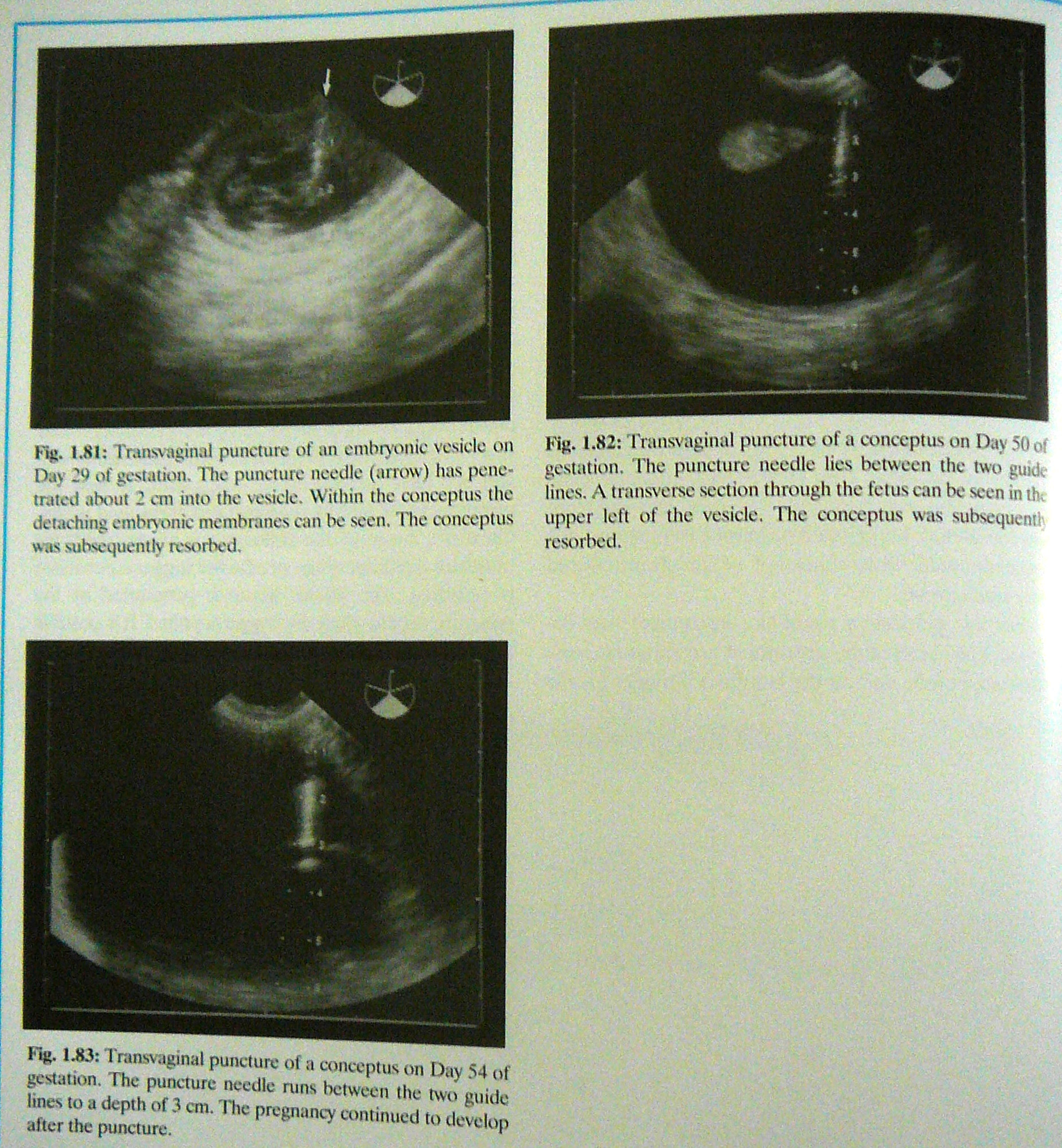
Fig. 1.81: Transvagina! puncture of an embryonic ycsiclc on Day 29 of gcstution. The puncture ncedlc (arrow) has pene-trated about 2 cm into the vesicle. Within the conceptus the detaching embryonic membranes can be seen. The conceptus was suhsequently resorbed.
Fig. 1.82: Transvaginal puncture of a conceptus on Day 50 of gestation. The puncture needle lies between the two gukk lines. A transversc section through the fetus can be scen in ih. upper left of the vesicle. The conceptus was subsequentl resorbed.
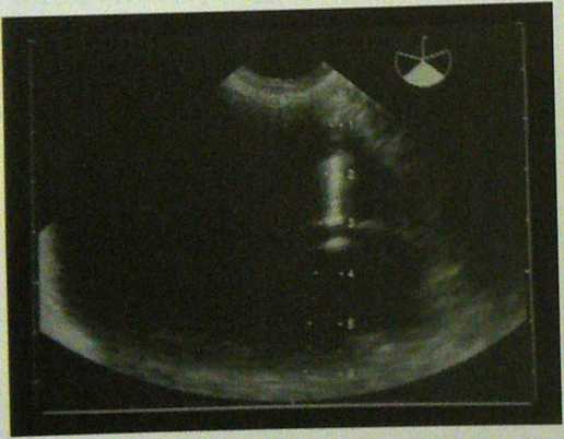
Fig. 1.83: Transvaginal puncture of a conceptus on Day 54 of gcstation. The puncture necdlc runs between the two guide lines to a depth of 3 cm. The prcgnancy continued to develop
afit*r thi* rainPhirft
Wyszukiwarka
Podobne podstrony:
P1130001 Fig. l.lfl: Transvagjnal sonographic puncture of an estrous foUichs of a fitiiffc foflowcd
P1130054 Fig- 1.105: Transverse section through the uterine hom (arrows) of a marę with chronię endo
P1130039 Ejcperiences with transvaginal punctures of concep-tuses in the horse are stiłl limitcd. In
45738 P1130048 Fig. 1.95: Oasct of embryonic mortality on Day 17 of gesta-tion. Signs of abnormality
P1130010 Fig. 13: Corpus hiteum of pregnancy (airows) in a marę on Fig. 130: Two c
P1130040 Fig. 1.84: Transrectal image of the eye and braincase of a fetus on Day 151 of gestation. I
czy CCD IMCD H Fig. 44-1 Segmenta! reabsorption of HC03“. The fraction of the fiitered load of HC03~
P1130005 Fig. 1.24: Intense echogenicily (arrows) at the site of the est-rous follicle one day after
więcej podobnych podstron