P1130040

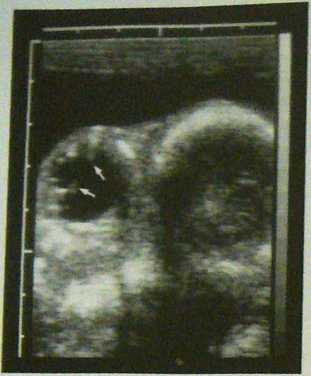
Fig. 1.84: Transrectal image of the eye and braincase of a fetus on Day 151 of gestation. In the anterior aspect of the eye thc caudal wali of the leas (arrows) is depicted.

Fig. 1.85: Longitudinal section through the neck of a fetus or Day 154 of pregnancy. The arches and bodies of three vene brae delineate the spinał canal (S). Behind them shadow arii facts extend into the depth of the image.

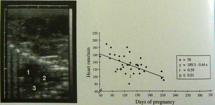
Fig. 1.86: Horizontal section through the thorax of a fetus on Day 134 of gestation. The echoes formed by the cross sections through the ribs of both hałves of the chest run in two lines to-wards each other. Between the two lines three cardiac cham-
Fig. 1.87: The heart ratę of cąuinc fetuses (Thoroughbrcd and Standardbred) during pregnancy (adapted from KAHN and Leidl 1987 a).
Wyszukiwarka
Podobne podstrony:
P1130054 Fig- 1.105: Transverse section through the uterine hom (arrows) of a marę with chronię endo
20503 P1130038 Fig. 1.81: Transvagina! puncture of an embryonic ycsiclc on Day 29 of gcstution. The
34424 P1130050 Fig. 1,96: Uterus 4 days afler the death of the fetus on about Day T) of pregnancy. A
carving?ce@ 40 Rangę of Expressions Fig. 82 (Dęta i I from Mariili on) Fig. 84 shows a variety of an
P1130010 Fig. 13: Corpus hiteum of pregnancy (airows) in a marę on Fig. 130: Two c
P1130005 Fig. 1.24: Intense echogenicily (arrows) at the site of the est-rous follicle one day after
45738 P1130048 Fig. 1.95: Oasct of embryonic mortality on Day 17 of gesta-tion. Signs of abnormality
więcej podobnych podstron