34424 P1130050

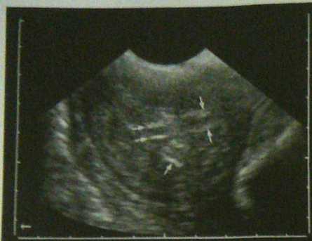
Fig. 1,96: Uterus 4 days afler the death of the fetus on about Day T) of pregnancy. At this ttme the feta! fluids had largely drsappeared. Hyperechoic feta! remnants (arrows) were stili detcctaNc by uttrasonogmphy for another 2 wecks.
Fig. 1.99: Hydrops of the placental membranes in a rnarc i day 230 of gęstation. The excessive accumulation of fluid cl be seen to cxtend beyond the maximal penctration depth the sound waves. The fetus could not be detected by transze tal ułtrasonography.

Fig. 1.101: Norma! invoiution of the uterus m a marc 15 hcnus post partum. The uterine lumen is largely dosed A swal amount of hypoechotc fluid is \isihk between the endom?' tria! fołds. __
Fig. 1.100: Uterus (arrws) of a marę with a norma! jx»tpar-tum period 84 hours (3.5 days) after parturition. Within the utcrus the anechok lochial secredon expanding over a few centuneters can be scen.
Wyszukiwarka
Podobne podstrony:
P1130054 Fig- 1.105: Transverse section through the uterine hom (arrows) of a marę with chronię endo
P1130003 Fig, 1.21: Solid corpus luteum (dots) betwecn several smali follieles on the ovaiy (arrows)
P1130029 1.3.2.3 Day 21 to 40 of pregnancy On about Day 21 thc bmhryo is detecłabie for the tirst «m
P1130052 Fig. 1.103: Postpaitiim uterus (arrows) 3 days after parturi-tion. There is hyperechoic loc
P1130005 Fig. 1.24: Intense echogenicily (arrows) at the site of the est-rous follicle one day after
20503 P1130038 Fig. 1.81: Transvagina! puncture of an embryonic ycsiclc on Day 29 of gcstution. The
45738 P1130048 Fig. 1.95: Oasct of embryonic mortality on Day 17 of gesta-tion. Signs of abnormality
więcej podobnych podstron