75 (101)
7: Viral dermatoses
i
■ft
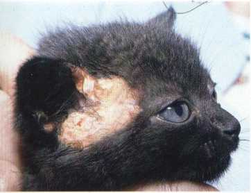
Figurę 7:9:Skinnecrosisofthepretemporalregion ina cat withfeline infectious peritonitis
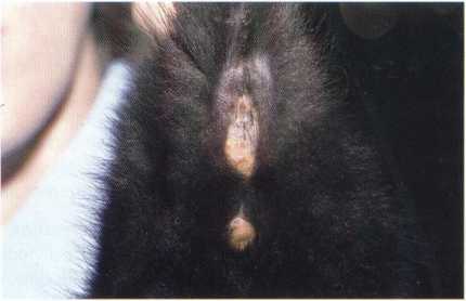
Figurę 7:10: Same cat as in figurę 7:9- notę skin necrosis around anus and genitals
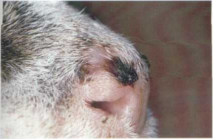
Figurę 7:11: Hyperkeratotic plaques on the nasal płanum ofa cat with multicentric squamous carcinoma in situ (Bowen ’s disease) (courtesy of E. Bensignor)
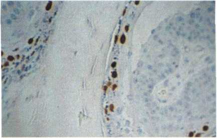
Figurę 7 : 12 : Immunohistochemistry - notę papillomavirus antigens within keratinocytes (x 250) (multicentric squamous celi carcinoma in situ) (AEC stain) (courtesy ofS. Leclercq)
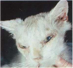
Figurę 7:13: Scaling and crusting on the face of a cat with a giant celi dermatosis associated with FeLV infection (courtesy of D.W. Scott)
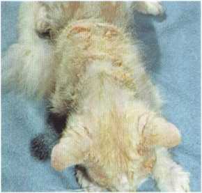
Figurę 7:14: Same cat as in figurę 7: 13 - notę scaling and crusting on the trunk (courtesy of D.W. Scott)
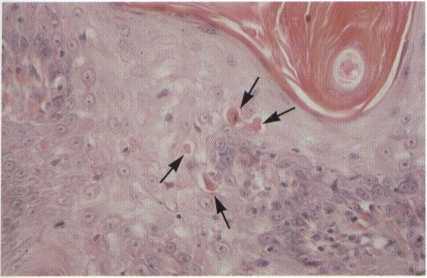
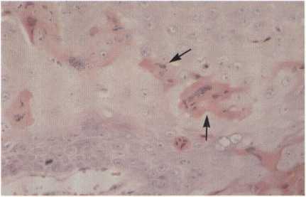
Figurę 7 : 15 : Histopathology - notę many apoptotic keratinocytes f—►) (giant celi dermatosis) (H &E,x 250) (courtesy ofD.W. Scott)
Figurę 7:16 : Histopathology - notę keratinocytic giant cells (—►) (giant celi dermatosis) (H &E,x 250) (courtesy of D.W. Scott)
Wyszukiwarka
Podobne podstrony:
73 (103) 7 : Viral dermatoses Figurę 7:1: Ulcerative lesions on the upper and lower lip ofa cat with
77 (104) 7: Viral dermatoses Figurę 7:17: Epidermal horns on a metacarpal footpad ofa cat infected w
Obraz 2 I ■i 1 ft >■%: < ffT3 WS H ®S I Uh K
IMGV85 75 Ool^bi* »now «i«? uknw»ly 40>. Koniku palne aJuiknly świerszcze na dr
I‘J0A W K K a 75 k iz : 15 xr. (» » » K (V (V I» » K R :t K K K (( n
IMAG0071 (3) BU i®® 3 M i fT f H1 > W % OT &WB BS3S9ja vo‘fioł-HE W v*X a •
•I *«••«* * » ft«>/ «• MH1 •» **•*••* te • • i•• ti (• ^
r •i u * O* b ft r c V;K J) P H A >t M u/m// itMpriitłinnł* KA* h>*.rt.u*ivnni f
DSC00102 (12) •I : l
slide0013 image051 ■i? & m ó 9 fT a s j e a a stji] a f-y.7 j_fi, 9 ■.rfjg a
tnsy1 Tamy (Teiuttłur* Vulgan)—i ft. tali
64 (132) 6: Bacterial dermatoses Figurę 6:2: Secondarypyoderma in a cat with atopic dermatitis Figur
68 (121) 6: Bacterial dermatoses Figurę6:9: Ulceratednodular lesion ina cat with leprosy (courtesy o
71 (116) E. Guaguere - J. DeclercąYiral dermatoses Viral dermatoses are a developing field in feline
QP Codę : 13839 (2 Vi Hours) [ Total Marks : 75 N.B*: ,(,1) ...Ali ąuestions are compuisory T :(£) F
więcej podobnych podstron