64 (132)

Figurę 6:2: Secondarypyoderma in a cat with atopic dermatitis
Figurę6:1:Abscesson thedistal limb, causedby Pasteurellamultocida
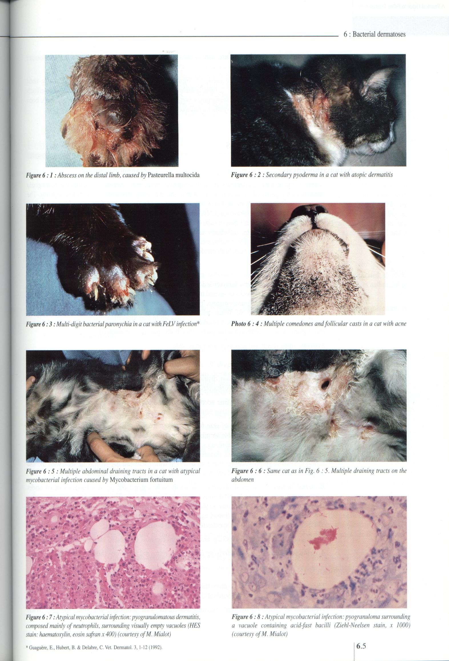
Figurę 6:3: Multi-digit bacterialparonychia in a cat with FeLVinfection*
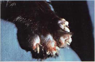
Photo 6:4: Multiple comedones and follicular casts in a cat with acne

Figurę 6:5: Multiple abdominal draining tracts in a cat with atypical mycobacterial infection caused by Mycobacterium fortuitum
Figurę 6:6: Same cat as in Fig. 6:5. Multiple draining tracts on the abdomen


Figurę 6:7: Atypical mycobacterial infection: pyogranulomatous dermatitis, composed mainly ofneutrophils, surrounding \isually empty vacuoles (HES stain: haematoxylin, eosin sąfran x 400) (courtesy ofM. Mialot)
Figurę 6:8: Atypical mycobacterial infection: pyogranuloma surrounding a vacuole containing acid-fast bacilli (Ziehl-Neelsen stain, x 1000) (courtesy ofM. Mialot)
6.5
Guagućre, E., Hubert, B. & Delabre, C. Vet. Dermatol. 3,1-12 (1992).
Wyszukiwarka
Podobne podstrony:
95 (100) 9 : Flea allergy dermatitis Figurę 9:9: Extensive eosinophilic plaques in a cat with FAD Fi
29 (352) 2: Diagnostic approach Figurę 2:17: Sclerosis in a cat with morphea (courtesy of E. Bensign
53 (169) 5 : Deep mycoses Figurę 5 :2 : Nasal nodule in a cat with phaeohyphomycosis Figurę 5:1: Ulc
38 (245) 3 : Ectoparasitic skin diseases Figurę 3 :17: Circular lesion of erythema and scaling in a
61 (139) R. S. MuellerBacterial dermatoses Bacterial dermatoses, also called pyodermas, are rare in
68 (121) 6: Bacterial dermatoses Figurę6:9: Ulceratednodular lesion ina cat with leprosy (courtesy o
43 (223) 4: Dermatophytosis Figurę 4:1: Erythematous blepharitis with comedones in a Persian cat wit
45 (221) Figurę4:9:Generalisedexfoliative dermatitis in a Persian cat with dermatophytosis caused by
66 (126) 6: Bacterial dermatoses antibiotics have been used in the treatment of atypical mycobacteri
73 (103) 7 : Viral dermatoses Figurę 7:1: Ulcerative lesions on the upper and lower lip ofa cat with
243 (19) 24 : Diagnostic approach to feline pododermatoses Figurę 24: 2 : Alopecia and rudimentary c
249 (17) 24 : Diagnostic approach to feline pododermatoses Figurę 24:26: Multiple bacterial paronych
25 (430) Figurę 2:1: Generalised facial erythema in a cat withfood intolerance2: Diagnostic approach
Flowerd r r ^bloem 64 Borduur eerst de takken ais in het voorbeeid en daarna de&nb
IMAG0149 (7) once everytwo seconds. In the stand by State only the receiver portion of the SART is p
132 Zbigwew //<ę* ___ negotiatc thc agrccmcnt in ąuestion and spedfy how i
Document 2 (11) Figurę 2.13: Pronounced gynecomastia in a man with ReifensteirTs syndrome. For cosme
więcej podobnych podstron