95 (100)
9 : Flea allergy dermatitis
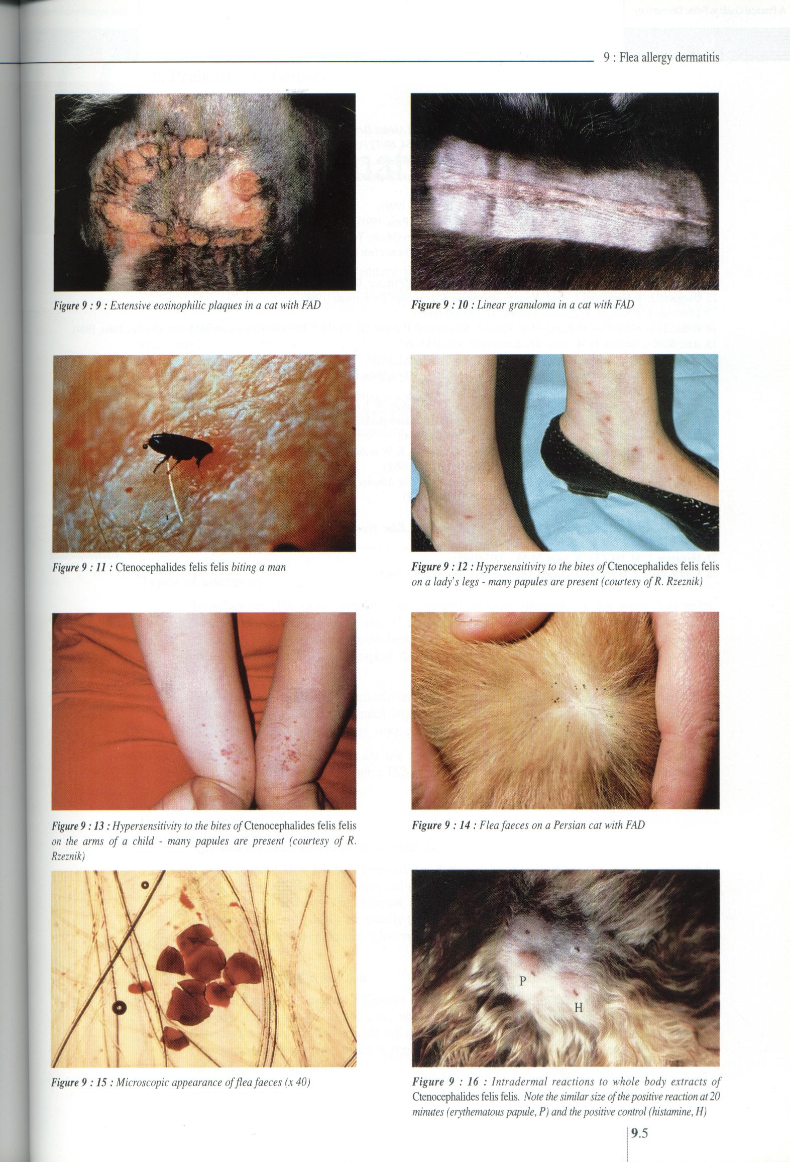
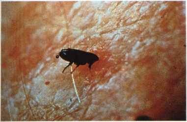
Figurę 9:9: Extensive eosinophilic plaques in a cat with FAD
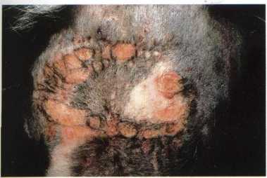
Figurę 9 :10: Linear granuloma in a cat with FAD
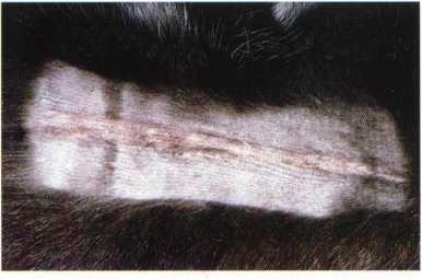
Figurę 9 :11: Ctenocephalides felis felis biting a man Figurę 9:12: Hypersensitivity to the bites of Ctenocephalides felis felis
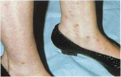
on a lady’s legs - many papules are present (courtesy ofR. Rzeznik)
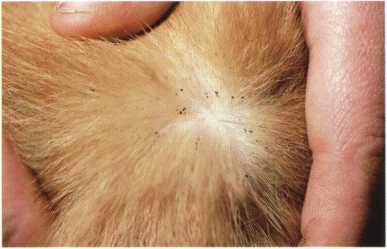
Figurę 9:14 : Fleafaeces on a Persian cat with FAD
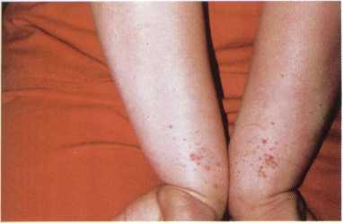
Figurę 9:13: Hypersensitmty to the bites of Ctenocephalides felis felis on the arms of a child - many papules are present (courtesy of R. Rzeznik)
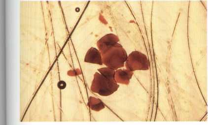
Figurę 9 :15: Microscopic appearance offleafaeces (x 40)
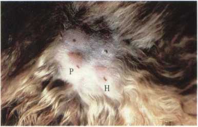
Figurę 9 : 16 : lntradermal reactions to whole body extracts of Ctenocephalides felis felis. Notę the similar size ofthe positive reaction at 20 minutes (erythematous papule, P) and the positive control (histaminę, H)
19.5
Wyszukiwarka
Podobne podstrony:
93 (100) 9 : Flea allergy dermatitis Figurę 9:1: Self-induced dorsolumbar alopecia in a Persian cal
91 (103) M. W. YroomFlea allergy dermatitis Flea allergy dermatitis (FAD) is the most common pruriti
64 (132) 6: Bacterial dermatoses Figurę 6:2: Secondarypyoderma in a cat with atopic dermatitis Figur
- 139 - a) 6 mgm/cm2 100 kV 5 m A 50 HOURS FIGURĘ 10. b) 7 mgm/cm2 100 k V 5 m A 50 HOURSA
29 (352) 2: Diagnostic approach Figurę 2:17: Sclerosis in a cat with morphea (courtesy of E. Bensign
53 (169) 5 : Deep mycoses Figurę 5 :2 : Nasal nodule in a cat with phaeohyphomycosis Figurę 5:1: Ulc
38 (245) 3 : Ectoparasitic skin diseases Figurę 3 :17: Circular lesion of erythema and scaling in a
skanuj0022 2 Duża 60 70 Ciężarne II. III trymestr 70 80 Karmiące 95 100 Powyżej 60
68 (121) 6: Bacterial dermatoses Figurę6:9: Ulceratednodular lesion ina cat with leprosy (courtesy o
73 (103) 7 : Viral dermatoses Figurę 7:1: Ulcerative lesions on the upper and lower lip ofa cat with
77 (104) 7: Viral dermatoses Figurę 7:17: Epidermal horns on a metacarpal footpad ofa cat infected w
83 (123) 8 : The cat flea: applied biology Figurę8:3: Severalfemale Ctenocephalides felis feeding on
43 (223) 4: Dermatophytosis Figurę 4:1: Erythematous blepharitis with comedones in a Persian cat wit
45 (221) Figurę4:9:Generalisedexfoliative dermatitis in a Persian cat with dermatophytosis caused by
47 (209) 4: Dermatophytosis Figurę 4 :17: Infected hairs in chloral lactophenol. Compare ectothrix i
P1030653 90 270 90 270 95 100 Mors dicit
10 1 Fascia lata Medial collateral ligament Patellar ligament Extensor retinacula Figurę 10-1 Anatom
10 41 Dorsal interossei Figurę 10-41 Anatomy of extensor digitorum brevis Extensor digitorum brevis
więcej podobnych podstron