1002889827
70 A. Pielesz i inni
Summary
Background. The formation of AGEs progressively increases with normal aging, even in the absence of disease (the patho-genesis of diabetes associated vascular disorders and neurodegenerative diseases, including Alzheimer’s disease, Parkinson’s disease). However, they are formed at accelerated rates in age-related diseases. The polysaccharides might play a role in wound healing, both internally and externally, and also that they could play a role against inflammation and may lead to the production of better medicines to be used as supplements in cancer treatment.
Objectives. The acid hydrolysis was studied with H2SO4 at 80% concentration to determine the most effective procedurę for total hydrolysis of [5-glucan. The standard of p-glucans acid hydrolysate were compared for commercial oat and oatmeal, mushrooms: Pleurotus ostreatus, Fungus and yeast Saccharomyces cerevisiae.
Materiał and Methods. The following materials and reagents were used in the examination: reference (3-(l—»3)-(l—»6)-glucan, oat and oatmeal, mushrooms: Pleurotus ostreatus, Fungus and yeast Saccharomyces cerevisiae.
The Raman spectra of the sample Solutions ((S-glucan acid hydrolysates) were recorded on a MAGNA-IR 860 with FT-Raman accessory. Sample was irradiated with a 1064 nm linę of the T10-8S Nd spectra-physics model: YAG laser and scattered radia-tion were collected at 180°, using 4 cm'1 resolution.
The polysaccharide was hydrolyzed into component monosaccharides with 80% H2S04 at 0°C for 30 minutes and monosac-charide derivatives were subjected to electrophoresis, as in a ealier authors study, on a strip of cellulose acetate membranę (CA-SYS-MINI Cellulose Acetate Systems) in 0.2 M Ca(OAc)2 (pH 7.5) at 10 mA, max. 240 V for 1.5 h. The strips were stained with 0.5% toluidine blue in 3% HOAc solution and then rinsed in distilled water and air-dried [1],
Results and conclusions. A part of the hexoses (for example glucose) are converted, to products such as 5-hydroxymethyl-furfural. Various coloured substances, through the Maillard reaction have been reported for saccharides. The resulting mono-and oligosaccharides were analysed by cellulose acetate membranę electrophoresis CAE and Raman spectroscopy.
Individual bands or CAE spots were selected to monitor the sugar content in medical plant celi walls and to conflrm the iden-tity of the analysed sample: oat and oatmeal, mushrooms: Pleurotus ostreatus, Fungus and yeast Saccharomyces cerevisiae.. The possibility of a taxonomic classification of products rich in celi-wali materials based on cellulose acetate membranę electrophoresis CAE and Raman spectroscopy for authentication and detection of adulteration of products are discussed (Polim. Med. 2012,42,1, 69-77).
Key words: Raman spectroscopy, cellulose acetate membranę electrophoresis, P-glucans, the Maillard reaction.

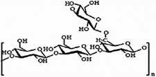
Fig. 1. The structure of P-glucans from Saccharomyces cer-evisiae
Wprowadzenie
Beta-glukany są typowymi polisacharydami występującymi w ścianach komórkowych [2-8] wielu zbóż, grzybów, drożdży a także ziół, alg i niektórych traw (np. opuncji, porostu islandzkiego). Występujące np. w ścianie komórkowej drożdży (Saccharomyces cerevisiae) glukany są polimerami D-glukozy, zbudowanymi z rdzenia glukozowego połączonego wiązaniami p-1,3, od którego odchodzą krótkie boczne łańcuchy połączone z nim za pomocą wiązań (3-1,6 (rycina 1):
Ryc. 1. Budowa P-glukanów występujących w ścianie komórkowej drożdży (Saccharomyces cerevisiae)
Pozostała część ściany komórkowej drożdży jest zbudowana z mannozy 31%, białek 13%, lipidów 9% oraz chityny stanowiącej 1-2% ogólnego składu ściany. W zależności od pochodzenia p-glukany wykazują różnice w swojej strukturze i tworzą cząstki liniowe, rozgałęzione oraz cykliczne, co ma istotny wpływ na biologiczną aktywność tych związków. Wyróżnia się dwa główne typy polisacharydów ściany komórkowej: sztywne włókienko-we polimery chitynowe lub celulozowe, oraz wypełniające wolne przestrzenie a- i p- glukany oraz glikoproteiny. Polisacharydy luźno związane z zewnętrzną warstwą ściany komórkowej, jako rozpuszczalne w wodzie mogą być wydzielane poza komórkę. Jak zaznacza M. Gibiński „P-glukany stanowią około 50% rozpuszczalnych związków błonnika. Błonnik składa się z celulozy, hemiceluloz i pektyn. Hemicelulozy mają złożoną budowę strukturalną i relatywnie niski ciężar molekularny, złożone są z pentozanów (łańcuchy ksylozy i arabinozy) oraz hekso-zanów (polimery glukozy, galaktozy i mannozy), których główną frakcją rozpuszczalną w wodzie są P-glukany” [5]. Inną, grupę polisacharydów biologicznie czynnych stanowią heteroglikany, które dzieli się na galaktany, fu-kany, ksylany i mannany na podstawie składników cukrowych tworzących ich szkielet. Łańcuchy boczne hete-roglikanów mogą zawierać arabinozę, mannozę, fukozę, galaktozę, ksylozę, kwas glukuronowy i glukozę jako główne składniki połączone ze sobą w różny sposób.
Wykazano [5, 9], że rozpuszczalny w wodzie błonnik, tj. p-glukan zmniejsza ryzyko chorób cywiliza-
Wyszukiwarka
Podobne podstrony:
•9 TJN DEBAT": LES MENTAUTE3 COLLECTIVES 639 The contribution of rural elements to the for
S5001378 (2) CELTIC SETTLEMENT IN SLOVAKIA YOUNO LA TŻNE PERIOD the Danube region stood at the forma
•9 TJN DEBAT": LES MENTAUTE3 COLLECTIVES 639 The contribution of rural elements to the for
51. B.111820 THE FORMATION of Capital / by Harold G. Moulton. - Washington : Center for Economic and
PRESENTATIONS Sebald, Brigita. 2012. "Social NetWork Sites and the Formation of Musical Taste,”
sulf of sulfate that is activated by the formation of adenosine phosphosulfate (APS) and its subsequ
Artykuły w języku angielskim: 1) P. Tosiek, The Formation of Political Groups in t
195 involved in complex reactions, sintering process results in the formation of new crystalline and
96 McAdoo, William Gibbs, 1863-1941. A leaguc to prcvcnt war: with a rcvicw of the tight against the
Zdjęcie032(2) 10 Km Diagram showing how sea water circulation through the crust might give ri*e to t
The Factors of Influence on the Formation of a Reg i o na I Transport-Logistic System The morę moder
The Factors of Influence on the Formation of a Regional Transport-Logistic SystemConclusions of the
From the Editor The format of the current issue of the journal is quite unusual due to the fact that
The Factors of Influence on the Formation of a Regional Transport-Logistic System Regional logistic
218 JAN KIENIEWICZ, MARCIN KULA Works dealing with problems of the formation of nations and national
J. P. Bakker who engaged himself in the formation of this Subcom-mission. Lectures on the principles
więcej podobnych podstron