
Cancers of the Female
Reproductive Tract
8
chapter
Key
TERMS
cervical cancer
cervical dysplasia
colposcopy
cone biopsy
cryotherapy
endometrial cancer
human papillomavirus
ovarian cancer
Papanicolaou (Pap) test
vaginal cancer
vulvar cancer
Learning
OBJECTIVES
After studying the chapter content, the student should be able to
accomplish the following:
1. Define the key terms.
2. Identify the major modifiable risk factors for reproductive tract cancers.
3. Discuss the risk factors, screening methods, and treatment modalities for
cancers of the reproductive tract.
4. Outline the nursing management needed for the most common malignant
reproductive disorders in women.
5. Describe lifestyle changes and health screenings needed to reduce risk or
prevent reproductive tract cancers.
6. List community resources available for the women undergoing surgery for a
malignant reproductive condition.
Key
Learning
3132-08_CH08.qxd 12/15/05 3:12 PM Page 167

ancer is the second leading
cause of death for women in the United States, surpassed
only by cardiovascular disease (Youngkin & Davis, 2004).
Obviously, cardiovascular disease is and must continue
to be a major focus of our efforts in women’s health.
However, we must not lose sight of the fact that a large
number of women between the ages of 35 and 74 are
developing and dying of cancer (NCI, 2004). Women
have a one in three lifetime risk of developing cancer, and
one out of every four deaths is from cancer (Alexander
et al., 2004). African-American women have the highest
death rates from both heart disease and cancer (Breslin
& Lucas, 2003).
It has been estimated that in the United States half of
all premature deaths, one third of acute disabilities, and
one half of chronic disabilities are preventable (NCI,
2004). Nurses need to put their energies into screening,
education, and early detection to reduce these statistics.
Because cancer risk is strongly associated with lifestyle and
behavior, screening programs are of particular importance
for early detection. There is evidence that prevention and
early detection have reduced both cancer mortality rates
and prevented reproductive cancers (Smith et al., 2004).
This chapter will cover selected cancers of the repro-
ductive system and will identify the appropriate screenings
needed. The reproductive cancers to be discussed are cer-
vical, endometrial, ovarian, vaginal, and vulvar.
Cervical Cancer
Cervical cancer
is cancer of the uterine cervix. The
American Cancer Society (ACS) estimates that over
10,000 cases of invasive cervical cancer will be diag-
nosed in the United States in 2005. Of that number,
approximately 4,000 women will die. Some researchers
estimate that noninvasive cervical cancer (carcinoma in
situ) is about four times more common than invasive
cervical cancer. The 5-year survival rate for all stages
of cervical cancer is 73% (ACS, 2005). The median age
at diagnosis for cervical cancer is 47 years, and nearly
half of all cases are diagnosed before the age of 35
(Waggoner, 2003).
Cervical cancer incidence and mortality rates have
decreased noticeably in the past several decades, with most
of the reduction attributed to the
Papanicolaou (Pap)
test,
which detects cervical cancer and precancerous
lesions. Cervical cancer is one of the most treatable can-
cers when detected at an early stage (ACS, 2005). Healthy
People 2010 (USDHHS, 2000) identifies two goals that
address cervical cancer (Healthy People 2010).
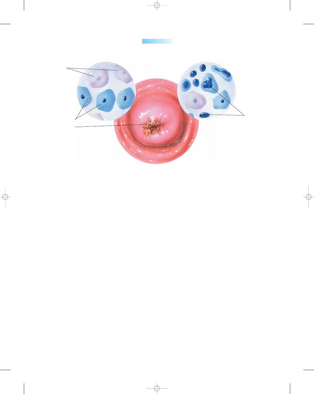
Pathophysiology
Cervical cancer starts with abnormal changes in the cel-
lular lining or surface of the cervix. Typically these
changes occur in the squamous–columnar junction of the
cervix. Here, cylindrically shaped secretory epithelial cells
(columnar) meet the protective flat epithelial cells (squa-
mous) from the outer cervix and vagina in what is termed
the transformation zone. The continuous replacement of
columnar epithelial cells by squamous epithelial cells in
this area makes these cells vulnerable to take up foreign or
abnormal genetic material (Adams, 2002). Figure 8-1
shows the pathophysiology of cervical cancer.
Etiology and Risk Factors
The primary factor in the development of cervical cancer
is
human papillomavirus
(HPV), which is acquired
through sexual activity (Roye et al., 2003). More than 90%
of squamous cervical cancers contain HPV DNA, and the
virus is now accepted as a major causative factor in the
development of cervical cancer and its precursor,
cervical
dysplasia
(disordered growth of abnormal cells).
Risk factors associated with cervical cancer include:
•
Early age at first intercourse (within 1 year of menarche)
•
Lower socioeconomic status
•
Promiscuous male partners
•
Unprotected sexual intercourse
C
HEALTHY PEOPLE
2010
National Health Goals Related to Cervical Cancer
Objective
Significance
Goal 3–4—Reduce the
death rate from cancer
of the uterine cervix
from 3 per 100,000
females (1998) to 2 per
100,000 females in 2010.
Goal 3–11—Increase the
proportion of women
who received a Pap
smear within the pre-
ceding 3 years from
79% to 90% by 2010.
This will help improve mor-
tality rates and quality
of life for women, and
reduce healthcare costs
related to treatment of
malignancies.
This will help to promote
screening and early
detection. The National
Institutes of Health (NIH)
reported that half of
women diagnosed with
invasive cervical can-
cer have never had a
Pap smear and 10%
have not had Pap
smears during the past
5 years (NIH, 2005).
168
3132-08_CH08.qxd 12/15/05 3:12 PM Page 168
•
Family history of cervical cancer (mother or sisters)
•
Sexual intercourse with uncircumcised men
•
Female offspring of mothers who took diethylstilbestrol
(DES)
•
Infections with genital herpes or chronic chlamydia
•
History of multiple sex partners
•
Cigarette smoking
•
Immunocompromised state
•
HIV infection
•
Oral contraceptive use
•
Moderate dysplasia on Pap smear within past 5 years
•
HPV infection (Grund, 2005)
Clinical Manifestations
Clinically, the first symptom is abnormal vaginal bleeding,
usually after sexual intercourse. Vaginal discomfort, mal-
odorous discharge, and dysuria are common manifesta-
tions also. Some women with cervical cancer have no
symptoms. Frequently it is detected at an annual gyne-
cologic examination and Pap test. Advanced symptoms of
cervical cancer may include pelvic, back, or leg pain, weight
loss, anorexia, weakness and fatigue, and bone fractures.
Diagnosis
Screening for cervical cancer is very effective because the
presence of a precursor lesion, cervical intraepithelial neo-
plasia (CIN), helps determine whether further tests are
needed. Lesions start as dysplasia and progress in a pre-
dictable fashion over a long period, allowing ample oppor-
tunity for intervention at a precancerous stage. Progression
from low-grade to high-grade dysplasia takes an average of
9 years, and progression from high-grade dysplasia to inva-
sive cancer takes up to 2 years (Jemal et al., 2005).
Widespread use of the Pap test (also known as a Pap
smear), a procedure used to obtain cells from the cervix for
cytology screening, is credited with saving tens of thou-
sands of women’s lives and decreasing deaths from cer-
vical cancer by more than 70% (ACS, 2005) (Nursing
Procedure 8-1: Assisting with Collection of a Pap Smear).
Despite its outstanding record of success as a screening
tool for cervical cancer (it detects approximately 90%
of early cancer changes), the conventional Pap smear
has a 20% false-negative rate. High-grade abnormalities
missed by human screening are frequently detected by
computerized instruments (Garcia & Bi, 2004). Thus,
many new technologies are being studied and introduced
clinically, including:
•
Automated slide thin-layer preparation (Thin-Prep): In this
liquid-based cervical cytology technique, the cervical
specimen is placed into a vial of fixative solution rather
than on the glass slide.
•
Computer-assisted automated Pap test rescreening (Autopap):
An algorithm-based decision-making technology that
identifies slides that should be rescreened by cytopathol-
ogists by selecting samples that exceed a certain threshold
for the likelihood of abnormal cells
•
HPV-DNA typing (Hybrid Capture): This system uses the
association between certain types of HPV (16, 18, 31, 33,
Chapter 8
CANCERS OF THE FEMALE REPRODUCTIVE TRACT
169
Carcinoma in situ
Normal cells
Pre-malignant cells
Ectocervical lesion
Squamous cell carcinoma
Malignant cells
●
Figure 8-1
Cervical cancer. (The Anatomical Chart Company. [2002]. Atlas of
pathophysiology. Springhouse, PA: Springhouse.)
3132-08_CH08.qxd 12/15/05 3:13 PM Page 169

170
Unit 2
WOMEN’S HEALTH THROUGHOUT THE LIFESPAN
Nursing Procedure
8-1
Assisting With Collection of a Pap Smear
Purpose: To Obtain Cells From the Cervix for Cervical Cytology Screening
1. Explain procedure to the client (Fig. A).
2. Instruct client to empty her bladder.
3. Wash hands thoroughly.
4. Assemble equipment, maintaining sterility of
equipment (Fig. B)
5. Position client on stirrups or foot pedals so that
her knees fall outward.
6. Drape client with a sheet for privacy, covering
the abdomen but leaving the perineal area
exposed.
7. Open packages as needed.
8. Encourage client to relax.
9. Provide support to client as the practitioner
obtains a sample by spreading the labia;
inserting the speculum; inserting the cytobrush
and swabbing the endocervix; and inserting
the plastic spatula and swabbing the cervix
(Fig. C–H).
10. Transfer specimen to container (Fig. I) or slide.
If a slide is used, spray the fixative on the slide.
11. Place sterile lubricant on the practitioner’s
fingertip when indicated for the bimanual
examination.
12. Wash hands thoroughly.
13. Label specimen according to facility policy.
14. Rinse reusable instruments and dispose of waste
appropriately (Fig. J).
15. Wash hands thoroughly.
A
B
C
D
E
F
3132-08_CH08.qxd 12/15/05 3:13 PM Page 170

35, 45, 51, 52, and 56) and the development of cervical
cancer. This system can identify high-risk HPV types and
improves detection and management.
•
Computer-assisted technology (Cytyc CDS-1000, AutoCyte,
AcCell): These computerized instruments can detect
abnormal cells that are sometimes missed by technolo-
gists (Anderson & Runowicz, 2002).
Other factors contributing to the high rate of false-
negative results include errors in sampling the cervix, in
preparing the slide, and in patient preparation. To opti-
mize conditions for Pap smear collection, nurses can offer
the instructions provided in Teaching Guidelines 8-1.
Although many professional medical organizations
disagree as to the frequency of screening for cervical can-
cer, the ACS 2003 guidelines suggest that women should
begin annual screening for cervical cancer via a Pap test
after they initiate sexual activity or at 21 years of age,
whichever comes first. If three consecutive Pap smears
are negative, a trained healthcare provider may suggest
that screening can be performed less frequently. Women
ages 65 to 70 with no abnormal tests in the previous
10 years may choose to stop screenings (ACS, 2003).
Chapter 8
CANCERS OF THE FEMALE REPRODUCTIVE TRACT
171
Used with permission from Klossner, N. J. (2006). Introductory maternity nursing. Philadelphia: Lippincott Williams & Wilkins.
Nursing Procedure
8-1
Assisting With Collection of a Pap Smear
(Continued)
G
H
I
J
T E A C H I N G G U I D E L I N E S 8 - 1
Strategies to Optimize Pap Smear Results
•
Schedule your Pap smear appointment about 2 weeks
(10 to 18 days) after the first day of your last menses
to increase the chance of getting the best sample of
cervical cells without menses.
•
Refrain from intercourse for 48 hours before the test
because additional matter such as sperm can obscure
the specimen.
•
Do not douche within 48 hours before the test to
prevent washing away cervical cells that might be
abnormal.
•
Do not use tampons, birth control foams, jellies,
vaginal creams, or vaginal medications for 72 hours
before the test, as they could cover up or obscure the
cervical cell sample.
•
Cancel your Pap appointment if vaginal bleeding
occurs, because the presence of blood cells
interferes with visual evaluation of the sample
(Ross, 2003).
3132-08_CH08.qxd 12/15/05 3:13 PM Page 171

For high-risk women, annual Pap smears should con-
tinue annually throughout their life (Table 8-1).
Pap smear results are classified using the Bethesda
System (Box 8-1), which provides a uniform diagnostic
terminology that allows clear communication between
the laboratory and the healthcare provider. The health-
care provider receives the laboratory information divided
into three categories: specimen adequacy, general cate-
gorization of cytologic findings, and interpretation/result
(ACS, 2005).
Treatment
Using the 2001 Bethesda system, the following manage-
ment guidelines were developed by the National Cancer
Institute (NCI) to provide direction to healthcare providers
and their patients to deal with abnormal Pap smear results:
•
ASC-US: Repeat the Pap smear in 4 to 6 months or
refer for colposcopy.
•
ASC-H: Refer for colposcopy without HPV testing.
•
Atypical glandular cells (AGC) and adenocarcinoma in
situ (AIS): Immediate colposcopy; follow-up is based
on the results of findings.
Colposcopy
is a microscopic examination of the
lower genital tract using a magnifying instrument called a
colposcope. Specific patterns of cells that correlate well
with certain histologic findings can be visualized. With the
woman in lithotomy position, the cervix is cleansed with
acetic acid solution. Acetic acid makes abnormal cells
appear white, which is referred to as acetowhite. These
white areas are then biopsied and sent to the pathologist
for tissue assessment. The examination is not painful, it
has no side effects, and it can be performed safely in the
healthcare provider’s office.
Treatment options available for abnormal Pap smears
depend on the severity of the results and the health history
of the woman. Therapeutic choices all involve destruction
of as many affected cells as possible. Box 8-2 describes
treatment options.
Nursing Management
The nurse’s role involves primary prevention through
education of women regarding risk factors and preventive
techniques to avoid cervical dysplasia. Secondary preven-
tion focuses on reducing or limiting the area of cervical
172
Unit 2
WOMEN’S HEALTH THROUGHOUT THE LIFESPAN
Table 8-1
First Pap
Age 21 or within 3 years of first
sexual intercourse
Until age 30
Yearly—using glass slide method
Every 2 years—using liquid-based
method
Age 30–70
Every 2–3 years if last 3 Paps were
normal
After age 70
May discontinue if:
- Past 3 Paps were normal and
- No Paps in the past 10 years were
abnormal
Table 8-1
Pap Smear Guidelines
American Cancer Society (ACS). (2005).
How Pap test results
are reported. American Cancer Society, Inc. [Online]
Available at: http://www.cancer.org/docroot/PED/
content/PED_2_3X_Pap_Test.asp.
Specimen Type: Conventional Pap smear vs. liquid-
based
Specimen Adequacy: Satisfactory or unsatisfactory
for evaluation
General Categorization: (optional)
• Negative for intraepithelial lesion or malignancy
• Epithelial cell abnormality. See interpretation/result
Automated Review: If case was examined by auto-
mated device or not
Ancillary Testing: Provides a brief description of the
test methods and report results so healthcare provider
understands
Interpretation/Result:
• Negative for intraepithelial lesion or malignancy
• Organisms: Trichomonas vaginalis; fungus; bacterial
vaginosis; herpes simplex
• Other non-neoplastic findings: Reactive cellular
changes associated with inflammation, radiation,
IUDs, atrophy
• Other: Endometrial cells in a woman >40 years of age
• Epithelial cell abnormalities:
• Squamous cell
- Atypical squamous cells
- Of undetermined significance (ASC-US)
- Cannot exclude HSIL (ASC-H)
- Low-grade squamous intraepithelial lesion (LSIL)
- Encompassing HPV/mild dysplasia/CIN-1
- High-grade squamous intraepithelial lesion (HSIL)
- Encompassing moderate and severe dysplasia
CIS/CIN-2 and CIN-3
- With features suspicious for invasion
- Squamous cell carcinoma
• Glandular Cell: Atypical
- Endocervical, endometrial, or glandular cells
- Endocervical cells—favor neoplastic
- Glandular cells—favor neoplastic
- Endocervical adenocarcinoma in situ
- Adenocarcinoma
- Endocervical, endometrial, extrauterine
• Other malignant neoplasms (specify)
Educational Notes and Suggestions: (optional)
BOX 8-1
THE 2001 BETHESDA SYSTEM FOR
CLASSIFYING PAP SMEARS
Sources: NIH, 2002; Apgar & Wright, 2003; ACS, 2005
3132-08_CH08.qxd 12/15/05 3:13 PM Page 172

dysplasia. Tertiary prevention focuses on minimizing dis-
ability or spread of cervical cancer. Specific areas of edu-
cation include:
•
Encourage prevention of STIs to reduce risk factors
(see Chapter 5: Sexually Transmitted Infections).
•
Counsel teenagers to avoid early sexual activity.
•
Encourage pelvic rest for a month after any cervical
treatment.
•
Screen for cervical cancer by annual Pap smears.
•
Identify high-risk behavior and how to reduce it.
•
Make sure the Pap smear is sent to an accredited labora-
tory for interpretation.
•
Encourage the faithful use of barrier methods of con-
traception.
•
Encourage cessation of smoking and drinking.
•
Reinforce guidelines for Pap smears and sample pre-
paration.
•
Remind all women about follow-up procedures and
times.
•
Explain in detail all procedures that might be needed.
•
Outline proper preparation before having a Pap smear.
•
Provide emotional support throughout the decision-
making process.
•
Inform all women of community resources available
to them.
Nursing Care Plan 8-1: Overview of a Woman With
Cervical Cancer highlights specific nursing interventions.
Endometrial Cancer
Endometrial cancer
(also known as uterine cancer) is
malignant neoplastic growth of the uterine lining. It is the
most common gynecologic malignancy and accounts for
6% of all cancers in women in the United States. The NCI
estimates that there will be over 40,000 new cases in 2005,
of which approximately 7,000 women will die (NCI,
2005). It is uncommon before the age of 40, but as
women age their risk of endometrial cancer increases.
Approximately 95% of these malignancies are carcinomas
of the endometrium. The most common symptom in up
to 90% of women is postmenopausal bleeding. Most
women recognize the need for prompt evaluation, so the
majority of women are diagnosed in an early stage of the
disease (Winter & Gosewehr, 2004).
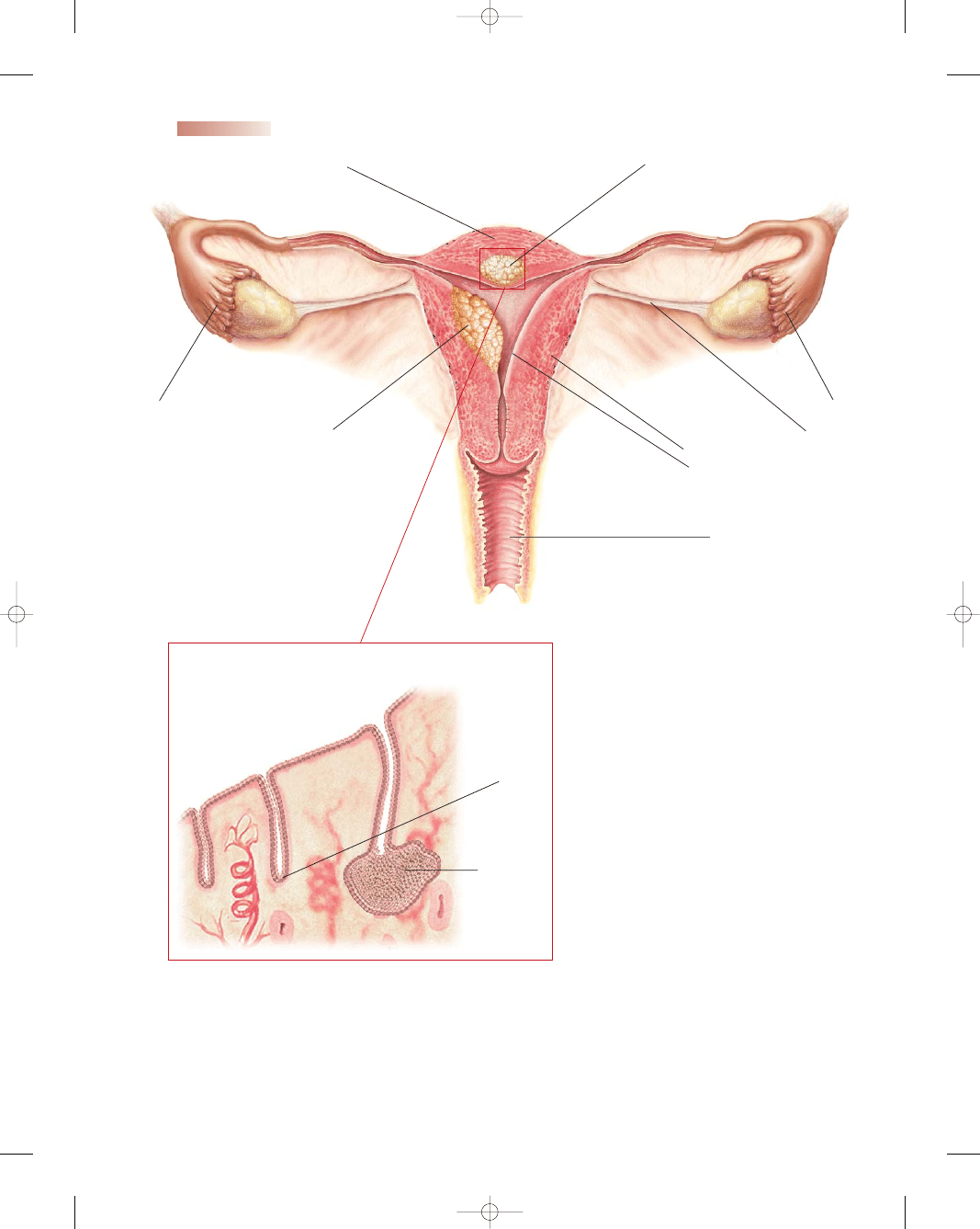
Pathophysiology
Endometrial cancer may originate in a polyp or in a diffuse
multifocal pattern. The pattern of spread partially depends
on the degree of cellular differentiation. Well-differentiated
tumors tend to limit their spread to the surface of the
endometrium. Metastatic spread occurs in a characteristic
pattern and most commonly involves the lungs, inguinal
and supraclavicular nodes, liver, bones, brain, and vagina
(NCI, 2005). Early tumor growth is characterized by
friable and spontaneous bleeding. Later tumor growth
is characterized by myometrial invasion and growth
toward the cervix (Fig. 8-2). Adenocarcinoma of the
endometrium is typically preceded by hyperplasia.
Carcinoma in situ is found only on the endometrial sur-
face. In stage I, it has spread to the muscle wall of the
uterus. In stage II, it has spread to the cervix. In stage
III, it has spread to the bowel or vagina, with metastases
to pelvic lymph nodes. In stage IV, it has invaded the
bladder mucosa, with distant metastases to the lungs,
liver, and bone (Brose, 2004).
Chapter 8
CANCERS OF THE FEMALE REPRODUCTIVE TRACT
173
• Cryotherapy—destroys abnormal cervical tissue by
freezing with liquid nitrogen, Freon, or nitrous oxide.
Studies show a 90% cure rate (Youngkin & Davis,
2004). Healing takes up to 6 weeks, and the client
may experience a profuse, watery vaginal discharge for
3 to 4 weeks.
• Cone Biopsy or conization—removes a cone-shaped
section of cervical tissue. The base of the cone is formed
by the ectocervix (outer part of the cervix) and the
point or apex of the cone is from the endocervical canal.
The transformation zone is contained within the cone
sample. The cone biopsy is also a treatment and can
be used to completely remove any precancers and
very early cancers. There are two methods commonly
used for cone biopsies:
▪ LEEP (loop electrosurgical excision procedure) or
LLETZ (large loop excision of the transformation
zone)—the abnormal cervical tissue is removed with
a wire that is heated by an electrical current. For this
procedure, a local anesthetic is used. It is performed
in the healthcare provider’s office in approximately
10 minutes. Mild cramping and bleeding may persist
for several weeks after the procedure.
▪ Cold knife cone biopsy—a surgical scalpel or a laser
is used instead of a heated wire to remove tissue.
This procedure requires general anesthesia and is
done in a hospital setting. After the procedure,
cramping and bleeding may persist for a few weeks.
• Laser therapy—destroys diseased cervical tissue by
using a focused beam of high-energy light to vaporize
it (burn it off). After the procedure, the woman may
experience a watery brown discharge for a few weeks.
Very effective in destroying precancers and preventing
them from developing into cancers.
• Hysterectomy—removes the uterus and cervix
surgically
• Radiation therapy—delivered by internal radium
applications to the cervix or external radiation therapy
that includes lymphatics of the pelvis
• Chemoradiation—weekly cisplatin therapy concur-
rent with radiation. Investigation of this therapy is
ongoing (ACS, 2005).
BOX 8-2
TREATMENT OPTIONS FOR CERVICAL CANCER
3132-08_CH08.qxd 12/15/05 3:13 PM Page 173

174
Unit 2
WOMEN’S HEALTH THROUGHOUT THE LIFESPAN
Outcome Identification and
evaluation
Client will demonstrate measures to cope with anxi-
ety
as evidenced by statements acknowledging
anxiety, use of positive coping strategies, and ver-
balization that anxiety level has decreased.
Interventions with
rationales
Encourage client to express her feelings and con-
cerns
to reduce her anxiety and to determine
appropriate interventions.
Assess the meaning of the diagnosis to the client, clar-
ify misconceptions, and provide reliable, realistic
information
to enhance her understanding of her
condition, subsequently reducing her anxiety level.
Assess client’s psychological status
to determine
degree of emotional distress related to diagnosis
and treatment options.
Identify and address verbalized concerns, providing
information about what to expect
to decrease
level of uncertainty about the unknown.
Assess the client’s use of coping mechanisms in the
past and their effectiveness
to foster use of posi-
tive strategies.
Teach client about early signs of anxiety and help
her recognize them (for example, fast heartbeat,
sweating, or feeling flushed)
to minimize escala-
tion of anxiety.
Provide positive reinforcement that the client’s con-
dition can be managed
to relieve her anxiety.
Molly, a 28-year-old, thin Native American woman, comes to the free health clinic com-
plaining of a thin, watery vaginal discharge and spotting after sex. She reports being home-
less and living “on the streets” for years. Molly admits to having multiple sex partners to
pay for her food and cigarettes. She had an abnormal Pap smear a while back but didn’t
return to the clinic for any follow-up. She hopes nothing “bad” is wrong with her because
she just found a job to get off the streets. Cervical cancer is suspected.
Nursing Care Plan
Nursing Diagnosis: Anxiety related to diagnosis and uncertainty of outcome
Nursing Care Plan
8-1
Overview of a Woman With Cervical Cancer
Client will demonstrate understanding of diagnosis,
as evidenced by making health-promoting
lifestyle choices, verbalizing appropriate health-
care practices, and adhering to measures to
comply with therapy.
Assess client’s current knowledge about her diagno-
sis and proposed therapeutic regimen
to establish
a baseline from which to develop a teaching
plan.
Nursing Diagnosis: Deficient knowledge related to diagnosis, prevention strategies, and treatment
3132-08_CH08.qxd 12/15/05 3:13 PM Page 174

Etiology and Risk Factors
Unopposed endogenous and exogenous estrogens are the
major etiologic risk factors associated with the develop-
ment of this cancer. Other risk factors for endometrial
cancer include:
•
Nulliparity
•
Obesity (>50 pounds overweight)
•
Liver disease
•
Infertility
•
Diabetes mellitus
•
Hypertension
•
History of pelvic radiation
•
Polycystic ovarian syndrome
•
Infertility
•
Early menarche (<12 years old)
•
High-fat diet
•
Use of prolonged exogenous unopposed estrogen with
an intact uterus
•
Endometrial hyperplasia
•
Family history of endometrial cancer
•
Personal history of hereditary nonpolyposis colon cancer
•
Personal history of breast or ovarian cancer
•
Late onset of menopause
•
Tamoxifen use
•
Anovulation (Smith et al., 2004)
Clinical Manifestations and Diagnosis
The major initial symptom of endometrial cancer is abnor-
mal and painless vaginal bleeding. Any episode of bright-
red bleeding that occurs after menopause should be
investigated. Abnormal uterine bleeding is rarely the result
of uterine malignancy in a young woman. In the post-
menopausal woman, however, it should be regarded with
suspicion. Additional clinical manifestations of advanced
disease may include dyspareunia, low back pain, purulent
genital discharge, dysuria, pelvic pain, weight loss, and
a change in bladder and bowel habits.
Screening for endometrial cancer is not routinely done
because it is not practical or cost-effective. The ACS rec-
ommends that women should be informed about the risks
and symptoms of endometrial cancer at the onset of
menopause and strongly encouraged to report any un-
Chapter 8
CANCERS OF THE FEMALE REPRODUCTIVE TRACT
175
Outcome Identification and
evaluation
Interventions with
rationales
Review contributing factors associated with devel-
opment of cervical cancer, including possible
associated lifestyle behaviors,
to foster an under-
standing of the the etiology of cervical cancer.
Review information provided about possible treat-
ments and procedures and recommendations for
healthy lifestyle, obtaining feedback frequently
to
validate adequate understanding of instructions.
Discuss strategies, including using condoms and limit-
ing the number of sexual partners,
to reduce the
risk of transmission of STIs, specifically human papil-
lomavirus (HPV), which is associated with causing
cervical cancer.
Encourage client to obtain prompt treatment of any
vaginal or cervical infections
to minimize the risk
for cervical cancer.
Urge the client to have an annual Pap smear
to
provide for screening and early detection.
Provide written material with pictures
to allow for
client review and help her visualize what is
occurring in her body.
Inform client about available community resources
and make appropriate referrals as needed
to
provide additional education and support.
Document details of teaching and learning
to allow
for continuity of care and further education, if
needed.
Overview of a Woman With Cervical Cancer
(continued)
3132-08_CH08.qxd 12/15/05 3:13 PM Page 175
176
Unit 2
WOMEN’S HEALTH THROUGHOUT THE LIFESPAN
Ovary
Ovarian
ligament
Endometrium
Uterine wall
Vagina
Ovary
Late
endometrial cancer
Fundus
Sarcoma
Normal
glandular
cells
Endometrial
cancer
Advanced endometrial cancer
●
Figure 8-2
Progression of endometrial cancer. (The Anatomical Chart Company. [2002].
Atlas of pathophysiology. Springhouse, PA: Springhouse.)
3132-08_CH08.qxd 12/15/05 3:13 PM Page 176

expected bleeding or spotting to their healthcare provider
(ACS, 2005). A pelvic examination is frequently normal in
the early stages of the disease. Changes in the size, shape,
or consistency of the uterus or its surrounding supporting
structures may exist when the disease is more advanced.
An endometrial biopsy is the procedure of choice to
make the diagnosis. It can be done in the healthcare pro-
vider’s office without anesthesia. A slender suction catheter
is used to obtain a small sample of tissue for pathology. It
can detect up to 90% of cases of endometrial cancer in the
woman with postmenopausal bleeding, depending on the
technique and experience of the healthcare provider
(Burke, 2005). The woman may experience mild cramping
and bleeding after the procedure for about 24 hours, but
typically mild pain medication will reduce this discomfort.
Transvaginal ultrasound can be used to evaluate the
endometrial cavity and measure the thickness of the
endometrial lining. It can be used to detect endometrial
hyperplasia. If the endometrium measures less than 4 mm,
then the client is at low risk for malignancy (Burke, 2005).
Because endometrial cancer is usually diagnosed in
the early stages, it has a better prognosis than cervical or
ovarian caner (Brose, 2004).
Treatment
Treatment of endometrial cancer depends on the stage of
the disease and usually involves surgery with adjunct ther-
apy based on pathologic findings. Surgery most often
involves removal of the uterus (hysterectomy) and the
fallopian tubes and ovaries (salpingo-oophorectomy).
Removal of the tubes and ovaries is recommended because
tumor cells spread early to the ovaries, and any dormant
cancer cells could be stimulated to grow by ovarian estro-
gen. In more advanced cancers, radiation and chemother-
apy are used as adjunct therapies to surgery. Routine
surveillance intervals for follow-up care are typically every
3 to 4 months for the first 2 years, since 85% of recur-
rences occur in the first 2 years after diagnosis (Winter &
Gosewehr, 2004).
Nursing Management
The nurse should make sure the woman understands
all the options available for treatment; listen to any sex-
ual concerns the woman expresses; ensure that follow-
up care appointments are scheduled appropriately;
refer the patient to a support group; and offer the fam-
ily explanations and emotional support throughout.
The nurse’s role is also to educate the patient about pre-
ventive measures or follow-up care if she has been
treated for cancer (Teaching Guidelines 8-2).
Ovarian Cancer
Ovarian cancer
is malignant neoplastic growth of the
ovary (Fig. 8-3). It is the seventh most common cancer
among women and the fourth most common cause of can-
cer deaths for women in the United States, accounting for
more deaths than any other cancer of the reproductive sys-
tem (ACS, 2005). The ACS estimates that about 23,000
new cases of ovarian cancer will be diagnosed in the United
States during 2005 and 16,000 deaths will occur. A
woman’s risk of getting ovarian cancer during her lifetime
is 1.7%, or about 1 in 58. About 77% of women with
ovarian cancer survive 1 year after diagnosis (ACS, 2005).
Older women are at highest risk. Ovarian cancer occurs
most frequently in women between 55 and 75 years of age,
and approximately 25% of ovarian cancer deaths occur in
women between 35 and 54 years old (Brose, 2004).
Etiology and Risk Factors
The cause of ovarian cancer is not known. Ovarian cancer
can originate from different cell types, although most orig-
inate in the ovarian epithelium. They usually present as
solid masses that have spread beyond the ovary and seeded
into the peritoneum prior to diagnosis. An inherited
genetic mutation is the causative factor in 5% to 10% of
cases of epithelial ovarian cancer. Two genes, BRCA-1
and BRCA-2, are linked with hereditary breast and ovar-
Chapter 8
CANCERS OF THE FEMALE REPRODUCTIVE TRACT
177
T E A C H I N G G U I D E L I N E S 8 - 2
Preventive and Follow-Up Measures
for Endometrial Cancer
•
Schedule regular pelvic examinations after the age of 21.
•
Visit healthcare practitioner for early evaluation of any
abnormal bleeding after menopause.
•
Maintain a low-fat diet throughout life.
•
Exercise daily.
•
Manage weight to discourage hyperestrogenic states,
which predispose to endometrial hyperplasia.
•
Pregnancy serves as a protective factor by reducing
estrogen.
•
Ask your doctor about the use of combination
estrogen and progestin pills.
•
When combination oral contraceptives are taken to
facilitate the regular shedding of the uterine lining,
take risk-reduction measures.
•
Be aware of risk factors for endometrial cancer and
make modifications as needed.
•
Report any of the following symptoms immediately:
▪
Bleeding or spotting after sexual intercourse
▪
Bleeding that lasts longer than a week
▪
Reappearance of bleeding after 6 months or more of
no menses
•
After cancer therapy, schedule follow-up appointments
for the next few years.
•
After cancer therapy, frequently communicate with
your healthcare provider concerning your status.
•
After surgery, maintain a healthy weight.
3132-08_CH08.qxd 12/15/05 3:13 PM Page 177

ian cancers. Blood tests can be performed to assess DNA
in white blood cells to detect mutations in the BRCA
genes. These genetic markers do not predict whether the
person will develop cancer; rather, they provide informa-
tion regarding the risk of developing cancer. If a woman is
BRCA positive, then her lifetime risk of developing ovar-
ian cancer increases to between 16% and 60% versus the
general population’s risk of 1.7% (O’Rourke & Mahon,
2003). Nurses must know the risk factors associated with
ovarian cancer so they can tailor patient care and teaching.
Risk factors for ovarian cancer include:
•
Nulliparity
•
Early menarche (<12 years old)
•
Late menopause (>55 years old)
•
Increasing age (>50 years of age)
•
High-fat diet
•
Obesity
•
Persistent ovulation over time
•
First-degree relative with ovarian cancer
•
Use of perineal talcum powder or hygiene sprays
•
Older than 30 years at first pregnancy
•
Positive BRCA-1 and BRCA-2 mutations
•
Personal history of breast or colon cancer
178
Unit 2
WOMEN’S HEALTH THROUGHOUT THE LIFESPAN
Uterus
Carcinoma of
the left ovary
Microscopic
view of ovarian
cancer cells
Fallopian tube
Ovary
●
Figure 8-3
Ovarian cancer. (The Anatomical Chart Company. [2002]. Atlas of pathophysiology.
Springhouse, PA: Springhouse.)
Consider
THIS!
I felt I was a lucky woman because I had been in remis-
sion from breast cancer for 12 years, and I had been given
the gift of life to share with my beloved family. Recently I
became ill with stomach problems: pain, indigestion,
bloating, and nausea. My doctor treated me for GERD
(acid reflux disease), but the symptoms persisted. I then
was referred to a gastroenterologist, an urologist, and then
a gynecologist, who did an ultrasound, which was nega-
tive. I received reassurance from all three that there was
nothing wrong with me. As time went by, I experienced
more pain, more symptoms, and increased frustration.
Six months after seeing all three specialists, a repeat
ultrasound revealed I had ovarian cancer, and I needed
surgery as soon as possible. I underwent a complete hys-
terectomy and my surgeon found I was in stage 3. Since
then, I have undergone chemotherapy and participated in
a clinical cancer study that wasn’t successful for me, and
now I am facing the fact that I am going to die soon.
Consider
•
Hormone replacement therapy for more than 10 years
•
Infertility (Claus et al., 2005)
Clinical Manifestations and Diagnosis
3132-08_CH08.qxd 12/15/05 3:13 PM Page 178

Ovarian cancers are considered the worst of all the gyne-
cologic malignancies, primarily because they sometimes
develop slowly and remain silent and without symptoms
until the cancer is far advanced. It has been described
as “the overlooked disease” or “the silent killer” because
women and health care practitioners often ignore or ratio-
nalize early symptoms. For example, women may attrib-
ute gastrointestinal problems to personal stress and
midlife changes. However, vague complaints may precede
more obvious symptoms by months. The most common
symptoms include unusual bloating, back pain, abdominal
fullness, fatigue, urinary frequency, constipation, and
abdominal pressure. The less common symptoms include
anorexia, dyspepsia, ascites, palpable abdominal mass,
weight loss or gain, pelvic pain, and vaginal bleeding (Goff
et al., 2004).
Seventy-five percent of ovarian cancers are not diag-
nosed until the cancer has advanced to stage III or IV,
primarily because there is still no adequate screening
test. There is no practical and certain way of detecting
early cancer of the ovary. Currently available tests are
not reliable, sensitive, or affordable enough to be useful
in mass screening of all women. Pap smears are gener-
ally ineffective, and the cancer is usually found by chance
in advanced stages.
Clinical guidelines for the diagnostic screening of
ovarian cancer have not been developed, which markedly
hinders the diagnosis of ovarian cancer until it is in later
stages. The U.S. Preventive Services Task Force (USP-
STF) recommends against routine screening for ovarian
cancer with serum CA-125 or transvaginal ultrasound
because earlier detection would have a small effect, at best,
on mortality. The USPSTF concluded that the potential
harm from the invasive nature of the diagnostic tests would
outweigh the potential benefits (USPSTF, 2004). CA-125
is a biologic tumor marker associated with ovarian can-
cer. Although levels are elevated in many women with
ovarian cancer, it is not specific for this cancer and may be
elevated with other malignancies (pancreatic, liver, colon,
breast, and lung cancers). Currently, it is not sensitive
enough to serve as a screening tool (Speroff & Fritz, 2005).
Women need to have yearly bimanual pelvic exami-
nations and a transvaginal ultrasound to identify ovarian
masses in their early stages. After menopause, a mass on
an ovary is not a cyst. Physiologic cysts can arise only from
a follicle that has not ruptured or from the cystic degener-
ation of the corpus luteum. There is no such thing as a
physiologic cyst in a postmenopausal woman, therefore,
because there are no follicles or luteal cysts in the post-
menopausal ovary. A small ovarian “cyst” found on ultra-
sound in an asymptomatic postmenopausal woman should
arouse suspicion. Any mass or ovary palpated in a post-
menopausal woman should be considered cancerous until
proven otherwise (DeGaetano, 2004).
Treatment
Treatment options for ovarian cancer vary depending on
the stage and severity of the disease. Usually a laparoscopy
(abdominal exploration with an endoscope) is performed
for diagnosis and staging, as well as evaluation for further
therapy. In stage I the ovarian cancer is limited to the
ovaries. In stage II the growth involves one or both
ovaries, with pelvic extension. Stage III cancer spreads to
the lymph nodes and other organs or structures inside the
abdominal cavity. In stage IV, the cancer has metastasized
to distant sites (Alexander et al., 2004). Figure 8-4 shows
the likely metastatic sites for ovarian cancer.
Surgical intervention remains the mainstay of treat-
ment in the management of ovarian cancer. Surgery gener-
ally includes a total abdominal hysterectomy, bilateral
salpingo-oophorectomy, peritoneal biopsies, omentectomy,
and pelvic para-aortic lymph node sampling to evaluation
cancer extension (Garcia, 2004). Because most women are
diagnosed with advanced-stage ovarian cancer, aggressive
management involving debulking or cytoreductive surgery
is the primary treatment. This surgery involves resecting all
visible tumors from the peritoneum, taking peritoneal biop-
sies, sampling lymph nodes, and removing all reproductive
organs and the omentum. This aggressive surgery has been
shown to improve long-term survival rates.
The most important variable influencing the prog-
nosis is the extent of the disease. Survival depends on the
stage of the tumor, grade of differentiation, gross findings
on surgery, amount of residual tumor after surgery, and
effectiveness of any adjunct treatment postoperatively.
Many women with ovarian cancer will experience recur-
rence despite the best efforts of eradicating the cancer
through surgery, radiation, or chemotherapy to eliminate
residual tumor cells. The likelihood of long-term survival
in the event of recurrence is dismal (Garcia, 2004). The
5-year survival rates (the percentage of women who live at
least 5 years after their diagnosis) are shown in Table 8-2
according to stage.
Nursing Management
Although ovarian cancer is a scary disease, a nurse with a
positive attitude can be reassuring to the client. The com-
plexities of ovarian cancer make a multidisciplinary ap-
Chapter 8
CANCERS OF THE FEMALE REPRODUCTIVE TRACT
179
Thoughts:
This woman has tried everything to
save her life, but, alas, time has run out for her with
advanced ovarian cancer. Women diagnosed with
breast cancer are at a significant risk for developing
ovarian cancer later in life. Of the string of doctors
she saw, one has to ponder why none ordered a
CA-125 blood test with her history of breast cancer.
We are haunted with the question: If they had and it
was elevated, would she be in stage 3 now? I guess we
will never know.
3132-08_CH08.qxd 12/15/05 3:13 PM Page 179

proach necessary for optimal management. With the insid-
ious nature and high risk of recurrence and mortality of this
condition, most women find it an emotionally exhausting
and devastating experience. The nurse should focus on
activities related to early detection of the disease, informa-
tion about ovarian cancer, and emotional support for
women and their families. The nurse can also carry out the
following interventions during all interactions with clients:
•
Educate women about the risk factors and common
early symptoms.
•
Avoid dismissing innocuous symptoms as “just a part of
aging.”
•
Encourage women to describe their nonspecific com-
plaints.
•
Advise women about screening options. Emphasize the
lack of good screening methods for ovarian cancer.
•
Direct women with high personal risk to the appropriate
screening strategies.
•
Assess the woman’s family and personal history for
risk factors.
•
Encourage genetic testing for women with affected
family members.
•
Outline screening guidelines for women with hereditary
cancer syndrome.
•
Advise women about risk reduction.
•
Explain that pregnancy and use of oral contraceptives
reduce the risk of ovarian cancer.
•
Stress the importance of maintaining a healthy weight
to reduce risk.
•
Encourage women to eat a low-fat diet.
•
Raise community awareness about risk-reducing
behaviors.
•
Encourage breastfeeding as a risk-reducing strategy
•
Instruct women to avoid the use of talc and hygiene
sprays to genitals.
•
Try to restore hope to women with ovarian cancer, and
stress treatment compliance.
•
Teach coping strategies to allow for the best quality of life.
•
Outline information about treatment options and the
implications of choices.
•
Provide one-to-one support for women facing treatment
for ovarian cancer.
•
Describe in simple terms the tests, treatment modalities,
and follow-up needed.
•
Discuss the hereditary factors BRCA-1 and BRCA-2
and lifetime risks.
•
Listen to and support women contemplating prophy-
lactic oophorectomy.
•
Encourage participation in clinical trials to offer hope
for all women.
•
Encourage open discussion of sexuality and the impact
of cancer.
•
Offer support for family members coping with grief and
sadness.
•
Refer the woman and family members to appropriate
community resources and support groups.
180
Unit 2
WOMEN’S HEALTH THROUGHOUT THE LIFESPAN
Table 8-2
Stage
Five-Year Relative Survival Rates
I
80% to 90%
II
65% to 70%
III
30% to 60%
IV
20%
Table 8-2
Five-Year Survival Rates
for Ovarian Cancer
American Cancer Society (ACS). (2005).
What are the key
statistics about ovarian cancer? American Cancer Society,
Inc. [Online] Available at: http://www.cancer.org/docroot/
CRI/content/CRI_2_4_1X_What_are_the_key_statistics_for_
ovarian_cancer_33.asp?sitearea
=&level=.
Diaphragm
Liver
Serosal bowel
implants
Colon
Nodes
Ovaries
Pleura
Omentum
Stomach
Pelvic peritoneal
implant
●
Figure 8-4
Common metastatic sites for ovarian
cancer. (The Anatomical Chart Company. [2002].
Atlas of pathophysiology. Springhouse, PA:
Springhouse.)
3132-08_CH08.qxd 12/15/05 3:13 PM Page 180

Vaginal Cancer
Vaginal cancer
is malignant tissue growth arising in the
vagina. It is rare, representing less than 3% of all genital
cancers. The ACS estimates that in 2005, over 2,000 new
cases of vaginal cancer will be diagnosed in the United
States, and approximately 800 will die of this cancer (ACS,
2005). Vaginal cancer can be effectively treated, and when
found early it is often curable. There are several types of
vaginal cancer. About 85% are squamous cell carcinomas
that begin in the epithelial lining of the vagina. They
develop slowly over a period of years, commonly in the
upper third of the vagina. They tend to spread early by
directly invading the bladder and rectal walls. They also
metastasize through blood and lymphatics. About 15% are
adenocarcinomas, which differ from squamous cell carci-
noma by an increase in pulmonary metastases and supra-
clavicular and pelvic node involvement (ACS, 2005).
Etiology and Risk Factors
The etiology of vaginal cancer has not been identified. It
usually occurs in women over age 50 and is usually of the
squamous cell variety. The peak incidence of vaginal can-
cer occurs at 60 to 65 years of age. Malignant diseases of
the vagina are either primary vaginal cancers or metastatic
forms from adjacent or distant organs. About 80% of vagi-
nal cancers are metastatic, primarily from the cervix and
endometrium. These cancers invade the vagina directly.
Cancers from distant sites that metastasize to the vagina
through the blood or lymphatic system are typically from
the colon, kidneys, skin (melanoma), or breast (Bardawil
& Manetta, 2004). Tumors in the vagina commonly occur
on the posterior wall and spread to the cervix or vulva.
Direct risk factors for the initial development of vagi-
nal cancer have not been identified. Associated risk factors
include advancing age (>60 years old), previous pelvic
radiation, exposure to diethylstilbestrol (DES) in utero,
vaginal trauma, history of genital warts (HPV infection),
HIV infection, cervical cancer, chronic vaginal discharge,
smoking, and low socioeconomic level (Lewis et al., 2004).
Clinical Manifestations and Diagnosis
Most women with vaginal cancer are asymptomatic. Those
who do present with symptoms have painless vaginal bleed-
ing (often after sexual intercourse), abnormal vaginal dis-
charge, dyspareunia, dysuria, constipation, and pelvic pain
(Bardawil & Manetta, 2004). Colposcopy with biopsy of
suspicious lesions confirms the diagnosis.
Treatment and Nursing Management
Treatment of vaginal cancer depends on the type of cells
involved and the stage of the disease. If the cancer is local-
ized, radiation, laser surgery, or both may be used. If the
cancer has spread, radical surgery might be needed, such
as a hysterectomy, or removal of the upper vagina with dis-
section of the pelvic nodes in addition to radiation therapy.
Women undergoing radical surgery need intensive
counseling about the nature of the surgery, risks, poten-
tial complications, changes in physical appearance and
physiologic function, and sexuality alterations. Nursing
management for this cancer is similar to that for other
reproductive cancers with emphasis on sexuality coun-
seling and referral to local support groups.
The prognosis of vaginal cancer depends largely on
the stage of disease and the type of tumor. The overall
5-year survival rate for squamous cell carcinoma is
about 42%; that for adenocarcinoma is about 78%
(Brose, 2004).
Vulvar Cancer
Vulvar cancer
is an abnormal neoplastic growth on the
external female genitalia (Fig. 8-5). It is responsible for 1%
of all malignancies in women and 4% of all female genital
cancers. It is the fourth most common gynecologic cancer,
after endometrial, ovarian, and cervical cancers (Youngkin
& Davis, 2004). The ACS estimates that in 2005, about
4,000 cancers of the vulva will be diagnosed in the United
States and about 870 women will die of this cancer (ACS,
2005). When detected early, it is highly curable. The over-
all 5-year survival rate when lymph nodes are not involved
is 90%, but it drops to 50% to 70% when the lymph nodes
have been invaded (ACS, 2005).
Etiology and Risk Factors
Vulvar cancer is found most commonly in older women in
their mid-60s to 70s, but the incidence in women younger
than 35 years old has increased over the past few decades.
The disease has been linked to the presence of genital
warts caused by HPV (types 16, 18, 31, 33, 35, and 51),
but the exact relationship is unknown (Lowdermilk &
Perry, 2004).
Approximately 90% of vulvar tumors are squamous
cell carcinomas. This type of cancer forms slowly over
several years and is usually preceded by precancerous
changes. These precancerous changes are termed vulvar
intraepithelial neoplasia (VIN). The two major types of
VIN are classic (undifferentiated) and simplex (differ-
entiated). Classic VIN, the more common one, is asso-
ciated with HPV infection and smoking. It typically
occurs in women between 30 and 40 years old. In con-
trast to classic VIN, simplex VIN usually occurs in post-
menopausal women and is not associated with HPV
(Edwards et al., 2005).
The following risk factors have been linked to the
development of vulvar cancer:
•
Exposure to HPV type 16
•
Age above 50
•
HIV infection
•
VIN
Chapter 8
CANCERS OF THE FEMALE REPRODUCTIVE TRACT
181
3132-08_CH08.qxd 12/15/05 3:13 PM Page 181

•
Lichen sclerosus
•
Melanoma or atypical moles
•
Exposure to HSV II
•
Multiple sex partners
•
Smoking
•
History of breast cancer
•
Immune suppression
•
Hypertension
•
Diabetes mellitus
•
Obesity (ACS, 2005)
Clinical Manifestations and Diagnosis
The diagnosis of vulvar cancer is often delayed signifi-
cantly because there is no single specific clinical symptom
that heralds it. The most common presentation is persis-
tent vulvar itching that does not improve with the use of
creams or ointments. Less common presenting symptoms
include vulvar bleeding, discharge, dysuria, and pain. The
most common presenting sign of vulvar cancer is a vulvar
lump or mass. The vulvar lesion is usually raised and may
be fleshy, ulcerated, leukoplakic, or warty (Naumann &
Higgins, 2004). The diagnosis of vulvar cancer is made
by a biopsy of the suspicious lesion, usually found on the
labia majora.
Treatment
Treatment varies depending on the extent of the disease.
Laser surgery, cryosurgery, or electrosurgical incision may
182
Unit 2
WOMEN’S HEALTH THROUGHOUT THE LIFESPAN
Vestibule
Clitoris
Urethral orifice
Vulvar lesions
Vaginal orifice
Anus
●
Figure 8-5
Vulvar cancer. (The Anatomical Chart Company. [2002]. Atlas of pathophysiology.
Springhouse, PA: Springhouse.)
3132-08_CH08.qxd 12/15/05 3:13 PM Page 182

be used. Larger lesions may need more extensive surgery
and skin grafting. The traditional treatment for vulvar
cancer has been radical vulvectomy, but more conserva-
tive techniques are being used to improve psychosexual
outcomes.
Nursing Management
Women with vulvar cancer must clearly understand their
disease, treatment options, and prognosis. To accom-
plish this, nurses must provide information and establish
effective communication with the patient and her family.
The nurse’s role is one of an educator and advocate.
Important teaching points are as follows:
•
Encourage smoking cessation.
•
Teach clients self-examination of genitals.
•
Advise clients to avoid tight undergarments.
•
Advise clients to avoid using perfumes and dyes in the
vulvar region.
•
Instruct clients to seek care for any suspicious lesions.
•
Educate women to use barrier methods of birth control
(e.g., condoms) to reduce the risk of contracting HIV,
HSV, and HPV.
•
Discuss changes in sexuality if radical surgery is
performed.
•
Encourage open communication between the client
and her partner.
•
Refer to appropriate community resources and support
groups.
•
Instruct clients to complete vulvar examinations monthly
between menstrual periods, looking for any changes in
appearance (e.g., whitened or reddened patches of skin);
changes in feel (e.g., areas of the vulva becoming itchy
or painful); or the development of lumps, moles (e.g.,
changes in size, shape, or color), freckles, cuts, or sores
on the vulva. These changes should be reported to the
healthcare provider (ACS, 2005).
Nursing Management
for Women With Cancer
of the Reproductive Tract
Neoplastic conditions often cause extreme emotional dis-
tress to women and their families. Nurses, therefore, can
play a vital role in the healing process for many patients.
Nurses can have a positive impact by providing answers
to clients to help guide them through the “medical maze”
of diagnostic tests and decision-making.
Assessment
Nurses may assess for cancers of the reproductive tract by
considering risk factors, prompting discussion of symp-
toms, and recording thorough medical and gynecologic
histories. Physical assessment centers on the collection of
data to rule out or confirm cancer of the reproductive tract.
A nurse might recommend further diagnostic procedures
or follow-up appointments.
Nursing Diagnosis
Applicable nursing diagnosis might include:
Disturbed body image related to:
•
Loss of body part
•
Loss of good health
•
Altered sexuality patterns
Anxiety related to:
•
Threat of malignancy
•
Potential diagnosis
•
Anticipated pain/discomfort
•
Effect of condition or treatment on future
Deficient knowledge related to:
•
Disease process and prognosis
•
Specific treatment options
•
Diagnostic procedures needed
Nursing Interventions
Nurses can arm patients with the facts, which helps to pre-
vent disease and enhance quality of life. Nurses should
educate women about the importance of consistent and
timely screenings to identify a neoplasm early to improve
their overall outcome. Nurses can be instrumental in
assisting women to identify lifestyle behaviors that need to
be altered to reduce their risk of developing various repro-
ductive tract cancers. Nursing interventions are not lim-
ited to preventive education; they also include informing
women about the consequences of “doing nothing” about
their conditions and what the long-range possibilities
might be without treatment. Other nursing interventions
for cancers of the reproductive tract may include:
•
Promote cancer awareness, prevention, and control.
•
Work to improve the availability of cancer-screening
services.
•
Provide public education about risk factors for pelvic
cancers.
•
Stress the importance of annual pelvic examinations by
a healthcare professional.
•
Stress the importance of visiting a healthcare profes-
sional if certain symptoms appear:
••
Blood in a bowel movement
••
Unusual vaginal discharge or chronic vulvar itching
••
Persistent abdominal bloating or constipation
••
Irregular vaginal bleeding
••
Persistent low backache not related to standing
••
Elevated or discolored vulvar lesions
••
Bleeding after menopause
••
Pain or bleeding after sexual intercourse
•
Validate the patient’s feelings and provide realistic hope.
•
Use basic communication skills in a sincere way during
all interactions.
Chapter 8
CANCERS OF THE FEMALE REPRODUCTIVE TRACT
183
3132-08_CH08.qxd 12/15/05 3:13 PM Page 183

•
Provide useful, nonjudgmental advice to all women.
•
Individualize care to address the client’s cultural
traditions.
•
Carry out postoperative care and instructions as
prescribed.
•
Discuss postoperative issues, including incision care,
pain, and activity level.
•
Instruct client on health maintenance activities after
treatment.
•
Inform the client and family about available support
resources.
Nurses have traditionally served as advocates in the
health care arena. They must continue to be on the fore-
front of health education and diagnosis and leaders in the
fight against malignancies. Over a half million women in
the United States will be diagnosed with cancer this year
alone, and more than half will die of it. It is important to
get the word out that not only are these deaths preventable,
but also many of the cancers themselves are preventable.
Nurses need to work to improve the availability and qual-
ity of cancer-screening services, as well as make them
accessible to underserved and socioeconomically dis-
advantaged patients. Through consistency, continuity, and
collaboration, nurses can offer quality care to all women
who experience a malignancy.
A reduction in malignant pelvic disorders can be
achieved through a unified effort between health care
professionals, health policy experts, government agen-
cies, health insurance companies, the media, educa-
tional institutions, and women themselves. Nurses can
have a tremendous impact on the lives of many women
and their families by stepping forward and meeting the
challenges ahead.
K E Y C O N C E P T S
●
Women have a one in three lifetime risk of develop-
ing cancer, and one out of every four deaths is from
cancer; thus, nurses must focus on screening and
educating all women regardless of risk factors.
●
Cervical cancer incidence and mortality rates have
decreased noticeably in the past several decades, with
most of the reduction attributed to the Pap test, which
detects cervical cancer and precancerous lesions.
●
The nurse’s role involves primary prevention of
cervical cancer through education of women regard-
ing risk factors and preventive techniques to avoid
cervical dysplasia.
●
Unopposed endogenous and exogenous estrogens
are the major etiologic risk factors associated with
the development of endometrial cancer.
●
The American Cancer Society (ACS) recommends
that women should be informed about risks and
symptoms of endometrial cancer at the onset of
menopause and strongly encouraged to report any
unexpected bleeding or spotting to their health care
providers.
●
Ovarian cancer is the seventh most common cancer
among women and the fourth most common cause
of cancer deaths for women in the United States,
accounting for more deaths than any other cancer of
the reproductive system.
●
Ovarian cancer has been described as “the over-
looked disease” or “silent killer,” because women
and/or health care practitioners often ignore or ratio-
nalize early symptoms. It is typically diagnosed in
advanced stages.
●
Vaginal cancer tumors can be effectively treated and,
when found early, are often curable.
●
Malignant diseases of the vagina are either primary
vaginal cancers or metastatic forms from adjacent or
distant organs.
●
Diagnosis of vulvar cancer is often delayed signifi-
cantly because there is no single specific clinical
symptom that heralds it. The most common presen-
tation is persistent vulvar itching that does not im-
prove with the application of creams or ointments.
●
Nurses should educate women about the importance
of consistent and timely screenings to identify a neo-
plasm early to improve their overall outcome.
●
Nurses can be very instrumental in assisting women
to identify lifestyle behaviors that need to be altered
to reduce their risk of developing various reproduc-
tive tract cancers.
References
Adams, K. L. (2002). Confronting cervical cancer: screening is the
key to stopping this killer. AWHONN Lifelines, 6(3), 216–222.
Alexander, L. L., LaRosa, J. H., Bader, H., & Garfield, S. (2004).
New dimensions in women’s health (3rd ed.). Sudbury, MA: Jones
and Bartlett Publishers
American Cancer Society (ACS) (2005). What are the key statistics
about cervical cancer? American Cancer Society, Inc. [Online]
Available at:
http://www.cancer.org/docroot/CRI/content/CRI_2_4_1X_
What_are_the_key_statistics_for_cervical_cancer_8.asp?sitearea
=
American Cancer Society (ACS) (2005). How Pap test results are
reported. American Cancer Society, Inc. [Online] Available at:
http://www.cancer.org/docroot/PED/content/PED_2_3X_Pap_
Test.asp
American Cancer Society (ACS) (2005). What are the key statistics
about ovarian cancer? American Cancer Society, Inc. [Online]
Available at: http://www.cancer.org/docroot/CRI/content/CRI_
2_4_1X_What_are_the_key_statistics_for_ovarian_cancer_33.asp?
sitearea
=&level=
American Cancer Society (ACS) (2005). What are the key statistics
about vaginal cancer? American Cancer Society, Inc. [Online]
Available at: http://www.cancer.org/docroot/CRI/content/CRI_
2_4_1X_What_are_the_key_statistics_for_vaginal_cancer_55.asp?
sitearea
=
American Cancer Society (ACS) (2005). What are the key statistics
about vulvar cancer? American Cancer Society, Inc. [Online]
Available at: http://www.cancer.org/docroot/cri/content/cri_2_
4_1x_what_are_the_key_statistics_for_vulvar_cancer_45.asp?sitear
ea
=&level=
184
Unit 2
WOMEN’S HEALTH THROUGHOUT THE LIFESPAN
3132-08_CH08.qxd 12/15/05 3:13 PM Page 184

American Cancer Society (ACS) (2005). Cancer prevention & early
detection cancer facts and figures 2005. Atlanta: ACS.
Anderson, P. S., & Runowicz, C. D. (2002). Beyond the Pap test:
new techniques for cervical cancer screening. Women’s Health, 2,
37–43.
Apgar, B. S., & Wright, T. C. (2003). The 2001 Bethesda System
terminology. American Family Physician, 68(10), 1992–1998.
Bardawil, T., & Manetta, A. (2004). Surgical treatment for vaginal
cancer. eMedicine. [Online] Available at:
htto://www.emedicine.com/med/topic3330.htm
Breslin, E. T., & Lucas, V. A. (2003). Women’s health nursing: toward
evidence-based practice. St. Louis, MO: Saunders.
Brose, M. S. (2004). Endometrial cancer. Medline Plus. [Online]
Available at: http://www.nlm.nih.gov/medlineplus/print/ency/
article/000910.htm
Brose, M. S. (2004). Ovarian cancer. Medline Plus. [Online] Available
at: http://www.nlm.nih.gov/medlineplus/print/ency/article/000889.
htm
Brose, M. S. (2004). Vaginal tumors. Medline Plus. [Online] Available
at: http://www.nlm.nih.gov/medlineplus/ency/article/001510.htm
Burke, C. (2005). Endometrial cancer and tamoxifen. Clinical Journal
of Oncology Nursing, 9(2), 247–249.
Claus, E. B., Petruzella, S., Matloff, E., & Carter, D. (2005).
Prevalence of BRCA1 and BRCA2 mutations in women diag-
nosed with ductal carcinoma in situ. JAMA, 293(8), 964–969.
DeGaetano, C. (2004). Ovarian cancer: It whispers . . . so listen.
[Online]. Available at: http://nsweb.nursingspectrum.com/ce/
ce237.htm
Edwards, Q. T., Saunders-Goldson, S., Morgan, P. D., et al. (2005).
Vulvar intraepithelial neoplasia. Advance for Nurse Practitioners,
13(3), 49–51.
Garcia, A. A., & Bi, J. (2004). Cervical cancer. eMedicine. [Online]
Available at: http://www.emedicine.com/med/topic324.htm
Garcia, A. A. (2004). Ovarian cancer. eMedicine [Online] Available at:
http://www.emedicine.com/med/topic1698.htm
Goff, B., Mandel, L. S., Melancon, C. H., & Muntz, H. G. (2004).
Frequency of symptoms of ovarian cancer in women presenting to
primary care clinics. JAMA, 291(22), 2705–2712.
Grund, S. (2005). Cervical cancer. Medline Plus. [Online] Available
at: http://www.nlm.nih.gov/medlineplus/ency/article/000893.htm
Jemal, A., Murray, T., Ward, E., et al. (2005). Cancer statistics,
2005. CA: A Cancer Journal for Clinicians, 55(1), 10–31.
Lewis, S. M., Heitkemper, M. M., & Dirksen, S. R. (2004). Medical-
surgical nursing: assessment and management of clinical problems
(6th ed.). St. Louis: Mosby.
Lowdermilk, D. L., & Perry, S. E. (2004). Maternity & women’s health
care (8th ed.). St. Louis: Mosby.
National Cancer Institute (NCI) (2004). Annual report on the status
of cancer. U.S. National Institutes of Health. [Online] Available
at: http://www.nci.nih.gov/newscenter/pressreleases/
ReportNation2004Release
National Cancer Institute (NCI) (2005). Endometrial cancer: treatment.
U.S. National Institutes of Health. [Online] Available at:
http://www.nci.hih.gov/cancertopics/pdq/treatment/endometrial/
HealthProfessional/page1/ . . .
National Institutes of Health (NIH) (2005). Cervical cancer: preven-
tion. National Cancer Institute. [Online] Available at: http://
www.nci.nih.gov/cancertopics/pdq/prevention/cervical/
HealthProfessional/page2
National Institutes of Health News Release. (2002). Bethesda 2001:
a revised system for reporting Pap test results aims to improve cervical
cancer screening. [Online] Available at: http://www.nih.gov/news/
pr/apr2002/nci-23.htm
Naumann, R. W., & Higgins, R. V. (2004). Surgical treatment of
vulvar cancer. eMedicine. [Online] Available at: http://www.
emedicine.com/med/topic3328.htm
O’Rourke, J., & Mahon, S. M. (2003). A comprehensive look at the
early detection of ovarian cancer. Clinical Journal of Oncology
Nursing, 7(1), 1–7.
Richart, R. M. (2002). A sea change in diagnosing and managing
HPV and cervical disease—part 1. Contemporary OB/GYN, 5,
42–56.
Ross, S. H. (2003). Cervical cancer prevention. [Online]. Available at:
http://nsweb.nursingspectrum.com/ce/ce170.htm
Roye, C. F., Nelson, J., & Stanis, P. (2003). Evidence of the need for
cervical cancer screening in adolescents. Pediatric Nursing, 29(3),
224–232.
Smith, R. A., Cokkinides, V., & Eyre, H. J. (2004). American Cancer
Society guidelines for early detection of cancer, 2004. CA: A
Cancer Journal for Clinicians, 54, 41–52.
Solomon, D., Davey, D., Kurman, R., et al. (2002). The 2001
Bethesda System: terminology for reporting results of cervical
cytology. JAMA, 287(16), 2114–2119.
Speroff, L., & Fritz, M. A. (2005). Clinical gynecologic endocrinology
and infertility (7th ed.). Philadelphia: Lippincott Williams &
Wilkins.
U.S. Department of Health and Human Services (USDHHS), Public
Health Service. (2000). Healthy People 2010 (conference edition,
in two volumes). U.S. Department of Health and Human
Services. Washington, D.C.: U.S. Government Printing Office.
U.S. Preventive Services Task Force (USPSTF). (2004). Screening for
ovarian cancer. U.S. Preventive Services Task Force Summary of
Recommendations. [Online] Available at: http://www.ahrq.gov/
clinic/uspstf/uspsovar.htm
Waggoner, S. E. (2003). Cervical cancer. Lancet, 361(9376),
2217–2226.
Winter, W. E., & Gosewehr, J. A. (2004). Uterine cancer. eMedicine.
[Online] Available at: http://www.emedicine.com/med/topic
2832.htm
Youngkin, E. Q., & Davis, M. S. (2004). Women’s health: a primary
care clinical guide (3rd ed.). New Jersey: Prentice Hall.
Web Resources
American Cancer Society: 1-800-ACS-2345, www.cancer.org
American Urological Association: (410) 727-1100, www.auanet.org
Cancer Care, Inc.: (212) 712-8080, www.cancercare.org
Gilda Radner Familial Ovarian Cancer Registry: (800) OVARIAN,
www.ovariancancer.com
Gynecologic Cancer Foundation: (800) 444-4441, www.wcn.org
Hysterectomy Educational Resource and Services (HERS): (215)
667-7757, www.ccon.com/hers
National Ovarian Cancer Coalition: (888) 682-7426.
www.ovarian.org
National Women’s Health Information Center: (800) 994-9662,
www.4women.gov
Oncology Nursing Society (ONS): (866) 257-4ONS, www.ons.org
Ovarian Cancer Research Fund, Inc.: (800) 873-9569, www.ocrf.org
Sexuality Information and Education Counsel of the United States:
(212) 819-9770, www.siecus.org
SHARE: Self-Help for Women with Breast or Ovarian Cancer: (866)
891-3431, www.sharecancersupport.org
Vulvar Health: www.vulvarhealth.org
Women’s Cancer Network: (312) 644-6610, www.wcn.org
Chapter 8
CANCERS OF THE FEMALE REPRODUCTIVE TRACT
185
3132-08_CH08.qxd 12/15/05 3:13 PM Page 185

186
Unit 2
WOMEN’S HEALTH THROUGHOUT THE LIFESPAN
Chapter
WORKSHEET
Chapter
●
M U L T I P L E C H O I C E Q U E S T I O N S
1.
Ovarian cancer is often not diagnosed early because:
a. The disease progresses very slowly
b. The early stages produce very vague symptoms
c. The disease usually is diagnosed only at autopsy
d. Clients don’t follow up on acute pelvic pain
2.
A postmenopausal woman reports that she has started
spotting again. The nurse should advise the client to:
a. Keep a menstrual calendar for the next few months
b. Not to worry, since this a common but not
serious event
c. Start warm-water douches to promote healing
d. Visit her doctor for an endometrial biopsy
3.
One of the key psychosocial needs of women diag-
nosed with cancer is:
a. Providing clear information
b. Hand-holding
c. Being cheerful
d. Offering hope
4.
The most effective screening tool for the early detec-
tion of cervical cancer is:
a. Fecal occult blood test
b. CA-125 blood test
c. Pap smear
d. Sigmoidoscopy
5.
The deadliest cancer of the female reproductive
system is:
a. Vulvar
b. Ovarian
c. Endometrial
d. Cervical
●
C R I T I C A L T H I N K I N G E X E R C I S E S
1.
Tammy Scott, a 27-year-old sexually active
Caucasian woman, visits the Health Department
family planning clinic and requests information
about the various methods available. In taking her
history, the nurse learns that she started having sex
at age 15 and has had multiple sex partners since
then. She smokes two packs of cigarettes daily.
Because she has been unemployed for a few
months, her health insurance policy has lapsed.
She has never previously obtained any gynecologic
care.
a. Based on her history, which risk factors for cervi-
cal cancer are present?
b. What recommendations would you make for her
and why?
c. What are this patient’s educational needs concern-
ing health maintenance?
2.
Jennifer Nappo, a 60-year-old nulliparous woman,
presents to the gynecologic oncology clinic after her
health care provider palpated an adnexal mass on
her right ovary. In taking her history, the nurse
learns that Mrs. Nappo has experienced mild
abdominal bloating and weight loss for the past sev-
eral months but felt fine otherwise. She was diag-
nosed with breast cancer 15 years ago and was
treated with a lumpectomy and radiation. She has
occasionally used talcum powder in her perineal
area over the past 20 years. A trans-
vaginal ultrasound revealed a complex mass in the
right adnexa. She underwent a total abdominal hys-
terectomy and bilateral salpingo-oophorectomy and
lymph node biopsy. Pathology confirmed a diagno-
sis of stage III ovarian cancer with abdominal
metastasis and positive lymph nodes.
a. Is Mrs. Nappo typical for a woman with this
diagnosis?
b. What in her history might increase her risk for
ovarian cancer?
c. What can the nurse do to increase awareness of
this cancer for all women?
3132-08_CH08.qxd 12/15/05 3:13 PM Page 186

Chapter 8
CANCERS OF THE FEMALE REPRODUCTIVE TRACT
187
●
S T U D Y A C T I V I T I E S
1.
During your surgical clinical rotation, interview a
female patient undergoing surgery for cancer of her
reproductive organs. Ask her to recall the symp-
toms that brought her to the healthcare provider.
Ask her what thoughts, feelings, and emotions went
through her mind before and after her diagnosis.
Finally, ask her how this experience will change her
life in the future.
2.
Visit an oncology and radiology treatment center to
find out about the various treatment modalities avail-
able for cancer. Contrast the various treatment meth-
ods and report your findings to your class.
3.
Visit one of the websites listed at the end of the
chapter to explore a topic of interest concerning
reproductive cancers. Critique the web site for cor-
rectness, currency, and level of content. Share your
assessment with your classmates.
4.
Taking oral contraceptives provides protection
against ___________________ cancer.
5.
Two genes, BRCA-1 and BRCA-2, are linked with
hereditary ___________________ and
___________________ cancers.
3132-08_CH08.qxd 12/15/05 3:13 PM Page 187

3132-08_CH08.qxd 12/15/05 3:13 PM Page 188
Wyszukiwarka
Podobne podstrony:
Essentials of Maternity Newborn and Women's Health 3132A 30 p780 781
Essentials of Maternity Newborn and Women's Health 3132A 29 p778 779
Essentials of Maternity Newborn and Women s Health 3132A 32 p785 808
Essentials of Maternity Newborn and Women s Health 3132A 23 p634 662
Essentials of Maternity Newborn and Women s Health 3132A 17 p428 446
Essentials of Maternity Newborn and Women s Health 3132A 16 p393 427
Essentials of Maternity Newborn and Women s Health 3132A 21 p585 612
Essentials of Maternity Newborn and Women s Health 3132A 09 p189 207
Essentials of Maternity Newborn and Women s Health 3132A 11 p235 252
Essentials of Maternity Newborn and Women s Health 3132A 20 p543 584
Essentials of Maternity Newborn and Women s Health 3132A 03 p042 058
Essentials of Maternity Newborn and Women s Health 3132A 27 p769 771
Essentials of Maternity Newborn and Women s Health 3132A 26 p729 768
Essentials of Maternity Newborn and Women s Health 3132A 28 p772 777
Essentials of Maternity Newborn and Women s Health 3132A 25 p717 728
Essentials of Maternity Newborn and Women s Health 3132A 05 p107 126
Essentials of Maternity Newborn and Women s Health 3132A 19 p496 542
Essentials of Maternity Newborn and Women s Health 3132A 22 p613 633
więcej podobnych podstron