
10681-19_UT7-CH19rev.qxd 6/19/07 3:08 PM Page 496
7
Childbearing at Risk
unit
10681-19_UT7-CH19rev.qxd 6/19/07 3:08 PM Page 497

10681-19_UT7-CH19rev.qxd 6/19/07 3:08 PM Page 498
Nursing Management of
Pregnancy at Risk: Pregnancy-
Related Complications
19
chapter
Key
TERMS
abortion
abruptio placenta
clonus
ectopic pregnancy
eclampsia
gestational trophoblastic
disease
gestational hypertension
high-risk pregnancy
hydramnios
hyperemesis gravidarum
oligohydramnios
multiple gestation
placenta previa
preeclampsia
preterm labor
premature rupture of
membranes (or PROM)
tocolytic
Learning
OBJECTIVES
After studying the chapter content, the student should be able to
accomplish the following:
1. Define the key terms.
2. Identify common factors that might place a pregnancy at high risk.
3. Outline nursing management for the pregnant woman experiencing vaginal
bleeding.
4. Describe the psychosocial impact of gestational diabetes and needed
educational components for the woman and her family.
5. Summarize the management of preeclampsia, eclampsia, and HELLP syndrome.
6. Identify factors in a woman’s prenatal history that place her at risk for preterm
labor and/or premature rupture of membranes (PROM).
7. Explain the pathophysiology of hydramnios and subsequent management.
8. Formulate a teaching plan for maintenance of health for pregnant women
experiencing a high-risk pregnancy.
Key
Learning
10681-19_UT7-CH19rev.qxd 6/19/07 3:08 PM Page 499

ost individuals view preg-
nancy as a natural process with a positive outcome—that
of the birth of a healthy newborn. Unfortunately, condi-
tions can occur that possibly result in negative outcomes
for the fetus, mother, or both. A
high-risk pregnancy
is one in which a condition exists that jeopardizes the
health of the mother, her fetus, or both. The condition
may be the sole result of the pregnancy or it may be a con-
dition that existed before the woman became pregnant.
Approximately one in four pregnant women is diag-
nosed with complications or is considered high risk
(Youngkin & Davis, 2004). Women who are considered
high risk have a higher incidence of morbidity and mortal-
ity compared with mothers in the general population. In
addition, the risk status of a woman and her fetus can
change during the months of her pregnancy, with a num-
ber of problems occurring during labor, birth, or afterward,
even in women without any known previous antepartal risk.
Examples of high-risk conditions include gestational
diabetes and ectopic pregnancy. These conditions are
specifically addressed in Healthy People 2010 (see Healthy
People 2010: National Health Goals Related to High-Risk
Pregnancy). Early identification of the woman at risk is
essential to ensure that appropriate interventions can be
instituted promptly, increasing the opportunity to change
the course of events and provide a positive outcome.
The term risk may mean different things to different
groups. For example, healthcare professionals may focus
on the disease processes and treatment to prevent com-
plications. Nurses may focus on nursing care and on
the psychosocial impact to the woman and her family.
Insurance companies may concentrate on the economic
issues related to the high-risk status, whereas the woman’s
attention may be focused on her own needs and those of
her family. Together, working as a collaborative team, the
ultimate goal of care is to ensure the best possible outcome
for the woman, her fetus, and her family.
Risk assessment begins with the first antepartal visit
and continues with each subsequent visit because addi-
tional factors may be identified in later visits that were not
apparent during earlier visits. For example, as the nurse
and client develop a trusting relationship, previously
unidentified or unsuspected factors (such as drug abuse
or intimate partner violence) may be revealed. Through
education and support, the nurse can encourage the client
to inform her healthcare provider of these concerns, and
necessary interventions or referrals can be made.
Diverse factors must be considered when determining
a woman’s risk for adverse pregnancy outcomes (Gupton,
Heaman, & Cheung, 2001). A comprehensive approach to
high-risk pregnancy is needed, and the factors associ-
ated with them are grouped into broad categories based
on threats to health and pregnancy outcome. Current
categories of risk are biophysical, psychosocial, socio-
demographic, and environmental (Gilbert & Harmon,
2003) (Box 19-1).
This chapter describes the major conditions related
directly to the pregnancy that can complicate a pregnancy,
possibly affecting maternal and fetal outcomes. These
include bleeding during pregnancy (spontaneous abor-
tion, ectopic pregnancy, gestational trophoblastic disease
[GTD]), cervical insufficiency, placenta previa, and abrup-
tio placenta), hyperemesis gravidarum, gestational hyper-
tension, HELLP syndrome, gestational diabetes, blood
incompatibility, hydramnios and oligohydramnios, multi-
ple gestation, premature rupture of membranes (PROM),
and preterm labor. Chapter 20 addresses preexisting con-
ditions that can complicate a woman’s pregnancy.
Bleeding During Pregnancy
Bleeding any time during pregnancy is serious and poten-
tially life-threatening. Bleeding can occur early or late in
the pregnancy and may result from numerous conditions.
Conditions commonly associated with early bleeding (first
Detours and bumps along the road of life can be managed,
but many cannot be entirely cured.
wow
500
M
HEALTHY PEOPLE
2010
National Health Goals Related to High-Risk Pregnancy
Objective
Significance
Decrease the propor-
tion of pregnant
women with gesta-
tional diabetes
Reduce ectopic
pregnancies
Will help to promote proper
prepregnant and pregnancy
glycemic control; foster care-
ful perinatal obstetric monitor-
ing, thereby helping to
reduce perinatal death and
congenital abnormalities
Will help to reinforce the impor-
tance of good nutrition during
pregnancy as paramount in
increasing better pregnancy
outcomes
Will help to focus attention on
the need for initiating prena-
tal care early and for contin-
ued monitoring throughout
pregnancy, thus helping to
decrease maternal mortality
related to ectopic pregnan-
cies through early detection
Source: U.S. DHHS, 2000.
10681-19_UT7-CH19rev.qxd 6/19/07 3:08 PM Page 500

half of pregnancy) include spontaneous abortion, ectopic
pregnancy, and GTD. Conditions associated with late
bleeding include placenta previa and abruptio placenta,
which usually occur after the 20th week of gestation.
Spontaneous Abortion
An
abortion
is the loss of an early pregnancy, usually
before week 20 of gestation. Abortion can be spontaneous
or induced. A spontaneous abortion refers to the loss of a
fetus resulting from natural causes—that is, not elective or
therapeutically induced by a procedure. The term mis-
carriage is often used by nonmedical people to denote an
abortion that has occurred spontaneously. A miscarriage
can occur during early pregnancy, and many women who
miscarry may not even be aware that they are pregnant.
About 80% of spontaneous abortions occur within the
first trimester.
The overall rate for spontaneous abortion in the
United States is reported as 15 to 20% of recognized preg-
nancies in the United States. However, with the develop-
ment of highly sensitive assays for hCG levels that detect
pregnancies prior to the expected next menses, the inci-
dence of pregnancy loss increases significantly—to about
60 to 70% (Puscheck & Pradhan, 2004).
Causes
The causes of spontaneous abortion are varied and often
unknown. The most common cause for first trimester abor-
tions is fetal genetic abnormalities, usually unrelated to the
mother. Those occurring during the second trimester are
more likely to have maternal causes, such as incompetent
cervix, congenital or acquired anomaly of the uterine cav-
ity, hypothyroidism, diabetes mellitus, chronic nephritis,
use of crack cocaine, lupus, and acute infection such as
rubella virus, cytomegalovirus, herpes simplex virus, bacte-
rial vaginosis, and toxoplasmosis (Marchiano, 2004).
Spontaneous abortions may be classified into six cate-
gories based on the signs and symptoms exhibited. These
categories include threatened abortion, inevitable abortion,
incomplete abortion, complete abortion, missed abortion,
and habitual abortion (Table 19-1).
Nursing Management
Nursing management of the woman with a spontaneous
abortion focuses on psychological support for the family
Chapter 19
NURSING MANAGEMENT OF PREGNANCY AT RISK: PREGNANCY-RELATED COMPLICATIONS
501
Biophysical Factors
• Genetic conditions
• Chromosomal abnormalities
• Multiple pregnancy
• Defective genes
• Inherited disorders
• ABO incompatibility
• Large fetal size
• Medical and obstetric conditions
• Preterm labor and birth
• Cardiovascular disease
• Chronic hypertension
• Incompetent cervix
• Placental abnormalities
• Infection
• Diabetes
• Maternal collagen diseases
• Pregnancy-induced hypertension
• Asthma
• Postterm pregnancy
• Hemoglobinopathies
• Nutritional status
• Inadequate dietary intake
• Food fads
• Excessive food intake
• Under- or overweight status
• Hematocrit value less than 33%
• Eating disorder
Psychosocial Factors
• Smoking
• Caffeine
• Alcohol
• Drugs
• Inadequate support system
• Situational crisis
• History of violence
• Emotional distress
• Unsafe cultural practices
Sociodemographic Factors
• Poverty status
• Lack of prenatal care
• Age younger than 15 years or older than 35 years
• Parity—all first pregnancies and more than five pregnancies
• Marital status—increased risk for unmarried
• Accessibility to health care
• Ethnicity—increased risk in nonwhite women
Environmental Factors
Exposure to
• Infections
• Radiation
• Pesticides
• Illicit drugs
• Industrial pollutants
• Secondhand cigarette smoke
• Personal stress (Gilbert & Harmon, 2003; Lee, 2003;
Mattson & Smith, 2004; Verklan & Walden, 2004)
BOX 19-1
FACTORS PLACING A WOMAN AT RISK DURING PREGNANCY
10681-19_UT7-CH19rev.qxd 6/19/07 3:08 PM Page 501

502
Unit 7
CHILDBEARING AT RISK
Table 19-1
Table 19-1
Categories of Abortion
Category
Assessment Findings
Diagnosis
Treatment
Threatened abortion
Inevitable abortion
Incomplete abortion
(passage of some
of the products of
conception)
Complete abortion
(passage of all
products of
conception)
Missed abortion
(nonviable embryo
retained in utero
for at least
6 weeks)
Habitual abortion
Vaginal bleeding (often
slight) early in a
pregnancy
No cervical dilation or
change in cervical
consistency
Mild abdominal cramping
Closed cervical os
No passage of fetal tissue
Vaginal bleeding (greater
than that associated
with threatened
abortion)
Rupture of membranes
Cervical dilation
Strong abdominal
cramping
Possible passage of
products of
conception
Intense abdominal
cramping
Heavy vaginal bleeding
Cervical dilation
History of vaginal
bleeding and
abdominal pain
Passage of tissue with
subsequent decrease
in pain and significant
decrease in vaginal
bleeding
Absent uterine
contractions
Irregular spotting
Possible progression to
inevitable abortion
History of three or more
consecutive
spontaneous abortions
Not carrying the
pregnancy to viability
or term
Vaginal ultrasound to
confirm if sac is empty
Declining maternal
serum hCG and
progesterone levels
to provide additional
information about
viability of pregnancy
Ultrasound and hCG
levels to indicate
pregnancy loss
Ultrasound confirmation
that products of
conception still in
uterus
Ultrasound
demonstrating an
empty uterus
Ultrasound to identify
products of
conception in uterus
Validation via client’s
history
Conservative supportive
treatment
Possible reduction in activity
in conjunction with
nutritious diet and
adequate hydration
Vacuum curettage if
products of conception
are not passed, to
reduce risk of excessive
bleeding and infection
Prostaglandin analogs such
as misoprostol to empty
uterus of retained tissue
(only used if fragments
are not completely
passed)
Client stabilization
Evacuation of uterus via
dilation and curettage
(D&C) or prostaglandin
analog
No medical or surgical
intervention necessary
Follow-up appointment to
discuss family planning
Evacuation of uterus (if
inevitable abortion does
not occur): suction
curettage during first
trimester, dilation and
evacuation during
second trimester
Induction of labor with
intravaginal PGE2
suppository to empty
uterus without surgical
intervention
Identification and treatment
of underlying cause
(possible causes such as
genetic or chromosomal
abnormalities,
reproductive tract
abnormalities, chronic
diseases or immunologic
problems)
Cervical cerclage in second
trimester if incompetent
cervix is the cause
10681-19_UT7-CH19rev.qxd 6/19/07 3:08 PM Page 502

experiencing an acute loss and grief. In addition, women
need reassurance that spontaneous abortions usually result
from an abnormality and that their actions did not cause
the abortion.
When a pregnant woman calls and reports vaginal
bleeding, she must be seen as soon as possible by a health
care professional to ascertain the etiology. Varying degrees
of vaginal bleeding, low back pain, abdominal cramping,
and passage of products of conception tissue may be
reported. Ask the woman about the color of the vaginal
bleeding (bright red is significant) and the amount—for
example, question her about the frequency with which she
is changing her peripads (saturation of one peripad hourly
is significant). Also, obtain a description of any other signs
and symptoms the woman may be experiencing, along with
a description of their severity and duration. It is important
to remain calm and listen to the woman’s description.
Assessment
When the woman arrives and is admitted, the priorities are
to assess her vital signs, amount and color of the bleeding,
and current pain rating on a scale of 1 to 10 points. Also,
evaluate the amount and intensity of the woman’s abdom-
inal cramping or contractions, and assess the woman’s
level of understanding about what is happening to her.
Nursing Interventions
Ongoing assessment is essential for the woman experi-
encing a spontaneous abortion. Nursing care focuses on
monitoring the amount of vaginal bleeding through pad
counts, observing for any passage of products of con-
ception tissue, and providing pain management to address
the cramping discomfort. In addition, the nurse plays a
major role in providing emotional support to the woman
and her family.
Keep in mind that the woman’s emotional reaction
may vary depending on her desire for this pregnancy and
her available support network. Provide both physical and
emotional support. In addition, prepare the woman and her
family for the assessment process, and answer her ongoing
questions regarding what is happening.
Offering a factual explanation about some of the causes
of spontaneous abortions can assist the woman to under-
stand what is happening and perhaps allay her fears and
guilt that she did something to cause this pregnancy loss.
Assist in preparing the woman for procedures and treat-
ment such as surgery to evacuate the uterus or medications
such as misoprostol or PGE2. If the woman is Rh negative
and not sensitized, expect to administer RhoGAM within
72 hours after the abortion is complete. See Drug Guide
19-1 for more information about these medications.
Most women will experience an acute sense of loss
and go through a grieving process with a spontaneous
abortion. Providing sensitive listening, counseling, and
anticipatory guidance to the woman and her family will
allow them to verbalize their feelings and ask questions
concerning future pregnancies.
The grieving period may last as long as 2 years after
a pregnancy loss, with each person grieving in his or her
own way. Encourage friends and family to be supportive,
but give the couple space and time to work through their
loss. Referral to a community support group for parents
who have experienced a miscarriage can be very helpful
during this grief process.
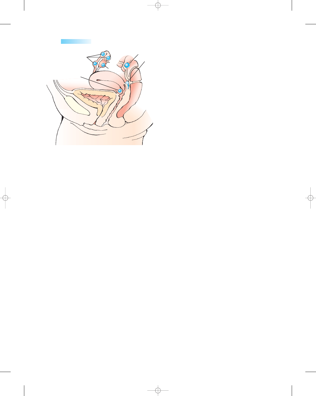
Ectopic Pregnancy
Normally, implantation of the fertilized ovum occurs in
the uterus. An
ectopic pregnancy
is any pregnancy in
which the fertilized ovum implants outside the uterine
cavity. The most common site for implantation is the fal-
lopian tubes, but some ova may implant in the cornua of
the uterus, ovary, cervix, or abdominal cavity (Fig. 19-1)
(Sepilian & Wood, 2004). Unfortunately, none of these
anatomic sites can accommodate placental attachment or
a growing embryo. Thus, the potential for rupture and
hemorrhage exists. A ruptured ectopic pregnancy is a true
medical emergency. It is a potentially life-threatening con-
dition and involves pregnancy loss. It is the leading cause
of maternal mortality in the first trimester and accounts
for 10 to 15% of all pregnancy-related deaths (Sepilian &
Wood, 2005).
Ectopic pregnancies occur from 1 in every 40 to 1 in
every 100 pregnancies in the United States (Marchiano,
2004). Their incidence has increased dramatically in the
past few decades as a result of improved diagnostic tech-
niques, such as more sensitive beta-hCG assays and the
availability of transvaginal ultrasound (Chen, 2004).
Causes
Ectopic pregnancies usually are caused by conditions that
obstruct or slow the passage of the fertilized ovum through
the fallopian tube to the uterus. This may be a physical
blockage in the tube, or failure of the tubal epithelium to
move the zygote (the cell formed after the egg is fertilized)
down the tube into the uterus. In the general population,
most cases are the result of tubal scarring secondary to
pelvic inflammatory disease. Organisms such as Neisseria
gonorrhoeae and Chlamydia trachomatis preferentially attack
the fallopian tubes, producing silent infections. Even with
early treatment, tubal damage can occur. Other contribut-
ing factors include
•
Previous ectopic pregnancy
•
History of STIs
•
Endometriosis
•
Previous tubal or pelvic surgery
•
Infertility and infertility treatments, including use of
fertility drugs
•
Uterine abnormalities such as fibroids
•
Presence of intrauterine device (IUD)
•
Use of progestin-only mini pill (slows ovum transport)
•
Postpartum or postabortion infection
•
Increasing age older than 35 years
•
Cigarette smoking (Owen, 2003)
Chapter 19
NURSING MANAGEMENT OF PREGNANCY AT RISK: PREGNANCY-RELATED COMPLICATIONS
503
10681-19_UT7-CH19rev.qxd 6/19/07 3:08 PM Page 503

Clinical Manifestations
Although onset may vary, clinical manifestations of an
unruptured ectopic pregnancy usually begin at about the
seventh or eighth week of gestation. A missed menses,
adnexal fullness, and tenderness may indicate an unrup-
tured tubal pregnancy. As the tube stretches, the pain
increases. The hallmark of ectopic pregnancy is abdomi-
nal pain with spotting within 6 to 8 weeks after a missed
menses. Other symptoms include breast tenderness, nau-
sea, and low back pain. Pain may be unilateral, bilateral,
or diffuse over the abdomen.
If rupture or hemorrhaging occurs before successfully
treating the pregnancy, symptoms may worsen and include
severe, sharp, and sudden pain in the lower abdomen as the
tube tears open and the embryo is expelled into the pelvic
cavity; feelings of faintness; referred pain to the shoulder
area indicating bleeding into the abdomen caused by
phrenic nerve irritation; hypotension; marked abdominal
tenderness with distension; and hypovolemic shock.
The use of transvaginal ultrasound to visualize the
misplaced pregnancy and low levels of serum beta-hCG
assist in diagnosing an ectopic pregnancy. The ultrasound
determines whether the pregnancy is intrauterine, assesses
the size of the uterus, and provides evidence of fetus via-
bility. Absence of an intrauterine gestational sac is diag-
nostic of ectopic pregnancy (Baines, 2003). In a normal
intrauterine pregnancy, beta-hCG levels typically double
every 2 to 4 days. Therefore, low beta-hCG levels are sug-
gestive of an ectopic pregnancy or impending abortion.
Other tests may be done to rule out other conditions such
as spontaneous abortion, ruptured ovarian cyst, appen-
dicitis, and salpingitis.
504
Unit 7
CHILDBEARING AT RISK
Drug Guide 19-1
Drug
Action
Indications
Nursing Implications
Misoprostol
(RU 486; Cytotec)
PGE2, dinoprostone
(Cervidil, Prepidil
Gel, Prostin E2)
Rh (D) immunoglobulin
(Gamulin, HydroRho-D,
RhoGAM)
Stimulates uterine
contractions to
terminate a
pregnancy
Stimulates uterine
contractions,
causing
expulsion of
uterine contents
Suppresses immune
response of
nonsensitized
Rh-negative
patients who are
exposed to Rh-
positive blood
Evacuate the uterus
after abortion to
ensure passage of all
the products of
conception
Expel uterine contents
in fetal death, missed
abortion during
second trimester, or
to efface and dilate
the cervix in
pregnancy at term
Prevent isoimmunization
in Rh-negative
women exposed to
Rh-positive blood
after abortions,
miscarriages, and
pregnancies
Monitor for side effects such as
diarrhea, abdominal pain,
nausea, vomiting, dyspepsia
Assess vaginal bleeding and report
any increased bleeding, pain,
or fever
Monitor for signs and symptoms of
shock, such as tachycardia,
hypotension, anxiety
Bring gel to room temperature
before administering
Avoid contact with skin
Use sterile technique to administer
Keep client supine 30 minutes after
administering
Document time of insertion and
dosing intervals
Remove insert with retrieval system
after 12 hours or at the onset of
labor
Explain purpose and expected
response to client
Administer intramuscularly in
deltoid area
Give only MICRhoGAM for
abortions and miscarriages
< 12 weeks unless fetus or father
is Rh negative (unless patient is
Rh positive, Rh antibodies are
present)
Educate woman that she will need
this after subsequent deliveries if
newborns are Rh positive; also
check lab study results prior to
administering the drug
Drug Guide 19-1
Medications Used With Spontaneous Abortions
10681-19_UT7-CH19rev.qxd 6/19/07 3:08 PM Page 504
Treatment
The therapeutic management of ectopic pregnancy
depends on whether the tube is intact or has ruptured.
Historically, the treatment of ectopic pregnancy was lim-
ited to surgery, but medical therapy is currently available.
If the fallopian tube is still intact, medical manage-
ment becomes an option. To be eligible for medical ther-
apy, the client must be hemodynamically stable, with no
signs of active bleeding in the peritoneal cavity, and the
mass (which must measure less than 4 cm as determined
by ultrasound) must be unruptured (Valley & Fly, 2005).
The potential advantages include avoidance of surgery,
the preservation of tubal patency and function, and a
lower cost. Methotrexate, prostaglandins, misoprostol,
and actinomycin have all been used in the medical (non-
surgical) management of ectopic pregnancy (Youngkin &
Davis, 2004).
Methotrexate, the agent most commonly used, is a
folic acid antagonist that inhibits cell division in the devel-
oping embryo. It typically has been used as a chemother-
apeutic agent in the treatment of leukemias, lymphomas,
and carcinomas. It has been shown to produce results
similar to that for surgical therapy, in terms of high suc-
cess rate, low complication rate, and good reproductive
potential (Simpson, 2002). Adverse effects associated
with methotrexate include nausea, vomiting, stomatitis,
diarrhea, gastric upset, increased abdominal pain, and
dizziness. Prior to receiving the single-dose intramuscu-
lar injection to treat unruptured pregnancies, the woman
should be counseled on the risks, benefits, adverse effects,
and the possibility of failure of medical therapy, which
would result in tubal rupture, necessitating surgery
(Sepilian & Wood, 2005). The woman is then instructed
to return weekly for follow-up lab studies for the next sev-
eral weeks until beta-hCG titers decrease.
Surgical management for the unruptured fallopian
tube might involve a linear salpingostomy to preserve the
tube—an important consideration for the woman want-
ing to preserve her future fertility.
With a ruptured ectopic pregnancy, surgery is nec-
essary as a result of possible uncontrolled hemorrhage.
A laparotomy with a removal of the tube (salpingectomy)
may be necessary. With earlier diagnosis and medial man-
agement, the focus has changed from prevention of mater-
nal death to facilitating rapid recovery and preserving
fertility.
Regardless of the treatment approach (medical or sur-
gical), the woman’s beta-hCG level is monitored until it is
undetectable to ensure that any residual trophoblastic tis-
sue that forms the placenta is gone. Also, all Rh-negative
unsensitized clients are administered Rh immunoglobulin
to prevent isoimmunization in future pregnancies.
Nursing Management
The woman with an ectopic pregnancy requires support
throughout diagnosis, treatment, and aftercare. If surgery
is needed, close assessment and monitoring of the client’s
vital signs, bleeding (peritoneal or vaginal), and pain sta-
tus are critical to identify hypovolemic shock that may
occur with tubal rupture. The client often experiences a
great deal of pain. Administer analgesics as ordered to
promote comfort and relieve discomfort from abdominal
pain. If the woman is treated medically on an outpatient
basis, it is important to outline the signs of ectopic rup-
ture (severe, sharp, stabbing, unilateral abdominal pain;
vertigo/fainting; hypotension; and increased pulse) and
advise the woman to seek medical help immediately.
Prepare the client physiologically and psychologically
for surgery or any procedure. Provide a clear explanation
of the expected outcome. Astute vigilance and early refer-
ral will help reduce short- and long-term morbidity.
A woman’s psychological reaction to an ectopic preg-
nancy is unpredictable. However, it is important to rec-
ognize she has experienced a pregnancy loss in addition
to undergoing treatment for a potentially life-threatening
condition. It can be difficult for the woman to compre-
hend what has happened to her because events occur so
quickly. In the woman’s mind, she had just started a
pregnancy and now it has ended abruptly. Assist her with
bringing this experience more into reality by encouraging
the woman and her family to express their feelings and
concerns openly, and validating that this is a loss of preg-
nancy and it is okay to grieve over the loss.
Provide emotional support, spiritual care, client edu-
cation, and information about community support groups
available (such as Resolve through Sharing) as the client
grieves the loss of her unborn child and comes to terms with
the medical complications of the situation. Acknowledge
the client’s pregnancy and allow her to discuss her feelings
about what the pregnancy means. Also, stress the need for
follow-up blood testing for several weeks to monitor hCG
Chapter 19
NURSING MANAGEMENT OF PREGNANCY AT RISK: PREGNANCY-RELATED COMPLICATIONS
505
Cervix
Ovary
Fallopian tube
Intestine
Abdomen
●
Figure 19-1
Ectopic pregnancy: possible sites for implantation.
10681-19_UT7-CH19rev.qxd 6/19/07 3:08 PM Page 505

titers until they return to zero, indicating resolution of the
ectopic pregnancy. Additionally, discuss her feelings and
concerns about her future fertility and provide teaching
about the need for using contraceptives at this time for at
least three menstrual cycles to allow time for her reproduc-
tive tract to heal and tissue to be repaired. Include the
woman’s partner in this discussion to make sure there is
understanding by both parties regarding what has hap-
pened, what intervention is needed, and what the future
holds for both of them regarding childbearing.
Prevention of ectopic pregnancies through screening
and client education is essential. Many can be prevented by
avoiding those conditions that might cause scarring of the
fallopian tubes. Such prevention education may include
•
Reducing risk factors such as sexual intercourse with
multiple partners or intercourse without a condom
•
Avoiding contracting STIs that lead to pelvic inflam-
matory disease (PID)
•
Obtaining early diagnosis and adequate treatment of
STIs
•
Avoiding the use of an IUD as a contraceptive method
to reduce the risk of repeat ascending infections respon-
sible for tubal scarring
•
Using condoms to decrease the risk of infections that
cause tubal scarring
•
Seeking prenatal care early if pregnant to confirm loca-
tion of pregnancy
Gestational Trophoblastic Disease
Gestational trophoblastic disease
(GTD) comprises
a spectrum of neoplastic disorders that originate in the
human placenta. Gestational tissue is present, but the
pregnancy is not viable. The incidence is about 1 in 1000
pregnancies (Cunningham, Gant, Leveno, Gilstrap,
Hauth, & Wenstrom, 2005). The two most common
types are hydatidiform mole (partial and complete) and
choriocarcinoma.
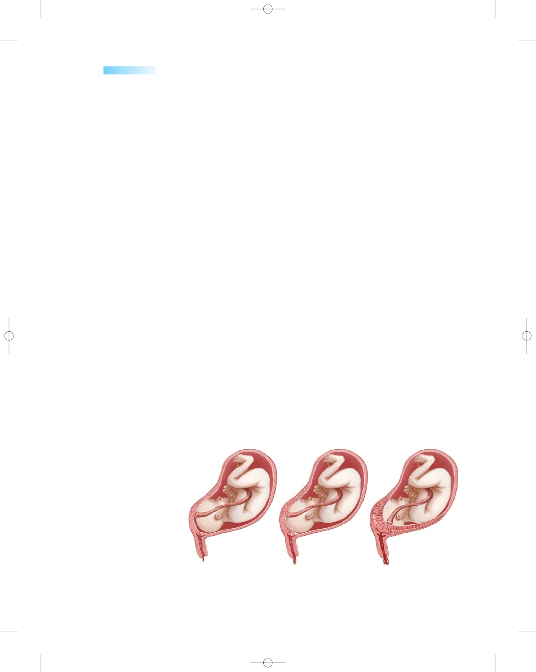
The hydatidiform mole is a benign neoplasm of the
chorion in which chorionic villi degenerate and become
transparent vesicles containing clear, viscid fluid. Hyda-
tiform mole is classified as complete or partial, distin-
guished by differences in clinical presentation, pathology,
genetics, and epidemiology (Gerulath, 2002). The com-
plete mole develops from an “empty egg,” which is fer-
tilized by a normal sperm (46 all-paternal chromosomes).
The embryo dies early, no circulation is established, and
no embryonic tissue is found. The complete mole is asso-
ciated with the development of choriocarcinoma. The
partial mole has a triploid karyotype (69 chromosomes),
because two sperm have provided a double contribution
by fertilizing the ovum (Fig. 19-2).
Having a molar pregnancy (partial or complete) results
in the loss of the pregnancy and the possibility of devel-
oping choriocarcinoma, a chorionic malignancy from the
trophoblastic tissue.
506
Unit 7
CHILDBEARING AT RISK
●
Figure 19-2
Complete hydatidiform mole.
Consider
THIS!
We had lived across the dorm hall from each other during
nursing school, but really didn’t get to know each other
except for a casual hello in passing. When we graduated,
Rose went to work in the emergency room and I in OB.
We saw each other occasionally in the employee cafeteria,
but a quick hello was all that was usually exchanged.
I heard she married one of the paramedics who worked in
the ER and was soon pregnant. I finally got to say more
than hello when she was admitted to the OB unit bleed-
ing during her fourth month of pregnancy. What was dis-
covered was gestational trophoblastic disease and not a
normal pregnancy. I remember holding her in my arms as
she wept. She was told she had a complete molar preg-
nancy after surgery, and she would need extensive follow-
up for the next year. I lost track of her that summer as my
life became busier. Around Thanksgiving time, I heard
she had died from choriocarcinoma. I attended her
funeral, finally, to get the time to say a final hello and
good-bye, but this time with sadness and tears.
Thoughts:
Rose was only 26 years old when she
succumbed to this very virulent cancer. I think
back and realize I missed knowing this brave young
woman and wished that I had taken the time to say
more than hello. Could her outcome have been
different? Why wasn’t it recognized earlier? Did
she not follow up after her diagnosis? I can only
speculate regarding the whom, what, and where.
She lived a short but purposeful life, and hopefully
continued research will change other women’s
outcomes in the future.
Consider
Causes
The exact cause of molar pregnancies is unknown, but
recent research is looking into a genetic basis for it. Studies
10681-19_UT7-CH19rev.qxd 6/19/07 3:08 PM Page 506

have revealed some remarkable features about molar preg-
nancies. They
•
Have the ability to invade into the wall of the uterus
•
Can metastasize to other organs
•
Recur in subsequent pregnancies
•
Can develop into choriocarcinoma, a virulent cancer
•
Occur more in Asia (1 in 120 pregnancies) compared
with the United States (1 in 1000 pregnancies)
•
Are influenced by nutritional factors, such as protein
deficiency
•
Tend to affect older women more than younger women
Clinical Manifestations
Clinical manifestations of GTD are very similar to that of
spontaneous abortion at about 12 weeks of pregnancy.
Signs and symptoms of a molar pregnancy include
•
Report of early signs of pregnancy, such as amenorrhea,
breast tenderness, fatigue
•
Brownish vaginal bleeding/spotting
•
Anemia
•
Severe morning sickness resulting from high hCG levels
•
Fluid retention and swelling
•
Larger size uterus when compared with that for preg-
nancy dates
•
Expulsion of grapelike vesicles possibly occurring in
some women
•
Extremely high hCG levels present; no single value con-
sidered diagnostic
•
Early development of
preeclampsia,
which usually is
not present until after 24 weeks
•
Absence of fetal heart rate or fetal activity
•
Ultrasonic evidence of characteristic molar pattern
The diagnosis is made by visualizing the characteris-
tic appearance of the vesicular molar pattern in the uterus
via transvaginal ultrasound and high levels of hCG.
Treatment
Treatment consists of immediate evacuation of the uter-
ine contents as soon as the diagnosis is made and long-term
follow-up of the client to detect any remaining trophoblas-
tic tissue that might become malignant. Dilation and suc-
tion curettage are used to empty the uterus. The tissue
obtained is sent to the laboratory for analysis to evaluate
for choriocarcinoma. Serial levels of hCG are used to
detect residual trophoblastic tissue for 1 year. If any tissue
remains, hCG levels will not regress. Because of this can-
cer risk, the client is advised to receive extensive follow-up
therapy for the next 12 months. The follow-up protocol
may include
•
Baseline hCG level, chest radiograph, and pelvic ultra-
sound
•
Weekly serum hCG levels until it drops to zero and
remains at that level for 3 consecutive weeks, then
monthly for 6 months, then every 2 months for the
remainder of the year
•
Chest radiograph every 6 months to detect pulmonary
metastasis
•
Regular pelvic examinations to assess uterine and ovar-
ian regression
•
Systemic assessments for symptoms indicative of lung,
brain, liver, or vaginal metastasis
•
Strong recommendation to avoid pregnancy for 1 year
because the pregnancy can interfere with the monitor-
ing of hCG levels
•
Use of reliable contraceptive (Gilbert & Harmon, 2003)
Nursing Management
Nursing management of the woman with GTD focuses on
educating her about the potential risk of cancer that may
develop after a molar pregnancy and the strict adherence
needed for the follow-up program. The woman must
understand the necessity for the continued follow-up care
regimen to improve her chances of future pregnancies and
to ensure her continued quality of life.
Assessment
The nurse plays a crucial role in identifying and bringing
this condition to the attention of the health care provider
based on sound knowledge of the typical clinical mani-
festations and through astute antepartal assessments.
Assess the woman for potential clinical manifestations at
each antepartal visit.
Nursing Interventions
After GTD is diagnosed, the nurse needs to educate the
client about the condition and appropriate interventions
that may be necessary to save her life. Explain each phase
of treatment accurately and provide support for the woman
and her family as they go through the grieving process.
Prepare the woman physically and psychologically for
D&C as indicated.
To aid the client and her family in coping with the
loss of the pregnancy and the possibility of a cancer diag-
nosis, use the following interventions:
•
Listen to their concerns and fears.
•
Allow them time to grieve for their pregnancy loss.
•
Acknowledge their loss and sad feelings (say you are
sorry for their loss).
•
Encourage them to express their grief; make it okay for
them to cry.
•
Provide them with as much factual information as pos-
sible to help them make sense of what is happening.
•
Enlist support from additional family and friends as
appropriate, and with the client’s permission.
As with any facet of health care, the nurse needs to
keep current on the latest research and new therapies.
Inform the client about her follow up care, which will prob-
Chapter 19
NURSING MANAGEMENT OF PREGNANCY AT RISK: PREGNANCY-RELATED COMPLICATIONS
507
10681-19_UT7-CH19rev.qxd 6/19/07 3:08 PM Page 507

ably involve a close clinical surveillance for approximately
1 year, and reinforce its necessity in monitoring the client’s
condition. Serial serum beta-hCG levels are used to detect
residual trophoblastic tissue. Continued high or increasing
hCG titers are abnormal and need further evaluation.
Anticipate the use of chemotherapy, such as meth-
otrexate, which may be started as prophylaxis. Inform the
client about the need for using a reliable contraceptive to
prevent pregnancy for 1 year. A positive pregnancy test
would interfere with tracking of the serial beta-hCG lev-
els used to identify a potential malignancy. Stress the
need for client cooperation and adherence to the plan of
therapy throughout this year-long follow-up.
Cervical Insufficiency
Cervical insufficiency describes a weak, structurally defec-
tive cervix that spontaneously dilates in the absence of con-
tractions in the second trimester, resulting in the loss of the
pregnancy. The incidence of cervical insufficiency is less
than 1% and ranges in estimation from 1 in 500 to 1 in
2000 pregnancies, accounting for approximately 20 to
25% of midtrimester losses (Ahn & Hibbard, 2003).
Causes
Cervical insufficiency may result from an in utero exposure
to diethylstilbestrol (DES), which was commonly used for
the treatment of recurrent pregnancy loss until the mid-
1970s; an acquired cause, such as trauma to the cervix
from pervious gynecologic or obstetric procedures (cone
biopsy, D&C); damage to the cervix from a previous dif-
ficult birth (cervical lacerations from forceps); increased
uterine volume (multiple gestation, hydramnios); or un-
known reasons (Creasy, Resnik, & Iams, 2004).
Clinical Manifestations
Commonly, with cervical insufficiency, the woman will
report a pink-tinged vaginal discharge or an increase in
pelvic pressure. History may reveal a previous loss of preg-
nancy around 20 weeks. Cervical dilation also occurs.
Continuation leads to rupture of the membranes, release
of amniotic fluid, and uterine contractions, subsequently
resulting in delivery of the fetus, often before the time of
viability.
Treatment
The diagnosis of cervical insufficiency remains difficult in
many circumstances. The cornerstone of diagnosis is a his-
tory of midtrimester pregnancy loss associated with pain-
less cervical dilatation without evidence of uterine activity.
Close surveillance of cervical length with transvaginal
ultrasound is typically started around 20 weeks’ gestation.
Regular evaluations are performed (particularly in women
with pelvic pressure, backache, or increased mucoid dis-
charge) every few days to avoid missing rapid changes in
cervical dilation or until the trend in cervical length can be
characterized (Ressel, 2004).
Cervical shortening occurs from the internal os out-
ward and can be viewed on ultrasound as funneling. The
amount of funneling can be determined on ultrasound by
dividing funnel length by cervix length. A cervical length
less than 25 mm is abnormal between 14 weeks and
24 weeks, and increases the risk of preterm labor. The
most common time at which a short cervix or funneling
develops is 18 to 22 weeks, so ultrasound screening should
be performed during this interval (Berghella, 2004).
Management for cervical insufficiency has been treated
in a variety of ways: bed rest; pelvic rest; avoidance of
heavy lifting; or surgically, via a procedure of a cervical cer-
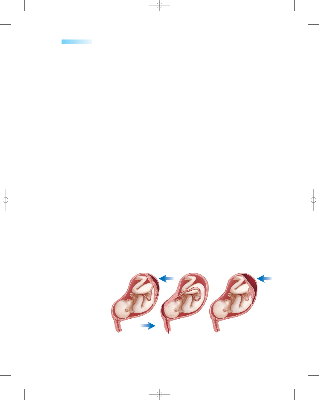
clage in the second trimester. Cervical cerclage involves
using a heavy purse-string suture to secure and reinforce
the internal os of the cervix (Fig. 19-3).
According to ACOG (2003a), if a short cervix is
identified at or after 20 weeks with absence of infection
(chorioamnionitis), the decision to proceed with cerclage
should be made with caution; there have been limited
numbers of well-designed randomized studies to support
its efficacy. Suture displacement, rupture of membranes,
and chorioamnionitis are the most common complications
associated with cerclage placement, and incidence varies
widely in relation to timing and indications for the cerclage
(Ressel, 2004). The optimal timing for cerclage removal is
unclear, according to ACOG (2003a).
Nursing Management
Nursing management related to women with cervical
insufficiency involves taking a very thorough history to be
alert to any risk factors that might have a bearing on this
pregnancy—previous cervical trauma, preterm labor, fetal
loss in second trimester, or previous surgeries or proce-
dures involving the cervix. Monitor the woman very closely
for signs of preterm labor: backache, increase in vaginal
discharge, rupture of membranes, and uterine contrac-
tions. Provide emotional and educational support to allay
the couple’s anxiety about the well-being of their fetus.
Continuing surveillance throughout the pregnancy is
important to promote a positive outcome for the family.
508
Unit 7
CHILDBEARING AT RISK
●
Figure 19-3
Cervical
cerclage.
10681-19_UT7-CH19rev.qxd 6/19/07 3:08 PM Page 508
Placenta Previa
Placenta previa
is a bleeding condition that occurs dur-
ing the last two trimesters of pregnancy. Placenta previa lit-
erally means “afterbirth first” and defines a condition in
which the placenta implants over the cervical os. It may
cause serious morbidity and mortality to fetus and mother.
It complicates approximately 5 of 1000 births or 1 in every
200 pregnancies and is associated with potentially seri-
ous consequences from hemorrhage, abruption (sepa-
ration) of the placenta, or emergency cesarean birth (Joy
& Lyon, 2004).
Placenta previa is generally classified according to
the degree of coverage or proximity to the internal os as
follows (Fig. 19-4):
•
Total placenta previa—occurs when the internal cervi-
cal os is completely covered by the placenta
•
Partial placenta previa—occurs when the internal os is
partially covered by the placenta
•
Marginal placenta previa—occurs when the placenta is
at the margin or edge of the internal os
•
Low-lying placenta previa—occurs when the placenta is
implanted in the lower uterine segment and is near the
internal os but does not reach it
Causes
The exact etiology of placenta previa is unknown. Placenta
previa is initiated by implantation of the embryo in the
lower uterus. With placental attachment and growth, the
cervical os may become covered by the developing pla-
centa. Placental vascularization is defective, allowing
the placenta to attach directly to the myometrium (acc-
reta), invade the myometrium (increta), or penetrate the
myometrium (percreta).
The condition may be multifactorial and is associ-
ated with the following risk factors:
•
Advanced maternal age (more than 30 years old)
•
Previous cesarean birth
•
Multiparity
•
Uterine insult or injury
•
Cocaine use
•
Prior placenta previa
•
Male infants (Thompson, 2005)
•
African-Americans and Asian cultural groups (Ko &
Yoon, 2005)
•
Multiple gestations
•
Previous induced abortion
•
Previous myomectomy to remove fibroids
•
Smoking (Ko & Yoon, 2005)
Clinical Manifestations
The classical clinical presentation is painless, bright-red
vaginal bleeding occurring during the second or third
trimester. The initial bleeding is not usually profuse and
it ceases spontaneously, only to recur again. The first
episode of bleeding occurs (on average) at 27 to 32 weeks’
gestation, and contractions may or may not occur with the
bleeding. The bleeding is thought to arise secondary to
the thinning of the lower uterine segment in preparation
for the onset of labor. When the bleeding occurs at the
implantation site in the lower uterus, the uterus is unable
to contract adequately and stop the flow of blood from the
open vessels. Typically with normal placental implantation
in the upper uterus, minor disruptive placental attachment
is not a problem, because there is a larger volume of
myometrial tissue able to contract and constrict bleeding
vessels. The client’s uterus is soft and nontender on exam-
ination. Auscultation of fetal heart tones is within normal
parameters, and fetal distress usually is not present unless
a cord accident occurs or vaginal blood loss has been heavy
enough to induce maternal shock or placenta abruption
(Gaudier, 2003).
To validate the position of the placenta, a transvaginal
ultrasound is done. In addition, MRI may be ordered in
preparing for delivery because it allows identification
of placenta accreta, increta, or percreta in addition to
Chapter 19
NURSING MANAGEMENT OF PREGNANCY AT RISK: PREGNANCY-RELATED COMPLICATIONS
509
Marginal
Partial
Complete
A
B
C
●
Figure 19-4
Classification of
placenta previa. (A) Marginal.
(B) Partial. (C) Complete.
10681-19_UT7-CH19rev.qxd 6/19/07 3:08 PM Page 509

placenta previa. These placental abnormalities, although
rare, carry a very high morbidity and mortality rate, neces-
sitating a possible hysterectomy at delivery.
Treatment
The treatment depends on the extent of bleeding, the
amount of placenta over the cervical os, whether the fetus
is developed enough to survive outside the uterus, the posi-
tion of the fetus, the parity for the mother, and the presence
or absence of labor (Gaudier, 2003).
If the mother and fetus are both stable, therapeutic
management may involve expectant (or wait-and-see) care.
This care can be carried out in the home or on an antepar-
tal unit in the healthcare facility. If there is no active bleed-
ing and the client has readily available access to reliable
transportation, is able to maintain bed rest in the home
setting, and has the ability to comprehend instructions,
expectant care at home is appropriate. However, if the
client requires continuous care and monitoring and is not
able to comply with needed home care requirements, the
antepartal unit is the best environment.
Nursing Management
Whether the care setting is in the client’s home or in the
healthcare facility setting, the nurse focuses on monitor-
ing the maternal–fetal dyad status, assessing for signs
and symptoms of vaginal bleeding and fetal distress, and
providing support and education to the client and her
family members, including what events and diagnostic
studies are being performed. For the majority of women,
a cesarean birth will be planned. See Nursing Care Plan
19-1 for application of the nursing process for the woman
with placenta previa.
Assessment
Assessing the woman’s degree of vaginal bleeding is para-
mount. Estimate and document the amount of bleeding.
Perform a peripad count on an ongoing basis, making
sure to report any changes in amount or frequency to the
healthcare provider. If the woman is experiencing active
bleeding, prepare for blood typing and crossmatching in
the event a blood transfusion is needed.
Assess fetal heart rates via Doppler to detect fetal
distress. Monitor the woman’s cardiopulmonary status,
reporting any difficulties in respirations, changes in skin
color, or complaints of difficulty breathing. Have oxy-
gen equipment readily available should fetal or mater-
nal distress develop.
If the woman has an IV inserted, inspect the IV site
frequently. Alternately, anticipate the insertion of an
intermittent IV access device such as a saline lock, which
can be used if quick access is needed for fluid restoration
and infusion of blood products.
Assess the woman’s level of understanding regarding
the condition of placenta previa and associated procedures
and treatment plan. Doing so is important to prevent con-
fusion and gain the woman’s cooperation. Provide infor-
mation related to the condition and make sure that all
information related is consistent with information from the
primary care provider.
If the woman will require prolonged hospitalization
or home bed rest, assess the physical and emotional
impact that this may have on her. Evaluate the woman’s
coping mechanisms to aid in determining how well she
will be able to adjust to and cooperate with the treatment
plan. In addition to emotional impact with prolonged bed
rest, thoroughly assess the woman’s skin to prevent skin
breakdown and to help alleviate the woman’s discomfort
secondary to limited physical activity.
Nursing Interventions
The following interventions would be appropriate for the
woman with placenta previa regardless of the setting:
•
Monitoring amount of blood loss, pain level, and uter-
ine contractility
•
Assessing maternal vital signs frequently
•
Ascertaining the client’s understanding and implica-
tions of this condition
•
Monitoring the results of all laboratory testing, such as
CBC, type and crossmatch, coagulation studies
•
Providing emotional support and listening to her fears
of the unknown
•
Explaining assessments and treatment measures needed
•
Assisting the client to remain on bed rest with bathroom
privileges
•
Counseling the client and family about all activities and
interventions
•
Providing opportunities for distraction—educational
videos, arts and crafts, computer games, reading books
•
Instructing the client to assess fetal activity via “kick
counts” daily
•
Acting as a client advocate in obtaining information for
the family
•
Monitoring the woman’s coping ability to comply with
activity restrictions
•
Preventing any vaginal examinations from being per-
formed, which might disrupt the placenta and cause
hemorrhage
•
Evaluating fetal heart rate via an external monitor or
Doppler
•
Encouraging a balanced, nutritious diet with plenty of
fluid intake
•
Administering Rh immunoglobulin if the client is Rh
negative at 28 weeks’ gestation
•
Preparing the client for cesarean birth when necessary
•
Informing the client and family of any status change
that requires intervention
•
Monitoring tocolytic medication if prevention of preterm
labor is needed
•
Educating the client regarding risk of reoccurrence of
this condition
510
Unit 7
CHILDBEARING AT RISK
10681-19_UT7-CH19rev.qxd 6/19/07 3:08 PM Page 510

Chapter 19
NURSING MANAGEMENT OF PREGNANCY AT RISK: PREGNANCY-RELATED COMPLICATIONS
511
Outcome identification and
evaluation
Client will maintain adequate tissue perfusion
as
evidenced by stable vital signs, decreased blood
loss, few or no uterine contractions, normal fetal
heart rate patterns and variability, and positive
fetal movement
Interventions with
rationales
Establish intravenous access to allow for administra-
tion of fluids, blood, and medications as necessary
Obtain type and crossmatch for at least 2 U blood
products
to ensure availability should bleeding
continue
Obtain specimens as ordered for blood studies, such
as CBC and clotting studies
to establish a baseline
and use for future comparison
Monitor output
to evaluate adequacy of renal
perfusion
Administer IV fluid replacement therapy as ordered
to maintain blood pressure and blood volume
Palpate for abdominal tenderness and rigidity
to
determine bleeding and evidence of uterine
contractions
Institute bed rest
to reduce oxygen demands
Assess for rupture of membrane
s to evaluate for
possible onset of labor
Avoid vaginal examinations
to prevent further bleed-
ing episodes
Complete an Rh titer t
o identify need for RhoGAM
Avoid nipple stimulation
to prevent uterine contractions
Continuously monitor for contractions or PROM
to
allow for prompt intervention
Administer tocolytic agents as ordered
to stall
preterm labor
Monitor vital signs frequently
to identify possible
hypovolemia and infection
Assess frequently for active vaginal bleeding
to
minimize risk of hemorrhage
Continuously monitor fetal heart rate with electronic
fetal monitor
to evaluate fetal status
Assist with fetal surveillance tests as ordered
to aid in
determining fetal well-being
Sandy, a 39-year-old G5, P4, multigravida client at 32 weeks’ gestation, was admitted to the
labor and birth suite with sudden vaginal bleeding. Sandy had no further active bleeding and
did not complain of any abdominal discomfort or tenderness. She did complain of occasional
“tightening” in her stomach. Her abdomen palpated soft. Fetal heart rates were in the 140s
with accelerations with movement. She was placed on bed rest with bathroom privileges.
Ultrasound identified a low-lying placenta with a viable, normal-growth fetus. She was diag-
nosed with placenta previa and admitted for observation and surveillance of fetal well-being.
Her history revealed two previous cesarean births, smoking half a pack of cigarettes per day,
and endometritis infection after birth of her last newborn. Additional assessment findings
included painless, bright-red vaginal bleeding with initial bleeding ceasing spontaneously;
irregular, mild, and sporadic uterine contractions; fetal heart rate and maternal vital signs
within normal range; fetus in transverse lie; anxiety related to the outcome of pregnancy; and
expression of feelings of helplessness.
Nursing Care Plan
Nursing Diagnosis: Ineffective tissue perfusion (fetal and maternal) related to blood loss
(continued )
Nursing Care Plan
19-1
Overview of the Woman With Placenta Previa
10681-19_UT7-CH19rev.qxd 6/19/07 3:08 PM Page 511

Abruptio Placenta
Abruptio placenta
refers to separation of a normally
located placenta after the 20th week of gestation and
prior to birth, producing hemorrhage. It is a significant
cause of third trimester bleeding with a high mortality
rate. It occurs in about 1% of all pregnancies throughout
the world (Gaufberg, 2004). The overall fetal mortality
rate for placenta abruption is 20% to 40%, depending on
the extent of the abruption. This is caused by the insult
of the abruption itself and by issues related to prematu-
rity when early birth is required to alleviate maternal or
fetal distress. Maternal mortality is approximately 6% in
abruptio placenta and is related to cesarean birth and/or
hemorrhage/coagulopathy (Deering & Satin, 2004).
Abruptio placenta is a major medical emergency. It
requires rapid, effective interventions to prevent maternal
and fetal morbidity and mortality.
Causes
The etiology of this condition is unknown. Several risk
factors are associated with it, such as maternal smoking,
extremes of age (<20 years or >35 years old), poor nutri-
tion, multiple gestation, excessive intrauterine pressure
caused by hydramnios, hypertension, prior abruption in
a previous pregnancy, severe trauma (such as an auto
accident or injury secondary to intimate partner violence),
cocaine and methamphetamine abuse, alcohol ingestion,
and multiparity (Chen, 2004). Other notable risk factors
include male fetal gender, chorioamnionitis, prolonged
512
Unit 7
CHILDBEARING AT RISK
Outcome identification and
evaluation
Interventions with
rationales
Observe for abnormal fetal heart rate patterns, such
as loss of variability, decelerations, tachycardia,
to
identify fetal distress
Position patient in side-lying position with wedge for
support
to maximize placental perfusion
Assess fetal movement
to evaluate for possible fetal
hypoxia
Teach woman to monitor fetal movement
to
evaluate well-being
Administer oxygen as ordered
to increase oxygena-
tion to mother and fetus
Overview of the Woman With Placenta Previa
(continued)
Client will experience a decrease in anxiety
as
evidenced by verbal reports of less anxiety, use of
effective coping measures, and calm demeanor
Provide factual information about diagnosis and
treatment, and explain interventions and the
rationale behind them
to provide client with
understanding of her condition
Answer questions about health status honestly
to
establish a trusting relationship
Speak calmly to patient and family members
to
minimize environmental stress
Encourage the use of past effective techniques
for coping
to promote relaxation and feelings of
control
Acknowledge and facilitate the woman’s spiritual
needs
to promote effective coping
Involve the woman and family in the decision-
making process
to foster self-confidence and
control over situation
Maintain a presence during stressful periods
to
allay anxiety
Use the sense of touch if appropriate to convey
caring and concern
Encourage talking
as a means to release tension
Nursing Diagnosis: Anxiety related to threats to self and fetus
10681-19_UT7-CH19rev.qxd 6/19/07 3:08 PM Page 512
premature ruptured membranes (>24 hours), oligohy-
dramnios, preeclampsia, and low socioeconomic status
(Deering & Satin, 2004).
Clinical Manifestations
Abruptio placenta is classified according to the extent of
separation and the amount of blood loss from the mater-
nal circulation. Classifications include
•
Mild (grade 1)—minimal bleeding (<500 mL), marginal
separation (10–20%), tender uterus, no coagulopathy,
no signs of shock, no fetal distress
•
Moderate (grade 2)—Moderate bleeding (1000–
1500 mL), moderate separation (20–50%), continuous
abdominal pain, mild shock
•
Severe (grade 3)—absent to moderate bleeding
(>1500 mL), severe separation (>50%), profound shock,
agonizing abdominal pain, and development of dis-
seminated intravascular coagulopathy (DIC) (Gilbert &
Harmon, 2003)
Abruptio placenta also may be classified as partial
or complete, depending on the degree of separation.
Alternately, it can be classified as concealed or appar-
ent, by the type of bleeding (Fig. 19-5).
As the placenta separates from the uterus, hemorrhage
ensues. It can be apparent, appearing as vaginal bleeding,
or it can be concealed. Classic manifestations include
painful, dark-red vaginal bleeding (port wine color); “knife-
like” abdominal pain; uterine tenderness; contractions; and
decreased fetal movement. Vaginal bleeding is present in
80% of women diagnosed with abruptio placenta and may
be significant enough to jeopardize both maternal and fetal
health within a short time frame. The remaining 20% of
abruptions are associated with a concealed hemorrhage
and the absence of vaginal bleeding. Decreased fetal move-
ment may be the presenting complaint resulting from fetal
jeopardy or fetal death (Deering & Satin, 2004).
Laboratory and diagnostic tests may be helpful in
diagnosing the condition and guiding management. These
studies may include
•
CBC—determines the current hemodynamic status;
however, it is not reliable for estimating acute blood loss
•
Fibrinogen levels—typically are increased in pregnancy
(hyperfibrinogenemia); thus, a moderate dip in fibrino-
gen levels might suggest coagulopathy (DIC) and, if
profuse bleeding occurs, the clotting cascade might be
compromised
•
Prothrombin time (PT)/activated partial thromboplas-
tin time (aPTT)—determines the client’s coagulation
status, especially if surgery is planned
•
Type and crossmatch—determines blood type if trans-
fusion is needed
•
Kleihauer–Betke test—detects fetal RBCs in the mater-
nal circulation, determines the degree of fetal–maternal
hemorrhage, helps calculate the appropriate dosage of
RhoGAM to give for Rh-negative clients
•
Ultrasound—helps to diagnose quickly the etiology of
bleeding and visualize active or concealed hemorrhage,
aids in identifying retroplacental hematoma
•
Nonstress test—demonstrates findings of fetal jeopardy
manifested by late decelerations or bradycardia
•
Biophysical profile—used to evaluate clients with chronic
abruption, provides information about possible fetal com-
promise by a low score (<6 points) (Cavanaugh, 2003)
Treatment
Treatment of abruptio placenta is designed to assess, con-
trol, and restore the amount of blood lost; to provide a
positive outcome for both mother and newborn; and to
prevent coagulation disorders, such as DIC (Box 19-2).
Emergency measures include starting two large-bore IV
lines with normal saline or lactated Ringer’s solution to
combat hypovolemia, obtaining blood specimens for
evaluating hemodynamic status values and for typing and
Chapter 19
NURSING MANAGEMENT OF PREGNANCY AT RISK: PREGNANCY-RELATED COMPLICATIONS
513
Partial abruption,
concealed hemorrhage
Partial abruption,
apparent hemorrhage
Partial abruption,
concealed hemorrhage
A
B
C
●
Figure 19-5
Classifications of
abruptio placenta. (A) Partial
abruption with concealed hemor-
rhage. (B) Partial abruption
with apparent hemorrhage.
(C) Complete abruption with
concealed hemorrhage.
10681-19_UT7-CH19rev.qxd 6/19/07 3:08 PM Page 513

crossmatching, and frequently monitoring fetal and
maternal well-being. After determining the severity of
abruption, and appropriate blood and fluid replacement
is given, cesarean birth is done immediately if fetal distress
is evident. If the fetus is not in distress, close monitoring
continues, with delivery planned at the earliest signs of
fetal distress. Because of the possibility of fetal blood loss
through the placenta, a critical care neonatal team should
be available during the birth process to assess and treat the
newborn immediately for shock, blood loss, and hypoxia.
If the woman develops DIC, treatment focuses on
determining the underlying cause of DIC and correcting it.
Replacement therapy of the coagulation factors is achieved
by transfusion of fresh frozen plasma along with cryopre-
cipitate to maintain the circulating volume and provide
oxygen to the cells of the body. Prompt identification and
early intervention are essential for a woman with acute
DIC associated with abruptio placenta to resolve DIC and
possibly save the woman’s life.
Nursing Management
Abruptio placenta warrants immediate care to provide
the best outcome for both mother and fetus.
Assessment
The nurse’s role in the assessment of the woman present-
ing with abdominal pain and/or experiencing vaginal bleed-
ing is critical. An accurate assessment forms the basis of
medical management and intervention, especially in a con-
cealed hemorrhage in which the extent of bleeding is not
recognized. Vital signs can be within normal range, even
with significant blood loss, because a pregnant woman can
lose up to 40% of her total blood volume without showing
signs of shock (Gilbert & Harmon, 2003).
Nursing assessment includes
•
Assessing all women for risk factors that might cause
abruption of the placenta: hypertension, drug use, mem-
brane status, smoking, etc.
•
Assessing maternal level of consciousness and signs of
shock
•
Monitoring the fetal heart rate continuously electronically
•
Assessing pain type, onset, and location
•
Assessing for abdominal tenderness, pain, and rigidity
•
Monitoring uterine contractions by frequent palpation
Nursing Interventions
When the woman arrives to the labor and birth suite, place
the woman on strict bed rest and in a left lateral position to
prevent pressure on the vena cava. This position provides
uninterrupted perfusion to the fetus. Expect to administer
oxygen therapy via nasal cannula to ensure adequate tissue
perfusion.
Additional nursing interventions include
•
Monitoring hourly intake and output after insertion of
indwelling urinary (Foley) catheter
•
Obtaining maternal blood pressure, pulse, respirations,
and pulse rate; and assessing pain level every 15 to 60
minutes, depending on maternal stability and amount
of blood loss
•
Observing for changes in vital signs suggesting hypov-
olemic shock and reporting them immediately
•
Observing and recording amount and time of bleeding
every 30 minutes
•
Initiating an IV line with a large-bore catheter and doc-
umenting fluid intake to prevent fluid overload
•
Avoiding vaginal examinations until placenta previa is
ruled out
•
Administering pain medication as ordered
•
Monitoring uterine contractions for increased rigidity
and tenderness
•
Monitoring the amount and nature (dark or bright red)
of vaginal bleeding
•
Evaluating the fundal height (increasing size would
indicate bleeding)
•
Observing for signs of DIC—bleeding gums, tachycar-
dia, and petechiae—and administering blood products
if DIC is apparent
•
Observing for fetal distress and tetanic contractions on
monitor
514
Unit 7
CHILDBEARING AT RISK
DIC is a bleeding disorder characterized by an abnor-
mal reduction in the elements involved in blood clotting
resulting from their widespread intravascular clotting
(O’Toole, 2005). This disorder can occur secondary to
abruptio placenta.
Simply, the clinical and pathologic manifestations of
DIC can be described as a loss of balance between the
clot-forming activity of thrombin and the clot-lysing
activity of plasmin. Therefore, too much thrombin tips
the balance toward the prothrombic state and the client
develops clots. Alternately, too much clot lysis (fibrinol-
ysis) results from plasmin formation and the client hem-
orrhages. Small clots form throughout the body, and
eventually the blood-clotting factors are used up, ren-
dering them unavailable to form clots at sites of tissue
injury. Clot-dissolving mechanisms are also increased,
which result in bleeding, which can be severe.
DIC can be stimulated by many factors including
sepsis, malignancy, and obstetric conditions such as
placenta abruption, missed abortion or retained dead
fetus, amniotic fluid embolism, and eclampsia.
Laboratory studies assist in the diagnosis and include
• Decreased fibrinogen and platelets
• PT and aPTT times
• Positive D-dimer tests and fibrin (split) degradation
products, which uncover objective evidence of the
simultaneous formation of thrombin and plasmin
(NANDA, 2005).
BOX 19-2
DISSEMINATED INTRAVASCULAR COAGULATION
10681-19_UT7-CH19rev.qxd 6/19/07 3:08 PM Page 514

•
Using pulse oximetry to monitor oxygen saturation lev-
els of circulating blood
•
Answering questions about fetal health status in an
honest manner
•
Acknowledging and facilitating the client’s spiritual and
cultural needs
•
Communicating empathy and understanding of the
client’s experience, and providing emotional support
throughout this frightening time
•
Remaining with the couple and acknowledging their
emotions and fears
•
Providing the couple with factual information about
projected management
•
Explaining thoroughly the care needed so they will
know what to expect
•
Informing the client and family about diagnostic tests
and surgery
•
Preparing the client and family for the possibility of
cesarean birth
•
Reducing family anxiety by reassuring fetal well-being
as appropriate based on test results
•
Helping the family to deal with loss or with infant in the
intensive care unit
Although abruptio placenta is not a preventable con-
dition, client education is important to help reduce the
risk for this condition. Educational topics would include
encouraging the woman to avoid drinking, smoking, or
using drugs during pregnancy, and to obtain rehabilita-
tion services to eliminate drug abuse prior to the preg-
nancy; to seek early and continuous prenatal care; to
recognize diabetes and hypertension early so that treat-
ment can be started; to identify intimate partner abuse to
prevent further abuse; and to receive prompt health care
if any symptoms are present in the future.
Hyperemesis Gravidarum
Approximately 80% or more women experience nausea
and vomiting during their pregnancy (Edelman & Logan,
2004). The term morning sickness is often used to describe
this condition when symptoms are relatively mild. Such
symptoms usually disappear after the first trimester. This
mild form mostly impacts the quality of life of the woman
and her family, whereas the severe form—hyperemesis
gravidarum—results in dehydration, electrolyte imbalance,
and the need for hospitalization (Koren & Maltepe, 2004).
Unlike morning sickness,
hyperemesis gravida-
rum
is a complication of pregnancy characterized by per-
sistent, uncontrollable nausea and vomiting before the
20th week of gestation. This complication can lead to dehy-
dration, acid–base imbalances, electrolyte imbalances, and
weight loss. If it continues, it jeopardizes fetal well-being
(Green & Wilkinson, 2004).
The incidence of hyperemesis is estimated to occur in
approximately 1% of pregnant women. The prevalence
increases in molar and multiple pregnancies. Its peak inci-
dence occurs between 8 weeks and 12 weeks of pregnancy,
usually resolving by week 16 (Garcia, 2003).
Causes
The cause of nausea and vomiting is unknown. Although
theories abound, few studies have produced scientific evi-
dence that determine the etiology of this condition. It is
likely that multiple factors contribute to it.
Elevated levels of hCG are present in all pregnant
women during early pregnancy, usually declining after 12
weeks. This corresponds to the usual duration of morn-
ing sickness. In hyperemesis gravidarum, the hCG levels
are often higher and extend beyond the first trimester.
Symptoms exacerbate the disease. Decreased fluid intake
and prolonged vomiting cause dehydration; dehydration
increases the serum concentration of hCG, which in turn
exacerbates the nausea and vomiting—a vicious cycle. A
few other theories that have been proposed to explain its
etiology include
•
Endocrine theory—high levels of hCG and estrogen
during pregnancy
•
Metabolic theory—vitamin B6 deficiency
•
Psychological theory—psychological stress increasing
the symptoms
Regardless of etiology, the client with hyperemesis gravi-
darum is extremely uncomfortable.
Risk factors associated with hyperemesis gravidarum
include young age, nausea and vomiting with previous
pregnancy, history of intolerance of oral contraceptives,
multiple gestation, emotional or psychological stress,
gastroesophageal reflux disease, primigravida status, obe-
sity, hyperthyroidism, and Helicobacter pylori seropositivity
(Swearingen, 2004).
Clinical Manifestations
Symptoms of hyperemesis gravidarum include, but are not
limited to, disturbed nutrition, severe vomiting, electrolyte
imbalance, ketosis, acetonuria, and weight loss greater
than 5% of body mass. If it progresses untreated, it may
cause neurologic disturbance, renal damage, retinal hem-
orrhage, or death (Christopher, 2003). In addition, it is
exhausting and emotionally distressing.
Other common symptoms include ptyalism (excessive
salivation), fatigue, weakness, and dizziness. Additionally,
sleep disturbance, depression, anxiety, irritability, mood
changes, and decreased ability to concentrate can add to
the woman’s emotional distress (Michelini, 2004).
The results of diagnostic studies may provide clues
to the severity of the disorder: These may include
•
Liver enzymes—elevations of aspartate aminotrans-
ferase (AST) and alanine aminotransferase (ALT) usu-
ally present
Chapter 19
NURSING MANAGEMENT OF PREGNANCY AT RISK: PREGNANCY-RELATED COMPLICATIONS
515
10681-19_UT7-CH19rev.qxd 6/19/07 3:08 PM Page 515

•
CBC—elevated levels of RBCs and hematocrit denot-
ing dehydration
•
Urine ketones—positive when the body breaks down fat
to provide energy in the absence of inadequate intake
•
Blood, urea, nitrogen (BUN)—increased in the presence
of salt and water depletion
•
Urine specific gravity—greater than 1.025, possibly
indicating concentrated urine linked to inadequate fluid
intake or excessive fluid loss
•
Serum electrolytes—decreased levels of potassium,
sodium, and chloride resulting from excessive vomiting
and loss of hydrochloric acid in stomach (Swearingen,
2004)
Treatment
Conservative management in the home is the first line of
treatment for the woman with hyperemesis gravidarum.
This usually focuses on dietary and lifestyle changes.
If conservative management fails to alleviate the
client’s symptoms, and nausea and vomiting continue,
hospitalization is necessary to reverse the effects of severe
nausea and vomiting.
On admission to the hospital baseline, blood tests
are ordered to assess the severity of the client’s dehydra-
tion, electrolyte imbalance, ketosis, and malnutrition.
Parenteral fluid and drug therapy are ordered to rehy-
drate and reduce the symptoms. The first choice for fluid
replacement is generally 5% dextrose in lactated Ringer’s
solution with vitamins and electrolytes added. Oral food
and fluids are withheld for the first 24 to 36 hours to allow
the GI tract to rest. Antiemetics may be administered
orally, rectally, or intravenously to control the nausea and
vomiting. Because the woman is initially considered
“NPO,” no food or fluids orally, her medication is admin-
istered parenterally or rectally until she is stabilized.
If the client does not improve after several days of bed
rest, “gut rest,” IV fluids, and antiemetics, total parenteral
nutrition or percutaneous endoscopic gastrostomy tube
feeding is instituted to prevent malnutrition.
The FDA has not approved any drugs for the treat-
ment of nausea and vomiting in pregnancy, but the risk of
administration must be balanced against the sequelae of
prolonged starvation and dehydration. Finding a drug
that works for any given client is largely a matter of trial
and error. A client for whom one drug is ineffective may
be helped by another class of drugs with a different mech-
anism of action. Promethazine (Phenergan) and prochlor-
perazine (Compazine) are among the older preparations
usually tried first. If they fail to relieve symptoms, newer
drugs such as ondansetron (Zofran) may be tried. Most
drugs are given parenterally or rectally (Garcia, 2003) (see
Drug Guide 19-2).
516
Unit 7
CHILDBEARING AT RISK
Drug Guide 19-2
Drug
Action
Indications
Nursing Implications
Promethazine
(Phenergan)
Prochlorperazine
(Compazine)
Odansetron
(Zofran)
Diminishes vestibular
stimulation and acts
on the chemoreceptor
trigger zone (CTZ)
Acts centrally to inhibit
dopamine receptors
in the CTZ and
peripherally to
block vagus nerve
stimulation in the
GI tract
Blocks serotonin
peripherally, centrally,
and in the small
intestine
Symptomatic relief of
nausea and vomiting,
and motion sickness
Controls severe nausea
and vomiting
Prevents nausea and
vomiting
Be alert for urinary retention, dizziness,
hypotension, and involuntary
movements
Institute safety measures to prevent
injury secondary to sedative effects
Offer hard candy and frequent rinsing
of mouth for dryness
Be alert for abnormal movements
and for neuroleptic malignant
syndrome such as seizures, hyper-/
hypotension, tachycardia, and
dyspnea
Assess mental status, intake/output
Caution patient not to drive as a
result of drowsiness or dizziness
Advise to change position slowly to
minimize effects of orthostatic
hypotension
Monitor for possible side effects
such as diarrhea, constipation,
abdominal pain, headache,
dizziness, drowsiness, and fatigue
Monitor liver function studies as ordered
Drug Guide 19-2
Medications Used for Hyperemesis Gravidarum
Sources: Skidmore-Roth, 2005; Spratto & Woods, 2005.
10681-19_UT7-CH19rev.qxd 6/19/07 3:08 PM Page 516

Few women receive complete relief of symptoms from
any one therapy. Complementary treatments appeal to
many to help supplement traditional ones. More popular
therapies include acupressure, massage, therapeutic touch,
ginger, and the wearing of seabands to prevent nausea and
vomiting. Recent research has reported a positive effect of
using acupressure over the Neiguan point on the wrist by
Sea-bands to control nausea and vomiting associated with
pregnancy (Steele et al., 2001).
Nursing Management
By the time clients are admitted to the hospital, many are
exhausted, frustrated, and anxious. Nursing care focuses
on providing comfort and emotional support to these dis-
traught clients. Offer reassurance that all interventions are
directed toward promoting positive pregnancy outcomes
for both the woman and her fetus. Providing information
about the expected plan of care may help to alleviate the
client’s anxiety. Attempting to provide the client a sense
of control may help her overcome the feeling that she is
“losing it” resulting from this condition.
Assessment
Nursing assessment related to hyperemesis gravidarum
would begin with identifying the duration and course of
the woman’s nausea and vomiting, medications or treat-
ments used and their efficacy, and specific signs and symp-
toms experienced. This information will help establish a
baseline to make progressive comparisons of the current
situation. Additional assessments will focus on identifying
signs and symptoms of fluid and electrolyte imbalances
and nutritional deficiencies by assessing the woman’s
mucus membranes for dryness, skin turgor for dehydra-
tion, low blood pressure, signs of weakness, and weight
loss since this condition started. Additionally, assess for
•
Anorexia, indigestion, abdominal pain or distension,
and passage of blood or mucus rectally
•
Daily weight, comparing prepregnancy weight with cur-
rent weight to determine total loss
•
Dietary intake by requesting a diet recall for past week
•
Activity tolerance through job activities or daily exercise
routine
•
Knowledge of basic four food group and typical con-
sumption patterns that may or may not trigger GI distress
•
Intake and output for the previous 24 hours
•
Type and amount of nutritional supplements taken
•
Laboratory and diagnostic test results to validate dehy-
dration and electrolyte imbalances
•
Pain-precipitating factors the woman may be experi-
encing
•
Client’s perception of situation and future needs
•
Support systems available to the client that can offer help
Nursing Interventions
Nursing interventions focus on controlling the woman’s
nausea and vomiting, promoting adequate nutrition,
improving the client’s fluid and electrolyte balance, and
providing comfort and support during this time. The fol-
lowing interventions may be included:
•
Administer antiemetics as ordered and assess their
effectiveness.
•
Administer and maintain IV fluid and electrolyte
replacements as ordered; monitor rate and sites of IV
therapy to prevent complications.
•
Monitor intake and output, including oral food and
fluid intake when allowed.
•
Evaluate the results of laboratory studies to determine
effectiveness of treatment.
•
Provide physical comfort measures, including environ-
mental, hygiene (such as keeping the area free from
pungent odors), and oral care.
•
Reassure the client that treatment plan is in the best
interest for the family unit.
•
Encourage therapeutic lifestyle changes, such as avoid-
ing stressors and fatigue that may trigger nausea and
vomiting.
•
Offer ongoing support and encouragement, empowering
the client and her family with knowledge and choices.
•
Involve the client and family in all decisions concern-
ing care.
•
Listen to concerns and feelings, and answer all ques-
tions asked honestly.
•
Educate about the probable etiology of her condition
and treatment options (see Teaching Guidelines 19-1).
•
Refer the client to a spiritual leader or counseling as
needed.
Chapter 19
NURSING MANAGEMENT OF PREGNANCY AT RISK: PREGNANCY-RELATED COMPLICATIONS
517
T E A C H I N G G U I D E L I N E S 1 9 - 1
Teaching to Minimize Nausea and Vomiting
•
Avoid noxious stimuli—such as strong flavors, per-
fumes, or strong odors such as frying bacon—that
might trigger nausea and vomiting.
•
Avoid tight waistbands to minimize pressure on
abdomen.
•
Eat small, frequent meals throughout the day—six
small meals.
•
Separate fluids from solids by consuming fluids in
between meals.
•
Avoid lying down or reclining for at least 2 hours
after eating.
•
Use high-protein supplement drinks.
•
Avoid foods high in fat.
•
Increase your intake of carbonated beverages.
•
Increase your exposure to fresh air to improve symptoms.
•
Eat when you are hungry, regardless of normal mealtimes.
•
Drink herbal teas containing peppermint or ginger.
•
Avoid fatigue and learn how to mange stress in life.
•
Schedule daily rest periods to avoid becoming overtired.
•
Eat foods that settle the stomach, such as dry crackers,
toast, or soda
10681-19_UT7-CH19rev.qxd 6/19/07 3:08 PM Page 517

•
Arrange for possible home care follow-up and reinforce
home care on discharge to ensure understanding.
•
Refer to local or national support organizations for
additional information.
•
Collaborate with community resources to ensure con-
tinuing care.
Gestational Hypertension
Gestational hypertension
is characterized by hyper-
tension without proteinuria after 20 weeks of gestation and
a return of the blood pressure to normal postpartum. Pre-
viously, gestational hypertension was known as pregnancy-
induced hypertension or toxemia of pregnancy, but these
terms are no longer used. Gestational hypertension is clin-
ically characterized by a blood pressure of 140/90 mmHg
or more, on two occasions at least 6 hours apart (Youngkin
& Davis, 2004). Gestational hypertension can be differen-
tiated from chronic hypertension, which appears before the
20th week of gestation; or hypertension before the current
pregnancy, which continues after giving birth.
Gestational hypertension is the leading cause of mater-
nal death in the United States and the most common
complication reported during pregnancy. Hypertension
complicates 12 to 20% of pregnancies and has increased
steadily, approximately 30 to 40% since 1990 for all ages,
races, and ethnic groups (Martin et al., 2002). The highest
rates of occurrence in women are those who are younger
than 20 years and older than 40 years old (Mattson &
Smith, 2004).
Classifications
The classification system used in the United States is based
on reports from ACOG (2002) and the National High
Blood Pressure Education Program (2000). Hypertension
may be a preexisting condition (chronic hypertension) or it
may present for the first time during pregnancy (gesta-
tional hypertension). Regardless of its onset, it jeopar-
dizes the well-being of the mother as well as the fetus.
The classification of hypertensive disorders in pregnancy
currently consists of four categories, as described in
Table 19-2.
Gestational hypertension can be classified as pre-
eclampsia or
eclampsia.
Each is associated with specific
criteria. Preeclampsia is clinically defined as a blood pres-
sure greater than 140/90 mmHg after 20 weeks’ gestation
plus proteinuria (300 mg/24 hours or greater than 1
+
protein on a dipstick sample of urine collected at random)
(Bridges, Womble, Wallace, & McCartney, 2003). With
severe preeclampsia, the blood pressure is higher than
160/110 mmHg on two occasions at least 6 hours apart.
Proteinuria is greater than 500 mg in 24-hour urine col-
lection, and oliguria (<500 mL in 24 hours) is present.
Other symptoms that also present in severe preeclampsia
include pulmonary edema or thrombocytopenia with or
without liver damage, cerebral or visual disturbances,
epigastric or right upper quadrant pain, and fetal growth
restriction (Johnson, 2003). Eclampsia is the onset of
seizure activity or coma in the woman diagnosed with
preeclampsia, with no history of preexisting pathology,
that can result in seizure activity (ACOG, 2002).
Causes
Gestational hypertension remains an enigma. The condi-
tion can be devastating to both the mother and her unborn
child, and yet the etiology still remains a mystery to med-
ical science, despite decades of research. Many different
theories regarding it exist, but none have truly explained the
widespread pathologic changes that result in pulmonary
edema, oliguria, seizures, thrombocytopenia, and abnor-
mal liver enzymes (Sibai, 2003). Despite the results of sev-
eral research studies, the use of aspirin or supplementation
518
Unit 7
CHILDBEARING AT RISK
Table 19-2
Adapted from ACOG, 2002, and Blackburn, 2003.
Type
Description
Gestational
hypertension
Preeclampsia
Eclampsia
Chronic hypertension
Transient hypertension
Chronic hypertension
with preeclampsia
Blood pressure elevation (140/90 mmHg) identified after midpregnancy without
proteinuria
Pregnancy-specific syndrome occurring after 20 weeks’ gestation with gestational
hypertension plus proteinuria
Seizure state in a woman with preeclampsia that cannot be attributed to any other
cause
Hypertension prior to the pregnancy or diagnosed before week 20 of gestation
Hypertension without preeclampsia at the time of birth, during labor, or within 24 hours
postpartum; resolution within 12 weeks postpartum to normotensive blood pressure
levels
Chronic hypertension with new proteinuria or exacerbation of previously controlled
blood pressure, or proteinuria, thrombocytopenia, or elevated liver enzymes
Table 19-2
Classification of Hypertensive Disorders
10681-19_UT7-CH19rev.qxd 6/19/07 3:08 PM Page 518

with calcium, vitamins C and E, magnesium, zinc, or fish
oils has not proved to prevent this destructive condition.
Factors associated with an increase risk for developing
gestational hypertension have been identified and include
•
Primigravida status
•
History of preeclampsia in a previous pregnancy
•
Excessive placental tissue, as is seen in women with
GTD and multiple gestations
•
Family history of preeclampsia (mother or sister)
•
Lower socioeconomic group
•
History of diabetes, hypertension, or renal disease
•
Women with poor nutrition
•
African-American ethnicity
•
Age extremes of younger than 17 years or older than
35 years old
•
Obesity (Green & Wilkinson, 2004)
Pathophysiologic Events
Vasospasm and hypoperfusion are the underlying mecha-
nisms involved with this disorder. Several other changes
are associated with gestational hypertension. Endothelial
injury occurs, leading to subsequent platelet adherence,
fibrin deposition, and the presence of schistocytes (frag-
ment of an erythrocyte). Generalized vasospasm results in
elevation of blood pressure and reduced blood flow to the
brain, liver, kidneys, placenta, and lungs. Decreased liver
perfusion leads to impaired liver function and subcapsular
hemorrhage. This is demonstrated by epigastric pain and
elevated liver enzymes in the maternal serum. Decreased
brain perfusion leads to small cerebral hemorrhages and
symptoms of arterial vasospasm such as headaches, visual
disturbances, blurred vision, and hyperactive deep tendon
reflexes (DTRs). A thromboxane/prostacyclin imbalance
leads to increased thromboxane (potent vasoconstrictor
and stimulator of platelet aggregation) and decreased
prostacyclin (potent vasodilator and inhibitor of platelet
aggregation), which contribute to the hypertensive state.
Decreased kidney perfusion reduces GFR, resulting in
decreased urine output and increased serum levels of
sodium, BUN, uric acid, and creatinine, which further
increases extracellular fluid and edema. Increased capillary
permeability in the kidneys allows albumin to escape,
which reduces plasma colloid osmotic pressure and moves
more fluid into extracellular spaces, and leads to pul-
monary edema and generalized edema. Poor placental per-
fusion resulting from prolonged vasoconstriction helps to
contribute to intrauterine growth restriction, premature
separation of the placenta (abruptio placenta), persistent
fetal hypoxia, and acidosis. In addition, hemoconcen-
tration (resulting from decreased intravascular volume)
causes increased blood viscosity and elevated hematocrit
(ACOG, 2002).
Clinical Manifestations
The significant signs of gestational hypertension—
proteinuria and hypertension—occur without the woman’s
awareness. Unfortunately, by the time symptoms are
noticed, gestational hypertension can be severe. The
absolute blood pressure (value that validates elevation)
of 140/90 mmHg should be obtained on two occasions
6 hours apart to be diagnostic of gestational hypertension.
Proteinuria develops later than hypertension and is
defined as 300 mg or more of urinary protein per 24 hours
or more than 1
+ protein by chemical reagent strip or dip-
stick of at least two random urine samples collected at least
6 hours apart with no evidence of UTI (ACOG, 2002).
Although edema is not a cardinal sign of preeclampsia,
weight should be monitored frequently to identify sudden
gains within a short time span. Current research relies less
on the classic triad of symptoms (hypertension, protein-
uria, and edema or weight gain) and more on decreased
organ perfusion, endothelial dysfunction (capillary leaking
and proteinuria), and elevated blood pressure as key indi-
cators (Mattson & Smith, 2004).
Preeclampsia is characterized by generalized vaso-
spasm, a decrease in circulating blood volume, and an
activation of the coagulation system. Clinically, these
changes present as hypertension and decreased perfusion
to the placenta, kidneys, liver, and brain (National High
Blood Pressure Education Program, 2000). The fetus
can be severely affected by poor perfusion, especially to
the uterus and thus the placenta.
Preeclampsia is categorized as mild or severe, or as
complicated by the HELLP syndrome (discussed later in
this chapter). In mild preeclampsia, the diastolic blood
pressure is less than 100 mmHg, proteinuria is 1 or 2
+ pro-
tein by dipstick, many women will demonstrate edema
in the face and hands, and weight gain is noted. Mild
preeclampsia can progress rapidly to the more severe form.
Preeclampsia can place the woman at risk for eclampsia,
abruptio placenta, DIC, liver and/or renal failure, pul-
monary edema, and cerebral hemorrhage (Brooks, 2005).
Women who have progressed to severe preeclampsia may
present with any of the following:
•
Diastolic blood pressure of 110 mmHg or higher
•
Proteinuria less than 5 g protein excreted in a 24-hour
specimen or a persistent greater than 2
+ or 3+ on a dip-
stick measurement
•
Increased hematocrit, creatinine, and uric acid levels
•
Thrombocytopenia with a platelet count less than
100,000 platelets/mm
3
•
Oliguria of less than 400 mL in 24 hours
•
Epigastric or right upper quadrant pain linked to swelling
of hepatic capsule
•
Cerebral or visual disturbances—altered level of con-
sciousness, headache, blurred vision, and scotomata
(blind spots)
•
Hyperreflexia of DTRs
•
Markedly elevated liver enzymes
•
Pulmonary edema with cyanosis
•
Fetal growth restriction (ACOG, 2002; Sibai, 2003)
Table 19-3 compares mild and severe preeclampsia.
Chapter 19
NURSING MANAGEMENT OF PREGNANCY AT RISK: PREGNANCY-RELATED COMPLICATIONS
519
10681-19_UT7-CH19rev.qxd 6/19/07 3:08 PM Page 519

Eclampsia describes a condition during which a
woman with preeclampsia develops seizures or goes into
a coma. Its development is a major concern because
maternal–fetal mortality or morbidity is very high. Signs
and symptoms of eclampsia include tonic–clonic seizure
activity, headache, hyperactive reflexes, marked protein-
uria, generalized edema, visual disturbances, and right
upper quadrant pain or epigastric pain. Twenty-five per-
cent of eclampsia cases occur prenatally, 50% occur during
labor, and 25% occur during the postpartal period (Fugate
& Chow, 2004).
Eclampsia can lead to cerebral hemorrhage, pul-
monary edema, and renal failure, and is associated with
HELLP syndrome (Morgan, 2002). It develops after the
20th week of gestation and is considered a complication
of severe preeclampsia. Eclampsia is a Greek word, mean-
ing “bolt from the blue,” which describes this condition
well. The woman may have one or more seizures, gener-
ally lasting 60 to 75 seconds. The mechanisms leading to
the development of seizures in women with eclampsia
may include cerebral edema, ischemia, hemorrhage, or
vasospasm. Severe headache and hyperreflexia are typ-
ically clinical precursors of eclamptic seizures (Longo,
Dola, & Pridjian, 2003).
Although preeclampsia is not preventable, theoreti-
cally eclampsia should be preventable by identifying most
cases of preeclampsia and swiftly delivering the newborn
before seizures start.
Treatment
Antepartum Management
Conservative strategies for mild preeclampsia are used if
the woman exhibits no signs of renal or hepatic dysfunc-
tions or coagulopathy. A woman with mild elevations
in blood pressure may be placed on rest at home. She
is encouraged to rest as much as possible in the lateral
recumbent position to improve uteroplacental blood flow,
reduce her blood pressure, and promote diuresis. In addi-
tion, frequency of antepartal visits and diagnostic testing—
such as CBC, clotting studies, liver enzymes, and platelet
levels—will increase. The woman will be asked to monitor
her blood pressure daily (every 4–6 hours while awake)
and report any increased readings, amount of protein found
in urine using a dipstick, and weight gain. Daily fetal move-
ment counts also are implemented. If there is any decrease
in movement, the woman needs to be evaluated by her
health care provider that day. A balanced, nutritional diet
with no sodium restriction is advised. In addition, she is
encouraged to drink six to eight 8-oz glasses of water daily.
If home management fails to reduce the blood pres-
sure, admission to the hospital is warranted and the treat-
ment strategy is individualized based on the severity of
the condition and gestational age at the time of diagno-
sis. During the course of hospitalization, the woman with
mild preeclampsia is monitored closely for signs and
symptoms of severe preeclampsia or impending eclamp-
sia (e.g., persistent headache, hyperreflexia). Blood pres-
sure measurements are frequently recorded along with
daily weights to detect excessive weight gain resulting
from edema. Fetal surveillance is instituted in the form of
daily fetal movement counts, nonstress testing, and ser-
ial ultrasounds to evaluate fetal growth and amniotic fluid
volume to confirm fetal well-being. Expectant manage-
ment usually continues until the pregnancy reaches term,
the fetal lung maturity is documented, or complications
develop that warrant immediate birth (Morley, 2004).
Severe preeclampsia may develop suddenly and bring
with it high blood pressure of more than 160/110 mmHg,
proteinuria of more than 5 g in 24 hours, oliguria of less
than 400 mL in 24 hours, cerebral and visual symptoms,
and rapid weight gain. This clinical picture signals severe
preeclampsia, and immediate hospitalization is needed.
After the woman is hospitalized for management of severe
preeclampsia, the course of treatment is highly individu-
alized and based on disease severity and fetal age. Birth of
the infant is the only cure, because preeclampsia depends
on the presence of trophoblastic tissue. Therefore, the
exact age of the fetus is assessed to determine viability.
Severe preeclampsia is treated aggressively, because
hypertension poses a serious threat to mother and fetus.
The goal of care is to stabilize the mother–fetus dyad and
prepare for birth. Therapy focuses on controlling hyper-
tension, preventing seizures, preventing long-term mor-
bidity, and preventing maternal, fetal, or newborn death
(Mattson & Smith, 2004). Intense maternal and fetal sur-
veillance starts when the mother enters the hospital and
continues throughout her stay.
Eclamptic seizures are life-threatening emergencies
and require immediate treatment to decrease maternal
morbidity and mortality. In the woman who develops an
eclamptic seizure, the convulsive activity begins with
facial twitching followed by generalized muscle rigidity.
Respirations cease for the duration of the seizure, result-
ing from muscle spasms, thus compromising fetal oxy-
genation. Coma usually follows the seizure activity with
respiration resuming.
520
Unit 7
CHILDBEARING AT RISK
Table 19-3
Clinical Picture
Mild
Severe
Blood pressure,
140/90
160/110
+
mmHg
Urine for protein
2
+
3
+ to 4+
Urinary output,
>30
<20
mL/hour
Pulmonary edema
Negative
Can be present
HELLP syndrome
Negative
Can be present
Table 19-3
Comparison of Clinical Manifestations:
Mild vs. Severe Preeclampsia
10681-19_UT7-CH19rev.qxd 6/19/07 3:08 PM Page 520

As with any seizure, the initial management is to
clear the airway and administer adequate oxygenation.
Positioning the woman on her left side and protecting her
from injury during the seizure are key. Suction equipment
must be readily available to remove secretions from her
mouth after the seizure is over. IV fluids should be admin-
istered after the seizure at a rate to replace urine output
and additional insensible losses. Fetal heart rate is moni-
tored closely. Magnesium sulfate is administered intra-
venously to decrease and prevent further seizures from
occurring. Hypertension is controlled with administration
of antihypertensive medications. After the seizures are
controlled, stability of the woman is assessed and birth via
induction or cesarean birth is planned (Brooks, 2004).
Intrapartum Management
Intrapartal management of a woman with mild pre-
eclampsia focuses on prevention of disease progression
by frequent, consistent monitoring of blood pressure. On
admission to the labor and birth suite, baseline vital signs,
weight, DTRs, fetal heart rate, edema amount and loca-
tion, proteinuria, and lung sounds are determined. Recent
laboratory test results also are reviewed.
A quiet environment is important to minimize the risk
of stimulation and to promote rest. IV magnesium sulfate
is infused to prevent any seizure activity, along with anti-
hypertensives if blood pressure values begin to elevate.
Calcium gluconate is kept at the bedside in case the mag-
nesium level becomes toxic. Continued close monitoring
of neurologic status is warranted to detect any signs or
symptoms of hypoxemia, impending seizure activity, or
increased intracranial pressure. An indwelling urinary
(Foley) catheter usually is inserted to allow for accurate
measurement of urine output.
Intrapartum management of a woman with severe
preeclampsia presents a challenge for the entire health-
care team. Oxytocin is used to stimulate uterine con-
tractions, and magnesium sulfate is infused to prevent
seizure activity. Oxytocin and magnesium sulfate can be
given simultaneously via infusion pumps to ensure both
are administered at the prescribed rate. The client is eval-
uated closely for magnesium toxicity. If at all possible, a
vaginal delivery is preferable to a cesarean birth. PGE2
gel may be used to ripen the cervix. A cesarean birth may
be performed if the client is seriously ill. A pediatrician/
neonatologist must be available in the birthing room to
care for the newborn. A newborn whose mother received
high doses of magnesium sulfate needs to be monitored
for respiratory depression, hypocalcemia, and hypotonia
(Shennan, 2003).
Postpartum Management
Postpartum management of the client with preeclampsia
or eclampsia continues until discharge. During this time,
the woman is monitored as closely as before because her
condition can still deteriorate. This risk can be enhanced
by normal postpartum diuresis, which decreases the level
of magnesium, subsequently leading to seizures. Usually
the client improves rapidly, but still needs careful moni-
toring. Infusion of magnesium sulfate continues for at
least 24 to 48 hours to prevent seizure activity. Frequent
close surveillance of the client for signs of preeclampsia–
eclampsia is continued in conjunction with routine post-
partum assessments. Special attention should be paid to
the fundal assessment because magnesium sulfate inhibits
uterine tone, placing the client at risk for postpartum hem-
orrhage. Close surveillance after birth is critical because
many complications, such as acute renal failure, pulmonary
edema, seizures, liver failure, DIC, or cerebral hemorrhage,
can occur. There is a direct correlation between severity
of the disease and the length of time for recovery (Gilbert
& Harmon, 2003).
Nursing Management
Assessment
Preventing hypertension-induced complications in preg-
nancy requires nurses to use their assessment, advocacy,
and counseling skills. Assessment begins with the accu-
rate measurement of the client’s blood pressure at each
encounter. In addition, nurses need to assess for subjective
complaints that may indicate progression of the disease—
visual changes, severe headaches, unusual bleeding or
bruising, or epigastric pain (Peters & Flack, 2004).
Take a thorough history during the first antepartal
visit to identify those women at risk for preeclampsia. In
addition, complete a nutritional assessment that includes
the woman’s usual intake of protein, calcium, daily calo-
ries, and fluids. Women at risk for preeclampsia require
more frequent prenatal visits throughout their pregnancy,
and they require teaching about problems so that they
can report them promptly.
At every antepartal visit, assess the fetal heart rate
with a Doppler device, obtain maternal blood pressure,
check a clean-catch urine specimen for protein using a
dipstick, obtain the client’s weight (noting gain since last
visit and amount), and assess for amount and location of
edema. Asking questions such as “Do your rings still fit
on your fingers?” or “Is your face puffy when you get up
in the morning?” will help to determine whether fluid
retention is present or if the woman’s status has changed
since her last visit.
The supine pressor test, or rollover test, can be given
between 28 weeks and 32 weeks’ gestation. It is a screen-
ing tool to help identify asymptomatic clients who will
likely develop preeclampsia. With this test, blood pressure
is obtained at the brachial artery with the woman in the
lateral recumbent position. She is then asked to roll over
onto her back and her blood pressure is measured imme-
diately and again in 5 minutes. An increase of 20 mmHg
or greater in diastolic blood pressure is a positive indica-
tor of potential preeclampsia (Harvey, 2004).
Chapter 19
NURSING MANAGEMENT OF PREGNANCY AT RISK: PREGNANCY-RELATED COMPLICATIONS
521
10681-19_UT7-CH19rev.qxd 6/19/07 3:08 PM Page 521

The tolerance–hyperbaric test (THT) is a more sensi-
tive test that can detect gestational hypertension and
preeclampsia risk at an earlier time than when clinical signs
appear. This test can be done during the first trimester to
identify a woman’s risk for the development of preeclamp-
sia, thus allowing for early intervention. The woman wears
a portable blood pressure cuff and monitor, which records
intermittent blood pressure readings during a 48-hour
period. The THT compares actual recordings with the
expected variation in blood pressure, allowing for detec-
tion of those women whose blood pressure is repeatedly
outside the expected range (Hermida & Ayala, 2003).
In addition, blood pressures must be carefully and
consistently measured to be meaningful. Obtain all mea-
surements with the woman in the same position (blood
pressure is highest in the sitting position and lowest in
the side-lying position) and by using the same technique
(automated vs. manual). This standardization in position
and technique will yield the most accurate readings for
determining treatment decisions (Bridges et al., 2003).
The woman with severe preeclampsia or who devel-
ops eclampsia is hospitalized. Nursing assessments are
carried out frequently to monitor disease progress and
the client’s response to the therapy. These include assess-
ing for edema, and evaluating the woman’s overall health
status, DTRs, and results of laboratory studies.
Assess edema for distribution, degree, and pitting.
Document your findings and identify whether the edema
is dependent or pitting. Dependent edema is present
on the lower half of the body if the client is ambula-
tory, where hydrostatic pressure is greatest. It is usually
observed in the feet and ankles or in the sacral area if the
client is on bed rest.
Pitting edema is edema that leaves a small depression
or pit after finger pressure is applied to a swollen area
(Lowdermilk & Perry, 2004). Record the depth of pitting
demonstrated when pressure is applied. Although sub-
jective, the following is used to record relative degrees:
•
1
+ pitting edema = 2-mm depression into skin; dis-
appears rapidly
•
2
+ pitting edema = 4-mm skin depression; disappears in
10 to 15 seconds
•
3
+ pitting edema = 6-mm depression into skin; lasts
more than 1 minute
•
4
+ pitting edema = 8-mm depression into skin; lasts 2
to 3 minutes
If the client is receiving magnesium sulfate to suppress
or control seizures, assess DTRs to determine the effec-
tiveness of therapy. Clients with preeclampsia commonly
present with hyperreflexia. Severe preeclampsia causes
changes in the cortex, which disrupts the equilibrium of
impulses between the cerebral cortex and the spinal cord.
Brisk reflexes (hyperreflexia) are the result of an irritable
cortex and indicate central nervous system involvement
(Nick, 2003). Diminished or absent reflexes occur when
the client develops magnesium toxicity. Because magne-
sium is a potent neuromuscular blockade, the afferent and
efferent nerve pathways do not relay messages properly
and hyporeflexia develops. Common sites used to assess
DTRs are biceps reflex, triceps reflex, patellar reflex,
Achilles reflex, and plantar reflex. Nursing Procedure 19-1
highlights the steps for assessing the patellar reflex.
The National Institute of Neurological Disorders
and Stroke (NINDS), a division of the National Institutes
522
Unit 7
CHILDBEARING AT RISK
Nursing Procedure
19-1
Assessing the Patellar Reflex
Purpose: To Evaluate for Nervous System Irritability Related to Preeclampsia
1. Place the woman in the supine position (or sit-
ting upright with the legs dangling freely over the
side of the bed or examination table).
2. If lying supine, have the woman flex her knee
slightly.
3. Place a hand under the knee to support the leg
and locate the patellar tendon. It should be mid-
line just below the knee cap.
4. Using a reflex hammer or the side of your hand,
strike the area of the patellar tendon firmly and
quickly.
5. Note the movement of the leg and foot. A patel-
lar reflex occurs when the leg and foot move
(documented as 2
+).
6. Repeat the procedure on the opposite leg.
10681-19_UT7-CH19rev.qxd 6/19/07 3:08 PM Page 522

Assess fetal well-being continuously via electronic
fetal monitoring, noting trends in baseline rate and pres-
ence or absence of accelerations or decelerations.
Nursing Interventions for Preeclampsia
Preeclampsia can have a rapid onset and quick progres-
sion. Be sure that all women are taught the signs and
symptoms of preeclampsia and know how to contact their
healthcare professional for immediate evaluation.
Home care can be offered to many women if their
condition is mild and they have a good understanding of
the disease process, are stable, have no abnormal labora-
tory test results, and demonstrate good fetal movement
(see Teaching Guidelines 19-2). The home care nurse
makes frequent visits and follow-up phone calls to assess
the woman’s condition, to assist with scheduling periodic
evaluations of the fetus (such as nonstress tests), and to
evaluate any changes that might suggest a worsening of
the woman’s condition.
Early detection and management of mild preeclampsia
is associated with the greatest success in reducing progres-
sion of this condition. As long as the client carries out the
guidelines of care as outlined by the health care provider,
and she remains stable, home care can continue to main-
tain the pregnancy until the fetus is mature. If disease pro-
gression occurs, hospitalization is required.
Nursing Interventions
for Severe Preeclampsia
To achieve a safe outcome for the fetus, prepare the woman
for possible testing to evaluate fetal status as preeclampsia
progresses. These may include the nonstress test, serial
ultrasounds to track fetal growth, amniocentesis to deter-
Chapter 19
NURSING MANAGEMENT OF PREGNANCY AT RISK: PREGNANCY-RELATED COMPLICATIONS
523
Table 19-4
Description of Finding
Grade
Reflex absent, no response detected
0
Hypoactive response, diminished
1
Reflex in lower half of normal range
2
Reflex in upper half of normal range
3
Hyperactive, brisk, clonus present
4
Table 19-4
Grading Deep Tendon Reflexes
Sources: Dillon, 2003; Nick, 2003; and Seidel, Ball, Dains, &
Benedict, 2003.
Nursing Procedure
19-2
Testing for Ankle Clonus
Purpose: To Evaluate for Nervous System Irritability Related to Preeclampsia
1. Place the woman in the supine position.
2. Dorsiflex the foot and then quickly release it.
3. Watch for the foot to rebound smoothly against
your hand. If the movement is smooth, then
clonus is not present; if the movement is jerky
and rapid, clonus is present.
4. Repeat on the opposite side.
of Health, published a scale in the early 1990s that,
although subjective, is used widely today. It grades reflexes
from 0 to 4
+. Grades 2+ and 3+ are considered normal
whereas grades 0 and 4 may indicate pathology (Table
19-4). Because these are subjective assessments, to improve
communication of reflex results it is recommended that
condensed descriptor categories such as absent, average,
brisk, or
clonus
be used, rather than numeric codes
(Nick, 2003).
Clonus is the presence of rhythmic involuntary con-
tractions, most often at the foot or ankle. Sustained clonus
confirms central nervous system involvement. Nursing
Procedure 19-2 highlights the steps when testing for
ankle clonus.
Also assess the woman for signs and symptoms of
pulmonary edema, such as crackles and wheezing heard
on auscultation, dyspnea, decreased oxygen saturation
levels, cough, neck vein distention, anxiety, and restless-
ness (Mattson & Smith, 2004).
10681-19_UT7-CH19rev.qxd 6/19/07 3:08 PM Page 523

mine fetal lung maturity, Doppler velocimetry to screen
for fetal compromise, and biophysical profile to evaluate
ongoing fetal well-being (London, Ladewug, Ball, &
Bindler, 2003).
Other laboratory tests may be performed to monitor
the disease process and to determine if it is progressing into
HELLP syndrome. These include liver enzymes such as
lactic dehydrogenase (LDH), ALT, and AST; chemistry
panel, such as creatinine, BUN, uric acid, and glucose;
CBC, including platelet count; coagulation studies, such
as PT, PTT, fibrinogen, and bleeding time; and a 24-hour
urine collection for protein and creatinine clearance.
In addition, the following are appropriate when caring
for the woman with severe preeclampsia:
•
Maintain the client on complete bed rest in the left lat-
eral lying position.
•
Monitor intake and output every hour.
•
Closely monitor for signs of labor.
•
Administer fluid and electrolyte replacements as ordered
based on laboratory test results.
•
Give sedatives as ordered to encourage quiet bed rest.
•
Offer a high-protein diet with 8 to 10 glasses of water
daily.
•
Administer glucocorticoid treatment as ordered to
enhance fetal lung maturity
•
Give parenteral magnesium sulfate as ordered to pre-
vent seizures and lower blood pressure.
•
Have calcium gluconate (antidote) readily available for
magnesium toxicity.
•
Administer antihypertensives as ordered to reduce blood
pressure (see Drug Guide 19-3).
•
Perform continuous electronic fetal monitoring to assess
fetal well-being. Observe for signs of fetal distress and
report immediately.
•
Provide a quiet, darkened room to stabilize the client.
•
Institute and maintain seizure precautions, such as
padding the side rails and having oxygen, suction equip-
ment, and call light readily available to protect the client
from injury.
•
Continue assessing edema and DTRs.
•
Monitor vital signs, fetal heart rate, vision, and level of
consciousness. Report any changes and any complaints
of headache or visual disturbances.
•
Prepare for labor induction if condition warrants.
•
Keep client and family informed of condition and pro-
vide necessary education regarding course of treatment.
•
Provide emotional support for the client and family.
With magnesium sulfate administration, the client is
at risk for magnesium toxicity. Closely assess the client
for signs and symptoms of toxicity, including absent or
weak DTRs, respirations less than 12 breaths/minute,
diminished level of consciousness, drowsiness, and uri-
nary output of less than 30 mL/hour. Also monitor serum
magnesium levels. Although exact levels may vary among
agencies, serum magnesium levels ranging from 4 to
7 mEq/L are considered therapeutic, whereas levels more
than 8 mg/dL are generally considered toxic. As levels
increase, the woman is at risk for severe problems:
•
10 mEq/L, possible loss of DTRs
•
15 mEq/L, possible respiratory depression
•
25 mEq/L, possible cardiac arrest (Yankowitz, 2004)
Severe preeclampsia is very frightening for the client
and her family, and most expectant mothers are very anx-
ious about their own health status as well as that of the
fetus. Use light touch to comfort and reassure her that the
necessary actions are being completed to help allay anxi-
ety. Actively listening to her concerns and fears, and com-
municating them to her health care provider is important
in keeping lines of communication open. Offering praise
for small accomplishments can provide positive reinforce-
ment of behaviors to be continued.
524
Unit 7
CHILDBEARING AT RISK
T E A C H I N G G U I D E L I N E S 1 9 - 2
Teaching for the Woman With Mild Preeclampsia
•
Rest in a quiet environment to prevent cerebral
disturbances.
•
Drink 8 to 10 glasses of water daily.
•
Consume a balanced, high-protein diet including
high-fiber foods.
•
Obtain intermittent bed rest to improve circulation to
the heart and uterus.
•
Limit your physical activity to promote urination and
subsequent decreased in blood pressure.
•
Enlist the aid of your family so that you can obtain
appropriate rest time.
•
Perform self-monitoring as instructed, including
•
Taking your own blood pressure twice daily
•
Checking and recording weight daily
•
Performing urine dipstick twice daily
•
Recording the number of fetal kicks daily
•
Contact the home health nurse if any of the following
occurs:
•
Increase in blood pressure
•
Protein present in urine
•
Gain of more than 1 lb in 1 week
•
Burning or frequency when urinating
•
Decrease in fetal activity or movement
•
Headache (forehead or posterior neck region)
•
Dizziness or visual disturbances
•
Increase in swelling in hands, feet, legs, and face
•
Stomach pain, excessive heartburn, or epigastric pain
•
Decreased or infrequent urination
•
Contractions or low back pain
•
Easy or excessive bruising
•
Sudden onset of abdominal pain
•
Nausea and vomiting
10681-19_UT7-CH19rev.qxd 6/19/07 3:08 PM Page 524

Chapter 19
NURSING MANAGEMENT OF PREGNANCY AT RISK: PREGNANCY-RELATED COMPLICATIONS
525
Drug Guide 19-3
Drug
Action
Indications
Nursing Implications
Magnesium
sulfate
Hydralazine
hydrochloride
(Apresoline)
Labetalol
hydrochloride
(Normodyne)
Nifedipine
(Procardia)
Sodium
nitroprusside
Furosemide
(Lasix)
Blockage of
neuromuscular
transmission,
vasodilation
Vascular smooth
muscle relaxant,
thus improving
perfusion to
renal, uterine,
and cerebral
areas
Alpha 1 and beta
blocker
Calcium channel
blocker/dilation
of coronary
arteries,
arterioles, and
peripheral
arterioles
Rapid vasodilation
(arterial and
venous)
Diuretic action,
inhibiting the
reabsorption of
sodium and
chloride from
the ascending
loop of Henle
Prevention and
treatment of
eclamptic seizures,
reduction in blood
pressure in
preeclampsia and
eclampsia
Reduction in blood
pressure
Reduction in blood
pressure
Reduction in blood
pressure,
stoppage of
preterm labor
Severe hypertension
requiring rapid
reduction in blood
pressure
Pulmonary edema
Administer IV loading dose of 4–6 g over
30 minutes, continue maintenance infusion
of 2–4 g/hour as ordered
Monitor serum magnesium levels closely
Assess DTRs and check for ankle clonus
Have calcium gluconate readily available in
case of toxicity
Monitor for signs and symptoms of toxicity,
such as flushing, sweating, hypotension,
and cardiac and central nervous system
depression
Administer 5–10 mg by slow IV bolus every
20 minutes
Use parenteral form immediately after opening
ampule
Withdraw drug slowly to prevent possible
rebound hypertension
Monitor for adverse effects such as
palpitations, headache, tachycardia,
anorexia, nausea, vomiting, and diarrhea
Be aware that drug lowers blood pressure
without decreasing maternal heart rate or
cardiac output
Administer IV bolus dose of 10–20 mg and then
administer IV infusion of 2 mg/minute until
desired blood pressure value achieved
Monitor for possible adverse effects such as
gastric pain, flatulence, constipation,
dizziness, vertigo, and fatigue
Administer 10 mg orally for three doses and
then every 4–8 hours
Monitor for possible adverse effects such as
dizziness, peripheral edema, angina,
diarrhea, nasal congestions, cough
Administer via continuous IV infusion with dose
titrated according to blood pressure levels
Wrap IV infusion solution in foil or opaque
material to protect from light
Monitor for possible adverse effects, such as
apprehension, restlessness, retrosternal
pressure, palpitations, diaphoresis,
abdominal pain
Administer via slow IV bolus at a dose of
10–40 mg over 1–2 minutes
Monitor urine output hourly
Assess for possible adverse effects such as
dizziness, vertigo, orthostatic hypotension,
anorexia, vomiting, electrolyte imbalances,
muscle cramps, and muscle spasms
Drug Guide 19-3
Medications Used With Preeclampsia and Eclampsia
Yankowitz, 2004.
10681-19_UT7-CH19rev.qxd 6/19/07 3:08 PM Page 525

Nursing Interventions for Eclampsia
Eclamptic seizures are generalized and typically start with
facial twitching. The body then becomes rigid, in a state
of tonic muscular contraction. The clonic phase of the
seizure finds the client alternating contraction and relax-
ation of all body muscles. Respirations stop during seizure
activity and resume shortly after it ends. Client safety is
the primary concern during eclamptic seizures. If possi-
ble, turn the client to her side and remain with her. Make
sure that the side rails are up and padded. Dim the lights
and keep the room quiet. Box 19-3 presents a helpful
acronym to use when intervening with a woman who is
experiencing an eclamptic seizure.
Document the time and sequence of events as soon
as possible. After the seizure activity has ceased, suction
the nasopharynx as necessary and administer oxygen.
Continue the magnesium sulfate infusion to prevent fur-
ther seizures. Ensure continuous electronic fetal moni-
toring, evaluating fetal status for changes. Also assess the
client for uterine contractions. After the client is stabi-
lized, delivery should be planned as soon as possible to
reduce the risk of perinatal mortality.
Nursing Interventions during
the Postpartum Period
Postpartum management of the woman with gestational
hypertension continues until discharge. Usually the
client improves rapidly, but still needs careful monitor-
ing. Continue assessing her for signs and symptoms of
preeclampsia/eclampsia for at least 48 hours. Expect to
continue to administer magnesium sulfate infusion for
24 hours to prevent possible seizure activity, and mon-
itor serum magnesium levels for possible toxicity.
Assess vital signs at least every 4 hours along with
routine postpartum assessments—fundus, lochia, breasts,
bladder, bowels, and emotional state. Pay special attention
when performing the fundal assessment and assessing
lochia. Magnesium sulfate inhibits uterine tone, placing the
client at risk for postpartum hemorrhage. Monitor urine
output closely. Diuresis is a positive sign that, along with a
decrease in proteinuria, signals resolution of the disease.
HELLP Syndrome
HELLP is an acronym for hemolysis, elevated liver en-
zymes, and low platelets. HELLP syndrome occurs in
about 20% of pregnant women diagnosed with severe
preeclampsia. Although it has been reported as early as
17 weeks’ gestation, most of the time it is diagnosed
between 22 weeks and 36 weeks’ gestation (Kidner &
Flanders–Stepans, 2004). This devastating maternal
hypertensive complication results in multisystem changes
that can rapidly cascade into organ failure and death.
Causes
The exact etiology of HELLP syndrome is unclear. The
hemolysis that occurs is termed microangiopathic hemolytic
anemia. It is thought to happen when RBCs become frag-
mented as they pass through small, damaged blood ves-
sels. Elevated liver enzymes are the result of reduced blood
flow to the liver secondary to obstruction from fibrin
deposits. Hyperbilirubinemia and jaundice result from
liver impairment. Low platelets result from vascular dam-
age, which are the result of vasospasm, and platelets aggre-
gate at sites of damage, resulting in thrombocytopenia in
multiple sites (Murray & McKinney, 2006).
Clinical Manifestations
Signs and symptoms of HELLP syndrome include
•
Nausea (with or without vomiting)
•
Malaise
•
Epigastric pain
•
Upper right quadrant pain
•
Demonstrable edema
•
Hyperbilirubinemia
•
Laboratory data
•
Low hematocrit that is not explained by any blood loss
•
Elevated LDH (liver impairment)
•
Elevated AST (liver impairment
•
Elevated ALT (liver impairment)
•
Elevated BUN
•
Elevated bilirubin level
•
Elevated uric acid and creatinine levels (renal
involvement)
•
Low platelet count of less than 100,000 cells/mm
3
526
Unit 7
CHILDBEARING AT RISK
Use the following acronym to help guide interventions
for the woman with eclampsia:
S—Safety: Place client in lateral position with side rails
up; remain with client throughout seizure activity.
E—Establish and maintain airway: Turn client’s head to
side; elevate head of bed 30
°; provide 100% oxygen
via facial mask
I—IV bolus: Administer ordered magnesium sulfate,
4- to 6-g loading dose over 15 minutes, followed by
2 to 3 g/hour; if seizure reoccurs, give 2 g over 3 to
5 minutes
Z—Zealous observation: Document seizure activity,
auras, and responses
U—Uterine activity: Observe for signs of precipitous
labor or placental abruption
R—Rapid resuscitation: Prepare client for further seda-
tion and possible mechanical ventilation in face of
continued seizure activity
E—Evaluate fetus: Monitor fetus for nonreassuring
patterns and appropriately intervene (Curran, 2003)
BOX 19-3
ACRONYM FOR INTERVENING IN ECLAMPSIA
10681-19_UT7-CH19rev.qxd 6/19/07 3:08 PM Page 526

A diagnosis of HELLP syndrome is made based on
laboratory test results. HELLP syndrome leads to an
increased maternal risk for developing liver hematoma or
rupture, stoke, cardiac arrest, seizure, pulmonary edema,
DIC, subendocardial hemorrhage, adult respiratory dis-
tress syndrome, renal damage, sepsis, hypoxic enceph-
alopathy, and maternal or fetal death (Kidner & Flanders–
Stepans, 2004).
Treatment
The treatment for HELLP syndrome is based on the
severity of the disease, gestational age of the fetus, and
the condition of the mother and fetus. The client should
be admitted or transferred to a tertiary center with a
neonatal intensive care unit available. In addition, addi-
tional treatments include magnesium sulfate, antihyper-
tensives, and correction of the woman’s coagulopathies
that accompany HELLP syndrome. After this syndrome
is diagnosed and the woman’s condition is stable, birth of
the infant is indicated.
Magnesium sulfate is used prophylactically to pre-
vent seizures. Antihypertensives such as hydralazine or
labetalol are given to control blood pressure. Blood com-
ponent therapy—such as fresh frozen plasma, packed
RBCs, or platelets—is transfused to address the micro-
angiopathic hemolytic anemia. Delivery may be delayed
up to 96 hours to administer dexamethasone (Decadron)
to stimulate fetal lung maturation in the preterm fetus.
All women experiencing this syndrome need to be
counseled about its high rate of reoccurrence (25%) in
subsequent pregnancies (Harvey, 2004).
Nursing Management
Nursing management of the woman diagnosed with
HELLP syndrome is the same as that for the woman with
severe preeclampsia. Systematic assessments are impor-
tant, with the frequency of assessments dictated by the
woman’s condition and response to therapy.
Gestational Diabetes
Gestational diabetes is a condition involving glucose
intolerance that occurs during pregnancy. It is discussed
in greater detail in Chapter 20.
Blood Incompatibility
Blood incompatibility most commonly involves blood
type or the Rh factor. Blood type incompatibility, also
known as ABO incompatibility, is not as severe a condition
as Rh incompatibility, because most antibodies to A and B
antigens are IgM antibodies, which do not cross the pla-
centa. It rarely causes significant hemolysis, and antepar-
tum treatment is not warranted. Usually, the mother is
blood type O, with anti-A and anti-B antibodies in her
serum; the infant is blood type A, B, or AB. The incom-
patibility arises as a result of the interaction of antibodies
present in maternal serum and the antigen sites on the fetal
RBCs. However, documentation of blood type (type O of
the mother and type A or B of the father) at the first pre-
natal visit is important to note. After birth, the newborn
will need careful evaluation and possible intervention for
hyperbilirubinemia if the incompatibility manifests itself
with jaundice.
Rh incompatibility is a condition that develops when
a woman with Rh-negative blood type is exposed to Rh-
positive blood cells and subsequently develops circulat-
ing titers of Rh antibodies. Individuals with Rh-positive
blood type have the D antigen present on their RBCs,
whereas individuals with an Rh-negative blood type do
not. The presence or absence of the Rh antigen on the
RBC membrane is genetically controlled.
In the United States, 15% of the population lack the
Rh surface antigen on the erythrocyte and are considered
Rh negative. The vast majority (85%) of individuals are
considered Rh positive.
The most common cause of Rh incompatibility is
exposure of an Rh-negative mother to Rh-positive fetal
blood during pregnancy or birth, during which erythro-
cytes from the fetal circulation leak into the maternal cir-
culation. After a significant exposure, alloimmunization or
sensitization occurs. As a result, maternal antibodies are
produced against the foreign Rh antigen. Rh sensitization
occurs in approximately 1 in 1000 births to Rh-negative
women (Hait, 2004).
Theoretically, fetal and maternal blood does not mix
during pregnancy. In reality, however, small placental acci-
dents (transplacental bleeds secondary to minor separa-
tion), abortions, ectopic pregnancy, abdominal trauma,
trophoblastic disease, amniocentesis, placenta previa, or
abruptio placenta allow fetal blood to enter the mater-
nal circulation and initiate the production of antibodies
to destroy Rh-positive blood. The amount of fetal blood
necessary to produce Rh incompatibility varies. In one
study, less than 1 mL Rh-positive blood was shown to
result in sensitization of women who are Rh negative
(Salem, 2005).
Once sensitized, it takes approximately a month for Rh
antibodies in the maternal circulation to cross over into the
fetal circulation. In 90% of cases, sensitization occurs dur-
ing delivery (Neal, 2001). Thus, most firstborn infants with
Rh-positive blood type are not affected because the short
period from first exposure of Rh-positive fetal erythrocytes
to the birth of the infant is insufficient to produce a signifi-
cant maternal IgG antibody response.
The risk and severity of alloimmune response increases
with each subsequent pregnancy involving a fetus with Rh-
positive blood. A second pregnancy with an Rh-positive
fetus often produces a mildly anemic infant, whereas suc-
ceeding pregnancies produce infants with more serious
hemolytic anemia.
Chapter 19
NURSING MANAGEMENT OF PREGNANCY AT RISK: PREGNANCY-RELATED COMPLICATIONS
527
10681-19_UT7-CH19rev.qxd 6/19/07 3:08 PM Page 527

Nursing Management
At the first prenatal visit, all women should have their
blood type and Rh status determined. While taking a his-
tory, note any reports of previous events involving hem-
orrhage to delineate risk for prior sensitization. When the
client’s history reveals an Rh-negative mother who may be
pregnant with an Rh-positive fetus, an antibody screen
(indirect Coombs test) is done to determine whether the
woman has developed isoimmunity to the Rh antigen.
This test detects unexpected circulating antibodies in a
woman’s serum that could be potentially harmful to the
fetus (Schnell, Van Leeuwen, & Kranpitz, 2003). If the
indirect Coombs test is negative (meaning no antibodies
present), then the woman is a candidate for RhoGAM. If
the test is positive, RhoGAM is of no value because isoim-
munization has occurred. In this case, the fetus is carefully
monitored for hemolytic disease.
The incidence of isoimmunization has declined dra-
matically as a result of prenatal and postnatal RhoGAM
administration after any event in which blood transfer
may occur. The standard dose is 300 µg, which is effec-
tive for 30 mL fetal blood. Rh immunoglobulin helps to
destroy any fetal cells in the maternal circulation before
sensitization occurs, thus inhibiting maternal antibody
production. This provides temporary passive immunity,
thereby preventing maternal sensitization. The current
recommendation is that every Rh-negative nonimmunized
woman receives RhoGAM at 28 weeks’ gestation and
again within 72 hours after giving birth. Other indications
for RhoGAM include
•
Ectopic pregnancy
•
Chorionic villus sampling
•
Amniocentesis
•
Prenatal hemorrhage
•
Molar pregnancy
•
Maternal trauma
•
Percutaneous umbilical sampling
•
Therapeutic or spontaneous abortion
•
Fetal death
•
Fetal surgery (Youngkin & Davis, 2004)
Despite the availability of RhoGAM and laboratory
tests to identify women and newborns at risk, isoimmu-
nization cases remain a serious clinical reality that con-
tinues to contribute to perinatal and neonatal mortality.
Nurses, as client advocates, are in a unique position to
make sure test results are brought to the health care
provider’s attention so appropriate interventions can be
initiated. In addition, nurses must stay abreast of current
literature and research regarding isoimmunization and its
management. Stress to all women that early prenatal care
can help identify and prevent this condition. Nurses can
make a tremendous impact to ensure positive outcomes
for the greatest possible number of pregnancies through
education.
Hydramnios
Amniotic fluid develops from several maternal and fetal
structures, including the amnion, chorion, maternal blood,
fetal lungs, GI tract, kidneys, and skin. Any alteration of
one or more of the various sources will alter the amount of
amniotic fluid.
Hydramnios
, also called polyhydramnios,
is a condition in which there is too much amniotic fluid
(>2000 mL) surrounding the fetus between 32 and
36 weeks. It occurs in approximately 3% to 4% of all preg-
nancies and is associated with fetal anomalies of develop-
ment (Creasy et al., 2004). It is associated with poor fetal
outcomes because of the increased incidence of preterm
births, fetal malpresentation, and cord prolapse.
Causes
There are several causes of hydramnios. Generally, too
much fluid is being produced, there is a problem with the
fluid being taken up, or both. It can be associated with
maternal disease and fetal anomalies, but it can also be
idiopathic in nature. Common factors associated with
hydramnios include
•
Maternal diabetes
•
Fetal esophageal atresia
•
Fetal intestinal atresia
•
Neural tube defects
•
Multiple gestation
•
Chromosomal deviations
•
Fetal hydrops
•
Central nervous defects
•
Cardiovascular anomalies
•
Hydrocephaly
Clinical Manifestations
With hydramnios, there is a discrepancy between fundal
height and gestational age, or a rapid growth of the uterus
is noted. The woman may complain of discomfort in her
abdomen, such as being severely stretched and tight, and
may also feel she is having uterine contractions result-
ing from overstretching of the uterus. She may experi-
ence shortness of breath resulting from pressure on her
diaphragm and have edema in her lower extremities
resulting from increased pressure on the vena cava. The
fetal parts and heart rate are often difficult to obtain with
the excess fluid present.
Diagnosis is made after a thorough history and physi-
cal examination with ultrasound by measuring pockets of
amniotic fluid to estimate the total volume. In some cases,
ultrasound is helpful in finding the etiology of hydramnios,
such as multiple pregnancy or a fetal structural anomaly.
Treatment
Treatment may include close monitoring and frequent
follow-up visits with the health care provider if the hydra-
528
Unit 7
CHILDBEARING AT RISK
10681-19_UT7-CH19rev.qxd 6/19/07 3:08 PM Page 528

mnios is mild to moderate. In severe cases in which the
woman is in pain and experiencing shortness of breath,
an amniocentesis or artificial rupture of the membranes
is done to reduce the fluid and the pressure. A noninva-
sive treatment may involve the use of a prostaglandin syn-
thesis inhibitor (indomethacin) to decrease amniotic fluid
volume by decreasing fetal urinary output (Cunningham
et al., 2005).
Nursing Management
Nursing management related to hydramnios focuses on
ongoing assessment and monitoring of the woman for
symptoms of abdominal pain, dyspnea, uterine contrac-
tions, and edema of the lower extremities. Explain to the
woman and her family that this condition can cause her
uterus to become overdistended and may lead to preterm
labor and PROM. Outline the signs and symptoms of
both conditions with instructions for the woman to con-
tact her health care provider if they do occur. If a thera-
peutic amniocentesis is performed, assist the health care
provider and monitor maternal and fetal status through-
out for any changes.
Oligohydramnios
Oligohydramnios
is a decreased amount of amniotic
fluid (<500 mL) between 32 weeks and 36 weeks’ gesta-
tion. This condition predisposes the fetus to increased risk
of perinatal morbidity and mortality (Creasy et al., 2004).
Reduction in amniotic fluid reduces the ability of the fetus
to move freely without risk of cord compression, which
increases the risk for fetal death and intrapartal hypoxia.
Causes
Oligohydramnios may result from several causes. Any con-
dition that prevents the fetus from making urine or blocks
it from going into the amniotic sac can lead to oligohy-
dramnios. Factors associated with oligohydramnios include
•
Uteroplacental insufficiency
•
PROM prior to labor onset
•
Hypertension of pregnancy
•
Maternal diabetes
•
Intrauterine growth restriction
•
Postterm pregnancy
•
Fetal renal agenesis
•
Polycystic kidneys
•
Urinary tract obstructions
Clinical Manifestations
Clinical manifestations associated with oligohydramnios
may include leaking of amniotic fluid from the woman’s
vagina when the cause is rupture of the amniotic sac or
suspected when the uterus is small for expected dates
of gestation. Typically, the reduced volume of amniotic
fluid is identified on ultrasound because the woman may
not present with any symptoms.
Treatment
The woman with oligohydramnios can be managed on
an outpatient basis with serial ultrasounds and fetal sur-
veillance through nonstress testing and biophysical pro-
files. As long as fetal well-being is demonstrated with
frequent testing, no intervention is necessary. If fetal
well-being is compromised, delivery is planned along
with amnioinfusion (the transvaginal infusion of crys-
talloid fluid to compensate for the lost amniotic fluid).
The infusion is administered in a controlled fashion to
prevent overdistension of the uterus.
Nursing Management
Nursing management related to the woman with oligo-
hydramnios involves continuous monitoring of fetal well-
being during nonstress testing or during labor and birth
by identifying nonreassuring patterns on the fetal moni-
tor. Variable decelerations indicating cord compression
is common. Changing the woman’s position might be
therapeutic in altering this fetal heart rate pattern. After
the birth, evaluate the newborn for signs of postmaturity,
congenital anomalies, and respiratory difficulty.
Multiple Gestation
Multiple gestation
is defined as more than one fetus
being born of a pregnant woman. This includes twins,
triplets, and higher-order multiples such as quadruplets on
up. In the past two decades, the number of multiple gesta-
tions in the United States has jumped dramatically because
of the widespread use of fertility drugs, older women hav-
ing babies, and assisted reproductive technologies to treat
infertility. In the United States, the overall prevalence of
twins is approximately 12 per 1000, and two thirds are
dizygotic (Zach & Pramanik, 2005). The increasing num-
ber of multiple gestations is a concern because women
who are expecting more than one infant are at high risk
for preterm labor, hydramnios, hyperemesis gravidarum,
anemia, preeclampsia, and antepartum hemorrhage. Fetal/
newborn risks or complications include prematurity,
respiratory distress syndrome, birth asphyxia/perinatal
depression, congenital anomalies (central nervous system,
cardiovascular, and GI defects), twin-to-twin transfusion
syndrome (transfusion of blood from one twin [i.e., donor]
to the other twin [i.e., recipient]), IUGR, and becoming
conjoined twins (Zach & Pramanik, 2005).
The two types of twins are monozygotic and dizy-
gotic (Fig. 19-6). Monozygotic twins develop when a sin-
gle, fertilized ovum splits during the first 2 weeks after
conception. Monozygotic twins also are called identical
twins. Two sperm fertilizing two ova produce dizygotic
twins, which are called fraternal twins. Separate amnions,
Chapter 19
NURSING MANAGEMENT OF PREGNANCY AT RISK: PREGNANCY-RELATED COMPLICATIONS
529
10681-19_UT7-CH19rev.qxd 6/19/07 3:08 PM Page 529
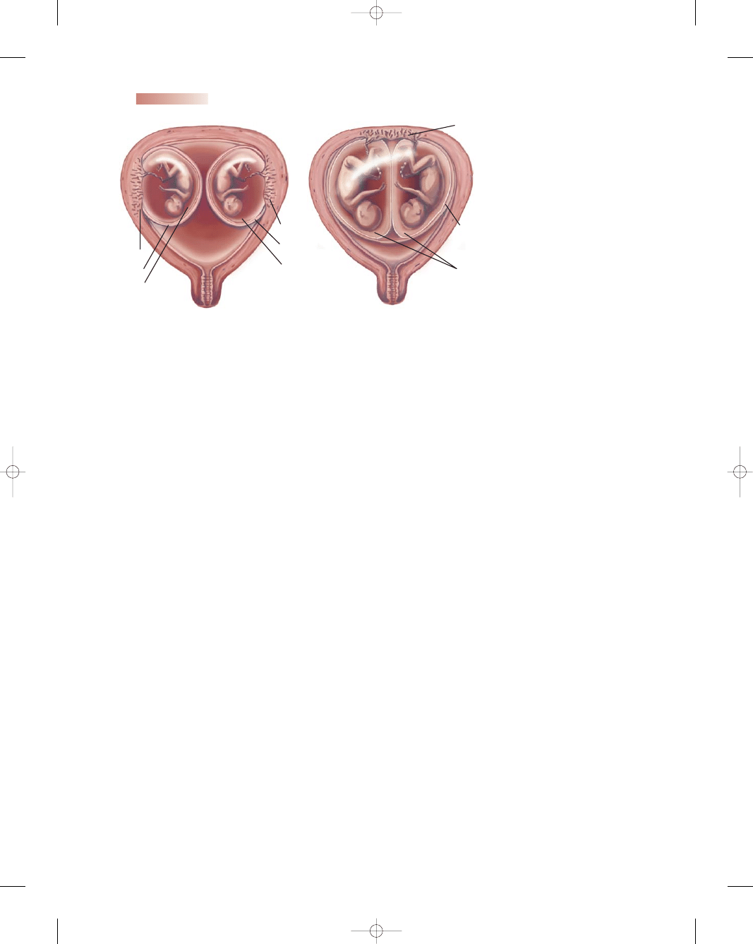
chorions, and placentas are formed in dizygotic twins.
Triplets can be monozygotic, dizygotic, or trizygotic.
Clinical Manifestations
Typically, with a multiple gestation, the woman’s uterus is
larger than that associated with her estimated date of birth.
Anemia, fatigue, and severe nausea and vomiting also may
be present. The diagnosis of a multiple gestation is typi-
cally made on ultrasound early in the pregnancy.
Treatment
From the point of confirmation on, the woman is followed
with serial ultrasounds to assess fetal growth patterns and
development. Biophysical profiles along with nonstress
tests are ordered to determine fetal well-being. Many
women are hospitalized in late pregnancy to prevent
preterm labor and receive closer surveillance. During
intrapartum, the woman is closely monitored, with a peri-
natal team available to assist after birth. Operative delivery
is frequently needed, resulting from fetal malpresentation.
Nursing Management
Nursing management during the prenatal period focuses
on education and support of the woman in areas of nutri-
tion, increased rest periods, and close observation for
pregnancy complications—anemia, excessive weight gain,
proteinuria, edema, vaginal bleeding, and hypertension.
Instruct the woman to be alert for and report immediately
any signs and symptoms of preterm labor—contractions,
uterine cramping, low back ache, increase in vaginal dis-
charge, loss of mucus plug, pelvic pain, and pressure.
When the woman with a multiple gestation is admit-
ted for labor and birth, expect to monitor fetal heart rates
continuously. Prepare the woman for an ultrasound to
assess the presentation of each fetus to determine the best
delivery approach. Ensure that extra nursing staff and the
perinatal team are available for any birth or newborn com-
plications. After giving birth, closely assess the woman for
hemorrhage by frequently assessing uterine involution.
Palpate the uterine fundus and monitor the amount and
characteristics of lochia. Throughout the entire pregnancy,
birth, and hospital stay, inform and support the woman
and her family regarding potential concerns that might
occur. Encourage them to ask questions and verbalize any
fears and concerns.
Premature Rupture
of Membranes
Premature rupture of membranes
(PROM) is
defined as the rupture of the bag of waters prior to the
onset of true labor. It carries a number of associated con-
ditions and complications, such as infection, prolapsed
cord, abruptio placenta, and preterm labor. This is the sin-
gle most common diagnosis associated with preterm births
(Moos, 2004).
The terminology pertaining to PROM can be confus-
ing. PROM is rupture of the membranes prior to the onset
of labor and is used appropriately when referring to a
woman who is beyond 37 weeks’ gestation, has presented
with spontaneous rupture of the membranes, and is not in
labor. Related terms include preterm premature rupture
of membranes (PPROM), which is defined as rupture of
membranes prior to the onset of labor in a woman who is
less than 37 weeks of gestation. Perinatal risks associated
with PPROM may stem from immaturity, including res-
piratory distress syndrome, intraventricular hemorrhage,
patent ductus arteriosus, and necrotizing enterocolitis.
Recent studies have shown clear benefits of antibiotics
to decrease neonatal morbidity associated with PPROM
(Wilkes & Galan, 2004).
The incidence of PROM is approximately 10% of
pregnancies (Wilkes & Galan, 2004). Prolonged rupture
of membranes consists of rupture of membranes for more
than 24 hours, and women are at increasing risk for infec-
tion (chorioamnionitis, endometritis, sepsis, and neonatal
530
Unit 7
CHILDBEARING AT RISK
Placenta
Chorion
Amnion
Amnions
Amnion
Chorion
Chorion
Placenta
Placenta
A
B
●
Figure 19-6
Multiple gesta-
tion with twins. (A) Monozygotic
twins where the fetuses share one
placenta, two amnions, and one
chorion. (B) Dizygotic twins,
where each fetus has its own pla-
centa, amnion, and chorion.
10681-19_UT7-CH19rev.qxd 6/19/07 3:08 PM Page 530

infections) as the duration of rupture increases. The time
interval from rupture of membranes to the onset of regu-
lar contractions is termed the latent period.
Cause
The exact mechanism of PROM is not known. However,
various factors have been associated with the condition,
including infection, increased uterine size (hydramnios,
macrosomia, multifetal gestation), uterine and fetal ano-
malies, lower socioeconomic status, STIs, incompetent
cervix, vaginal bleeding, and cigarette smoking during
pregnancy. In many cases, however, PROM may occur in
the absence of any recognized risk factors. Women who do
not go into labor immediately are at increasing risk of infec-
tion as the duration of rupture increases. Chorioamnionitis,
endometritis, sepsis, and neonatal infections are common
sequelae.
Clinical Manifestations
The clinical picture presented by the woman with PROM
may include labor symptoms (cramping, pelvic pressure,
or back pain), history or symptoms of UTI (frequency,
urgency, dysuria, or flank pain), history of or current
symptoms of pelvic or vaginal infection (pain or vagi-
nal discharge), and/or vital signs reflective of infection
(temperature elevation and white blood cell elevation
> 18,000 cells/mm
3
) (Mattson & Smith, 2004).
To determine the diagnosis of PROM, several proce-
dures may be used: the Nitrazine test, fern test, or ultra-
sound. After the insertion of a sterile speculum, a sample
of the fluid found in the vaginal area is obtained. With a
Nitrazine test, pH of the fluid is tested; amniotic fluid is
more basic (7.0) compared with normal vaginal secretions
(4.5). Nitrazine paper turns blue in the presence of amni-
otic fluid. Unfortunately, many false positives can occur if
blood, urine, semen, or antiseptic chemicals are also pre-
sent. All will increase the pH.
For the fern test, a sample of vaginal fluid is place on
a slide to be viewed directly under a microscope. Amniotic
fluid will develop a fern-like pattern when it dries, result-
ing from the amount of NaCl crystallization that occurs. If
both previous tests are inconclusive, a transvaginal ultra-
sound can also be used to determine whether membranes
have ruptured by demonstrating a decreased amount of
amniotic fluid (oligohydramnios) in the uterus (Wilkes &
Galan, 2004).
Treatment
Treatment of PROM typically depends on the gestational
age. Under no circumstances is a digital cervical exami-
nation done until the woman enters active labor, to min-
imize infection exposure. If the fetal lungs are mature,
induction of labor is initiated. PROM is not an indica-
tion, in and of itself, for surgical birth. If the fetal lungs
are immature, expectant management is carried out with
adequate hydration, reduced physical activity, pelvic rest,
and close observation for possible infection, such as with
frequent monitoring of vital signs and checking the results
of laboratory tests (e.g., the white blood cell count).
Corticosteroids may be given to enhance fetal lung matu-
rity, although this remains controversial.
Nursing Management
Nursing management of PROM usually depends on the
gestational age and presence or absence of an intraamniotic
infection (chorioamnionitis). An accurate assessment of
the gestational age and knowledge of the maternal, fetal,
and neonatal risks are essential to appropriate evaluation,
counseling, and management of women with PROM.
Assessment
After obtaining the woman’s medical and obstetric history,
set up and perform fetal heart rate monitoring to check for
fetal well-being, assess the woman’s labor status, and con-
duct a vaginal examination to ascertain the cervical status
in PROM. If PPROM exists, a sterile speculum examina-
tion is done rather than a digital cervical examination
because it may diminish latency (period of time from rup-
ture of membranes to birth) and increase newborn mor-
bidity (Cunningham et al., 2005). Key assessment areas
are highlighted in Box 19-4.
Assessing the characteristics of the amniotic fluid is
important. Abnormal findings would include presence
Chapter 19
NURSING MANAGEMENT OF PREGNANCY AT RISK: PREGNANCY-RELATED COMPLICATIONS
531
For the woman with PROM, the following assessments
are essential:
• Determining the date, time, and duration of membrane
rupture by client interview
• Ascertaining gestational age of the fetus based on date
of mother’s LMP, fundal height, and ultrasound dating
• Questioning the woman about possible history of or
recent UTI or vaginal infection that might have con-
tributed to PROM
• Assessing for any associated labor symptoms, such as
back pain or pelvic pressure
• Assisting with or performing diagnostic tests to vali-
date leakage of fluid, such as Nitrazine test, “ferning”
on slide, and ultrasound
• Continually assessing for signs of infection including
• Elevation of maternal temperature and pulse rate
• Abdominal/uterine tenderness
• Fetal tachycardia more than 160 bpm
• Elevated white blood cell count and C-reactive protein
• Cloudy, foul-smelling amniotic fluid
BOX 19-4
KEY ASSESSMENTS WITH PREMATURE
RUPTURE OF MEMBRANES
10681-19_UT7-CH19rev.qxd 6/19/07 3:08 PM Page 531

of meconium, minimal amount, and a foul odor. When
meconium is present in the amniotic fluid, it typically indi-
cates fetal distress related to hypoxia. Meconium stains the
fluid yellow to greenish brown, depending on the amount
present. A decreased amount of amniotic fluid reduces the
cushioning effect, thereby making cord compression a pos-
sibility. A foul odor of amniotic fluid indicates infection.
Nursing Interventions
Nursing interventions for the woman with PROM or
PPROM focus on infection prevention and identification
of preterm labor contractions. The risk for infection is
great, resulting from the break in the amniotic fluid mem-
brane and its close proximity to vaginal bacteria. Therefore,
monitor maternal vital signs closely. Be alert for a temper-
ature elevation or an increase in pulse, which would indi-
cate infection. Also monitor fetal heart rate, reporting any
fetal tachycardia, which could indicate a maternal infection.
Evaluate the results of laboratory tests such as a CBC. An
elevation in white blood cells would suggest infection.
Additional nursing interventions include
•
Administering antibiotics if ordered
•
Monitoring fetal heart rate patterns continuously,
reporting any variable decelerations suggesting cord
compression
•
Assisting with amnioinfusion to alter variable deceler-
ation pattern per orders or according to institutional
protocol
•
Educating the woman and her partner on the purpose
of the protective membranes and implications related to
early rupture
•
Continually informing the woman and her partner of
planned interventions, including potential complications
and required therapy
•
Encouraging the client and her partner to verbalize feel-
ings and concerns
•
Preparing the woman for induction or augmentation of
labor as appropriate if the woman is near term
Client education is key, especially if the woman is to
be discharged home. This may be the case for the woman
with PPROM. Therefore, teach the couple about the signs
and symptoms of infection if the client is to be discharged
home, including who and when to call for problems or
concerns (see Teaching Guidelines 19-3).
Preterm Labor
Preterm labor
is defined as the occurrence of regular
uterine contractions accompanied by cervical efface-
ment and dilation before 37 weeks’ gestation. It occurs
between 20 weeks and 37 weeks’ gestation. Preterm
births remain one of the most problematic situations
contributing to perinatal morbidity and mortality in the
world. According to the March of Dimes (March of
Dimes, 2005), one in eight infants born in the United
States is born prematurely.
Preterm birth is one of the most common obstetric
complications, and its sequelae have a profound effect on
the survival and health of about one in every eight infants
born in the United States annually (March of Dimes,
2004). The rate of preterm births in the United States has
increased 27% in the past 20 years. Preterm births account
for 75% of neurodevelopmental disorders and other seri-
ous morbidities, as well as behavioral and social problems.
In addition, up to $28 billion is spent on maternal and
infant care related to prematurity (March of Dimes Birth
Defects Foundation, 2003). Infants born prematurely also
are at risk for serious sequelae such as respiratory dis-
tress syndrome, infections, congenital heart defects, ther-
moregulation problems that can lead to acidosis and
weight loss, intraventricular hemorrhage, feeding difficul-
ties resulting from diminished stomach capacity and under-
developed suck reflex, and neurologic disorders related
to hypoxia and trauma at birth (Newton, 2004). Although
great strides have been made in neonatal intensive care,
prematurity remains the leading cause of death within the
first month of life and is the second leading cause of all
infant deaths (March of Dimes, 2005).
Cause
The exact cause of preterm labor and birth is not known.
Prevention is the goal. However, prevention of all preterm
births is not possible because numerous risk factors exist.
532
Unit 7
CHILDBEARING AT RISK
T E A C H I N G G U I D E L I N E S 1 9 - 3
Teaching for the Woman With PPROM
•
Monitor your baby’s activity by performing fetal kick
counts daily.
•
Check your temperature daily and report any temper-
ature increases to your healthcare provider.
•
Watch for signs related to the beginning of labor.
Report any tightening of the abdomen or contractions.
•
Avoid any touching or manipulating of your breasts,
which could stimulate labor.
•
Do not insert anything into your vagina or vaginal area.
•
Maintain any specific activity restrictions as
recommended.
•
Wash your hands thoroughly after using the bathroom
and make sure to wipe from front to back each time.
•
Keep your perineal area clean and dry.
•
Take your antibiotics as directed if your healthcare
provider has prescribed them.
•
Call your health care provider with changes in your
condition, including fever, uterine tenderness, feeling
like your heart is racing, and foul-smelling vaginal
discharge.
10681-19_UT7-CH19rev.qxd 6/19/07 3:08 PM Page 532

Box 19-5 highlights some of the risk factors associated
with preterm labor and birth.
Clinical Manifestations
Frequently, women are unaware that uterine contractions,
effacement, and dilation are occurring, thus making early
intervention and treatment ineffective in arresting preterm
labor and preventing the birth of a premature infant.
Recognizing preterm labor at an early stage requires that
the expectant mother and her healthcare provider identify
the subtle symptoms of preterm labor. These may include
•
Change or increase in vaginal discharge
•
Pelvic pressure (pushing down sensation)
•
Low, dull backache
•
Menstrual-like cramps
•
Uterine contractions, with or without pain
•
Intestinal cramping, with or without diarrhea (AAP &
ACOG, 2003)
Treatment
Predicting risk or preterm labor is only valuable if there is
an available intervention that is likely to improve the situ-
ation and, according to ACOG, many factors should be
considered before selecting an intervention. Many factors
influence the decision to intervene when women present
with symptoms of preterm labor, including the probabil-
ity of progressive labor, gestational age, and the risks of
treatment. ACOG (2003) recommends the following as
guidelines:
•
There are no clear “first-line”
tocolytic
drugs (drugs
that promote uterine relaxation by interfering with uter-
ine contraction) to manage preterm labor. Clinical cir-
cumstances and healthcare provider preference should
dictate treatment.
•
Antibiotics do not appear to prolong gestation and should
be reserved for group B streptococcal prophylaxis in
women in whom birth is imminent.
•
Tocolytic drugs may prolong pregnancy for 2 to 7 days,
which may allow for administration of steroids to improve
fetal lung maturity and transporting the woman to a
tertiary care center.
Despite the recommendations of ACOG (2003),
health care providers continue to prescribe pharmacologic
treatment for preterm labor at home and in the hospital set-
ting. This treatment often includes oral or IV tocolytics and
varying degrees of activity restriction. Antibiotics may also
be prescribed to treat presumed or confirmed infections.
Steroids may be given to enhance fetal lung maturity
between 24 weeks and 34 weeks’ gestation. Commonly
used tocolytics include
•
Beta-adrenergics, such as ritodrine (Yutopar) and terbu-
taline (Brethine)
•
Calcium channel blockers, such as nifedipine (Procardia)
•
Prostaglandin synthetase inhibitors, such as indo-
methacin (Indocin; not used after 32 weeks’ gestation,
because of its effects on the fetus)
•
Magnesium sulfate, which reduces muscular ability to
contract
Nursing Management
Nurses play a key role in reducing preterm labor and births
to improve pregnancy outcomes for both mothers and
their infants. Early detection of preterm labor is currently
the best strategy to improve outcomes. Because of the
numerous factors associated with preterm labor, it is chal-
lenging to identify and address all of them, especially when
women experiencing contractions are frequently falsely
reassured and not assessed thoroughly to determine the
cause. This delay impedes initiation of interventions to
reduce infant death and morbidity.
Preterm birth prevention programs for women at
high risk have used self-monitoring of symptoms and pat-
terns, weekly cervical examinations, telephone monitor-
Chapter 19
NURSING MANAGEMENT OF PREGNANCY AT RISK: PREGNANCY-RELATED COMPLICATIONS
533
• African-American race (double the risk)
• Maternal age extremes less than 16 years and more
than 40 years old
• Low socioeconomic status
• Alcohol or other drug use, especially cocaine
• Poor maternal nutrition
• Maternal periodontal disease
• Cigarette smoking
• Low level of education
• History of prior preterm birth (triples the risk)
• Uterine abnormalities, such as fibroids
• Low pregnancy weight for height
• Preexisting diabetes or hypertension
• Multiple gestation
• PROM
• Late or no prenatal care
• Short cervical length
• STIs: gonorrhea, Chlamydia, trichomoniasis
• Bacterial vaginosis (50% increased risk) (Newton, 2004)
• Chorioamnionitis
• Hydramnios
• Gestational hypertension
• Cervical insufficiency
• Short interpregnancy interval: less than 1 year
between births
• Placental problems, such as placenta previa and
abruption placenta
• Maternal anemia
• UTI
• Domestic violence
• Stress, acute and chronic (Moos, 2004)
BOX 19-5
RISK FACTORS ASSOCIATED WITH
PRETERM LABOR AND BIRTH
10681-19_UT7-CH19rev.qxd 6/19/07 3:08 PM Page 533

ing, home visiting, and home uterine activity monitoring,
along or in combination, with mixed results (Weiss, Saks,
& Harris, 2002).
Assessment
Signs and symptoms occurring between 20 weeks and
37 weeks of pregnancy that need to be assessed and eval-
uated further for preterm include
•
Pelvic pressure (feels like the fetus is pushing down)
•
Complaints of low, dull back pain
•
Feelings of menstrual-like cramps
•
Change or increase in vaginal discharge
•
Heaviness or aching in the thighs
•
Uterine contractions occurring every 10 minutes or more
frequently
•
Intestinal cramping, with or without diarrhea
•
Fetal engagement into maternal pelvis
•
Tachycardia (Freda & Patterson, 2004)
The diagnosis of preterm labor requires both uterine
contractions and cervical change. The contractions must
be persistent, such that four contractions occur every
20 minutes or eight contractions occur in 1 hour. Cervical
effacement is 80% or greater and cervical dilation is greater
than 1 cm (AAP & ACOG, 2003).
Diagnostic Methods
Currently, four tests are commonly used for preterm labor
prediction: fetal fibronectin testing, cervical length evalu-
ation by transvaginal ultrasound, salivary estriol, and home
monitoring of uterine activity to recognize preterm con-
tractions. Fetal fibronectin and cervical length examina-
tions have a high negative predictive value and are thus
better at predicting which pregnant women are unlikely to
have a preterm birth as opposed to predicting those who
will (Bernhardt & Dorman, 2004).
Other diagnostic testing used in preterm labor risk
assessment include a CBC to detect the presence of
infection, which may be a contributing factor to preterm
labor; urinalysis to detect bacteria and nitrites, which are
indicative of a UTI; and an amniotic fluid analysis to
determine fetal lung maturity and presence of subclinical
chorioamnionitis.
Fetal Fibronectin
Fetal fibronectin, a glycoprotein produced by the chorion,
is found at the junction of the chorion and decidua (fetal
membranes and uterus). It is present in cervicovaginal
fluid prior to delivery, regardless of gestational age. It is not
found in vaginal secretions unless there has been a disrup-
tion between the chorion and decidua. The test is a use-
ful marker for impending membrane rupture within 7 to
14 days if the level increases to greater than 0.05 µg/mL.
Conversely, if fetal fibronectin is not present, there is a
98% chance that the woman will not go into preterm
labor (Schnell, Van Leeuwen, & Kranpitz, 2003).
A sterile applicator is used to collect a cervicovaginal
sample during a speculum examination. The result is
either positive (fetal fibronectin is present) or negative
(fetal fibronectin is not present). Interpretation of fetal
fibronectin results should always be used in conjunction
with clinical findings and not used in isolation for preterm
labor prediction.
Transvaginal Ultrasound
Transvaginal ultrasound of the cervix has been used as a
tool to predict preterm labor in high-risk pregnancies
and to differentiate between true and false preterm labor.
Three parameters are evaluated during the transvaginal
ultrasound: cervical length and width, funnel width and
length, and percentage of funneling. Measurement of the
closed portion of the cervix visualized during the trans-
vaginal ultrasound is the single most reliable parameter
for prediction of preterm delivery in high-risk women
(Iams, 2003).
Cervical length varies during pregnancy. A cervi-
cal length of 3 cm or more indicates that delivery within
14 days is unlikely. Women with a short cervical length
of 2.5 cm during the mid trimester have a substantially
greater risk of preterm birth prior to 35 weeks’ gestation
(Iams & Creasy, 2004). As with fetal fibronectin testing,
negative results can be reassuring and prevent unneces-
sary interventions (Abrahams & Katz, 2002).
Salivary Estriol
Salivary estriol is another biochemical marker that has
been shown to increase before preterm labor. The woman
can perform the test at home and send the sample to the
lab. A value of estriol greater than 2.1 ng/mL is considered
positive.
Observational studies have shown that maternal levels
of estriol increase in their saliva and blood serum before the
onset of spontaneous term and preterm labor. According to
ACOG (2002), however, no current data support using
salivary estriol screening as a strategy to identify or prevent
preterm labor. The test carries a high percentage of false-
positive results and has failed to establish its usefulness
for anything more than investigational purposes (Ressel,
2004). Additional research is needed.
Home Uterine Activity Monitoring
Home monitoring of uterine activity can identify women
in preterm labor at an early stage to reduce the rate of
preterm birth (Moore, 2003). Home uterine activity mon-
itoring does not prevent preterm labor or birth; it provides
supplemental client education and clinical data for the
healthcare provider in an effort to prolong gestation and
maximize pregnancy outcomes through timely clinical
interventions (Fig. 19-7). It involves client education, daily
client assessment, data collection, data interpretation
(Morrison et al., 2004).
The woman is asked to wear an ambulatory toco-
dynamometer and transmit data to the nurse or health-
534
Unit 7
CHILDBEARING AT RISK
10681-19_UT7-CH19rev.qxd 6/19/07 3:08 PM Page 534

care provider daily via telephone lines. Uterine and fetal
activity data are transmitted to perinatal nurses who are
available around-the-clock for detection of uterine con-
tractions, in addition to picking up the fetal heart rate.
Women are also asked to record uterine activity (uter-
ine pressure, back pain, or cramps) they experience for
an hour twice a day and then speak to a perinatal nurse
who analyzes the results.
If the number of contractions exceeds a predeter-
mined threshold, the woman drinks 8 to 12 oz of water,
rests, empties her bladder, and then repeats uterine mon-
itoring. This modality of screening remains somewhat
controversial as to its real impact on preventing pre-
term birth.
Nursing Interventions
A reduction in the preterm birthrate cannot be achieved
until there are effective strategies to predict risk for preterm
birth as well as effective methods to prevent preterm births.
Because the etiology is often multifactorial, an individual-
ized approach is needed.
The signs or preterm labor are subtle and maybe dis-
regarded by the client as well as the health care professional.
Because risk assessment does not identify many women
who will develop preterm labor, ensure that every pregnant
woman receives basic education about preterm labor,
including information about harmful lifestyles, the signs of
genitourinary infections and preterm labor, and the appro-
priate response to these symptoms. Teach the client how to
palpate for and time uterine contractions. Provide written
materials to support this education at a level and in a lan-
guage appropriate for the client. Also, educate clients about
the importance of prenatal care, risk reduction, and recog-
nizing the signs and symptoms of preterm labor. Teaching
Guidelines 19-4 highlights important instructions related
to preventing preterm labor.
Additional nursing interventions include the follow-
ing activities:
•
Assessing maternal and fetal vital signs frequently
•
Stressing good hydration and consumption of a nutri-
tious diet
•
Advising against any activity, such as sexual activity or
nipple stimulation, that might stimulate oxytocin release
and initiate uterine contractions
•
Encouraging clients to access their healthcare provider
for reassurance
•
Assessing stress levels of client and family, and making
appropriate referrals
Chapter 19
NURSING MANAGEMENT OF PREGNANCY AT RISK: PREGNANCY-RELATED COMPLICATIONS
535
●
Figure 19-7
The mother with preterm labor resting in bed
at home.
T E A C H I N G G U I D E L I N E S 1 9 - 4
Teaching to Prevent Preterm Labor
•
Avoid traveling for long distances in cars, trains,
planes, or buses.
•
Avoid lifting heavy objects, such as laundry, groceries,
or a young child.
•
Avoid performing hard, physical work, such as yard
work, moving of furniture, or construction.
•
Visit a dentist in early pregnancy to evaluate and treat
periodontal disease.
•
Enroll in a smoking cessation program if you are
unable to quit on your own.
•
Curtail sexual activity until after 37 weeks if experi-
encing preterm labor symptoms.
•
Consume a well-balanced nutritional diet to gain
appropriate weight.
•
Avoid the use of substances such as marijuana,
cocaine, and heroin.
•
Identify factors and areas of stress in your life, and use
stress management techniques to reduce them.
•
If you are experiencing intimate partner violence, seek
resources to modify the situation.
Recognize the signs and symptoms of preterm labor and
notify your birth attendant if any occur:
•
Uterine contractions, cramping, or low back pain
•
Feeling of pelvic pressure or fullness
•
Increase in vaginal discharge
•
Nausea, vomiting, and diarrhea
•
Leaking of fluid from vagina
•
If you are experiencing any of these signs or symptoms,
do the following:
•
Stop what you are doing and rest for 1 hour.
•
Empty your bladder.
•
Lie down on your side.
•
Drink two to three glasses of water.
•
Feel your abdomen and make note of the hardness of
the contraction. Call your health care provider and
describe the contraction as
•
Mild if it feels like the tip of the nose
•
Moderate if it feels like the tip of the chin
•
Strong if it feels like your forehead (Mattson &
Smith, 2004)
10681-19_UT7-CH19rev.qxd 6/19/07 3:08 PM Page 535

•
Providing emotional support and client empowerment
throughout
•
Emphasizing the possible need more frequent super-
vision and office visits
•
Raising public awareness of the problem of prematurity
Medication Administration
When preterm labor is identified because of PPROM or
persistent uterine contractions accompanied by cervical
change, treatment has commonly consisted of IV hydra-
tion, often accompanied by tocolytic therapy. Neither
therapy has been shown to be effective for more than
48 hours. However, delaying birth for 48 hours allows time
for the administration of corticosteroids to accelerate fetal
lung maturity and client transfer to a medical facility where
there is a neonatal intensive care unit (Newton, 2004).
Tocolytic Therapy
Tocolytics are agents that promote uterine relaxation by
interfering with uterine contraction. Tocolytic therapy does
not typically prevent preterm birth, but it may delay it. It is
contraindicated for abruptio placenta, acute fetal dis-
tress or death, eclampsia or severe preeclampsia, active
vaginal bleeding, dilation more than 6 cm, chorioamnioni-
tis, and maternal hemodynamic instability (Harvey, 2003).
The decision to stop preterm labor is individualized,
based on risk factors, extent of cervical dilation, membrane
status, fetal gestational age, and presence or absence of
infection. It is most likely ordered if preterm labor occurs
before the 34th week of gestation, attempting to delay birth
and thereby helping to reduce the severity of respiratory
distress syndrome and other complications associated with
prematurity. Medications most commonly used for tocol-
ysis include magnesium sulfate, ritodrine (Yutopar), terbu-
taline (Brethine), indomethacin (Indocin), and nifedipine
(Procardia). Ritodrine is the only FDA-approved agent for
arresting preterm labor. The other drugs are used “off label.
This means they are effective for this purpose, but have not
been officially tested and developed for this purpose by the
FDA (Lowdermilk & Perry, 2004). All these medications
have serious side effects and warrant close supervision
when being administered (see Drug Guide 19-4).
Corticosteroids
Corticosteroids given to the mother in preterm labor can
help prevent or reduce the frequency and severity of res-
piratory distress syndrome in premature infants delivered
between 24 weeks and 34 weeks’ gestation. The benefi-
cial effects of corticosteroids on fetal lung maturation
have been reported within 48 hours of initial administra-
tion (Slattery & Morrison, 2002). These drugs require at
least 24 hours to become effective, so timely administra-
tion is crucial.
Psychological Support
The birth of an preterm newborn can be a devastating
event for family members, who may be faced with enor-
mous long-term health and social implications during
childhood, and beyond. Nurses are on the front lines to
provide this high-risk family with support, education, and
expert nursing care.
Preterm labor and birth present multifactorial chal-
lenges for everyone involved with this crisis event. If the
woman is restricted in her activities, additional stresses
may be placed on the family, contributing to the crisis.
Every case of spontaneous preterm labor is unique,
requiring individualization of care in light of the clinical
circumstances, and the full and informed consent of the
woman and her partner. Half of all women who ulti-
mately give birth prematurely have no identifiable risk
factors. Nurses need to take all women’s complaints seri-
ously and complete a thorough assessment to validate
their etiology.
Work to ensure that all pregnant women have access to
nurses who are sensitive to any complaint and are able to
provide appropriate information and follow-up. Sensitivity
to the subtle differences between normal pregnancy sensa-
tions and the prodromal symptoms of preterm labor is a key
factor in ensuring timely access to care. Offer clarification
and validation of their symptoms.
K E Y C O N C E P T S
●
Identifying risk factors early on and ongoing through-
out the pregnancy is important to ensure the best out-
come for every pregnancy. Risk assessment should
start with the first prenatal visit and continue with
subsequent visits.
●
The three most common causes of hemorrhage early
in pregnancy (first half of pregnancy) are spontaneous
abortion, ectopic pregnancy, and GTD.
●
Ectopic pregnancies occur in about 1 in 50 pregnan-
cies and have increased dramatically during the past
few decades.
●
Having a molar pregnancy results in the loss of
the pregnancy and the possibility of developing
choriocarcinoma, a chronic malignancy from the
trophoblastic tissue.
●
The classic clinical picture presentation for placenta
previa is painless, bright-red vaginal bleeding occur-
ring during the third trimester.
●
Treatment of abruptio placenta is designed to assess,
control, and restore the amount of blood lost; to
provide a positive outcome for both mother and
infant; and to prevent coagulation disorders.
●
DIC can be described in simplest terms as a loss of
balance between the clot-forming activity of thrombin
and the clot-lysing activity of plasmin.
●
Hyperemesis gravidarum is a complication of
pregnancy characterized by persistent, uncontrol-
lable nausea and vomiting before the 20th week of
gestation.
536
Unit 7
CHILDBEARING AT RISK
10681-19_UT7-CH19rev.qxd 6/19/07 3:08 PM Page 536

Chapter 19
NURSING MANAGEMENT OF PREGNANCY AT RISK: PREGNANCY-RELATED COMPLICATIONS
537
Drug Guide 19-4
Drug
Action/Indication
Nursing Implications
Magnesium
sulfate
Ritodrine
(Yutopar)
Relaxes uterine muscles to stop
irritability and contractions,
to arrest uterine contractions
for preterm labor (off-label
use)
Has aided in seizure prophylaxis
and treatment of seizures
in preeclamptic and
eclamptic patients for
almost 100 years (Rideout,
2005)
Relaxes smooth muscles to
calm the uterus, inhibits
uterine activity to arrest
preterm labor
Administer intravenously with a loading dose of 4–6 g over
15–30 minutes initially, and then maintain infusion at
1–4 g per hour
Assess vital signs and DTRs hourly; report any hypotension
or depressed or absent DTRs
Monitor level of consciousness; report any headache,
blurred vision, dizziness, or altered level of
consciousness
Perform continuous electronic fetal monitoring; report any
decreased fetal heart rate variability, hypotonia, or
respiratory depression
Monitor intake and output hourly; report any decrease in
output (<30 mL/hour)
Assess respiratory rate; report respiratory rate less than
12 breaths/minute; auscultate lung sounds for evidence
of pulmonary edema
Monitor for common maternal side effects including
flushing, nausea and vomiting, drug mouth, lethargy,
blurred vision, and headache
Assess for complaints of nausea, vomiting, transient
hypotension, lethargy
Assess for signs and symptoms of magnesium toxicity,
such as decreased level of consciousness, depressed
respirations and DTRs, slurred speech, weakness, and
respiratory and/or cardiac arrest
Have calcium gluconate readily available at the bedside
to reverse magnesium toxicity
Assess maternal vital signs frequently
Monitor the woman for possible adverse effects including
tachycardia, hypotension, palpitations, tremors, chest
pain, hypokalemia, water retention, nausea, vomiting,
diarrhea, hyperglycemia, decreased urinary output,
and nervousness
Monitor fetal status, being alert for tachycardia, heart
failure, hyperglycemia, hypotension, and jaundice
Be aware that side effects are dose related and will
increase as dose is increased
Perform cardiac assessment of woman to rule out
preexisting cardiac disease and diabetes
Perform continuous fetal monitoring
Administer as IV infusion, at prescribed rate, increasing
rate every 10–20 minutes as necessary; reduce rate
gradually to lowest possible rate to stop contractions;
maintain for 12–24 hours
Expect to begin oral dose an hour before IV infusion is
stopped; administer oral dose every 3–4 hours with
meals
Discontinue IV infusion if the following occur: maternal
tachycardia >120 bpm, hypotension <90/60 mmHg,
fetal heart rate >180 bpm, signs of maternal pulmonary
edema
Have propranolol (Inderal) available to reverse cardiac
adverse effects
Drug Guide 19-4
Medications Used With Preterm Labor
(continued)
10681-19_UT7-CH19rev.qxd 6/19/07 3:08 PM Page 537

538
Unit 7
CHILDBEARING AT RISK
Drug Guide 19-4
Drug
Action/Indication
Nursing Implications
Drug Guide 19-4
Medications Used With Preterm Labor
(continued)
Terbutaline sulfate
(Brethine)
Indomethacin
(Indocin)
Nifedipine
(Procardia)
Betamethasone
(Celestone)
Relaxes smooth muscles to
calm uterus, inhibits uterine
activity to arrest preterm
labor
Inhibits prostaglandins, which
stimulate contractions;
inhibits uterine activity to
arrest preterm labor
Blocks calcium movement into
to muscle cells, inhibits
uterine activity to arrest
preterm labor
Promotes fetal lung maturity
by stimulating surfactant
production, prevents or
reduces risk of respiratory
distress syndrome and
intraventricular hemorrhage
in the preterm neonate less
than 34 weeks’ gestation
Be aware that this drug is usually effective in delaying birth
for up to 48 hours (Rideout, 2005)
Monitor the mother for possible adverse effects such as
tachycardia, hypotension, palpitations, tremors, chest
pain, hypokalemia, water retention, nausea, vomiting,
diarrhea, hyperglycemia, decreased urinary output,
and nervousness
Assess fetal well-being, noting any possible adverse
effects such as tachycardia, heart failure,
hyperglycemia, hypotension, newborn jaundice
Be aware that adverse effects are dose related and will
increase as dose is increased
Perform cardiac assessment of woman to rule out
preexisting cardiac disease and diabetes
Institute continuous fetal monitoring
Assess vital signs frequently for changes
Discontinue drug for the following: maternal tachycardia
>120 bpm, hypotension <90/60 mmHg, fetal heart rate
>180 bpm, signs of maternal pulmonary edema
Have propranolol (Inderal) available to reverse cardiac
adverse effects
Continuously assess vital signs, uterine activity, and fetal
heart rate
Administer oral form with food to reduce GI irritation
Do not give to women with peptic ulcer disease
Schedule ultrasound to assess amniotic fluid volume and
function of ductus arteriosus before initiation of
therapy; monitor for signs of maternal hemorrhage
Be alert for possible maternal adverse effects such as
nausea, vomiting, heartburn, rash, prolonged bleeding
time, oligohydramnios, and hypertension
Monitor for possible fetal adverse effects including
constriction of ductus arteriosus, premature ductus
closure, necrotizing enterocolitis, oligohydramnios, and
pulmonary hypertension
Use caution if giving this drug with magnesium sulfate
because of increased risk for hypotension
Monitor blood pressure hourly if giving with magnesium
sulfate; report a pulse rate >110 bpm
Monitor for possible fetal effects such as decreased
uteroplacental blood flow and fetal bradycardia,
which can lead to fetal hypoxia
Monitor for possible adverse effects, such as flushing of the
skin, headache, transient tachycardia, palpitations,
postural hypertension, peripheral edema, and transient
fetal tachycardia
Administer two doses intramuscularly 24 hours apart
Monitor for possible maternal infection or pulmonary edema
Educate parents about potential benefits of drug to
preterm infant
Assess maternal lung sounds and monitor for signs of
infection
10681-19_UT7-CH19rev.qxd 6/19/07 3:08 PM Page 538

●
Gestational hypertension is the leading cause of
maternal death in the United States and the most
common complication reported during pregnancy.
●
HELLP is an acronym for hemolysis, elevated liver
enzymes, and low platelets.
●
Rh incompatibility is a condition that develops when
a woman of Rh-negative blood type is exposed to
Rh-positive fetal blood cells and subsequently devel-
ops circulating titers of Rh antibodies.
●
Hydramnios occurs in approximately 3 to 4% of all
pregnancies and is associated with fetal anomalies of
development.
●
Nursing care related to the woman with oligohy-
dramnios involves continuous monitoring of fetal
well-being during nonstress testing or during labor
and birth by identifying nonreassuring patterns on
the fetal monitor.
●
The increasing number of multiple gestations is a
concern because women who are expecting more
than one infant are at high risk for preterm labor,
hydramnios, hyperemesis gravidarum, anemia,
preeclampsia, and antepartum hemorrhage.
●
Nursing care related to PROM centers around infec-
tion prevention and identification of preterm labor
contractions.
●
Early identification of preterm labor would allow
for appropriate interventions that may prolong the
pregnancy, such as transferring the woman to a
facility with a neonatal intensive care unit for pre-
natal care, administering glucocorticoids to the
mother to promote fetal lung maturity, and giving
appropriate antibiotics to treat infections to arrest
the labor process.
●
It is essential that nurses teach all pregnant women
how to detect the early symptoms of preterm labor
and what to do if they experience contractions or
cramping that does not go away.
References
Abrahams, C. & Katz, M. (2002). A perspective on the diagnosis of
preterm labor. Journal of Perinatal and Neonatal Nursing, 16(1), 1–11.
Ahn, J. T., Hibbard, J. U. (2003). The short cervix in pregnancy:
Which therapy reduces preterm birth? OBG Management.
[Online] Available at www.obgmanagement.com/content/obg_
featurexml.asp?file
=203/08/obg_0803_00.
American Academy of Pediatrics, American College of Obstetricians
and Gynecologists. (2003). Guidelines for perinatal care (5th ed.).
Washington, DC: Author.
American College of Obstetricians and Gynecologists. (2002). ACOG
practice bulletin: Diagnosis and management of preeclampsia and
eclampsia. Number 33. Washington, DC: Author.
American College of Obstetricians and Gynecologists. (2003a).
ACOG practice bulletin: Cervical insufficiency. Obstetrics and
Gynecology, 102, 1091–1099.
American College of Obstetricians and Gynecologists. (2003b).
ACOG practice bulletin: Management of preterm labor. Obstetrics
and Gynecology, 101, 1039–1047.
Baines, E. (2003). A test for ectopic pregnancy. GP: General
Practitioner, February, 74–76.
Berghella, V. (2004). Grand rounds: The short and funneled cervix:
What do I do now? Contemporary OB/GYN, 49, 26–34.
Bernhardt, J. & Dorman, K. F. (2004). Pre-term birth risk assessment
tools: Exploring fetal fibronectin and cervical length for validating
risk. JOGNN, 8(1), 38–44.
Blackburn, S. (2003). Maternal, fetal, & neonatal physiology: A clinical
perspective (2nd ed.). St. Louis: WB Saunders.
Bridges, E. J., Womble, S., Wallace, M., & McCartney, J. (2003).
Hemodynamic monitoring in high-risk obstetrics patients II:
Pregnancy-induced hypertension and preeclampsia. Critical Care
Nurse, 23, 52–56.
Brooks, M. B. (2005). Pregnancy, preeclampsia. eMedicine. [Online]
Available at http://emedicine.com/emergy/topic480.htm.
Brooks, M. B. (2004). Pregnancy, eclampsia. eMedicine. [Online]
Available at http://emedicine.com/emerg/topic796.htm.
Cavanaugh, B. M. (2003). Nurse’s manual of laboratory and diagnostic
tests (4th ed.). Philadelphia: FA Davis.
Chen, P. (2004). Ectopic pregnancy. Medline Plus. [Online] Available
at www.nlm.nih.gov/medlineplus/ency/article/000895.htm.
Chen, P. (2004). Placenta abruptio. Medline Plus. [Online] Available
at www.nlm.nih.gov/medlineplus/ency/article/000901.htm.
Christopher, K. L. (2003). Hyperemesis gravidarum. Journal of
Obstetric, Gynecologic, and Neonatal Nursing, 32, 435–436.
Creasy, R., Resnik, R., & Iams, J. (Eds.), (2004). Maternal fetal medi-
cine (5th ed.). Philadelphia: WB Saunders.
Cunningham, F. G., Gant, N. F., Leveno, K. J., Gilstrap, L. C.,
Hauth, J. C., & Wenstrom, K. D. (2005). Williams’ obstetrics
(22nd ed.). New York: McGraw-Hill.
Curran, C. A. (2003). Intrapartum emergencies. Journal of Obstetric,
Gynecologic, and Neonatal Nursing, 32, 802–813.
Deering, S. H., & Satin, A. (2004). Abruptio placenta. eMedicine.
[Online] Available at www.emedicine.com/med/topic6.htm.
Dillon, P. M. (2003). Nursing health assessment: A critical thinking, case
studies approach. Philadelphia: FA Davis.
Edelman, A. & Logan, J. R. (2004). Pregnancy, hyperemesis gravi-
darum. eMedicine. [Online] Available at www.emedicine.com/
emerg/topic479.htm.
Freda, M. C., & Patterson, E. T. (2004). Preterm labor: Prevention
and nursing management. In March of Dimes nursing module.
White Plains, NY: March of Dimes Education and Health
Promotion.
Fugate, S. R., & Chow, G. E. (2004). Eclampsia. eMedicine. [Online]
Available at www.emedicine.com/med/topic633.htm.
Garcia, R. (2003). Hyperemesis gravidarum: More than morning
sickness. Nursing Spectrum. [Online] Available at http://nsweb.
nursingspectrum.com/ce/ce273.htm.
Gaudier, F. L. (2003). Placenta previa. Medline Plus. [Online]
Available at www.nlm.nih.gov/medlineplus/ency/article/
000900.htm.
Gaufberg, S. V. (2004). Abruptio placenta. eMedicine. [Online]
Available at www.emedicine.com/emerg/topic12.htm.
Gerulath, A. H. (2002). Gestational trophoblastic disease. Journal of
Obstetric & Gynecology of Canada, 24, 434–439.
Gilbert, E., & Harmon, J. (2003). Manual of high-risk pregnancy and
delivery (3rd ed.). Saint Louis: Mosby.
Goldenberg, R. L. (2002). The management of preterm labor.
Obstetrics and Gynecology, 100, 1020–1037.
Green, C. J., & Wilkinson, J. M. (2004). Maternal newborn nursing
care plans. St. Louis: Mosby.
Gupton, A., Heaman, M., & Cheung, L. W. (2001). Complicated
and uncomplicated pregnancies: Women’s perception of risk.
Journal of Obstetric, Gynecologic, and Neonatal Nursing, 30,
192–201.
Hail, E. (2004). Infantile reflexes. Medline Plus. [Online] Available at:
http://www.nim.nih.gov/medlineplus/ency/article/003292.htm.
Harvey, E. A. (2004). Hypertensive disorders of pregnancy: Patho-
physiology, symptoms and treatment. Nursing Spectrum. [Online]
Available at http://nsweb.nursingspectrum.com/ce/m23c-1.htm.
Hermida, R. C., & Ayala, D. E. (2003). Sampling requirements for
ambulatory blood pressure monitoring in the diagnosis of hyper-
tension in pregnancy. Hypertension, 42, 619–626.
Iams, J. D. (2003). Prediction and early detection of preterm labor.
Obstetrics and Gynecology, 101, 402–412.
Iams, J. D. & Creasy, R. K. (2004). Preterm labor and delivery. In
R. K. Creasy, R. Resnik & J. D. Iams (Eds.), Maternal-fetal medicine:
Chapter 19
NURSING MANAGEMENT OF PREGNANCY AT RISK: PREGNANCY-RELATED COMPLICATIONS
539
10681-19_UT7-CH19rev.qxd 6/19/07 3:08 PM Page 539

Principles and practice (5th ed., pp. 623–661). Philadelphia:
Saunders.
Johnson, K. D. (2003). Preeclampsia and eclampsia. Medical Library
Home. [Online] Available at www.medical-library.org/journals/
secure/01110/preeclampsiaeclampsia.htm.
Joy, S., & Lyon, D. (2004). Placenta previa. eMedicine. [Online]
Available at www.emedicine.com/med/topics3271.htm.
Kidner, M. C., & Flanders–Stepans, B. (2004). A model for the
HELLP syndrome: The maternal experience. Journal of Obstetric,
Gynecologic, and Neonatal Nursing, 33, 44–53.
Ko, P., & Yoon, Y. (2005). Placenta previa. eMedicine. [Online]
Available at www.emedicine.com/emerg/topic427.htm.
Koren, G., & Maltepe, C. (2004). Preemptive therapy for severe nau-
sea and vomiting of pregnancy and hyperemesis gravidarum.
Journal of Obstetrics and Gynecology, 24, 530–533.
Lee, R. V. (2003). What maternal mortality statistics tell us: High-risk
pregnancy, a consequence of medical progress. Journal of Obstetrics
and Gynecology, 23, 532–534.
London, M. L., Ladewug, P. W., Ball, J. W., & Bindler, R. C. (2003).
Maternal–newborn & child nursing. Upper Saddle River, NJ:
Prentice Hall.
Longo, S. A., Dola, C. P., & Pridjian, G. (2003). Preeclampsia and
eclampsia revisited. Southern Medical Journal, 96, 891–899.
Lowdermilk, D. L., & Perry, S. E. (2004). Maternity and women’s
health care (8th ed.). St. Louis: Mosby.
March of Dimes (MOD). (2005). Preterm birth. [Online] Available
at: http://www.marchofdimes.com/prematurity/5196_5799.asp.
March of Dimes Birth Defects Foundation. (2003). Taking action
against prematurity. Contemporary Obstetrics and Gynecology, 48,
92–104.
Marchiano, D. (2004). Ectopic pregnancy. Medline Plus. [Online]
Available at www.nlm.nih.gov/medlineplus/ency/article/
000895.htm.
Martin, J. A., Hamilton, B. E., Ventura, S. J., Menacker, F., Park,
M. M., & Sutton, P. (2002). Births: Final data for 2001. National
Vital Statistics Report, 50, 1–104.
Mattson, S., & Smith, J. E. (2004). Core curriculum for maternal–
newborn nursing (3rd ed.). St. Louis: Elsevier Saunders.
Michelini, G. A. (2004). Hyperemesis gravidarum. eMedicine.
[Online] Available at www.emedicine.com/med/topic1075.htm.
Moore, M. L. (2003). Preterm labor and birth: What have we learned
in the past two decades? JOGNN, 32(5), 638–649.
Moos, M. (2004). Understanding prematurity: Sorting fact from fic-
tion. AWHONN Lifelines, 8(1), 32–37.
Morgan, E. (2002). Action stat: Eclampsia. Nursing 2002, 32, 104.
Morley, A. (2004). Pre-eclampsia: Pathophysiology and its management.
British Journal of Midwifery, 12(1), 30–37.
Morrison, J. C., Roberts, W. E., Jones, J. S., Istwan, N., Rhea, D., &
Stanziano, G. (2004). Frequency of nursing, physician and hospi-
tal interventions in women at risk for preterm delivery. Journal of
Maternal–Fetal and Neonatal Medicine, 16, 102–105.
Murray, S. S. & McKinney, E. S. (2006). Foundations of
maternal–newborn nursing (4th ed.). Philadelphia: WB Saunders.
National High Blood Pressure Education Program. (2000). Working
Group in High Blood Pressure in Pregnancy report. American
Journal of Obstetrics and Gynecology, 183, S1–S22.
Neal, J. L. (2001). RhD isoimmunization and current management
modalities. Journal of Obstetric, Gynecologic, and Neonatal Nursing,
30, 589–606.
Newton, E. R. (2004). Preterm labor. eMedicine. [Online] Available
at: http://www.emedicine.com/med/topic3245.htm.
Nick, J. M. (2003). Deep tendon reflexes: The what, why, where, and
how of tapping. Journal of Obstetric, Gynecologic, and Neonatal
Nursing, 32, 297–306.
O’Toole, M. T. (2005). Miller–Keane encyclopedia and dictionary of
medicine, nursing and allied health (7th ed.). Philadelphia: Saunders.
Owen, P. (2003). Management options in ectopic pregnancy. Clinical
Pulse, June, 68–71.
Peters, R. M., & Flack, J. M. (2004). Hypertensive disorders of preg-
nancy. Journal of Obstetric, Gynecologic, and Neonatal Nursing, 33,
209–220.
Puscheck, E., & and Pradhan, A. (2004). Complete abortion. [Online]
Available at www.emedicine.com/med/topic3310.htm.
Ressel, G. W. (2004). Practice guidelines: ACOG releases bulletin on
managing cervical insufficiency. American Family Physician.
[Online] Available at www.aafp.org/afp/20040115/practice.html.
Rideout, S. L. (2005). Tocolytics for preterm labor: What nurses
need to know. AWHONN Lifelines, 9(1), 56–61.
Salem, L. (2005). Rh incompatibility. eMedicine. [Online] Available at
www.emedicine.com/emerg/topic507.htm.
Schmaier, A. H. (2003). Disseminated intravascular coagulation.
eMedicine. [Online] Available at www.emedicine.com/med/
topic577.htm.
Schnell, Z. B., Van Leeuwen, A. M., & Kranpitz, T. R. (2003).
Davis’s comprehensive handbook of laboratory and diagnostic tests with
nursing implications. Philadelphia: FA Davis.
Seidel, H. M., Ball, J. W., Dains, J. E., & Benedict, G. W. (2003).
Mosby’s guide to physical examination (5th ed.). St. Louis: Mosby.
Sepilian, V., & Wood, E. (2005). Ectopic pregnancy. eMedicine.
[Online] Available at www.emedicine.com/med/topic3212.htm.
Shennan, A. H. (2003). Recent developments in obstetrics. British
Medical Journal, 327, 604–609.
Sibai, B. M. (2003). Diagnosis and management of gestational hyper-
tension and preeclampsia. Obstetrics and Gynecology, 102, 181–192.
Simpson, J. (2002). Fetal wastage. In S. Gabbe, J. Niebyl, & J.
Simpson (Eds.), Obstetrics: Normal and problem pregnancies
(4th ed.). New York: Churchill Livingstone.
Skidmore–Roth, L. (2005). Mosby’s 2005 nursing drug reference.
St. Louis: Mosby.
Slattery, M. M. & Morrison, J. J. (2002). Review: Preterm delivery.
Lancet, 360(9344), 1489–1497.
Spratto, G. R., & Woods, A. L. (2005). PDR nurse’s drug handbook.
Clifton Park, NY: Delmar Learning.
Steele, N. M., French, J., Gatherer–Boyles, J., Newman, S., Leclaire,
S. (2001). Effect of acupressure by sea-bands on nausea and vom-
iting of pregnancy. Journal of Obstetric, Gynecologic, and Neonatal
Nursing, 30, 61–70.
Swearingen, P. L. (2004). All-in-one care planning resource. St. Louis:
Mosby.
Thompson, S. R. (2005). Placenta previa. Medline Plus. [Online]
Available at: http://www.nlm.nih.gov/medlineplus/ency/article/
000900.htm.
US Department of Health and Human Services. (2000). Healthy peo-
ple 2010 (conference edition, in two volumes). Washington, DC:
Author.
Valley, V. T., & Fly, C. A. (2005). Ectopic pregnancy. eMedicine.
[Online] Available at www.emedicine.com/EMERG/topic478.htm.
Verklan, M. T., & Walden, M. (2004). Core curriculum for neonatal
intensive care nursing (3rd ed.). St. Louis: Elsevier Saunders.
Weiss, M. E., Saks, N. P. & Harris, S. (2002). Resolving the uncer-
tainty of preterm symptoms: Women’s experiences with the onset
of preterm labor. JOGNN, 31, 66–76.
Wilkes, P. T., & Galan, H. (2004). Premature rupture of membranes.
eMedicine. [Online] Available at: http://www.emedicine.com/med/
topic3246.htm.
Yankowitz, J. (2004). Pharmacologic treatment of hypertensive dis-
orders during pregnancy. Journal of Perinatal and Neonatal
Nursing, 18, 230–240.
Youngkin, E. Q., & Davis, M. S. (2004). Women’s health: A primary
care clinical guide (3rd ed.). Upper Saddle River, NJ: Pearson
Prentice Hall.
Zach, T., & Pramanik, A. (2005). Multiple births. eMedicine. [Online]
Available at www.emedicine.com/ped/topic2599.htm.
Web Resources
American Academy of Pediatrics, www.aap.org
American College of Obstetricians and Gynecologists, www.acog.org
Association of Women’s Health, Obstetric & Neonatal Nurses,
www.awhonn.org
March of Dimes, www.modimes.org
Resolve through Sharing, www.ectopicpregnancy.com
Sidelines High-Risk Pregnancy Support Office, www.sidelines.org
540
Unit 7
CHILDBEARING AT RISK
10681-19_UT7-CH19rev.qxd 6/19/07 3:08 PM Page 540

Chapter 19
NURSING MANAGEMENT OF PREGNANCY AT RISK: PREGNANCY-RELATED COMPLICATIONS
541
Chapter
WORKSHEET
Chapter
●
M U L T I P L E C H O I C E Q U E S T I O N S
1.
The nurse understands that the purpose of
administering magnesium sulfate to a client with
preeclampsia is to
a. Reduce central nervous system irritability to
prevent seizures
b. Provide supplementation of an important mineral
she needs
c. Prevent constipation during and after the birthing
process
d. Decrease musculoskeletal tone to augment labor
2.
Which of the following women would the nurse iden-
tify as being at the greatest risk for preterm labor?
a. A woman who had twins in a previous pregnancy
b. A woman who lives in a big metropolitan city
c. A woman who works full-time as a computer
programmer
d. A woman who has a history of preterm birth
3.
The signs and symptoms of abruptio placenta
depend on the amount of placental separation and
type of abruption. Which of the following would the
nurse assess as a classic symptom?
a. Painless, bright-red bleeding
b. “Knife-like” abdominal pain
c. Excessive nausea and vomiting
d. Hypertension and headache
4.
Which of the following medications would the nurse
expect the physician to order for tocolysis?
a. Diazepam (Valium)
b. Phenobarbital
c. Nifedipine (Procardia)
d. Butorphanol (Stadol)
5.
RhoGAM is given to Rh-negative women to prevent
maternal sensitization from occurring. The nurse is
aware that in addition to pregnancy, Rh-negative
women would also receive this medication after
which of the following?
a. Therapeutic or spontaneous abortion
b. Head injury from a car accident
c. Blood transfusion after a hemorrhage
d. Unsuccessful artificial insemination procedure
●
C R I T I C A L T H I N K I N G E X E R C I S E S
1.
Suzanne, a 16-year-old primigravida, presents to the
maternity clinic complaining of continual nausea and
vomiting for the past 3 days. She states she is approx-
imately 15 weeks pregnant and she has been unable
to hold anything down or take any fluids in without
throwing up for the past 3 days. She reports she is
dizzy and weak. On examination, Suzanne appears
pale, anxious, mucus membranes are dry, skin turgor
is poor, and her lips are dry and cracked.
a. What is your impression of this condition?
b. What risk factors does Suzanne have?
c. What intervention is appropriate for this woman?
2.
Betty, a 20-year-old African-American primigravida
client, comes to the prenatal clinic for her first visit.
She is 4 months pregnant based on dates. As the
nurse assigned to her, you interview her and start to
record her history as follows:
• Is single, unmarried, lives with father of child
• Smokes 1.5 packs of cigarettes a day
• Works as a laborer in a nursery 12 hours daily
• Quit high school in the ninth grade and has no
plans to get GED
• Eats poorly, is underweight for height, and is anemic
• Reports she has frequent UTIs
a. Based on her history, what might this client be at
risk for? Why?
b. What client education is needed at this visit?
c. What specific nursing interventions might help
reduce her risk?
10681-19_UT7-CH19rev.qxd 6/19/07 3:08 PM Page 541

542
Unit 7
CHILDBEARING AT RISK
●
S T U D Y A C T I V I T I E S
1.
Identify a woman hospitalized on bed rest with
preterm labor. Ask her about her experience associ-
ated with the hospitalization and how nurses could
be helpful to her throughout her experience.
2.
Ask a public health maternity nurse how the signs
and symptoms of preterm labor are taught, and how
effective they have been in reducing the incidence in
their area.
3.
Search the Internet for a Web site to help parents
who have suffered a pregnancy loss and critique it for
current information and audience level.
4.
A pregnancy in which the blastocyst implants outside
the uterus describes an ___________________
pregnancy.
5.
The gravest complication of hydatidiform mole is the
development of ___________________ afterward.
6.
Discuss various activities a woman with a multiple
gestation could engage in to help pass the time when
ordered to be on bed rest at home for 2 months.
7.
The type of medication used to stop preterm labor is
called ___________________.
10681-19_UT7-CH19rev.qxd 6/19/07 3:08 PM Page 542
Wyszukiwarka
Podobne podstrony:
Essentials of Maternity Newborn and Women's Health 3132A 30 p780 781
Essentials of Maternity Newborn and Women's Health 3132A 29 p778 779
Essentials of Maternity Newborn and Women s Health 3132A 32 p785 808
Essentials of Maternity Newborn and Women s Health 3132A 23 p634 662
Essentials of Maternity Newborn and Women s Health 3132A 17 p428 446
Essentials of Maternity Newborn and Women s Health 3132A 16 p393 427
Essentials of Maternity Newborn and Women s Health 3132A 21 p585 612
Essentials of Maternity Newborn and Women s Health 3132A 09 p189 207
Essentials of Maternity Newborn and Women s Health 3132A 11 p235 252
Essentials of Maternity Newborn and Women s Health 3132A 20 p543 584
Essentials of Maternity Newborn and Women s Health 3132A 08 p167 188
Essentials of Maternity Newborn and Women s Health 3132A 03 p042 058
Essentials of Maternity Newborn and Women s Health 3132A 27 p769 771
Essentials of Maternity Newborn and Women s Health 3132A 26 p729 768
Essentials of Maternity Newborn and Women s Health 3132A 28 p772 777
Essentials of Maternity Newborn and Women s Health 3132A 25 p717 728
Essentials of Maternity Newborn and Women s Health 3132A 05 p107 126
Essentials of Maternity Newborn and Women s Health 3132A 22 p613 633
więcej podobnych podstron