
3132-03_UT2-Ch03.qxd 12/15/05 3:02 PM Page 42
2
Women’s Health
Throughout the Lifespan
unit
3132-03_UT2-Ch03.qxd 12/15/05 3:02 PM Page 43

3132-03_UT2-Ch03.qxd 12/15/05 3:02 PM Page 44
Anatomy and Physiology of the
Reproductive System
Key
TERMS
breasts
cervix
endometrium
estrogen
fallopian tubes
follicle-stimulating
hormone (FSH)
luteinizing hormone (LH)
menstruation
ovaries
ovulation
penis
progesterone
testes
uterus
vagina
vulva
Learning
OBJECTIVES
After studying the chapter content, the student should be able to
accomplish the following:
1. Define the key terms.
2. Discuss the structure and function of the major external and internal female
genital organs.
3. Outline the phases of the menstrual cycle, dominant hormones involved, and
changes taking place in each phase.
4. Identify external and internal male reproductive structures and the function of
each in hormonal regulation.
Key
Learning
3
chapter
3132-03_UT2-Ch03.qxd 12/15/05 3:02 PM Page 45

he reproductive system consists
of organs that function in the production of offspring. In
humans and other mammals, the female reproductive sys-
tem produces the female reproductive cells (the eggs, or
ova) and contains an organ (uterus) in which development
of the fetus takes place; the male reproductive system pro-
duces the male reproductive cells (the sperm) and contains
an organ (penis) that deposits the sperm within the female.
Nurses need to have a thorough understanding of anatomy
and physiology of the male and female reproductive sys-
tems to be able to care for them and the conditions that
might affect their reproductive organs. This chapter will
review the female and male reproductive systems and the
menstrual cycle as it relates to reproduction.
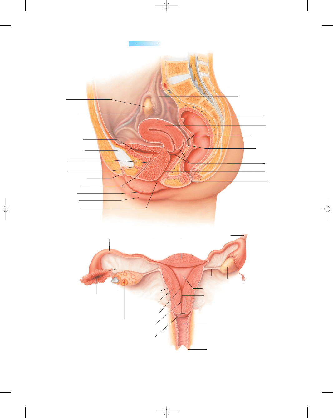
Female Reproductive Anatomy
and Physiology
The female reproductive system is composed of both inter-
nal and external reproductive organs.
Internal Female Reproductive Organs
The internal female reproductive organs consist of the
vagina, the uterus, the fallopian tubes, and the ovaries.
These structures develop and function according to the
specific hormone influences that affect fertility and child-
bearing (Fig. 3-1).
Vagina
The
vagina
is a highly distensible musculomembra-
nous canal situated in front of the rectum and behind the
bladder. It is a tubular, fibromuscular organ lined with
mucous membrane that lies in a series of transverse folds
called rugae. The rugae allow for extreme dilatation of the
canal during labor and birth. The vagina is a canal that
connects the external genitals to the uterus. It receives the
penis and the sperm ejaculated during sexual intercourse,
and it serves as an exit passageway for menstrual blood and
for the fetus during childbirth. The front and back walls
normally touch each other so that there is no space in the
vagina except when it is opened (e.g., during a pelvic
examination or intercourse). In the adult, the vaginal cav-
ity is 3 to 4 inches long. Muscles that control its diameter
surround the lower third of the vagina. The upper two
thirds of the vagina lies above these muscles and can be
easily stretched. During a woman’s reproductive years, the
mucosal lining of the vagina has a corrugated appearance
and is resistant to bacterial colonization. Before puberty
and after menopause (if the woman is not taking estrogen),
the mucosa is smooth secondary to lower levels of estrogen
(Venes, 2005).
Uterus
The
uterus
is a pear-shaped muscular organ at the top of
the vagina. It lies behind the bladder and in front of the rec-
tum and is anchored in position by eight ligaments. It is not
firmly attached or adherent to any part of the skeleton.
A full bladder tilts it backward; a distended rectum, for-
ward. It alters its position by gravity or with change of pos-
ture. It is the size and shape of an inverted pear. It is the
site of menstruation, implantation of a fertilized ovum,
development of the fetus during pregnancy, and labor.
Before the first pregnancy, it measures approximately
3 inches long, 2 inches wide, and 1 inch thick. After a preg-
nancy, the uterus remains larger than before the pregnancy.
After menopause, it becomes smaller and atrophies.
The uterine wall is relatively thick and composed
of three layers: the endometrium (innermost layer), the
myometrium (muscular middle layer), and the peri-
metrium (outer serosal layer that covers the body of the
uterus). The
endometrium
is the mucosal layer that
lines the uterine cavity in nonpregnant women. It varies
in thickness from 0.5 mm to 5 mm and has an abundant
supply of glands and blood vessels (Cunningham et al.,
2004). The myometrium makes up the major portion of
the uterus and is composed of smooth muscle linked by
connective tissue with numerous elastic fibers. During
pregnancy, the upper myometrium undergoes marked
hypertrophy, but there is limited change in the cervical
muscle content.
Anatomic subdivisions of the uterus include the con-
vex portion above the uterine tubes (the fundus); the
central portion (the corpus or body) between the fundus
and the cervix; and the cervix, or neck, which opens into
the vagina.
Cervix
The
cervix,
the lower part of the uterus, opens into the
vagina and has a channel that allows sperm to enter the
uterus and menstrual discharge to exit. It is composed of
fibrous connective tissue. During a pelvic examination, the
part of the cervix that protrudes into the upper end of the
vagina can be visualized. Like the vagina, this part of the
cervix is covered by mucosa, which is smooth, firm, and
doughnut-shaped, with a visible central opening called the
external os (Fig. 3-2). Before childbirth, the external cervi-
cal os is a small, regular, oval opening. After childbirth, the
opening is converted into a transverse slit that resembles
lips (Fig. 3-3). Except during menstruation or ovulation,
the cervix is usually a good barrier against bacteria.
The canal or channel of the cervix is lined with mucus-
secreting glands. This mucus is thick and impenetrable
to sperm until just before the ovaries release an egg
All nurses should take care of and respect the human body,
for it is a wondrous, precision machine.
wow
T
46
3132-03_UT2-Ch03.qxd 12/15/05 3:02 PM Page 46
Chapter 3
ANATOMY AND PHYSIOLOGY OF THE REPRODUCTIVE SYSTEM
47
Ureter
Rectum
Uterus
Posterior fornix
of vagina
Rectouterine
pouch
Cervix
Vagina
Anus
Ovary
Fallopian tube
Urinary bladder
Symphysis pubis
Urethra
Clitoris
Prepuce of clitoris
Urethral orifice
Labia minora
Labia majora
Vaginal orifice
Corpus of the uterus
Fallopian tube
Fundus of uterus
Suspensory
ligament of ovary
Ovary
Ovarian
ligament
Fimbria
Corpus luteum
of menstruation
Abdominal
opening of
fallopian tube
Secondary
oocyte
Uterus
Perimetrium
Myometrium
Endometrium
Cervical canal
External os
Internal os of cervix
Cervix
Vagina
Labia minora
Vesicular
appendix
A
B
●
Figure 3-1
The internal female reproductive organs. (A) Lateral view. (B) Anterior view.
(Source: The Anatomical Chart Company [2001]. Atlas of human anatomy. Springhouse,
PA: Springhouse.)
3132-03_UT2-Ch03.qxd 12/15/05 3:02 PM Page 47
els of hormones secreted by the ovaries: it is thickest dur-
ing the part of the menstrual cycle in which a fertilized egg
would be expected to enter the uterus and is thinnest just
after menstruation. If fertilization does not take place dur-
ing this cycle, most of the endometrium is shed and bleed-
ing occurs, resulting in the monthly period. If fertilization
does take place, the embryo attaches to the wall of the
uterus, where it becomes embedded in the endometrium
(about 1 week after fertilization); this process is called
implantation (Condon, 2004). Menstruation then ceases
during the 40 weeks (280 days) of pregnancy. During
labor, the muscular walls of the corpus contract to push
the baby through the cervix and into the vagina.
Fallopian Tubes
The
fallopian tubes
are hollow, cylindrical structures
that extend 2 to 3 inches from the upper edges of the
uterus toward the ovaries. Each tube is about 7 to 10 cm
long (4 inches) and approximately 0.7 cm in diameter.
The end of each tube flares into a funnel shape, providing
a large opening for the egg to fall into when it is released
from the ovary. Cilia (beating, hair-like extensions on
cells) line the fallopian tube and the muscles in the tube’s
wall. The fallopian tubes convey the ovum from the ovary
to the uterus and sperm from the uterus toward the ovary.
This movement is accomplished via ciliary action and
peristalsis. If sperm is present in the fallopian tube as a
result of sexual intercourse or artificial insemination, fer-
tilization of the ovum can occur. If the egg is fertilized, it
will divide over a period of 4 days while it moves slowly
down the fallopian tube and into the uterus.
Ovaries
The
ovaries
are a set of paired glands resembling
unshelled almonds set in the pelvic cavity below and to
either side of the umbilicus. They are usually pearl-colored
and oblong. They are homologous to the testes. Each ovary
weighs from 2 to 5 grams and is about 4 cm long, 2 cm
wide, and 1 cm thick (Speroff & Fritz, 2005). Several liga-
ments help hold each ovary in position. The ovaries link the
reproductive system to the body’s system of endocrine
glands, as they produce the ova (eggs) and secrete, in
cyclic fashion, the female sex hormones
estrogen
and
progesterone.
After an ovum matures, it passes into the
fallopian tubes. The ovaries are not attached to the fallop-
ian tubes but are suspended nearby from a ligament.
External Female Reproductive Organs
The external female reproductive organs collectively are
called the
vulva
(which means “covering” in Latin). The
vulva serves to protect the urethral and vaginal openings
and is highly sensitive to touch to increase the female’s
pleasure during sexual arousal (Sloane, 2002). The struc-
tures that make up the vulva include the mons pubis,
the labia majora and minora, the clitoris, the structures
within the vestibule, and the perineum (Fig. 3-4).
48
Unit 2
WOMEN’S HEALTH THROUGHOUT THE LIFESPAN
●
Figure 3-2
Appearance of normal cervix. Note: This is the
cervix of a multipara female. (Photo by B. Proud.)
A
B
●
Figure 3-3
(A) Nulliparous cervical os. (B) Parous cervical os.
(
ovulation
). At ovulation, the consistency of the mucus
changes so that sperm can swim through it, allowing fer-
tilization. At the same time, the mucus-secreting glands of
the cervix actually become able to store live sperm for 2 or
3 days. These sperm can later move up through the cor-
pus and into the fallopian tubes to fertilize the egg; thus,
intercourse 1 or 2 days before ovulation can lead to preg-
nancy. Because some women do not ovulate consistently,
pregnancy can occur at varying times after the last men-
strual period. The channel in the cervix is narrow, too
narrow for the fetus to pass through during pregnancy,
but during labor it stretches to let the newborn through.
Corpus
The corpus, or the main body of the uterus, is a highly
muscular organ that enlarges to hold the fetus during
pregnancy. The inner lining of the corpus (endometrium)
undergoes cyclic changes as a result of the changing lev-
3132-03_UT2-Ch03.qxd 12/15/05 3:02 PM Page 48
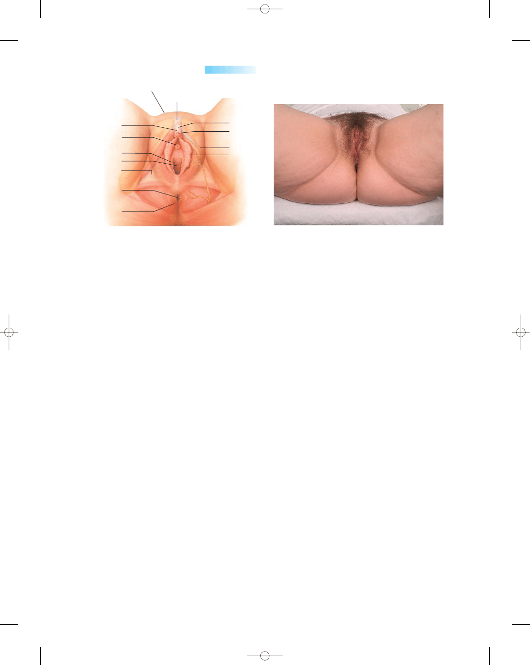
Mons Pubis
The mons pubis is the elevated, rounded fleshy promi-
nence over the symphysis pubis. This fatty tissue and skin
is covered with pubic hair after puberty. It protects the
symphysis pubis during sexual intercourse.
Labia
The labia majora (large lips), which are relatively large
and fleshy, are comparable to the scrotum in males. The
labia majora contain sweat and sebaceous (oil-secreting)
glands; after puberty, they are covered with hair. Its func-
tion is to protect the vaginal opening. The labia minora
(small lips) are the delicate hairless inner folds of skin that
can be very small or up to 2 inches wide. They lie just
inside the labia majora and surround the openings to the
vagina and urethra. The labia minora grow down from the
anterior inner part of the labia majora on each side. They
are highly vascular and abundant in nerve supply. They
lubricate the vulva, swell in response to stimulation, and
are highly sensitive.
Clitoris and Prepuce
The clitoris is a small, cylindrical mass of erectile tissue
and nerves. It is located at the anterior junction of the labia
minora. There are folds above and below the clitoris. The
joining of the folds above the clitoris forms the prepuce, a
hood-like covering over the clitoris; the junction below the
clitoris forms the frenulum. A rich supply of blood vessels
gives it a pink color. The clitoris, like the penis, is very
sensitive to touch, stimulation, and temperature and can
become erect. The word “clitoris” is from the Greek word
for key, which in ancient times was thought to be the key
to a woman’s sexuality. For its small size, it has a generous
blood and nerve supply. There are more free nerve endings
of sensory reception located on the clitoris than on any
other part of the body, and it is, unsurprisingly, the most
erotically sensitive part of the genitalia for most females. Its
function is sexual stimulation (Mattson & Smith, 2004).
Vestibule
The vestibule is an oval area enclosed by the labia minora
laterally. It extends from the clitoris to the fourchette and
is perforated by six openings. Opening into the vestibule
are the urethra from the urinary bladder, the vagina, and
two sets of glands. The opening to the vagina is called
the introitus, and the half-moon-shaped area behind the
opening is called the fourchette. Through tiny ducts
beside the introitus, Bartholin’s glands, when stimulated,
secrete mucus that supplies lubrication for intercourse.
Skene’s glands are located on either side of the opening
to the urethra. They secrete a small amount of mucus to
keep the opening moist and lubricated for the passage of
urine (Olds et al., 2004).
The vaginal opening is surrounded by the hymen
(maidenhead). The hymen is a tough, elastic, perforated,
mucosa-covered tissue across the vaginal introitus. In a
virgin, the hymen may completely cover the opening, but
it usually encircles the opening like a tight ring. Because
the degree of tightness varies among women, the hymen
may tear at the first attempt at intercourse, or it may be so
soft and pliable that no tearing occurs. In a woman who
is not a virgin, the hymen usually appears as small tags of
tissue surrounding the vaginal opening, but the presence
or absence of the hymen can neither confirm nor rule out
sexual experience (Mattson & Smith, 2004).
Perineum
The perineum is the most posterior part of the external
female reproductive organs. This external region is located
between the vulva and the anus. It is made up of skin, mus-
cle, and fascia. The perineum can become lacerated or
incised during childbirth and needs to be repaired with
Chapter 3
ANATOMY AND PHYSIOLOGY OF THE REPRODUCTIVE SYSTEM
49
Symphysis pubis
Mons pubis
Prepuce
Urethral orifice
Vaginal orifice
Perineal
membrane
Anus
External anal
sphincter
Clitoris
Body
Glans
Labia majora
Labia minora
Hymen
A
B
●
Figure 3-4
(A) The external female reproductive organs. (B) Normal appearance of external
structures. (Photo by B. Proud.)
3132-03_UT2-Ch03.qxd 12/15/05 3:02 PM Page 49
sutures. Incising the perineum area to provide more space
for the presenting part is called an episiotomy. Although
still a common obstetric procedure, the use of episiotomy
has decreased over the past 25 years. The procedure
should be applied selectively rather than routinely. An
episiotomy can add to postpartum discomfort, perineal
trauma, and potential fecal incontinence (Cunningham
et al., 2004).
Erection, Lubrication, and Orgasm
With sexual stimulation, tissues in the clitoris, in the
breasts, and around the vaginal orifice fill with blood and
the erectile tissues swell. At the same time, the vagina
begins to expand and elongate to accommodate the penis.
As part of the whole vasocongestive reaction, the labia
majora and minor swell and darken in color. As sexual
stimulation intensifies, the vestibular glands secrete mucus
to moisten and lubricate the tissues to facilitate insertion
of the penis.
The zenith of intense stimulation is orgasm, the spas-
modic and involuntary contractions of the muscles in the
region of the vulva, the uterus, and the vagina that pro-
duce a pleasurable sensation to the woman. Typically the
woman feels warm and relaxed after an orgasm. Within a
short time after orgasm, the two physiologic mechanisms
that created the sexual response, vasocongestion and
muscle contraction, rapidly dissipate.
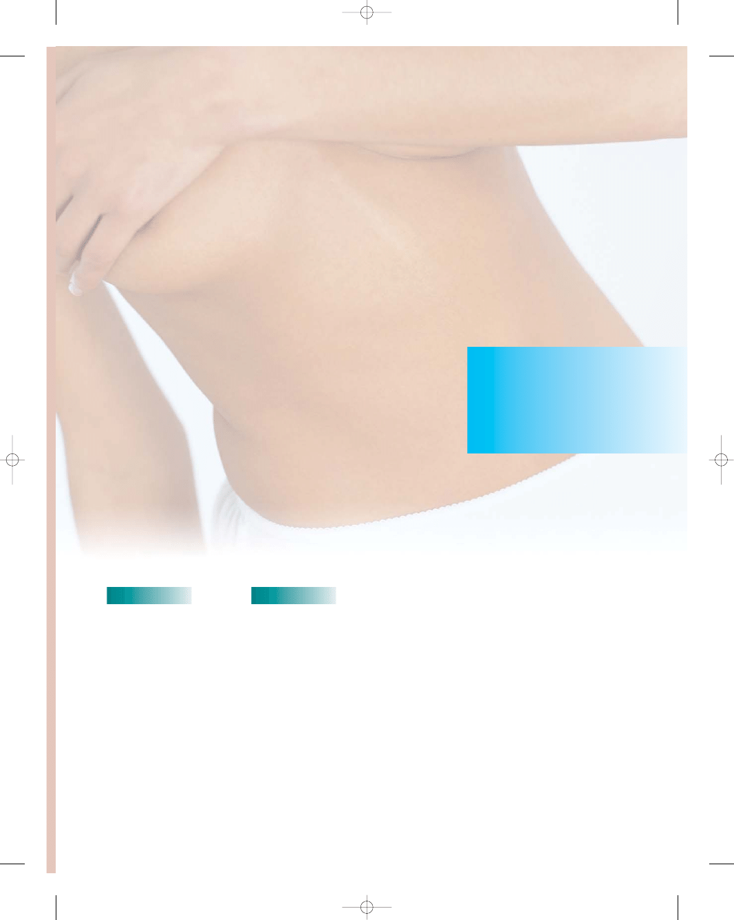
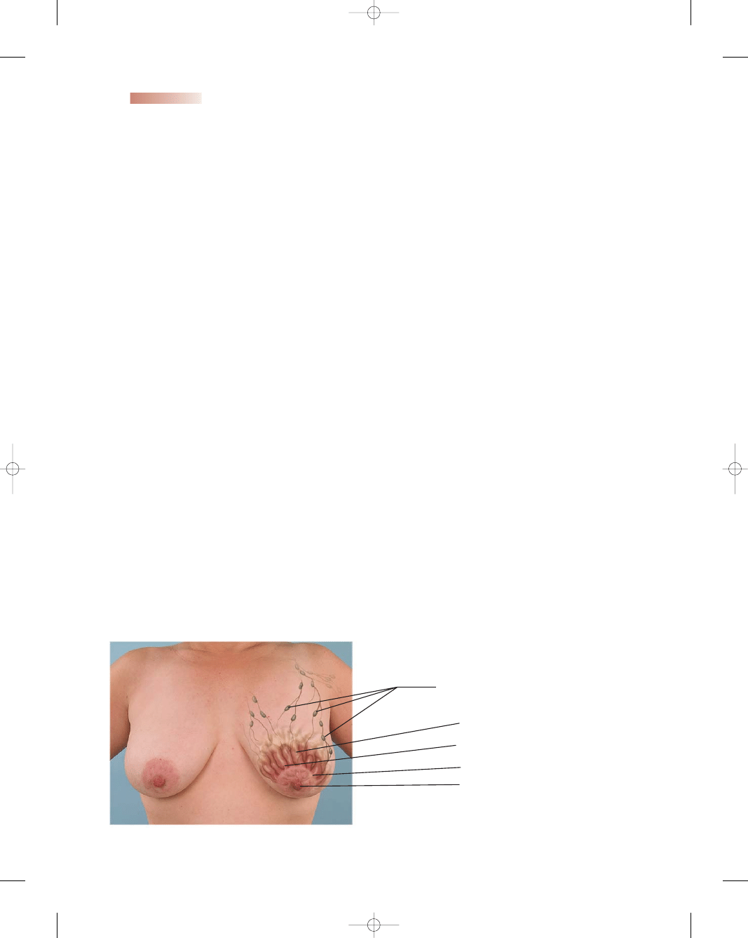
Breasts
The two mammary glands, or
breasts,
are accessory
organs of the female reproductive system that are spe-
cialized to secrete milk following pregnancy. They over-
lie the pectoralis major muscles and extend from the
second to the sixth ribs and from the sternum to the
axilla. Each breast has a nipple located near the tip, which
is surrounded by a circular area of pigmented skin called
the areola. Each breast is composed of 15 to 20 lobes,
which contain glands (alveolar) and a duct (lactiferous)
that leads to the nipple and opens to the outside (Fig. 3-5).
The lobes are separated by dense connective and adi-
pose tissues, which also help support the weight of the
breasts.
During pregnancy, placental estrogen and proges-
terone stimulate the development of the mammary glands.
Because of this hormonal activity, the breasts may double
in size during pregnancy. At the same time, glandular tis-
sue replaces the adipose tissue of the breasts.
Following childbirth and the expulsion of the placenta,
levels of placental hormones (progesterone and lactogen)
fall rapidly, and the action of prolactin (milk-producing
hormone) is no longer inhibited. Prolactin stimulates the
production of milk within a few days after childbirth, but in
the interim, a deep yellow fluid called colostrum is secreted.
Colostrum contains more minerals and protein but less
sugar and fat than mature breast milk. Colostrum secretion
may continue for approximately a week after childbirth,
with gradual conversion to mature milk. Colostrum is
rich in maternal antibodies, especially immunoglobulin A
(IgA), which offers protection for the newborn against
enteric pathogens.
The Female Reproductive Cycle
The female reproductive cycle is a complex process that
encompasses an intricate series of chemical secretions and
reactions to produce the ultimate potential for fertility and
birth. The female reproductive cycle is a general term
encompassing the ovarian cycle, the endometrial cycle,
the hormonal changes that regulate them, and the cyclical
changes in the breasts. The endometrium, ovaries, pitu-
itary gland, and hypothalamus are all involved in the
cyclic changes that help to prepare the body for fertiliza-
tion. Absence of fertilization results in
menstruation,
the monthly shedding of the uterine lining. Menstruation
marks the beginning and end of each menstrual cycle.
50
Unit 2
WOMEN’S HEALTH THROUGHOUT THE LIFESPAN
Alveoli
Ducts
Areola
Nipple
Lymph nodes
●
Figure 3-5
Anatomy of the
breasts. (Photo by B. Proud.)
3132-03_UT2-Ch03.qxd 12/15/05 3:02 PM Page 50

In the United States, the average age at menarche is
12.8 years, with a range between 8 and 18. Most women
will experience 300 to 400 menstrual cycles within their
lifetime (Youngkin & Davis, 2004). Events preceding
the first menses have an orderly progression: thelarche, the
development of breast buds; adrenarche, the appearance
of pubic and then axillary hair, followed by a growth
spurt; and menarche, a girl’s first menses. Cycles vary in
frequency from 21 to 36 days, bleeding lasts 3 to 8 days,
and blood loss averages 20 to 80 mL (Mattson & Smith,
2004). The average cycle is 28 days long. Irregular
menses can be associated with irregular ovulation, stress,
disease, and hormonal imbalances (Cunningham et al.,
2004).
Menopause refers to the cessation of regular men-
strual cycles. It is the end of menstruation and child-
bearing capacity. It is usually marked by atrophy of the
breasts, uterus, tubes, and ovaries (Bachmann, 2004).
Many women pass through menopause without untoward
symptoms. These women remain active and in good
health with little interruption of their daily routines. Other
women experience vasomotor symptoms, which give rise
to sensations of heat, cold, sweating, headache, insomnia,
and irritability (Kessenich, 2004). The average age of nat-
ural menopause—defined as 1 year without a menstrual
period—is 51 (Alexander et al., 2004). (See Chapter 4 for
more information.)
Although menstruation is a normal process, the vari-
ous world cultures have taken a wide variety of attitudes
toward it, seeing it as everything from a sacred time to an
unclean time. In a society where menstruation is viewed
negatively, nurses can help women develop a more pos-
itive image of this natural physiologic process.
The female reproductive cycle involves two cycles
that occur simultaneously: the ovarian cycle, during
which ovulation occurs, and the endometrial cycle, dur-
ing which menstruation occurs. Ovulation divides these
two cycles at midcycle. Ovulation occurs when the ovum
is released from its follicle; after leaving the ovary, the
ovum enters the fallopian tube and journeys toward
the uterus. If sperm fertilizes the ovum during its jour-
ney, pregnancy occurs. Figure 3-6 summarizes the men-
strual cycle.
Ovarian Cycle
The ovarian cycle is the series of events associated with a
developing oocyte (ovum or egg) within the ovaries. While
men manufacture sperm daily, often into advanced age,
women are born with a single lifetime supply of ova that
are released from the ovaries gradually throughout the
childbearing years. In the female ovary, 2 million oocytes
are present at birth, and about 400,000 follicles are still
present at puberty. The excess follicles are depleted dur-
ing the childbearing years, with only 400 follicles ovulated
during the reproductive period (Speroff & Fritz, 2005).
The ovarian cycle begins when the follicular cells (ovum
and surrounding cells) swell and the maturation process
starts. The maturing follicle at this stage is called a graafian
follicle. The ovary raises many follicles monthly, but usu-
ally only one follicle matures to reach ovulation. The ovar-
ian cycle consists of three phases: the follicular phase,
ovulation, and the luteal phase.
Follicular Phase
This phase is so named because it is when the follicles
in the ovary grow and form a mature egg. This phase
starts on day 1 of the menstrual cycle and continues until
ovulation, approximately 10 to 14 days. The follicular
phase is not consistent in duration because of the time
variations in follicular development. These variations
account for the differences in menstrual cycle lengths
(Breslin and Lucas, 2003). The hypothalamus is the
initiator of this phase. Increasing levels of estrogen secreted
from the maturing follicular cells and the continued growth
of the dominant follicle cell induce proliferation of the
endometrium and myometrium. This thickening of the
uterine lining supports an implanted ovum if pregnancy
occurs.
Prompted by the hypothalamus, the pituitary gland
releases
follicle-stimulating hormone (FSH),
which
stimulates the ovary to produce 5 to 20 immature folli-
cles. Each follicle houses an immature oocyte or egg.
The follicle that is targeted to mature fully will soon
rupture and expel a mature oocyte in the process of ovu-
lation. A surge in
luteinizing hormone (LH)
from the
anterior pituitary gland is actually responsible for affect-
ing the final development and subsequent rupture of the
mature follicle.
Ovulation
At ovulation, a mature follicle ruptures in response to a
surge of LH, releasing a mature oocyte (ovum). This usu-
ally occurs on day 14 in a 28-day cycle. When ovulation
occurs, there is a drop in estrogen. Typically ovulation
takes place approximately 10 to 12 hours after the LH
peak and 24 to 36 hours after estrogen levels peak (Speroff
and Fritz, 2005). The distal ends of the fallopian tubes
become active near the time of ovulation and create cur-
rents that help carry the ovum into the uterus. The lifes-
pan of the ovum is only about 24 hours; unless it meets a
sperm on its journey within that time, it will die.
During ovulation, the cervix produces thin, clear,
stretchy, slippery mucus that is designed to help the
sperm travel up through the cervix to meet the ovum
for fertilization. Some women can feel a pain on one
side of the abdomen around the time the egg is released.
This is known as mittelschmerz, a German word mean-
ing “middle pain.” The one constant, whether a women’s
cycle is 28 days or 120 days, is that ovulation takes
place 14 days before menstruation (Mattson & Smith,
2004).
Chapter 3
ANATOMY AND PHYSIOLOGY OF THE REPRODUCTIVE SYSTEM
51
3132-03_UT2-Ch03.qxd 12/15/05 3:02 PM Page 51

Luteal Phase
The luteal phase begins at ovulation and lasts until the
menstrual phase of the next cycle. After the follicle ruptures
as it releases the egg, it closes and forms a corpus luteum.
The corpus luteum secretes increasing amounts of the hor-
mone progesterone, which interacts with the endometrium
to prepare it for implantation. At the beginning of the luteal
phase, progesterone induces the endometrial glands to
secrete glycogen, mucus, and other substances. These
glands become tortuous and have large lumens due to
increased secretory activity. The progesterone secreted by
the corpus luteum causes the temperature of the body
to rise slightly until the start of the next period. A signif-
icant increase in temperature, usually 0.5 to 1 degrees
Fahrenheit, is generally seen within a day or two after
ovulation has occurred; the temperature remains elevated
for 12 to 16 days, until menstruation begins (Youngkin &
Davis, 2004). This rise in temperature can be plotted on a
graph and gives an indication of when ovulation has
occurred. In the absence of fertilization, the corpus luteum
52
Unit 2
WOMEN’S HEALTH THROUGHOUT THE LIFESPAN
FSH
LH
Controlled by hypothalamus
Anterior pituitary
LH peak triggers ovulation
Maturing follicle
Degenerating corpus luteum
Estrogen
Progesterone
Day 1-5
Day 6-14
Day 15-26
Days 27
and 28
Glands
Arteries
Veins
Menstrual
Proliferative
Secretory
Isch-
emic
Men-
strual
Follicular
Luteal
Uterine
phases
Ovarian
phases
Thickness of endometrial lining
during the menstrual cycle
Days
Ovarian hormones
Ovulation
Corpus
luteum
1
5
14
26
28
●
Figure 3-6
Menstrual cycle summary
based on a 28-day (average) menstrual cycle.
Consider
THIS!
I had been married 2 years when my husband and I
decided to start a family. I began thinking back to my high-
school biology class and tried to remember about ovulation
and what to look for. I also used the Internet to find the
answers I was seeking. As I was reading, it all started to
come into place. During ovulation, a woman’s cervical
mucus increases and she experiences a ‘wet sensation’ for
several days midcycle. The mucus also becomes stretch-
able during this time. In addition, her temperature rises
slightly and then falls if no conception takes place. Armed
with this knowledge, I began to check my temperature
daily before arising and checking the consistency of my
mucus. I figured that if these signs could help prevent
pregnancy by warning of the unsafe time, they could help
me discover the best time to conceive. Within 3 months I
became pregnant using my body’s natural signals.
Thoughts:
How does knowledge of the reproductive
system help nurses take care of couples who are
trying to become pregnant?
Consider
3132-03_UT2-Ch03.qxd 12/15/05 3:02 PM Page 52

begins to degenerate, and consequently ovarian hormone
levels decrease. As estrogen and progesterone levels
decrease, the endometrium undergoes involution. In a
28-day cycle, menstruation then begins approximately
14 days after ovulation in the absence of pregnancy. FSH
and LH are generally at their lowest levels during the luteal
phase and highest during the follicular phase.
Endometrial Cycle
The endometrial cycle occurs in response to cyclic hor-
monal changes. The three phases of the endometrial cycle
are the proliferative phase, the secretory phase, and the
menstrual phase.
Proliferative Phase
The proliferative phase starts with enlargement of the
endometrial glands in response to increasing amounts
of estrogen. The blood vessels become dilated and the
endometrium increases in thickness dramatically. It lasts
from about day 5 of the menstrual cycle to the time of
ovulation. This phase depends on estrogen stimulation
resulting from ovarian follicles.
Secretory Phase
The secretory phase follows ovulation to about 3 days
before the next menstrual period. Under the influence of
progesterone, the endometrium becomes thickened and
more vascular (growth of the spiral arteries) and glandu-
lar (secreting more glycogen and lipids). These dramatic
changes are all in preparation for implantation, if it were
to occur. Estrogen levels drop sharply during this phase
as progesterone dominates.
Menstrual Phase
The menstrual phase begins as the spiral arteries rupture
secondary to ischemia, releasing blood into the uterus,
and the endometrium is sloughed off. If fertilization does
not take place, the corpus luteum degenerates. As a result,
both estrogen and progesterone levels fall and the thick-
ened endometrial lining sloughs away from the uterine
wall and passes out via the vagina. The beginning of the
menstrual flow marks the end of one menstrual cycle and
the start of a new one. Most women report bleeding for
an average of 3 to 5 days (Mattson & Smith, 2004).
Menstrual Cycle Hormones
The menstrual cycle involves a complex interaction
of hormones. The predominant hormones include
gonadotropin-releasing hormone (GnRH), FSH, LH,
estrogen, progesterone, and prostaglandins. Box 3-1
summarizes menstrual cycle hormones.
Gonadotropin-Releasing
Hormone (GnRH)
Gonadotropin-releasing hormone (GnRH) is secreted
from the hypothalamus in a pulsatile manner throughout
the reproductive cycle. It pulsates slowly during the follic-
ular phase and increases during the luteal phase. GnRH
induces the release of FSH and LH to assist with ovulation.
Follicle-Stimulating Hormone (FSH)
FSH is secreted by the anterior pituitary gland and is pri-
marily responsible for the maturation of the ovarian folli-
cle. FSH secretion is highest and most critical during the
first week of the follicular phase of the reproductive cycle.
Luteinizing Hormone (LH)
LH is secreted by the anterior pituitary gland and is
required for both the final maturation of preovulatory fol-
licles and luteinization of the ruptured follicle. As a result,
estrogen production declines and progesterone secretion
continues. Thus, estrogen levels fall a day before ovula-
tion, and progesterone levels begin to rise.
Estrogen
Estrogen is secreted by the ovaries and is crucial for the
development and maturation of the follicle. Estrogen is
predominant at the end of the follicular phase, directly
preceding ovulation. After ovulation, estrogen levels drop
sharply as progesterone dominates. In the endometrial
cycle, estrogen induces proliferation of the endometrial
glands. Estrogen also causes the uterus to increase in size
and weight because of increased glycogen, amino acids,
electrolytes, and water. Blood supply is expanded as well.
Estrogen inhibits FSH production and stimulates LH
production.
Progesterone
Progesterone is secreted by the corpus luteum. Proges-
terone levels increase just before ovulation and peak 5 to
Chapter 3
ANATOMY AND PHYSIOLOGY OF THE REPRODUCTIVE SYSTEM
53
• Luteinizing hormone (LH) rises and stimulates the
follicle to produce estrogen.
• As estrogen is produced by the follicle, estrogen levels
rise, inhibiting the output of LH.
• Ovulation occurs after an LH surge damages the
estrogen-producing cells, resulting in a decline in
estrogen.
• The LH surge results in establishment of the corpus
luteum, which produces estrogen and progesterone.
• Estrogen and progesterone levels rise, suppressing
LH output.
• Lack of LH promotes degeneration of the corpus
luteum.
• Cessation of the corpus luteum means a decline in
estrogen and progesterone output.
• The decline of the ovarian hormones ends their
negative effect on the secretion of LH.
• LH is secreted, and the menstrual cycle begins again.
BOX 3-1
SUMMARY OF MENSTRUAL CYCLE HORMONES
3132-03_UT2-Ch03.qxd 12/15/05 3:02 PM Page 53
7 days after ovulation. During the luteal phase, proges-
terone induces swelling and increased secretion of the
endometrium. This hormone is often called the hormone
of pregnancy because of its calming effect (reduces uter-
ine contractions) on the uterus, allowing pregnancy to be
maintained.
Prostaglandins
Prostaglandins are a closely related group of oxygenated
fatty acids that are produced by the endometrium, with a
variety of effects throughout the body. Although they
have regulatory effects and are sometimes called hor-
mones, prostaglandins are not technically hormones
because they are produced by all tissues rather than by
special glands (Sloane, 2002). Prostaglandins increase
during follicular maturation and play a key role in ovula-
tion by freeing the ovum inside the graafian follicle. Large
amounts of prostaglandins are found in menstrual blood.
Research is ongoing as to the various roles prostaglandins
have on the menstrual cycle (Cunningham et al., 2004).
Male Reproductive System
The male reproductive system, like that of the female, con-
sists of those organs functioning to produce a new indi-
vidual. The male organs are specialized to produce and
maintain the male sex cells, or sperm; to transport them,
along with supporting fluids, to the female reproductive
system; and to secrete the male hormone testosterone. The
organs of the male reproductive system include the two
testes (where sperm cells and testosterone are made), the
penis, the scrotum, and the accessory organs (epididymis,
vas deferens, seminal vesicles, ejaculatory duct, urethra,
bulbourethral glands, and prostate gland).
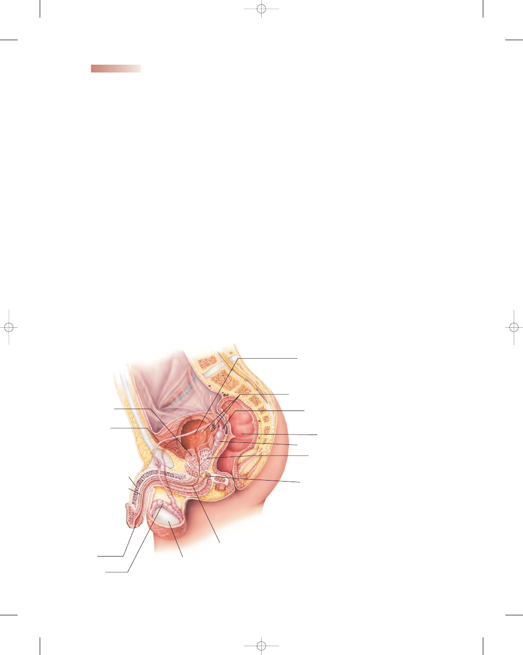
Internal Male Reproductive Organs
The internal structures include the testes, the ductal sys-
tem, and accessory glands (Fig. 3-7).
Testes
The
testes
are oval bodies the size of large olives that lie
in the scrotum; usually the left testis hangs a little lower
than the right one. The testes have two functions: produc-
ing sperm and synthesizing testosterone (the primary male
sex hormone). Sperm is produced in the seminiferous
tubules of the testes. The testes also produce the male hor-
mone testosterone and a portion of the seminal fluid, the
liquid in which sperm are carried. The epididymis, which
lies against the testes, is a coiled tube almost 20 feet long.
It collects sperm from the testes and provides the space
and environment for sperm to mature (Fig. 3-8).
54
Unit 2
WOMEN’S HEALTH THROUGHOUT THE LIFESPAN
Prostate gland
Vas deferens
Urinary bladder
Openings of ureter
Ampulla of
vas deferens
Rectum
Seminal vesicle
Ejaculatory
duct
Bulbourethral
gland and duct
Corpus cavernosum
Corpus spongiosum
Epididymis
External
urethral
opening
Urethra
Testis
●
Figure 3-7
Lateral view of the
internal male reproductive organs.
(Source: The Anatomical Chart
Company. [2001]. Atlas of human
anatomy. Springhouse, PA:
Springhouse.)
3132-03_UT2-Ch03.qxd 12/15/05 3:02 PM Page 54
The Ductal System
The vas deferens is a cordlike duct that transports sperm
from the epididymis. One such duct travels from each
testis up to the back of the prostate and enters the urethra
to form the ejaculatory ducts. Other structures, such as
blood vessels and nerves, also travel along with each vas
deferens and together form the spermatic cord. The ure-
thra is the terminal duct of the reproductive and urinary
systems, serving as a passageway for semen (fluid con-
taining sperm) and urine. It passes through the prostate
gland and the penis and opens to the outside.
Accessory Glands
The seminal vesicles, which produce nutrient seminal
fluid, and the prostate gland, which produces alkaline
prostatic fluid, are both connected to the ejaculatory duct
leading into the urethra. The paired seminal vesicles are
convoluted pouchlike structures lying posterior to and at
the base of the urinary bladder in front of the rectum.
They secrete an alkaline fluid that contains fructose and
prostaglandins. The fructose supplies energy to the sperm
on its journey to meet the ovum, and the prostaglandins
assist in sperm mobility.
The prostate gland lies just under the bladder in the
pelvis and surrounds the middle portion of the urethra.
Usually the size of a walnut, this gland enlarges with age.
The prostate and the seminal vesicles above it produce
fluid that nourishes the sperm. This fluid provides most
of the volume of semen, the secretion in which the sperm
is expelled during ejaculation. Other fluid that makes up
the semen comes from the vas deferens and from mucous
glands in the head of the penis.
The bulbourethral glands (Cowper’s glands) are two
small structures about the size of peas, located inferior to
the prostate gland. They are composed of several tubes
whose epithelial linings secrete a mucuslike fluid. It is
released in response to sexual stimulation and lubricates
the head of the penis in preparation for sexual intercourse.
Their existence is said to be constant, but they gradually
diminish in size with advancing age.
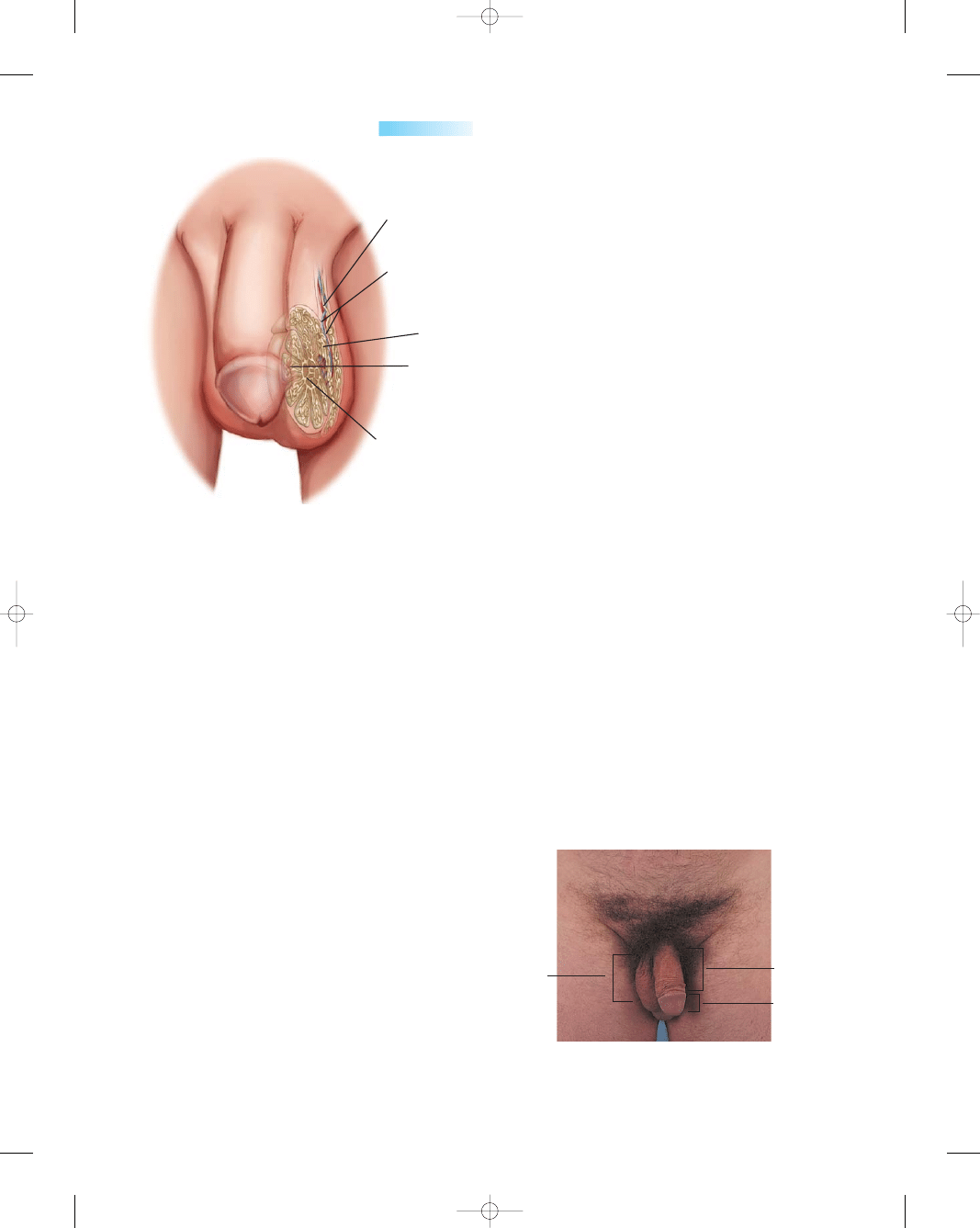
External Male Reproductive Organs
The penis and the scrotum form the external genitalia in
the male (Fig. 3-9).
Penis
The
penis
is the organ for copulation and serves as the
outlet for both sperm and urine. The skin of the penis is
thin, with no hairs. The prepuce (foreskin) is a circular fold
of skin that extends over the glans unless it is removed
by circumcision shortly after birth. The urinary meatus,
located at the tip of the penis, serves as the external open-
ing to the urethra (Fig. 3-10). The penis is composed
mostly of erectile tissue. Most of the body of the penis con-
sists of three cylindrical spaces (sinuses) of erectile tissue.
The two larger ones, the corpora cavernosa, are side by
side. The third sinus, the corpus spongiosum, surrounds
the urethra. Erection results when nerve impulses from the
autonomic nervous system dilate the arteries of the penis,
allowing arterial blood to flow into the erectile tissues of
the organ.
Scrotum
The scrotum is the thin-skinned sac that surrounds and
protects the testes. The scrotum also acts as a climate-
control system for the testes, because they need to be
slightly cooler than body temperature to allow normal
Chapter 3
ANATOMY AND PHYSIOLOGY OF THE REPRODUCTIVE SYSTEM
55
Testicular artery
and vein
Epididymis
Seminiferous
tubules
Rete testis
Vas deferens
●
Figure 3-8
Internal structures of a testis.
Scrotum
Penis shaft
Glans penis
●
Figure 3-9
The external male reproductive organs. (Photo
by B. Proud.)
3132-03_UT2-Ch03.qxd 12/15/05 3:02 PM Page 55
sperm development. The cremaster muscles in the scrotal
wall relax or contract to allow the testes to hang farther
from the body to cool or to be pulled closer to the body for
warmth or protection (Sloane, 2002). A medial septum
divides the scrotum into two chambers, each of which
encloses a testis.
Erection, Orgasm, and Ejaculation
With sexual stimulation, the arteries leading to the penis
dilate and increase blood flow into erectile tissues. At the
same time, the erectile tissue compresses the veins of the
penis, reducing blood flow away from the penis. Blood
accumulates, causing the penis to swell and elongate and
producing an erection. As in women, the culmination of
sexual stimulation is an orgasm, a pleasurable feeling of
physiologic and psychological release.
Orgasm is accompanied by emission (movement of
sperm from the testes and fluids from the accessory
glands) into the urethra, where it is mixed to form semen.
As the urethra fills with semen, the base of the erect penis
contracts, which increases pressure and forces the semen
through the urethra to the outside (ejaculation). During
ejaculation, the ducts of the testes, epididymis, and vas
deferens contract, causing expulsion of sperm into the
urethra, where the sperm mixes with the seminal and
prostatic fluids. These substances, together with mucus
secreted by accessory glands, form the semen, which is
discharged from the urethra.
K E Y C O N C E P T S
●
The female reproductive system produces the female
reproductive cells (the eggs, or ova) and contains an
organ (uterus) where the fetus develops. The male
reproductive system produces the male reproductive
cells (the sperm) and contains an organ (penis) that
deposits the sperm within the female.
●
The internal female reproductive organs consist of
the vagina, the uterus, the fallopian tubes, and the
ovaries. The external female reproductive organs
make up the vulva. These include the mons pubis,
the labia majora and minora, the clitoris, structures
within the vestibule, and the perineum.
●
The breasts are accessory organs of the female
reproductive system that are specialized to secrete
milk following pregnancy.
●
The main function of the reproductive cycle is to
stimulate growth of a follicle to release an egg and
prepare a site for implantation if fertilization occurs.
●
Menstruation, the monthly shedding of the uterine
lining, marks the beginning and end of the cycle if
fertilization does not occur.
●
The ovarian cycle is the series of events associated
with a developing oocyte (ovum or egg) within
the ovaries.
●
At ovulation, a mature follicle ruptures in response
to a surge of LH, releasing a mature oocyte (ovum).
●
The endometrial cycle is divided into three phases:
the follicular or proliferative phase, the luteal or
secretory phase, and the menstrual phase.
●
The menstrual cycle involves a complex interaction
of hormones. The predominant hormones are
gonadotropin-releasing hormone (GnRH),
follicle-stimulating hormone (FSH), luteinizing
hormone (LH), estrogen, progesterone, and
prostaglandins.
●
The organs of the male reproductive system include
the two testes (where sperm cells and testosterone
are made), penis, scrotum, and accessory organs
(epididymis, vas deferens, seminal vesicles, ejacula-
tory ducts, urethra, bulbourethral glands, and
prostate gland).
References
Alexander, L. L., LaRosa, J. H., Bader, H., & Garfield, S. (2004).
New dimensions in women’s health (3rd ed.). Boston: Jones and
Bartlett.
Bachmann, G. (2004), Menopause. eMedicine. [Online] Available:
http://www.emedicine.com/med/topic3289.htm.
Breslin, E. T., & Lucas, V. A. (2003). Women’s health nursing: toward
evidence-based practice. St. Louis, MO: Saunders.
Condon, M. C. (2004). Women’s health: an integrated approach to
wellness and illness. Upper Saddle River, NJ: Prentice Hall.
Cunningham, F. G., Leveno, K. J., Bloom, S. L., et al. (2004).
Williams obstetrics (22nd ed.). New York: McGraw-Hill.
Kessenich, C. R. (2004). Inevitable menopause. Nursing Spectrum.
[Online] Available: http://nsweb.nursingspectrum.com/ce/
ce232.htm.
Mattson, S., & Smith, J. E. (2004). Core curriculum for maternal-
newborn nursing (3rd ed.). St. Louis, MO: Elsevier Saunders.
Olds, S. B., London, M. L., Ladewig, P. W., & Davidson, M. R.
(2004). Maternal-newborn nursing and women’s health (7th ed.).
Upper Saddle River, NJ: Pearson Prentice Hall.
Sloane, E. (2002). Biology of women (4th ed.). New York: Delmar.
56
Unit 2
WOMEN’S HEALTH THROUGHOUT THE LIFESPAN
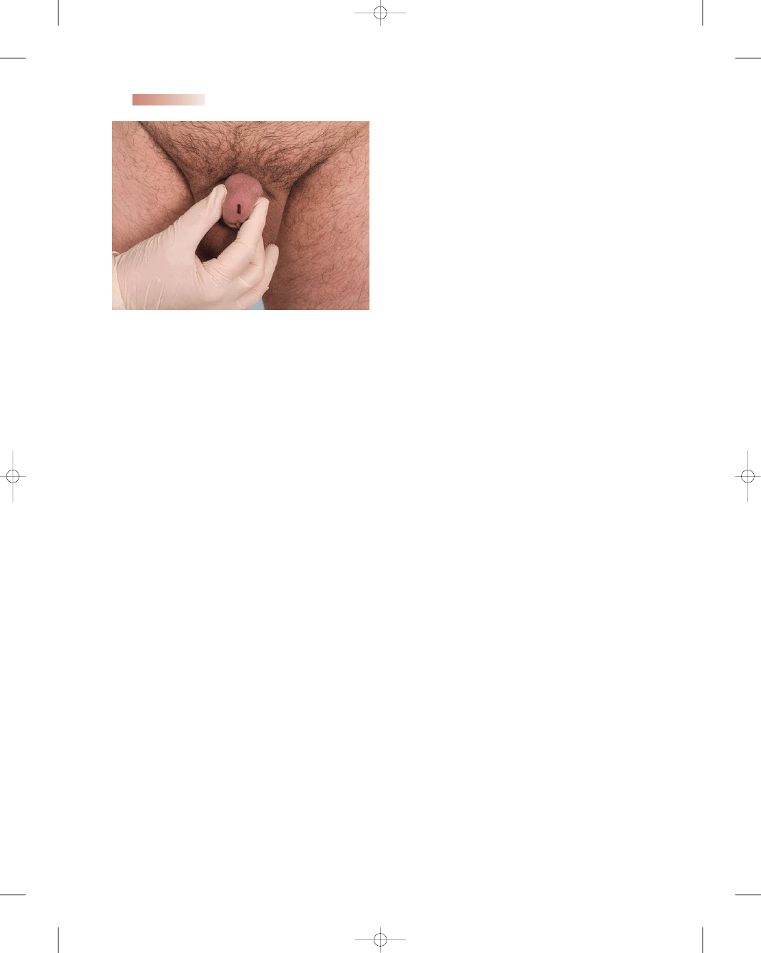
●
Figure 3-10
The urinary meatus. (Photo by B. Proud.)
3132-03_UT2-Ch03.qxd 12/15/05 3:02 PM Page 56

Speroff, L., & Fritz, M. A. (2005). Clinical gynecologic endocrinology
and infertility (7th ed.). Philadelphia: Lippincott Williams &
Wilkins.
Venes, D. (2005) Taber’s cyclopedia medical dictionary (20th ed.).
Philadelphia: F. A. Davis.
Writing Group for the Women’s Health Initiative Investigators.
(2002). Risks and benefits of estrogen plus progestin in healthy
postmenopausal women: principal results from the Women’s
Health Initiative randomized controlled trial. JAMA, 288(3),
321–333.
Youngkin, E. Q., & Davis, M. S. (2004) Women’s health: A primary
care clinical guide (3rd ed.). Upper Saddle River, NJ: Prentice Hall.
Web Resources
Alan Guttmacher Institute: www.agi-usa.org
American Society for Reproductive Medicine:
www.asrm.com
Kinsey Institution: www.indiana.edu/
∼kinsey/index.html
Sexuality Information of the United States: www.siecus.org
National Women’s Health Information Center:
www.4woman.gov
National Women’s Health Resource Center:
www.healthywomen.org
Society for Women’s Health Research: www.womens-health.org
Chapter 3
ANATOMY AND PHYSIOLOGY OF THE REPRODUCTIVE SYSTEM
57
3132-03_UT2-Ch03.qxd 12/15/05 3:02 PM Page 57

58
Unit 2
WOMEN’S HEALTH THROUGHOUT THE LIFESPAN
Chapter
WORKSHEET
Chapter
●
M U L T I P L E C H O I C E Q U E S T I O N S
1.
The predominant anterior pituitary hormones that
orchestrate the menstrual cycle include:
a. Thyroid-stimulating hormone (TSH)
b. Follicle-stimulating hormone (FSH)
c. Corticotropin-releasing hormone (CRH)
d. Gonadotropin-releasing hormone (GnRH)
2.
Which glands are located on either side of the female
urethra and secrete mucus to keep the opening moist
and lubricated for urination?
a. Cowper’s
b. Bartholin’s
c. Skene’s
d. Seminal
3.
The ovarian cycle comprises all of the following
phases except:
a. Secretory
b. Follicular
c. Ovulation
d. Luteal
4.
Which hormone is produced in high levels to prepare
the endometrium for implantation just after ovula-
tion by the corpus luteum?
a. Estrogen
b. Prostaglandins
c. Prolactin
d. Progesterone
5.
Sperm maturation and storage in the male reproduc-
tive system occurs in the:
a. Testes
b. Vas deferens
c. Epididymis
d. Seminal vesicles
●
C R I T I C A L T H I N K I N G E X E R C I S E
1.
The school health nurse was asked to speak to the
10th-grade biology class in the local high school about
menstruation. The teachers felt that the students mis-
understood this monthly event and wanted to dispel
some myths about it. After explaining the factors
influencing the monthly menses, one girl asks,
“Could someone get pregnant if she had sex
during her period?”
a. How should the nurse respond to this question?
b. What factor regarding the menstrual cycle was not
clarified?
c. What additional topics might this question lead
into that might be discussed?
●
S T U D Y A C T I V I T I E S
1.
Select a website under Web Resources to explore to find
information concerning a topic of interest regarding
women’s health. Be prepared to discuss it in class.
2.
List the predominant hormones and their function in
the menstrual cycle.
3.
The ovarian cycle describes the series of events asso-
ciated with the development of the _____________
within the ovaries.
4.
Sperm cells and the male hormone testosterone are
made in which of the following structures? Select all
that apply:
a. Vas deferens
b. Penis
c. Scrotum
d. Ejaculatory ducts
e. Prostate gland
f. Testes
g. Seminiferous tubules
h. Bulbourethral glands
3132-03_UT2-Ch03.qxd 12/15/05 3:02 PM Page 58
Wyszukiwarka
Podobne podstrony:
Essentials of Maternity Newborn and Women's Health 3132A 30 p780 781
Essentials of Maternity Newborn and Women's Health 3132A 29 p778 779
Essentials of Maternity Newborn and Women s Health 3132A 32 p785 808
Essentials of Maternity Newborn and Women s Health 3132A 23 p634 662
Essentials of Maternity Newborn and Women s Health 3132A 17 p428 446
Essentials of Maternity Newborn and Women s Health 3132A 16 p393 427
Essentials of Maternity Newborn and Women s Health 3132A 21 p585 612
Essentials of Maternity Newborn and Women s Health 3132A 09 p189 207
Essentials of Maternity Newborn and Women s Health 3132A 11 p235 252
Essentials of Maternity Newborn and Women s Health 3132A 20 p543 584
Essentials of Maternity Newborn and Women s Health 3132A 08 p167 188
Essentials of Maternity Newborn and Women s Health 3132A 27 p769 771
Essentials of Maternity Newborn and Women s Health 3132A 26 p729 768
Essentials of Maternity Newborn and Women s Health 3132A 28 p772 777
Essentials of Maternity Newborn and Women s Health 3132A 25 p717 728
Essentials of Maternity Newborn and Women s Health 3132A 05 p107 126
Essentials of Maternity Newborn and Women s Health 3132A 19 p496 542
Essentials of Maternity Newborn and Women s Health 3132A 22 p613 633
więcej podobnych podstron