
BioTechnologia
vol. 92(2) C pp. 159-165 C 2011
Journal of Biotechnology, Computational Biology and Bionanotechnology
REVIEW PAPER
Association between body iron stores
and level of oxidatively modified DNA bases
T
OMASZ
D
ZIAMAN
*,
M
AREK
J
URGOWIAK
,
R
YSZARD
O
LIŃSKI
Department of Clinical Biochemistry, Ludwik Rydygier Collegium Medicum in Bydgoszcz, Nicolaus Copernicus University in Toruń
* Corresponding author: tomekd@cm.umk.pl
Abstract
It appears that the presence of labile iron pool (LIP, iron not bound to proteins) in cells can result in the pro-
duction of reactive oxygen species namely
C
OH radical which may be responsible for the formation of 8-oxo-7,8-di-
hydro-2N-deoxyguanosine (8-oxodG) in the cellular DNA. This oxidatively modified molecule is regarded as a good
biomarker of cancer risk and a general index of oxidative stress in relation to other diseases. There are numerous
data suggesting that oxidative stress may be involved in the development of cardiovascular diseases and cancer.
It has been observed that heterozygosity for hereditary hemochromatosis (a disease with abnormal iron storage)
is a risk factor for vascular diseases. Previously we have demonstrated higher levels of LIP in a group of athero-
sclerotic patients when compared with the control group. This suggests that LIP may increase the risk of disease
development. The aforementioned condition may lead to oxidative stress, which is manifested by a higher level
of 8-oxodG in blood lymphocytes, and may be one of the factors responsible for the development of cardiovascular
diseases. We have also reported the relationship between LIP and the endogenous level of 8-oxodG in human
lymphocytes of the colon cancer patients. Good correlation has been determined between LIP and oxidatively mo-
dified nucleoside. The results of our studies on piglets supplemented with iron dextran (FeDex) also show an in-
crease in the 8-oxodG level in hepatic DNA. These findings confirm the possibility that iron overload may favor
the persistence of harmful LIP which may catalyze generation of the potentially carcinogenic 8-oxodG moiety in
the cellular DNA.
Key words: labile iron pool, oxidative stress, 8-oxodG
Introduction to iron metabolism and generation
of Reactive Oxygen Species
Iron plays a pivotal role in many crucial biological
processes, because it can serve either as an electron do-
nor or electron acceptor, alternating between ferrous
(Fe
2+
) and ferric (Fe
3+
) ion, but this ability makes this
metal both profitable and dangerous. Due to its features
of a transition metal, it is a useful component of many
enzymes and proteins (McCord, 1998) involved in mito-
chondrial respiration, electron transfer, oxygen trans-
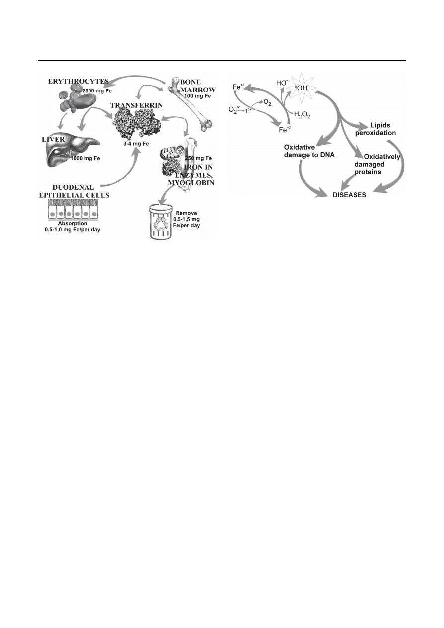
port, DNA synthesis and repair. The distribution and
amount of iron in tissues of the human body are shown
in Figure 1.
Due to its features, iron is a precious element for
the organism, and for that reason iron metabolism is
tightly controlled in mammalian cells by proteins such as
transferrin, ferritin or hemosiderin that bind iron. Iron
ions that circulate in the blood plasma are bound to
plasma transferrin, whereas the excess of iron in cells is
accumulated in complex with ferritin (Epsztejn et al.,
1997; Bolann and Ulvik, 1990; Olinski and Jurgowiak,
1996). If necessary, iron can be released as a result of
either ferritin degradation in lysosomes or directly from
native protein. Also, a mitochondrial form of ferritin
exists in cells. An increased concentration of this protein
is observed in the erythroid cells from patients with si-
deroblastic anemia, where mitochondria are overloaded
with iron (Cazzola et al., 2003). Hepcidin, a peptide, has
an influence on iron absorption in enterocytes, while its
recirculation from macrophages plays a special role in
iron metabolism. Excessive synthesis of this peptide
may induce accumulation of iron ions in these cells
(Rossi, 2005).
Iron bound to proteins is safe for organisms and in
this form it is involved in different physiological func-
tions. Inside the cell, iron can also exist in another form,
as a “free” or “labile” iron (LIP, iron not bound to prote-
ins). LIP-associated iron is in a dynamic equilibrium with
sequestered iron forms in cell and is bound to cytosolic
low-molecular weight ligands that have not yet been
T. Dziaman, M. Jurgowiak, R. Oliński
160
Fig. 1. Iron storage in human body
(adapted and modified from Oliński and Jurgowiak, 2002)
identified. When the amount of iron in the serum exce-
eds the binding capacities of transferrin, then ions of
this metal remain in a pool described as “non transferrin-
bound iron” (NTBI), which may influence the level of cel-
lular labile iron pool. This iron form is catalytically active
and participates in the production of toxic reactive oxy-
gen species (ROS) via Fenton reaction. Interaction of
ROS with cellular components may result in damage to
biomolecules, including DNA, lipids and proteins (Eme-
rit, Beaumont, Trivin, 2001) – see Figure 2, which in
turn may lead to an increased risk of cancer, coronary
heart or neurodegenerative diseases (Olinski and Jurgo-
wiak, 2003; Olinski et al., 2002; Andrews, 2000). The
risk of these pathologies development is higher when
iron concentration exceeds the binding capacities of the
aforementioned proteins.
ROS are the products of partial reduction of oxygen.
These species, which include superoxide anion (O
2
•!
),
hydrogen peroxide (H
2
O
2
) and hydroxyl radical (
•
OH),
are continuously produced in living cells as by-products
of normal metabolism (McCord, 1998; Olinski and Jurgo-
wiak, 1996; Bartosz, 1995). During mitochondrial respi-
ration, up to 1-5 % of oxygen undergoes single electron
transfer that generates the superoxide anion radical in
amounts corresponding to about 2 kg per year for a hu-
man being (Olinski and Jurgowiak, 2003). ROS have
been postulated to play a significant role in the etiology
Fig. 2. Fenton reaction and its repercussions. Generated
•
OH
radical can damage lipids, proteins and nucleic acids. It may lead
to dysfunctions of the cells and development of diseases
of at least 50 diseases including rheumatoid arthritis,
cancer, atherosclerosis, myocardial infraction, Parkin-
son’s disease and AIDS (Olinski and Jurgowiak, 2003;
Olinski et al., 2002). Reperfusion of ischemic tissues and
chronic inflammation also lead to ROS generation.
Furthermore, UV and ionizing irradiation, a wide variety
of drugs and xenobiotics can also stimulate the forma-
tion of ROS. A variety of carcinogens, including benzene,
aflatoxin and benzopyrene may exert their effect partly
through the generation of ROS during their metabolism.
The superoxide radical is degraded by superoxide dis-
mutase (SOD) and hydrogen peroxide by catalase. Ho-
wever, the reaction of hydrogen peroxide with transition
metal ions, like iron, leads to highly reactive hydroxyl
radical (
•
OH) and interaction of this radical with cellular
components may result in damage to biomolecules inclu-
ding DNA (Olinski and Jurgowiak, 1999).
There are experimental data which demonstrate the
existence of a free iron pool in the sera of patients with
hemochromatosis -- the disease with abnormally high
iron storage resulting from excessive iron absorption.
Several types of this disease are known: type 1 caused
by inactivation of the
HFE
gene and four types (2A, 2B,
3, 4) characterized as non-
HFE
hereditary hemochroma-
tosis (Roetto and Camaschella, 2005). This disease pre-
disposes to cancer and cardiovascular diseases (CVD).
Epidemiological data also suggest that elevation of the
body iron levels may increase the risk of cancer and
atherosclerosis (Andrews, 2000; Rossi et al., 2000). Our
results suggest a mechanism that may directly link iron
Association between body iron stores and level of oxidatively modified DNA bases
161
overload with carcinogenesis and atherosclerosis. Spe-
cifically, iron overload may favor the persistence of
harmful LIP, which may be responsible for LDL oxida-
tion as well as may catalyze generation of the potentially
carcinogenic oxidatively modified DNA bases in the cel-
lular DNA (Gackowski et al., 2001).
Oxidative damage to DNA
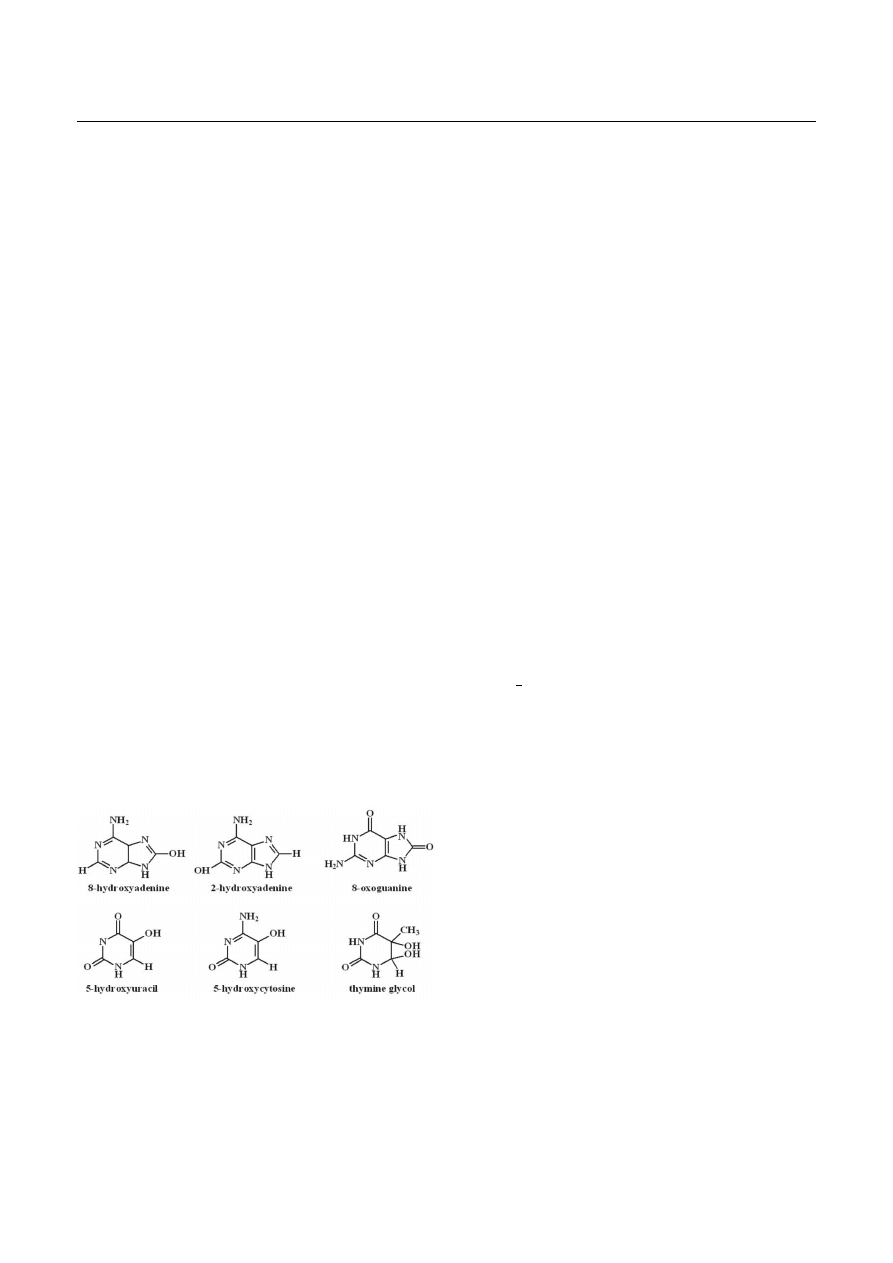
In living cells, there is a steady formation of DNA le-
sions arising from normal cellular metabolism as well as
pathophysiological processes and extracellular sources.
A substantial number of these lesions are formed by
endogenous factors that damage DNA on a continuous
basis. A free radical attack upon DNA generates a whole
series of DNA damage, including modified DNA bases.
Certainly, not all ROS can damage DNA directly (Halli-
well and Cross, 1994). For example, H
2
O
2
and O
2
•!
may
initiate DNA damage by interacting with transition metal
ion, in particular, iron and copper, in Haber-Weiss type
reaction, producing
•
OH. The hydroxyl radical is one of
the most reactive species responsible for the formation
of a large number of pyrimidine and purine-derived le-
sions in DNA (reviewed in: Dizdaroglu, 1993). Examples
of such lesions are presented in Figure 3. Some of these
modified DNA bases have a considerable potential to da-
mage the integrity of nuclear and mitochondrial genomes
(reviewed in: Floyd, 1990; Jackson and Loeb, 2001). The
levels of oxidative damage to mtDNA are several times
higher than those of nuclear DNA (Sastre et al., 2000).
Fig. 3. Examples of oxidatively modified DNA bases
One of the most widely studied lesions is 7.8-dihydro-
8-oxoguanine (8-oxoGua) and its nucleoside (8-oxodG).
The presence of 8-oxo-7,8-dihydro-2'-deoxyguanosine resi-
dues in DNA leads to GC 6 TA transversions unless re-
paired prior to DNA replication (Cheng et al., 1992).
Therefore, the presence of 8-oxodG in cells may lead to
point mutations. This oxidative DNA adduct is regarded
as a good biomarker of cancer risk from oxidative stress,
for the investigations of effectiveness of dietary antioxi-
dants and as a general index of oxidative stress in relation
to other diseases such as CVD.
Several other modified bases such as 2-hydroxyade-
nine (2-OH-Ade), 8-hydroxyadenine (8-OHAde), 5-hydro-
xycytosine (5-OH-Cyt), and 5-hydroxyuracil (5-OH-Ura)
have also been shown to possess miscoding potentials
and may be premutagenic as well (reviewed in: Wallace,
1998), but their biological properties are not yet fully
recognized.
Labile iron pool and atherosclerosis
The possibility that iron overload may play an impor-
tant role in CVD was put forward in 1981 by J. L. Sulli-
van (Sullivan, 1981). Now there are increasing epidemio-
logical evidences concerning the role of iron in athero-
sclerosis (Kiechl et al., 1997; de Valk and Marx, 1999).
Interestingly, it was found that heterozygosity for type 1
hereditary hemochromatosis is a risk factor for vascular
diseases (Kiechl et al., 1994). There are also some evi-
dences that iron depletion protects from atherosclerosis
and CVD (Roest et al., 1999). In this context, it is inte-
resting to note that in premenopausal women the inci-
dence of atherosclerosis and CVD is less than half that
of age-matched men (McCord, 1998; Emerit, Beaumont
and Trivin, 2001). One of the possible explanations for
this finding may be that depletion of iron stores by regu-
lar menstrual blood loss may be a source of protection of
premenopausal subjects.
Our data also suggest that iron metabolism may have
some influence on atherosclerosis development. In agre-
ement with previous studies, we have found that ferritin
concentration was higher in atherosclerotic patients than
in the control group (although these changes were statisti-
cally insignificant) (Gackowski et al., 2001). In this con-
text, it should be noted that increased ferritin concentra-
tion generally reflects iron stores but may also be rela-
ted to high alcohol consumption, cancer and inflamma-
tion (Looker and Johnson, 1998). In our study, we also
decided to analyze labile iron pool (LIP) in human lym-
phocytes – which is involved in the production of harm-

T. Dziaman, M. Jurgowiak, R. Oliński
162
ful ROS. Interestingly, LIP in lymphocytes of the athero-
sclerotic patient group was about two times higher than
that in the control group.
Moreover, we found that the level of 8-oxodG in lym-
phocytes of atherosclerotic patients was significantly hi-
gher than in DNA of the control group (12.78 and 9.80
lesions/10
6
dG respectively. In another study which invol-
ved healthy people (Nakano et al., 2003), a positive cor-
relation between urinary 8-oxodG and serum ferritin was
shown, which suggested that iron status may influence
the generation of 8-oxodG, in vivo. Experimental eviden-
ce suggests that urinary excretion of 8-oxodG represents
repair processes, namely removal of oxidatively modified
base/nucleoside at the level of whole body (Olinski et al.,
2003).
These changes may have some influence on the de-
velopment of atherosclerosis in the following way. The
plaques of the arterial walls, among other components,
contain lymphocytes (Gackowski et al., 2001). Since
8-oxodG has mutagenic properties and is a block to
transcription in mammalian cells (Le Page, 2000), it is
possible that lymphocytes with higher amount of this
modified base trapped into the plaque, can more easily
be involved in initiation-promoting process. There are
suggestions that this process may be responsible for the
formation of the atherosclerotic lesion. Thereafter, the
higher amount of trapped cells can lead to more advan-
ced lesions. Moreover, there are some experimental
data which strongly suggest that the elevated level of
8-oxodG found in the lesion of the aorta walls of athero-
sclerotic patients, may be one of the events directly in-
volved in the development of the disease (Collins et al.,
1998).
Our data confirms the hypothesis that higher levels
of LIP may increase the risk of the atherosclerosis de-
velopment. Progress of this pathology is associated with
inflammatory condition and oxidative stress, which is, in
turn, manifested by a higher level of 8-oxodG in blood
lymphocytes. It is possible that this condition may be a
factor responsible for the development of atherosclero-
sis (Gackowski et al., 2001).
Body iron stores and cancer
Iron-induced free radical damage to DNA seems to
be important for the development of cancer. Tumor cells
are known as the rapidly growing cells in response to
iron (Ullen et al., 1997). Carcinogenicity of iron overload
was demonstrated clearly in many animal experiments
(Campbell, 1940; Richmond, 1959; Li et al., 1987; Liu
and Okada, 1994).
Body iron stores and dietary iron intake have been
shown to be positively correlated with the risk of human
colon cancer (Nelson et al., 1994; Stevens et al., 1998).
In our study, carcinoma patients showed statistically sig-
nificant lower values of transferrin saturation, total iron
binding capacity and serum iron level when compared
with the control group (Gackowski et al., 2002A). Func-
tional iron deficiency can occur during the cancer and
chronic inflammation states and is often associated with
relatively high iron levels in the liver as an effect of en-
hanced hepcidine biosynthesis (Weiss, 2009).
In our above-mentioned work (Gackowski et al.,
2002a), it was reported that mean values of LIP in
lymphocytes of the patient group were higher than in the
control ones, but these differences were not statistically
significant. Rather insignificant differences in LIP and
ferritin concentrations observed in this study may be
explained, at least in part, by a large individual variability
of those parameters. Such huge individual differences in
ferritin concentration have also been reported by others
(Nelson et al., 1994). Since this was the first
in vivo
stu-
dy of LIP in human lymphocytes, it was difficult to com-
pare the obtained results with other data reported in lite-
rature. However, the concentration found in our study is
comparable to the levels estimated for different types of
mammalian cultured cells (Lipinski et al., 2000). It is
possible that higher concentrations of LIP in the lympho-
cytes of patients with carcinoma may be explained by the
distinctive behaviour of ferritin observed in our report,
i.e. high level of intracellular LIP may cause an increase
in ferritin synthesis and determine high plasma concen-
trations of ferritin. Since LIP can influence the produc-
tion of ROS, our results support a recent observation
that oxidative stress may be responsible for impaired
lymphocyte function in cancer patients (Schmielau and
Finn, 2001).
Usually, in malignant diseases plasma iron level falls
due to cytokines activity (Halliwell and Gutterige, 1999).
Our observations support an interesting hypothesis
which suggests that the observed changes lead to the
restriction of iron availability for tumour cells thus slo-
wing tumor growth (Weinberg, 1996).

Association between body iron stores and level of oxidatively modified DNA bases
163
Iron supplementation and level
of oxidatively damaged DNA
Iron supplementation is a frequently practised treat-
ment in the occurrence of iron deficiency anaemia (IDA),
which is most prevalent during the neonatal period and in
early childhood. IDA is probably the most prevalent
micronutrient deficiency disorder in newborn piglets,
due to their rapid growth (Svoboda and Drabek, 2005).
Intramuscular administration of large amounts of iron
compounds can cure those deficiencies. However, iron
overload is dangerous, because unbound ions are cataly-
tically active. As it was mentioned above, in our previous
study, we observed a strong correlation between iron
content and the levels of oxidatively modified nucleoside
in lymphocytes’ DNA (Gackowski et al., 2002b). There-
fore, besides the efficacy in curing iron deficiency, one
of the main criteria for selecting an iron supplementa-
tion protocol is that the supplemental iron should pro-
duce minimal toxicity.
In our recent studies (Lipinski et al., 2010), anaemic
neonatal pigs were supplemented with iron dextran
(FeDex). They were allotted to one of three different ex-
perimental groups on the basis of body weight (b.w.) at
the given experimental age, control piglets receiving no
iron supplementation; piglets intramuscularly injected in
the neck with 100 mg Fe/kg b.w. on day 3 postpartum
(traditional supplementation); piglets injected with
40 mg Fe/kg b.w. on day 3 and again on day 10 postpar-
tum (modified supplementation). The results demonstra-
ted that points with the highest iron concentrations were
related to the highest 8-oxodG levels. Furthermore, the
modified iron supplementation was linked with a signi-
ficantly smaller increase in 8-oxodG level in hepatic DNA
when compared with the traditional protocol.
Although the oxidative stress observed in newborn
piglets may be, partly, as a result of a sudden increase in
oxygenation after birth (Dziaman et al., 2007), the intra-
muscular injection resulted in elevated amounts of iron
in the colon and subsequent increase in oxidative stress,
as measured by increased levels of 8-oxodG in colon DNA
and increased urinary excretion of 8-oxoGua (Langie et
al., 2010). Similar observations upon injection with
FeDex were previously reported in rats (Wellejus, Poul-
sen, Loft, 2000).
In the study with young children (1-4 years old) with
IDA that were orally supplemented with Fe
3+
preparation
for 12 weeks, an increase in the oxidative damage to
DNA was observed. However, this growth was free from
the corresponding changes in the serum level of iron and
ferritin. These changes could have been a result of an in-
creased oxidative stress due to an accelerated metabolic
rate by the rehabilitation of oxygenation in the organism
(Aksu et al., 2010).
It appears that misregulation of iron administration
influences organism’s homestasis. The presence of an
excessive amount of iron in cells or/and extracellular
spaces can result in the production of ROS and induction
of oxidatively damaged DNA. Specifically, iron overload
may favor the persistence of harmful LIP, which may
catalyze the generation of potentially carcinogenic
8-oxodG moiety in the cellular DNA.
References
Aksu B.Y., Hasbal C., Himmetoglu S., Dincer Y., Koc E.E.,
Hatipoglu S., Akcay T. (2010)
Leukocyte DNA damage in
children with iron deficiency anemia: effect of iron supple-
mentation.
Eur. J. Pediatr. 169(8): 951-956.
Andrews N.C. (2000)
Iron homeostasis: insights from genetics
and animal models.
Nature Rev. Genet. 1: 208-217.
Bartosz G. (1995)
Druga twarz tlenu
. Wydawnictwo Naukowe
PWN, Warszawa.
Bolann E.J., Ulvik R.J. (1990)
On the limited ability of super-
oxide to release iron from ferritin
. Eur. J. Biochem. 193:
899-904.
Campbell J.A. (1940)
Effects of precipitated silica and of iron
oxide on the incidence of primary lung tumours in mice.
Br. Med. J. 275-280.
Cazzola M., Invernizzi R., Bergamaschi G., Levi S., Corsi B.,
Travaglino E., Rolandi V., Biasiotto G., Drysdale J., Aro-
sio P. (2003)
Mitochondrial ferritin expression in ery-
throid cells from patients with sideroblastic anemia.
Blood
101(5): 1996-2000.
Cheng K.C., Cahill D.S., Kasai H., Nishimura S., Loeb L.A.
(1992)
8-Hydroxyguanine, an abundant form of oxidative
DNA damage, causes G 6 T and A 6 C substitution.
J. Biol. Chem. 267: 166-172.
Collins A.R., Gedik C.M., Olmedilla B., Southon S., Belizzi M.
(1998)
Oxidative DNA damage measured in human lym-
phocytes: large differences between sexes and between
countries and correlation with heart disease mortality
rates.
FASEB J. 12: 1397-1400.
Dizdaroglu M. (1992)
Oxidative damage to DNA in mammalian
chromatin.
Mutat. Res. 275: 331-342.
Dziaman T., Gackowski D., Rozalski R., Siomek A., Szulczyn-
ski J., Zabielski R., Olinski R. (2007)
Urinary excretion
rates of 8-oxoGua and 8-oxodG and antioxidantvitamins
level as ameasure of oxidative status in healthy, full-term
newborns
., Free Radic. Res. 41: 997-1004.
Emerit J., Beaumont C., Trivin F. (2001)
Iron metabolism,
free radicals, and oxidative injury.
Biomed. Pharmacother.
55: 333-339.

T. Dziaman, M. Jurgowiak, R. Oliński
164
Epsztejn S., Kakhlon O., Glickstein H., Breuer W., Caban-
tchick Z.I. (1997)
Fluorescence analysis of the labile iron
pool in mammalian cells.
Anal. Biochem. 248: 31-40.
Floyd R.A. (1990)
The role of 8-hydroxyguanine in carcino-
genesis,
Carcinogenesis 11: 1447-1450.
Gackowski D., Kruszewski M., Jawień A., Ciecierski M., Oliń-
ski R. (2001)
Further evidence that oxidative stress may
be a risk factor responsible for the development of athero-
sclerosis.
Free Rad. Biol. Med. 31: 542-547.
Gackowski D., Kruszewski M., Banaszkiewicz Z., Jawien A.,
Olinski R. (2002a)
Lymphocyte labile iron pool, plasma
iron, transferrin saturation and ferritin levels in colon can-
cer patients.
Acta Bioch. Pol. 49: 269-272.
Gackowski D., Kruszewski M., Bartlomiejczyk T., Jawien A.,
Ciecierski M., Olinski R. (2002b)
The level of 8-oxo-7,8-
dihydro-2’-deoxyguanosine is positively correlated with
the size of the labile iron pool in human lymphocytes.
J. Biol. Inorg. Chem. 7: 548-550.
Halliwell B., Cross C.E. (1994)
Oxygen-derived species: their
relation to human disease and environmental stress
. En-
viron. Health Perspect. 102(Suppl 10): 5-12.
Halliwell B.M., Gutterige J.M.C. (1999)
Free Radicals in Biolo-
gy and Medicine.
Oxford University Press Inc., New York.
Jackson A.L., Loeb L.A. (2001)
The contribution of endogeno-
us sources of DNA damage to the multiple mutations in
cancer.
Mutat. Res. 477: 7-21.
Kiechl S., Aichner F., Gerstenbrand F., Egger G., Mair A.,
Runnger G., Spogler F., Jarosch E., Oberhollenzer F.,
Willeit J. (1994)
Body iron stores and presence of carotid
atherosclerosis: results from the Bruneck study.
Arterio-
scler. Thromb. 14: 1625-1630.
Kiechl S., Willeit J., Egger G., Egge G., Poewe W. (1997)
Body
iron stores and the risk of carotid atherosclerosis. Pros-
pective results from the Bruneck study.
Circulation 96:
3300-3307.
Langie S.A., Kowalczyk P., Tudek B., Zabielski R., Dziaman T.,
Oliński R., van Schooten F.J., Godschalk R.W. (2010)
The
effect of oxidative stress on nucleotide-excision repair in
colon tissue of newborn piglets.
Mutat. Res. 695(1-2):
75-80.
Li J.L., Okada S., Hamazaki S., Ebina Y., Midorikawa O. (1987)
Subacute nephrotoxicity and induction of renal cell carci-
noma in mice treated with ferric nitrilotriacetate.
Cancer
Res. 47: 1867-1869.
Lipinski P., Drapier J.-C., Oliveira L., Retmanska H., Sochano-
wicz B., Kruszewski M. (2000)
Intracellular iron status as
a hallmark of mammalian cell susceptibility to oxidative
stress: A study of L5178Y mouse lymphoma cell lines dif-
ferentially sensitive to H2O2.
Blood, 95: 2960-2966.
Lipinski P., Starzyński R.R., Canonne-Hergaux F., Tudek B.,
Oliński R., Kowalczyk P., Dziaman T., Thibaudeau O., Gra-
lak M.A., Smuda E. et al. (2010)
Benefits and risks of iron
supplementation in anemic neonatal pigs.
Am. J. Pathol.
177(3): 1233-1243.
Liu M., Okada S. (1994)
Induction of free radicals and tumors
in the kidneys of Wistar rats by ferric ethylenediamine-
N,N'-diacetate.
Carcinogenesis, 15: 2817-2821.
Looker A.C., Johnson C.L. (1998)
Prevalence of elevated se-
rum transferrin saturation in adults in United States.
Ann.
Intern. Med. 129: 940-945.
McCord J.M. (1998)
Sem. Iron, free radicals, and oxidative
injury.
Hematology 35: 5-12.
Nakano M., Kawanishi Y., Kamohara S., Uchida Y., Shiota M.,
Inatomi Y., Komori T., Miyazawa K., Gondo K., Yama-
sawa I. (2003)
Oxidative DNA damage (8-hydroxydeoxy-
guanosine) and body iron status: a study on 2507 healthy
people.
Free Radic. Biol. Med. 35(7): 826-832.
Nelson R.L., Davis F.G., Sutter E., Sobin L.H., Kikendall J.W.,
Bowen P. (1994)
Body iron stores and risk of colonic neo-
plasia.
J. Natl. Cancer Inst. 86: 455-460.
Oliński R., Jurgowiak M. (1996)
Reaktywne formy tlenu – uni-
wersalny czynnik patogenny?
[in:]
Nowe tendencje w bio-
logii molekularnej i inżynierii genetycznej oraz medy-
cynie
, ed. Barciszewski J., Łastowski K., Twardowski T.,
Wydawnictwo Sorus, Poznań: 373-400.
Oliński R., Jurgowiak M. (1999)
Oksydacyjne uszkodzenia
DNA (8-oksodG) – biomarkerem niektórych chorób czło-
wieka
. Kosmos, 48: 329-338.
Olinski R., Gackowski D., Foksinski M., Rozalski R., Roszkow-
ski K., Jaruga P. (2002)
Oxidative DNA damage: assess-
ment of the role in carcinogenesis, atherosclerosis, and
acquired immunodeficiency syndrome.
Free Radic. Biol.
Med. 33(2): 192-200.
Oliński R., Jurgowiak M. (2002)
Iron metabolism, oxidative
DNA damage and atherosclerosis
. Acta Angiol. 8(2): 37-44.
Olinski R., Jurgowiak M. (2003)
Uszkodzenia DNA przez wol-
ne rodniki tlenowe – konsekwencje biologiczne i impli-
kacje kliniczne.
[in:]
Na pograniczu chemii i biologii
, vol.
VII, Wydawnictwo Naukowe UAM, Poznań.
Olinski R., Gackowski D., Rozalski R., Foksinski M., Bialkow-
ski K. (2003)
Oxidative DNA damage in cancer patients:
a cause or a consequence of the disease development?
Mutat. Res. 531: 177-190.
Le Page F., Kwoh E.E., Avrutskaya A., Gentil A., Leadon S.A.,
Sarasin A., Cooper P.K. (2000)
Transcription-coupled re-
pair of 8-oxoguanine, requirement for XPG, TFIIH, and
CSB, and implications for Cockayne Syndrom.
Cell, 101:
158-171.
Richmond H.G. (1959)
Induction of sarcoma in the rat by iron-
dextran complex.
Br. Med. J. 947-949.
Roest M., van der Schouw Y.T., de Valk B., Marx J.J., Tempel-
man M.J., de Groot P.G., Sixma J.J., Banga D. (1999)
Heterozygosity for a hereditary hemochromatosis gene is
associated with cardiovascular mortality in women.
Cir-
culation 100: 1268-1273.
Roetto A., Camaschella C. (2005)
New insights into iron ho-
meostasis through the study of non-HFE hereditary haemo-
chromatosis.
Best Pract. Res. Clin. Haematol. 18(2):
235-250.
Rossi E., Brendan M., McQuillan M., Hung J., Thompson P.L.,
Kuek C., Beilby J.P. (2000)
Serum ferritin and C282Y mu-
tation of the hemochromatosis gene as predictors of asym-
ptomatic carotid atherosclerosis in a community popu-
lation.
Stroke 31: 3015-3020.

Association between body iron stores and level of oxidatively modified DNA bases
165
Rossi E. (2005)
Hepcidin – the Iron Regulatory Hormone.
Clin. Biochem. Rev. Vol. 26: 47-49.
Sastre J., Pallardó F.V., García de la Asunción J., Viña J.
(2000)
Mitochondria, oxidative stress and aging.
Free
Radic. Res. 32(3): 189-198.
Schmielau J., Finn O.J. (2001)
Activated granulocytes and gra-
nulocyte-derived hydrogen peroxide are the underlying
mechanisms of suppression of T-cell function in advanced
cancer patients.
Cancer Res. 61: 4756-4760.
Stevens R.G., Jones D.Y., Micozzi M.S., Taylor P.R. (1998)
Body iron stores and the risk of cancer.
N. Engl. J. Med.,
319: 1047-1052.
Sullivan J.L. (1981)
Iron and the sex difference in heart dise-
ase risk.
Lancet 1: 1293-1294.
Svoboda M., Drabek J. (2005)
Iron deficiency in suckling
piglets: etiology, clinical aspects and diagnosis
. Folia Vet.
49: 104-111.
Ullen H., Augustsson K., Gustavsson C., Steineck G. (1997)
Supplementary iron intake and risk of cancer: reversed
causality?
Cancer Lett. 114: 215-216.
de Valk B., Marx J.J.M. (1999)
Iron, atherosclerosis and ische-
mic heart disease.
Arch. Intern. Med. 159: 1542-1548.
Wallace S.S. (1998)
Enzymatic processing of radiation-induced
free radical damage in DNA.
Radiat. Res. 150: 60-S79.
Weinberg E.D. (1996)
The role of iron in cancer.
Eur. J.
Cancer Prevent. 5: 19-36.
Weiss G. (2009)
Iron metabolism in the anemia of chronic
disease
. Biochim. Biophys. Acta 1790: 682-693.
Wellejus A., Poulsen H.E., Loft S. (2000)
Iron-induced oxi-
dative DNA damage in rat sperm cells in vivo and in vitro
.
Free Radic. Res. 32: 75-83.
Wyszukiwarka
Podobne podstrony:
14 5id 15201 Nieznany (2)
chemia lato 12 07 08 id 112433 Nieznany
Literaturoznawstwo (08 04 2013) Nieznany
08 02bid 7351 Nieznany (2)
86 Nw 08 Lampy oscyloskopowe V Nieznany (2)
08 Programowanie w srodowisku j Nieznany (2)
08 Projektowanie i realizacja z Nieznany (2)
03 5id 4121 Nieznany
08 2id 7222 Nieznany
2007 08 Szkola konstruktorowid Nieznany
2 modul 5id 20554 Nieznany (2)
1 5id 8373 Nieznany (2)
19 5id 18138 Nieznany (2)
05 5id 5463 Nieznany (2)
CW 08 id 122562 Nieznany
2002 08 Osla laczka Nieznany
08 vimid 7592 Nieznany (2)
713[05] Z1 08 Wykonywanie posad Nieznany
więcej podobnych podstron