
Review
Understanding the Basis for Down Syndrome
Phenotypes
Randall J. Roper, Roger H. Reeves*
ABSTRACT
D
own syndrome is a collection of features that are
caused by trisomy for human Chromosome 21.
While elevated transcript levels of the more than 350
genes on the chromosome are primarily responsible, it is
likely that multiple genetic mechanisms underlie the
numerous ways in which development and function diverge
in individuals with trisomy 21 compared to euploid
individuals. We consider genotype–phenotype interactions
with the goal of producing working concepts that will be
useful for approaches to ameliorate the effects of trisomy.
Introduction
Trisomy 21 occurs in 1/750 live births. The frequency of
Down syndrome (DS) is much higher at conception, given
that up to 75% and 50% of DS fetuses identified during the
first and second trimester, respectively, are lost before term
[1,2]. Trisomy for some other autosomes occurs more
frequently than trisomy 21, nearly always resulting in
prenatal loss [3]. The relatively high frequency of postnatal
survival for trisomy 21 is thought to be principally a function
of the small number of genes on human Chromosome 21
(Hsa21), the smallest and least gene-dense of the autosomes.
Phenotypes
The clinical presentation of DS is complex and variable. A
few features occur to some degree in every individual with
trisomy 21, including characteristic facial dysmorphology, a
small and hypocellular brain, and the histopathology of
Alzheimer disease, which is present by the fourth decade.
Individuals with DS are invariably cognitively impaired,
though the severity is highly variable. Hypotonia occurs
frequently in newborns, and most have atypical
dermatoglyphic features, though the specific subset of these is
again individually variable.
Trisomy 21 is also a risk factor for a number of diseases.
For example, it is among the leading causes of congenital
heart disease (CHD), some form of which occurs in 40%–50%
of those with DS [4]. The incidence of childhood onset
leukemia and Hirschsprung disease are both significantly
elevated in individuals with trisomy 21. Health-care
guidelines for individuals with DS include more than 80
clinical features that occur more frequently than in the
population at large [5]. Three critical points for this
discussion arise from these basic observations: (1) the
incidence of most phenotypes seen in DS is variable; (2) the
severity of a given feature is highly variable; and (3) none of
the features diagnosed in DS is unique to people with trisomy
21. For ‘‘DS features’’ that also occur in euploid individuals,
we assume that there is some commonality of etiology
regardless of ploidy, but this must be proven for any specific
case.
A central challenge of genetic research in humans is to
precisely define phenotype. This is especially critical in DS,
which is a product of genetic effects on different cells,
structures, and functions throughout development, many of
which may have cascading effects to produce clinically
observed phenotypic end points in a given individual with
trisomy 21 [6]. A first step in this process is to separate those
effects of trisomy that disturb development from those that
alter function of cells that have reached an end point of
differentiation. These are obviously not independent
concepts; any ‘‘developmental’’ perturbation derives from
alteration of some function in a developing cell. However,
understanding when trisomy causes a divergence from
normal patterns of development in a cell that exists only for a
defined period during embryogenesis requires a different
experimental approach (and, ultimately, a different
therapeutic approach) than measuring how trisomy affects a
steady-state function (e.g., a signaling or metabolic pathway,
neuronal response to stimulation, etc.) in a terminally
differentiated cell. Indeed, the altered functions of a mature
cell may have little or nothing to do with up-regulation of
trisomic genes in that cell, but rather could reflect a
developmental error caused by trisomy that has downstream
consequences that affect function. That is, a specific
phenotype may be a consequence of but not a direct product of
trisomic gene expression (developmental versus functional
effects).
Genetic Models for DS
Because understanding the impact of elevated gene
expression throughout development is essential in DS
research, animal models play a critical role, especially for
correlating the direct and cascading effects of trisomic gene
Citation: Roper RJ, Reeves RH (2006) Understanding the basis for Down syndrome
phenotypes. PLoS Genet 2(3): e50.
DOI: 10.1371/journal.pgen.0020050
Copyright:
Ó 2006 Roper and Reeves. This is an open-access article distributed
under the terms of the Creative Commons Attribution License, which permits
unrestricted use, distribution, and reproduction in any medium, provided the
original author and source are credited.
Abbreviations: AMKL, acute megakaryoblastic leukemia; CHD, congenital heart
disease; DS, Down syndrome; DSCR, Down syndrome critical (or chromosomal)
region; Hsa21, human Chromosome 21; Mmu16, mouse Chromosome 16; TMD,
transient myeloproliferative disorder
Randall J. Roper and Roger H. Reeves are in the Department of Physiology at Johns
Hopkins University School of Medicine, and at McKusick-Nathans Institute for
Genetic Medicine, Baltimore, Maryland, United States of America.
* To whom correspondence should be addressed. E-mail: rreeves@jhmi.edu
PLoS Genetics | www.plosgenetics.org
March 2006 | Volume 2 | Issue 3 | e50
0231
expression on development and function. The best-
characterized mouse models to date are trisomic for segments
of mouse Chromosome 16 (Mmu16) conserved with Hsa21.
The Ts65Dn mouse is trisomic for a segment that contains
orthologs of about half of Hsa21 genes while Ts1Cje mice are
trisomic for about two-thirds of the genes that are trisomic in
Ts65Dn [7,8]. A variety of DS phenotypes have been assessed
quantitatively in these models, providing the basis for tracing
their origins in development.
Trisomic gene content can be manipulated by chromosome
engineering to add or subtract trisomic segments in mice [9].
Recently, a transchromosomal DS mouse model was reported
that inherits a copy of a nearly intact Hsa21 [10]. While
mosaicism due to random loss of the human chromosome
from subsets of mouse cells during development represents
an important consideration in making genotype–phenotype
correlations in these mice, the gene content of the cells that
remain trisomic provides a nearly ideal representation of the
genetic condition in DS. Indeed, these mice demonstrate a
number of developmental problems analogous to those in DS,
including similar defects in heart development that are not
seen in the models with trisomy only for Mmu16 orthologs of
Hsa21 genes.
Manipulating the set of genes that are trisomic in a mouse
can be used to build powerful models. The availability of
complete genome sequences for Hsa21 and its mouse
orthologs supports a gene catalog to further understand the
genetic contributions to DS phenotypes in the mouse. These
models provide one of the few ways to systematically study
the prenatal consequences of trisomy 21.
Mechanisms of Gene Action
Mouse models of DS show elevated expression of most
triplicated genes across a wide range of tissues throughout
development, maturation, and aging [11,12]. The ways in
which genes that are present in three copies might contribute
to changes in cell function directly or by modification of
disomic gene expression to cause specific DS phenotypes is
likely to represent the full range of genetic mechanisms seen
in other complex traits, with some additional aspects specific
to trisomy (Figure 1). We consider here effects of single
dosage-sensitive genes, alone or in combination; the possible
contributions of multiple recessive alleles and heterotrisomy;
small additive or coincident effects of dozens of genes; and
roles for disomic modifier genes.
Dosage-sensitive genes. The simplest model for gene action
in DS is of a single dosage-sensitive gene that acts by itself to
produce a phenotype, independent of effects by other genes
or the environment to either buffer or exacerbate its dosage
effect. In this sense, the gene is Mendelian in its function. A
number of transgenic mice have been engineered to express
elevated levels of Hsa21 genes or their mouse orthologs (see
[13]). For the most part, these models have not been used to
compare quantitatively the phenotypes in mice with
segmental trisomy for the same (plus other) genes. A number
of early transgenic studies used constitutive promoters to
obtain high levels of expression without regard to normal
spatial and temporal patterns for that gene. These types of
studies may provide some insights into possible roles for a
gene, but they are at best several steps removed from the
conditions that produce specific phenotypes in DS.
The single dosage-sensitive gene model underlies
hypotheses of ‘‘critical regions’’ on Hsa21, chromosome
segments believed to include a dosage-sensitive gene or genes
that are responsible for a given aspect of the DS phenotype.
Shortly after the discovery of trisomy 21 as the cause of DS in
1959 [14], rare individuals with partial trisomy 21 were
identified who had two complete copies of Hsa21 and a third
copy of a subset of genes from this chromosome due to
cytogenetic rearrangements [15,16]. Comparison of the
triplicated regions from individuals who shared a given
phenotype of DS could sometimes identify a common region
of overlap believed to contain the ‘‘critical’’ gene(s) in a Down
syndrome critical (or chromosomal) region (DSCR). The best-
described DSCR extended about 5.4 Mb on Hsa21 [17,18].
This region was associated with several of the major DS
phenotypes, including protruding tongue and flat facies
(largely a function of hypoplastic mandible and craniofacial
skeleton, respectively), short stature, mental retardation, joint
hyperlaxity, muscle hypotonia, and a variety of
dermatoglyphic abnormalities. The DSCR hypothesis
predicted that a gene or genes in this region were sufficient to
produce these DS features when present in three copies.
Several features attributed to this DSCR have direct
DOI: 10.1371/journal.pgen.0020050.g001
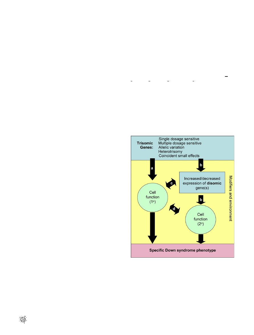
Figure 1. Possible Phenotypic Consequences of Gene Action in Down
Syndrome
(A) A trisomic gene or genes might directly affect cellular function in a
fully differentiated cell to cause a functional phenotype of DS or in an
immature cell to produce a developmental phenotype. (B) Trisomic
genes may alter expression of disomic genes, leading to a cellular
manifestation and a DS phenotype. A trisomy-induced change in cellular
function altering the relationship of that cell to surrounding cells leads to
a secondary distortion of (C) disomic gene expression or (D) function in
neighboring cells. Modifier genes or environment (yellow box) might
interact at multiple points to initiate, ameliorate, or exacerbate
phenotypes.
PLoS Genetics | www.plosgenetics.org
March 2006 | Volume 2 | Issue 3 | e50
0232

parallels that can be measured precisely in Ts65Dn mice.
With these phenotypic ‘‘readouts’’ for the predicted
functions of a critical region gene or genes, Olson et al. [9]
made a critical region model by re-engineering Mmu16 such
that the region corresponding to this DSCR was duplicated or
deleted. Mice carrying the duplication, which had segmental
trisomy involving only the critical region genes, did not
display the effects on stature nor the midface hypoplasia,
small mandible, or dysmorphology of the skull predicted by
the DSCR hypothesis. Thus, no gene(s) from this region was
sufficient to produce these phenotypes. Next, mice deleted
for the critical region segment were crossed to Ts65Dn mice
(which display all of these DS characteristics), thus returning
critical region gene dosage to normal in an animal that
carried the majority of Ts65Dn segment genes in three
copies. These mice had a somewhat attenuated presentation
of phenotypes seen in Ts65Dn, indicating that while critical
region genes made some contribution when present in three
copies, they were largely not necessary for these effects. This
result suggests that for those specific phenotypes, the DSCR
hypothesis of single gene effects is not correct. Rather,
multiple genes are required to produce these complex
alterations to structures that are the products of intricate
developmental processes.
Some aspects of DS may in fact be due primarily to the
effects of a single dosage-sensitive gene on Hsa21. For
example, elevated expression of endostatin, a protein that
inhibits angiogenesis required for tumor growth, may explain
at least part of the cancer resistance seen in DS [19]. However,
it seems to us unlikely that many aspects of the DS phenotype
that show highly variable presentation and derive from
changes in structures that are the product of a long span of
development are likely to reflect the effects of a single
dosage-sensitive gene. Indeed, the classical understanding of
Mendelian ‘‘single-gene’’ mutations as independently acting
elements has been qualified with the greater appreciation for
the roles of modifier genes on the phenotype.
Interacting genes of major effect. A simple extension of the
single dosage-sensitive gene model is to imagine additive
effects of multiple dosage-sensitive genes interacting in a
specific cell type during development. This could occur due
to co-expression of two or more genes of major effect in the
same cell at the same time or at different stages in the
developmental history of that cell population. The effects of
multiple dosage-sensitive genes might be amplified (or
attenuated) when they occur within the same biochemical
pathway. Possible trisomy 21 effects on a number of pathways
have been posited [20], prioritizing them as targets for
molecular analysis. However, the functions and interactions
of most Hsa21 (and other) genes are not catalogued to this
level. The combinatorial possibilities for testing groups of
genes present an obvious challenge to direct interrogation by
undirected screens. A further complication is that even in the
mouse, few phenotypes are defined with sufficient precision
to consistently detect small changes if one or two genes make
an incremental contribution to the trisomic phenotype.
Ultimately, it may be less important to tease out ‘‘sub-
phenotypic’’ consequences of individual genes than to
identify the pathways and processes that are perturbed by
trisomy. Correcting unbalanced pathways, regardless of the
precise genetic cause, is a logical approach to attenuation of
the phenotypic consequences [21].
Allelic variation on Hsa21. Dosage sensitivity may be
manifested in another fashion. Allelic variants of Hsa21 genes
are present in different ratios in an individual with trisomy
than in the diploid state. In the case where a mutant allele
results in lower levels of gene product, this mutation will
display recessive inheritance when the presence of one wild-
type allele is sufficient to carry on normal function. A
trisomic condition resulting in two copies of the loss-of-
function mutation plus one wild-type copy would probably
not alter the phenotypic outcome in this case. However, a
recessively inherited phenotype can also occur when a
mutant allele produces a gain or change of function, one copy
of which does not produce a detrimental effect in the
presence of a single wild-type allele, but two copies of which
may be sufficient to ‘‘overcome’’ the buffering of a normal
allele in a trisomic individual.
Another possible manifestation of trisomy at the molecular
level is heterotrisomy, in which alleles from three
grandparents are present in every cell [22]. This will occur
when trisomy results from an error in meiosis I, the most
frequent origin of the extra chromosome in DS [3]. For
multimeric proteins assembled from multiple peptides, such
as the collagens, the combinatorial possibilities become large.
(COL6A1, COL6A2, and COL18A1 are all encoded on distal
Hsa21.) Individuals with trisomy will produce combinations
of multimers that cannot occur in euploid individuals.
Baptista et al. described a region of Hsa21 between D21S167
and HMG14 that was frequently heterotrisomic in individuals
with DS and CHD [22].
Both ‘‘recessive dosage’’ and heterotrisomy should be
amenable to genetic analysis. However, standard statistical
methods do not account for the possibility of three alleles in
one individual. Sherman, Feingold, and colleagues have
established statistical methodologies for genetic association
studies to identify genes that affect the DS phenotype when
triplicated [23].
Coincident small effects. The preceding examples describe
situations in which phenotype is altered due to increased
expression of one or a few trisomic genes of major impact.
However, small coincident or additive effects of the many
genes over-expressed in every trisomic cell may also
contribute to trisomic phenotypes. Recent observations
confirm that transcript levels are elevated about 1.5-fold for
the majority of trisomic genes in a few tissues from humans
with trisomy 21 [24,25] and across a broad range of tissues
that can be measured in trisomic mouse models [11,12].
In this model, an individual triplicated gene might have no
demonstrable impact on phenotype by itself, whereas the
collective effect of dozens of genes affecting multiple cellular
processes is sufficient to result in a significant impact on
phenotype (see [26,27]). Proving this model presents
significant experimental challenges, but might be approached
by considering quantifiable phenotypes in animal models.
Several phenotypes have been measured precisely in trisomic
mouse models, allowing comparison between the Ts65Dn
mouse, with about 130 Hsa21 orthologs in three copies, and
Ts1Cje mice (91 triplicated genes). Behavioral, structural, and
functional brain phenotypes, dysmorphology of the skull, and
gene expression in the cerebellum all show patterns in
Ts65Dn that are similar but attenuated when fewer genes are
trisomic in Ts1Cje [6,28–31]. Attenuation of the phenotype
PLoS Genetics | www.plosgenetics.org
March 2006 | Volume 2 | Issue 3 | e50
0233

when fewer genes are triplicated is consistent with (but not
proof of) additive small effects by neighboring trisomic genes.
Note that not only trisomic genes show altered expression
in tissues from individuals with trisomy. In some but not all
studies, the perturbation of gene expression levels has been
demonstrated to extend beyond trisomic genes to those that
are disomic, affecting expression levels of a substantial
proportion of transcripts in trisomic tissues in mice [32,33].
In one study of trisomic mouse cerebellum, up to one-third of
disomic gene transcript levels were subtly altered [32]. Very
few of these genes showed a statistically significant difference
with euploid when considered individually, but collectively,
disomic gene expression robustly distinguished the trisomic
and euploid cerebellar transcriptomes. Conflicting results
regarding the question of perturbation of disomic gene
expression have been reported in human studies [24,25,34].
The controversy is perpetuated by the use of different
analytical approaches for array analysis in different studies.
Modifier genes. Most of the features that occur frequently
in DS are variable in severity (expressivity) and, except for a
few characteristic phenotypes, in occurrence (penetrance).
None of the commonly described DS phenotypes are unique
to DS or other chromosomal abnormalities but also may
occur in euploid individuals [35]. This wide degree of
variation suggests that a particular phenotype in a given
individual is affected by genetic and environmental variation,
and it is reasonable to assume that genetic background (the
specific allele set inherited by an individual) affects the
severity of outcome.
Preliminary data support the supposition that genetic
modifiers contribute to CHD, for which trisomy 21 is the
largest risk factor. About half of all individuals with DS have
some form of CHD, and most of these involve septal defects.
Complete atrioventricular canal occurs in one of five
individuals with trisomy 21, compared to 1/10,000 in the
euploid population [4]. However, since 80% of those with DS
do not have complete atrioventricular canal and 50% have no
clinical presentation of heart defects, trisomy 21 is not
sufficient to cause CHD by itself. Evidence from patient
studies now links the occurrence of CHD in individuals with
DS to mutations in disomic genes known to affect septation
in mouse models and in non-syndromic atrioventricular
canal (C. Maslen, S. Sherman, G. Capone and R. Reeves,
unpublished data).
The increased frequency of several important medical
conditions in DS, including CHD, childhood onset leukemia,
and Hirschsprung disease, suggests that additional genetic
factors may be involved. That is, the occurrence of these
diseases is greatly increased—but not caused—by trisomy 21.
Predisposing modifier genes may combine with the effects of
trisomy 21 to reach a threshold effect, resulting in an
observable phenotype (see Box 1). Genetic studies in
‘‘sensitized’’ DS populations can be especially effective for
identifying genetic variation that contributes to these
conditions, regardless of ploidy.
Not all predisposing conditions caused by trisomy have a
negative impact. Individuals with DS have reduced
frequencies of solid tumors [36,37] and may have a lower
incidence of atherosclerosis as well [38,39]. Characterization
of these effects could indicate approaches to reducing cancer
incidence or cardiovascular disease in all people.
The Fourth Developmental Dimension: Time
The demonstration that transcripts of trisomic genes are
elevated about 50% in a variety of cells and tissues and at
several developmental stages is a reasonable indicator that,
for the most part, this level of over-expression will occur in all
cells where that gene is expressed throughout development.
For those genes whose elevated expression alters a function in
fully differentiated cells, the presence of elevated expression
in adults may be considered directly in determining the
mechanism by which over-expression of that gene contributes
to a phenotype of DS. However, over-expression of a given
gene will not necessarily affect development and function in
every cell type and at every developmental time point when it
is expressed at elevated levels. It is likely that over-expression
of some genes is detrimental only at a specific time during
development, and then only in a specific cell type. Further, a
trisomy-induced change in one cell population could affect
neighboring cells, resulting in aberrant development as a
secondary consequence of trisomy (Figure 2).
For example, in a case where a threshold signal by ligand is
required to trigger a step in differentiation, elevated
expression of a cell-surface receptor encoded by a trisomic
gene could result in the cell experiencing that threshold at a
lower concentration of ligand than would be required for a
euploid cell. This might cause an early differentiation of cells
that would otherwise undergo additional cell divisions before
differentiating, resulting in a smaller anlage for subsequent
morphogenesis as a primary consequence of trisomy. If this
now depauperate cell population normally produces a ligand
to signal adjacent cells, the diminished signal produced by
fewer cells could have further consequences secondary to
those initiated by trisomy. Processing of transcripts or
protein can be differentially regulated throughout
development as well (e.g., stage-specific switches to alternative
splice forms of a message or different phosphorylation states
of a variety of proteins). Small alterations of almost any
cellular process as a result of aneuploidy could contribute to
deviations from normal patterns of development. In
Box 1. Trisomy 21 and GATA1
The incidence of childhood onset leukemia is elevated in DS and acute
megakaryoblastic leukemia (AMKL) occurs almost 500-fold more often
than in the euploid population [43]. This is associated with an unusual
feature of DS, transient myeloproliferative disorder (TMD). About 10% of
newborns with DS exhibit TMD, an expansion of immature
megakaryoblasts [44] that usually undergoes spontaneous remission
shortly after birth without clinical consequences. However, 20%–30% of
those who have TMD will develop AMKL later in life. Somatic mutations in
the GATA1 transcription factor gene, encoded on the X chromosome,
occur in most AMKL and almost every case of TMD in persons with DS,
but not in other leukemias that occur in DS; further, GATA1 mutations
have never been seen when AMKL occurs in euploid individuals except
when the expanded blasts are trisomic for Hsa21 [45]. Thus the
relationship between trisomy 21 and GATA1 is complex. It appears that
trisomy 21 makes megakaryoblasts highly sensitive to GATA1 mutations.
The same mutations presumably occur in euploid individuals, but have
no deleterious consequence unless Hsa21 becomes triplicated in those
cells. It would be interesting to know if the frequency of GATA1 mutations
is the same in megakaryoblasts trisomic for Hsa21 as it is in euploid
blasts.
PLoS Genetics | www.plosgenetics.org
March 2006 | Volume 2 | Issue 3 | e50
0234
particular, dosage effects of regulatory genes could have a
wide range of effects [40].
Ameliorating Consequences of Trisomy 21
Defining the etiology of genetic mechanisms in DS requires
knowledge of the trisomic genes, their expression patterns in
time and space, and their downstream effects, direct and
indirect, on the expression of other genes. This information
must be linked to a precise description of phenotypic
consequences, not only in fully differentiated cells, but also at
all stages where euploid and trisomic developmental
processes diverge. Animal models, including critically
important segmental trisomies and monosomies in mice,
provide a substrate for testing hypotheses about how over-
expression of genes individually or in concert can affect
development. The precision with which a phenotype and its
etiology can be explained in mice points to a difficulty with
extrapolation to humans, where phenotypes are defined
clinically for practical applications, and not necessarily with
the precision required for genetic studies.
Recent advances suggest that the origins of trisomic
phenotypes are perhaps even more complicated than
assumed for many decades. What then is the most effective
way to understand and, more importantly, to ameliorate the
effects of trisomy 21 on development and function? No single
approach will uncover the myriad sources of divergence from
normal development and function initiated by trisomy. One
area of research that may be currently under-represented is
an approach based in defining etiology—essentially, the
interface of genotype and phenotype.
As an example, Ts65Dn mice were shown to have a
disproportionately small cerebellum, comparable to a
broadly defined phenotype of DS [41]. Closer examination of
the trisomic mice demonstrated decreased neuronal density
in the Purkinje and granule cell layers, and this new aspect of
DS pathology was confirmed in humans. This phenotype was
followed through development in mice to identify the earliest
stage at which trisomic and euploid cerebellar development
diverged. While the number of granule cell precursors was
the same at the day of birth, the number of mitotic cells was
significantly reduced in trisomic mice. Genetic marker
crosses and primary culture assays identified a deficit in the
mitogenic response of granule cell precursors to the mitogen,
Sonic hedgehog. Treatment with an agonist of the hedgehog
pathway corrected the granule cell deficit through (at least)
the first third of cerebellar development [42]. This
‘‘phenotype-based’’ approach identified the basis for a
method to ameliorate structural deficits in cerebellum and
perhaps other brain regions, even though the Mmu16 gene or
genes responsible for the mitogenesis response deficit remain
to be identified.
Conclusions
Trisomy 21 is among the most complex genetic conditions
compatible with substantial survival beyond birth. This
complexity reflects a variety of genetic mechanisms, and the
sheer number of genes involved suggests that the primary
consequences of trisomic gene over-expression will be
amplified throughout development. Ameliorative strategies
for DS can be profitably pursued by studying the interface of
developmental processes and genetic mechanisms in order to
understand the etiology of processes that diverge as a
consequence of trisomy.
“
Acknowledgments
The requirement for brevity in this review has meant that a
substantial amount of important work is not covered here. Omissions
in no way reflect on the quality and importance of research in other
areas related to DS.
Competing interests. The authors have declared that no competing
interests exist.
Funding. This work was supported by NRSA fellowship HD 43614
(RJR) and PHS award HD 38384 (RHR).
References
1.
Morris JK, Wald NJ, Watt HC (1999) Fetal loss in Down syndrome
pregnancies. Prenat Diagn 19: 142–145.
2.
Spencer K (2001) What is the true fetal loss rate in pregnancies affected by
trisomy 21 and how does this influence whether first trimester detection rates
are superior to those in the second trimester? Prenat Diagn 21: 788–789.
3.
Hassold T, Hunt P (2001) To err (meiotically) is human: The genesis of
human aneuploidy. Nat Rev Genet 2: 280–291.
4.
Ferencz C, Neill CA, Boughman JA, Rubin JD, Brenner JI, et al. (1989)
Congenital cardiovascular malformations associated with chromosome
abnormalities: An epidemiologic study. J Pediatr 114: 79–86.
5.
Cohen WI (1999) Health care guidelines for individuals with Down
syndrome: 1999 revision. Down Syndrome Quarterly 4: 1–16.
6.
Potier MC, Rivals I, Mercier G, Ettwiller L, Moldrich RX, et al. (2006)
Transcriptional disruptions in Down syndrome: A case study in the Ts1Cje
mouse cerebellum during post-natal development. J Neurochem. E-pub 17
January 2006.
7.
Gardiner K, Fortna A, Bechtel L, Davisson MT (2003) Mouse models of
Down syndrome: How useful can they be? Comparison of the gene content
of human chromosome 21 with orthologous mouse genomic regions. Gene
318: 137–147.
8.
Antonarakis SE, Lyle R, Dermitzakis ET, Reymond A, Deutsch S (2004)
Chromosome 21 and down syndrome: From genomics to pathophysiology.
Nat Rev Genet 5: 725–738.
9.
Olson LE, Richtsmeier JT, Leszl J, Reeves RH (2004) A chromosome 21
critical region does not cause specific down syndrome phenotypes. Science
306: 687–690.
10. O’Doherty A, Ruf S, Mulligan C, Hildreth V, Errington ML, et al. (2005) An
aneuploid mouse strain carrying human chromosome 21 with down
syndrome phenotypes. Science 309: 2033–2037.
11. Kahlem P, Sultan M, Herwig R, Steinfath M, Balzereit D, et al. (2004)
Transcript level alterations reflect gene dosage effects across multiple
tissues in a mouse model of down syndrome. Genome Res 14: 1258–1267.
12. Lyle R, Gehrig C, Neergaard-Henrichsen C, Deutsch S, Antonarakis SE
(2004) Gene expression from the aneuploid chromosome in a trisomy
mouse model of down syndrome. Genome Res 14: 1268–1274.
13. Kola I, Herzog PJ (1998) Down syndrome and mouse models. Curr Opinion
Gen Dev 8: 316–321.
14. Lejeune J, Gauthier M, Turpin R (1959) Etudes des chromosomes
somatiques de neuf enfants mongoliens. CR Acad Sci (Paris) 248: 1721–1722.
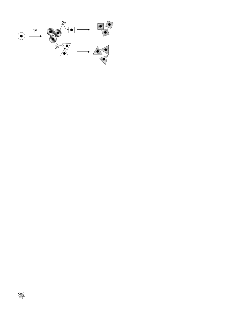
DOI: 10.1371/journal.pgen.0020050.g002
Figure 2. A Primary (18) Effect of Trisomy Produces an Aberrant
Phenotype as the Cells Proliferate
Trisomy causes a primary defect in (circular) cells as they proliferate. This
primary defect results in a signaling error to neighboring (square and
triangular) cells, resulting in their aberrant development as a secondary
(28) consequence of trisomy. Plain background indicates normal cells;
striped background indicates an aberrant phenotype.
PLoS Genetics | www.plosgenetics.org
March 2006 | Volume 2 | Issue 3 | e50
0235

15. Niebuhr E (1974) Down Syndrome. The possibility of a pathogenetic
segment on chromosome 21. Humangenetik 21: 99–101.
16. McCormick MK, Schinzel A, Petersen MB, Stetten G, Driscoll DJ, et al.
(1989) Molecular approach to the characterization of the Down syndrome
region of chromosome 21. Genomics 5: 325–331.
17. Delabar JM, Theophile D, Rahmani Z, Chettouh Z, Blouin JL, et al. (1993)
Molecular mapping of twenty-four features of Down syndrome on
chromosome 21. Eur J Hum Genet 1: 114–124.
18. Korenberg J (1991) Down syndrome phenotype mapping. In: Epstein C,
editor. Progress in clinical and biological research. New York: Wiley-Liss.
337 p.
19. Zorick TS, Mustacchi Z, Bando SY, Zatz M, Moreira-Filho CA, et al. (2001)
High serum endostatin levels in Down syndrome: Implications for
improved treatment and prevention of solid tumours. Eur J Hum Genet 9:
811–814.
20. Gardiner K (2003) Predicting pathway perturbations in Down syndrome. J
Neural Transm Suppl 67: 21–37.
21. Gardiner K, Davisson MT, Crnic LS (2004) Building protein interaction
maps for Down’s syndrome. Brief Funct Genomic Proteomic 3: 142–156.
22. Baptista MJ, Fairbrother UL, Howard CM, Farrer MJ, Davies GE, et al.
(2000) Heterotrisomy, a significant contributing factor to ventricular septal
defect associated with Down syndrome? Hum Genet 107: 476–482.
23. Kerstann KF, Feingold E, Freeman SB, Bean LJ, Pyatt R, et al. (2004)
Linkage disequilibrium mapping in trisomic populations: Analytical
approaches and an application to congenital heart defects in Down
syndrome. Genet Epidemiol 27: 240–251.
24. Mao R, Zielke CL, Ronald Zielke H, Pevsner J (2003) Global up-regulation of
chromosome 21 gene expression in the developing down syndrome brain.
Genomics 81: 457–467.
25. FitzPatrick DR, Ramsay J, McGill NI, Shade M, Carothers AD, et al. (2002)
Transcriptome analysis of human autosomal trisomy. Hum Mol Genet 11:
3249–3256.
26. Pritchard MA, Kola I (1999) The ‘‘gene dosage effect’’ hypothesis versus the
‘‘amplified developmental instability’’ hypothesis in Down syndrome. J
Neural Transm Suppl 57: 293–303.
27. Shapiro B (1983) Down syndrome—A disruption of homeostasis. Amer J
Medical Genet 14: 241–269.
28. Kleschevnikov AM, Belichenko PV, Villar AJ, Epstein CJ, Malenka RC, et al.
(2004) Hippocampal long-term potentiation suppressed by increased
inhibition in the Ts65Dn mouse, a genetic model of Down syndrome. J
Neurosci 24: 8153–8160.
29. Olson LE, Roper RJ, Baxter LL, Carlson EJ, Epstein CJ, et al. (2004) Down
syndrome mouse models Ts65Dn, Ts1Cje, and Ms1Cje/Ts65Dn exhibit
variable severity of cerebellar phenotypes. Dev Dyn 230: 581–589.
30. Richtsmeier JT, Zumwalt A, Carlson EJ, Epstein CJ, Reeves RH (2002)
Craniofacial phenotypes in segmentally trisomic mouse models for Down
syndrome. Am J Med Genet 107: 317–324.
31. Siarey RJ, Villar AJ, Epstein CJ, Galdzicki Z (2005) Abnormal synaptic
plasticity in the Ts1Cje segmental trisomy 16 mouse model of Down
syndrome. Neuropharmacology 49: 122–128.
32. Saran NG, Pletcher MT, Natale JE, Cheng Y, Reeves RH (2003) Global
disruption of the cerebellar transcriptome in a Down syndrome mouse
model. Hum Mol Genet 12: 2013–2019.
33. Dauphinot L, Lyle R, Rivals I, Dang MT, Moldrich RX, et al. (2005) The
cerebellar transcriptome during postnatal development of the Ts1Cje
mouse, a segmental trisomy model for Down syndrome. Hum Mol Genet 14:
373–384.
34. Amano K, Sago H, Uchikawa C, Suzuki T, Kotliarova SE, et al. (2004)
Dosage-dependent over-expression of genes in the trisomic region of
Ts1Cje mouse model for Down syndrome. Hum Mol Genet 13: 1333–1340.
35. Epstein CJ (2001) Down syndrome (Trisomy 21). In: Scriver CR, Beaudet
AL, Sly WS, Valle D, editors. The metabolic and molecular bases of
inherited disease. New York: McGraw-Hill. pp. 1223–1256.
36. Yang Q, Rasmussen SA, Friedman JM (2002) Mortality associated with
Down’s syndrome in the USA from 1983 to 1997: A population-based study.
Lancet 359: 1019–1025.
37. Hasle H (2001) Pattern of malignant disorders in individuals with Down’s
syndrome. Lancet Oncol 2: 429–436.
38. Yla-Herttuala S, Luoma J, Nikkari T, Kivimaki T (1989) Down’s syndrome
and atherosclerosis. Atherosclerosis 76: 269–272.
39. Murdoch JC, Rodger JC, Rao SS, Fletcher CD, Dunnigan MG (1977) Down’s
syndrome: An atheroma-free model? Br Med J 2: 226–228.
40. Birchler JA, Riddle NC, Auger DL, Veitia RA (2005) Dosage balance in gene
regulation: Biological implications. Trends Genet 21: 219–226.
41. Baxter LL, Moran TH, Richtsmeier JT, Troncoso J, Reeves RH (2000)
Discovery and genetic localization of Down syndrome cerebellar
phenotypes using the Ts65Dn mouse. Hum Mol Genet 9: 195–202.
42. Roper RJ, Baxter LL, Saran NG, Klinedinst DK, Beachy PA, et al. (2006)
Defective cerebellar response to mitogenic Hedgehog signaling in Down
syndrome mice. Proc Natl Acad Sci U S A 103: 1452–1456.
43. Lange B (2000) The management of neoplastic disorders of haematopoiesis
in children with Down’s syndrome. Br J Haematol 110: 512–524.
44. Zipursky A (2003) Transient leukaemia—A benign form of leukaemia in
newborn infants with trisomy 21. Br J Haematol 120: 930–938.
45. Crispino JD (2005) GATA1 mutations in Down syndrome: Implications for
biology and diagnosis of children with transient myeloproliferative
disorder and acute megakaryoblastic leukemia. Pediatr Blood Cancer 44:
40–44.
PLoS Genetics | www.plosgenetics.org
March 2006 | Volume 2 | Issue 3 | e50
0236
Wyszukiwarka
Podobne podstrony:
puchar swiata 2006 www prezentacje org
Gospodarka płynami kwiecień 2006
Znaki taktyczne i szkice obrona, natarcie,marsz maj 2006
Prowadzenie kliniczne pacjentów z dobrym widzeniem M Koziak 2006
prezentacja cwiczen 2006
Wyklad 09 2006
Wyk 2 WE Polityka monetarna 2006 2
3 DOWN STREAM PROCESSING
urazy kl piersiowej 04 2006
Wyk 6 Model klasyczny 2006
ADHD 2006
1 zaburzenia krążenia 1 2006 07 III
więcej podobnych podstron