
ABC of diseases of liver, pancreas, and biliary system
Pancreatic tumours
P C Bornman, I J Beckingham
Neoplasms of the pancreas may originate from both exocrine
and endocrine cells, and they vary from benign to highly
malignant. Clinically, 90% of pancreatic tumours are malignant
ductal adenocarcinomas, and most of this article concentrates
on this disease.
Ductal adenocarcinoma
Incidence and prognosis
Carcinoma of the pancreas has become more common in most
Western countries over the past three decades, and although
there is evidence of plateauing in some countries such as the
United States, it still ranks as the sixth commonest cause of
cancer death in the United Kingdom. Most patients are over the
age of 60 years (80%) and many will have concurrent medical
illnesses that complicate management decisions, particularly
because the median survival from diagnosis is less than six
months.
Clinical presentation
Two thirds of pancreatic cancers develop in the head of the
pancreas, and most patients present with progressive,
obstructive jaundice with dark urine and pale stools. Pruritus,
occurring as a result of biliary obstruction, is often troublesome
and rarely responds to antihistamines. Back pain is a poor
prognostic sign, often being associated with local invasion of
tumours. Severe cachexia, as a result of increased energy
expenditure mediated by the tumour, is also a poor prognostic
indicator. Cachexia is the usual presenting symptom in patients
with tumours of the body or tail of the pancreas.
Examination
The commonest sign is jaundice, with yellowing of the sclera
and, once the bilirubin concentration exceeds 35
ìmol/l, the
skin. Many patients with high bilirubin concentrations will have
skin scratches associated with pruritus. Patients with advanced
disease have severe weight loss accompanied by muscle wasting
and occasionally an enlarged supraclavicular lymph node. A
palpable gall bladder suggests pancreatic malignancy, but it can
be difficult to detect when displaced laterally or covered by an
enlarged liver. The presence of ascites or a palpable epigastric
mass usually indicates end stage disease. Full assessment of the
patient’s general fitness is essential to develop an individualised
management plan.
Investigation
Because of the poor prognosis, care should be taken not to
overinvestigate or embark on treatment strategies based on the
unrealistic expectations of patients, their families, or the
referring doctor. An increasing number of investigations are
available, and the aim is to select patients who will not benefit
from major resection by use of the fewest, least invasive, and
least expensive means. The choice of investigation will vary
according to local availability, particularly of newer
investigations such as laparoscopic and endoscopic
ultrasonography, and it remains to be seen if these techniques
offer major advantages over the latest generation of computed
tomography and magnetic resonance imaging scanners. Early
cooperation between a gastroenterologist, radiologist, and
Types of pancreatic neoplasms
x Benign exocrine
Serous cyst adenoma
Mucinous cyst adenoma
x Malignant exocrine
Ductal adenocarcinoma
Mucinous cyst adenocarcinoma
x Endocrine
Gastrinoma
Insulinoma
Other
Factors predicting poor prognosis
x Back pain
x Rapid weight loss
x Poor performance status—for example, World Health Organization
or Karnofsky scoring systems
x Ascites and liver metastases
x High C reactive protein and low albumin concentrations
Rarer presentations of pancreatic carcinoma
x Recurrent or atypical venous thromboses (thrombophlebitis
migrans)
x Acute pancreatitis
x Late onset diabetes mellitus
x Upper gastrointestinal bleeding
Patient with jaundice, bruising, and weight loss due
to pancreatic carcinoma
Clinical review
721
BMJ VOLUME 322 24 MARCH 2001 bmj.com
surgeon should avoid inappropriate investigations and
treatment that might interfere with patients’ quality of life.
Endoscopic retrograde cholangiopancreatography is an
important investigation in patients with obstructive jaundice. As
well as showing biliary and pancreatic strictures, the pathology
can be confirmed by taking brushings for cytology or biopsy
specimens of the duct for histology. The technique can also be
used to place a stent to relieve biliary obstruction. However, it is
important not to use this approach before patients are properly
selected for treatment.
The diagnosis can also be confirmed by fine needle
aspiration guided by ultrasonography or computed
tomography, but this investigation has a high rate of false
negative results and is rarely necessary. Fine needle aspiration
should be avoided in patients with potentially resectable
tumours as it can cause seeding and spread of the tumour.
Treatment
Surgical resection does not improve survival in patients with
locally advanced or metastatic disease. Tumour stage and the
patient’s fitness for major surgical resection are the main factors
in determining optimal treatment.
Resectable tumours
Surgical resection, usually a pancreaticoduodenectomy
(Whipple’s procedure), is the only hope for cure. Less than 15%
of tumours are suitable for resection. Very few tumours of the
body and tail are resectable (3%) as patients usually present late
with poorly defined symptoms.
The outcome of resection has been shown to be better in
specialised pancreatobiliary centres that perform the procedure
regularly than in small units. Mortality has fallen to 5-10% in
dedicated units. The overall five year survival rate of 10-15%
after resection remains disappointing, although survival is as
high as 20-30% in some subgroups such as patients with small
( < 2 cm), node negative tumours. Furthermore, the median
survival of patients who have resection is 18 months compared
with six months for patients without metastatic disease who do
not have resection.
Preoperative biliary drainage remains controversial. The
reduced complications from resolution of jaundice are offset by
more inflammatory tissue at surgery and higher rates of biliary
sepsis after stenting. Ideally patients with minimal jaundice
should be operated on without stenting whereas those with
higher bilirubin concentrations ( > 100
ìmol/l) probably benefit
from endoscopic stenting and reduction of bilirubin
concentrations before surgery.
Locally advanced disease
Several options are available for the 65% of patients who have
locally advanced disease. These depend on factors such as age,
disease stage, and the patient’s fitness. Endoscopic insertion of a
plastic or metal wall stent relieves jaundice in most patients.
Plastic stents are cheaper but have a median half life of three to
four months compared with six months for metal stents.
Blockage of a stent results in rigors and jaundice, and patients
should be given antibiotics and have the stent replaced.
Surgical exploration and bypass should be used in
patients who are predicted to survive longer than six months,
in whom it is not certain that the tumour cannot be resected,
in areas with limited access to endoscopic retrograde
cholangiopancreatography, or with recurrent stent blockage or
obstruction of the gastric outlet. Surgical bypass (open or
laparoscopic) now has a low mortality and has the advantage of
long term palliation of jaundice with a low risk of recurrence.
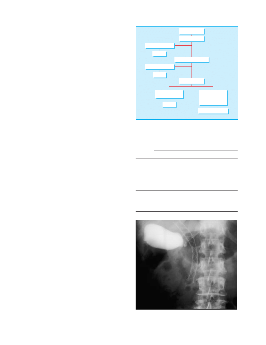
Treatment of pancreatic ductal carcinoma
Fitness
of
patient
Tumour stage
Resectable
Locally
advanced
Metatstatic
Low risk
Pancreatoduodectomy
Surgical
bypass or
endoscopic
stent
Palliative
care + /
−
endoscopic
stent
High risk
Endoscopic stent
Endoscopic
stent
Palliative
care
Tumours suitable for resection
x < 4 cm in diameter
x Confined to pancreas
x No local invasion or metastases
Obstructive jaundice
Ultrasonography
Spiral computed tomography
Laparoscopy
Peritoneal metastases
liver metastases
Resectable tumour
no metastases
no local invasion
no vascular involvement
Pancreatoduodenectomy
Liver metastases, ascites
Liver metastases, ascites
Palliation
Palliation
Palliation
Palliation = endoscopic stent, surgical bypass, or medical
palliation alone depending on patient's general health and symptoms
Investigation and management of pancreatic ductal carcinoma
Metal wall stent in common bile duct of patient with pancreatic carcinoma.
(Note contrast in gall bladder from endoscopic retrograde
cholangiopancreatography)
Clinical review
722
BMJ VOLUME 322 24 MARCH 2001 bmj.com

Metastatic disease
Patients with metastatic disease are often cachectic and rarely
survive more than a few weeks. Treatment should focus on
alleviation of pain and improving quality of life with input from
palliative care teams. Patients with less advanced metastatic
disease may require endoscopic stenting, especially if they have
intractable pruritus.
Radiotherapy and chemotherapy
Despite numerous trials, radiotherapy with or without
chemotherapy has not been shown to prolong survival. The
search for new chemotherapeutic and immunotherapeutic drugs
continues, but they currently have little role outside clinical trials.
Symptom control
A liberal policy of pain control with paracetamol, non-steroidal
anti-inflammatory drugs, and opiate analgesics should be
followed. In difficult cases and when increasingly large doses of
opiates are required, patients should be referred to a specialist
pain clinic for consideration of coeliac plexus block or
thoracoscopic splanchnicectomy. Early referral to the palliative
care team and Macmillan nurses, who can bridge the gap
between hospital and community care, is beneficial.
Cachexia is an important cause of disability in many
patients. Nutritional supplementation rarely combats weight
loss, and pancreatic replacement therapy is also of doubtful
benefit. Encouraging results have recently been reported with
polyunsaturated fatty acids (fish oil) and non-steroidal
anti-inflammatories, which seem to inhibit the inflammatory
response provoked by the tumour and reduce the speed of
weight loss with some survival benefit. Impaired gastric
emptying is generally underdiagnosed and may be functional or
mechanical in origin.
Cystic tumours
These rare tumours (1% of all pancreatic neoplasms) are mostly
benign, but the mucinous type (about 50%) is premalignant.
They are important because they occur predominantly in young
women and usually have a good prognosis when resected.
Cystic tumours may be mistaken for benign pseudocysts,
although they can usually be differentiated on the basis of
history and computed tomographic findings (tumours have
septa within the cyst and calcification of the rim of the cyst wall
without calcification in the rest of the pancreas). If the diagnosis
is in doubt, surgical resection with frozen section at the time of
definitive surgery is the optimal management.
Endocrine tumours
Tumours arising from the islets of Langerhans can produce
high concentrations of the hormones normally produced by the
islets (insulin, glucagon, somatostatin, etc) or non-pancreatic
hormones (such as gastrin or vasoactive peptide). Endocrine
tumours are rare, with an incidence of around 1-2 per million
population. The commonest forms are gastrinoma and
insulinoma. They may be sporadic or occur as part of the
multiple endocrine neoplasia syndrome, when they are
associated with tumours of the pituitary, parathyroid, thyroid,
and adrenal glands.
Patients usually present with a clinical syndrome produced
by hormonal excess, typically of a single peptide. With the
exception of insulinomas most endocrine tumours are
malignant. Treatment is by surgical excision, and survival is
generally good; 10 year survival rates for patients with
malignant lesions are around 50%.
Summary points
x 6000 people die from pancreatic cancer each year in the United
Kingdom
x Presentation is usually with painless insidious jaundice
x Median survival from diagnosis is less than six months
x Less than 15% of all pancreatic tumours are resectable, and five
year survival after resection is 10-15%
x Endoscopic retrograde cholangiopancreatography and surgical
biliary drainage offer good palliation of jaundice
x Cystic and endocrine pancreatic tumours are uncommon but have
a better prognosis
Further reading
Trede M, Carter DC. Surgical options for pancreatic cancer. In: Surgery
of the pancreas. Edinburgh: Churchill Livingstone, 1997:383-515
Cameron JL, Grochow LB, Milligan FD, Venbrux AC. Pancreatic
cancer. In: Pitt HA, Carr-Locke DL, Ferrucci JT, eds. Hepatobiliary and
pancreatic disease—the team approach to management. Boston: Little
Brown, 1995:475-86
Cotton P, Williams C. ERCP and therapy. In: Practical gastrointestinal
endoscopy. Oxford: Blackwell Science, 1996:167-86
Cystic tumour in head of pancreas with calcified rim
Necrolytic erythema migrans is pathognomic in patients with glucagonoma
P C Bornman is professor of surgery, University of Cape Town,
South Africa.
The ABC of diseases of liver, pancreas, and biliary system is edited by
I J Beckingham, consultant hepatobiliary and laparoscopic surgeon,
department of surgery, Queen’s Medical Centre, Nottingham
(Ian.Beckingham@nottingham.ac.uk). The series will be published as
a book later this year.
BMJ 2001;322:721-3
Clinical review
723
BMJ VOLUME 322 24 MARCH 2001 bmj.com
Wyszukiwarka
Podobne podstrony:
ABC Liver tumours
ABC Liver tumours
ABC Of Liver,Pancreas and Gall Bladder
ABC Liver and pancreatic trauma
ABC Transplantation of the liver and pancreas
ABC Acute pancreatitis
ABC Chronic pancreatitis
2014 ABC DYDAKTYKIid 28414 ppt
Amortyzacja pozycki ABC
ABC mądrego rodzica droga do sukcesu
ABC praw konsumenta demo
abc 56 58 Frezarki
ABC Madrego Rodzica Inteligencja Twojego Dziecka
ABC Neostrada
ABC trzylatka przewodnik
abc systemu windows xp 47IMHOQVXQT6FS4YTZINP4N56IQACSUBZSUF7ZI
więcej podobnych podstron