
Journal of Chromatography A, 902 (2000) 167–194
www.elsevier.com / locate / chroma
Review
Solid-phase microextraction in biomedical analysis
*
S. Ulrich
Institute of Clinical Pharmacology
, University Hospital, Otto-von-Guericke University, Leipziger Strasse 44, D-39120 Magdeburg,
Germany
Abstract
Chromatographic methods are preferred in the analysis of organic molecules with lower molecular mass (,500 g / mol) in
body fluids, i.e., the assay of drugs, metabolites, endogenous substances and poisons as well as of environmental exposure by
gas chromatography (GC) and liquid chromatography (LC), for example. Sample preparation in biomedical analysis is
mainly performed by liquid–liquid extraction and solid-phase extraction. However, new methods are investigated with the
aim to increase the sample throughput and to improve the quality of analytical methods. Solid-phase microextraction
(SPME) was introduced about a decade ago and it was mainly applied to environmental and food analysis. All steps of
sample preparation, i.e., extraction, concentration, derivatization and transfer to the chromatograph, are integrated in one step
and in one device. This is accomplished by the intelligent combination of an immobilized extraction solvent (a polymer) with
a special geometry (a fiber within a syringe). It was a challenge to test this novel principle in biomedical analysis. Thus, an
introduction is provided to the theory of SPME in the present paper. A critical review of the first applications to biomedical
analyses is presented in the main paragraph. The optimization of SPME as well as advantages and disadvantages are
discussed. It is concluded that, because of some unique characteristics, SPME can be introduced with benefit into several
areas of biomedical analysis. In particular, the application of headspace SPME–GC–MS in forensic toxicology and
environmental medicine appears to be promising. However, it seems that SPME will not become a universal method. Thus,
on-line SPE–LC coupling with column-switching technique may be a good alternative if an analytical problem cannot be
sufficiently dealt with by SPME.
2000 Elsevier Science B.V. All rights reserved.
Keywords
: Reviews; Solid-phase microextraction; Forensic analysis; Pharmaceutical analysis; Antidepressants; Beta-
blockers; Benzodiazepines; Amphetamine; Phencyclidine; Polynuclear aromatic hydrocarbons; Pesticides; Volatile organic
compounds; Polychlorinated biphenyls; Antihistamines
Contents
1. Introduction ............................................................................................................................................................................
168
2. Solid-phase microextraction – a new principle in sample preparation ..........................................................................................
169
3. Theory of solid-phase microextraction ......................................................................................................................................
171
3.1. Thermodynamics ............................................................................................................................................................
171
3.2. Kinetics..........................................................................................................................................................................
173
3.3. Solid-phase microextraction in biological fluids ................................................................................................................
174
4. Application of solid-phase microextraction in biomedical analysis ..............................................................................................
176
*Tel.: 149-391-6713-060; fax: 149-391-6713-062.
E-mail address
: sven.ulrich@medizin.uni-magdeburg.de (S. Ulrich).
0021-9673 / 00 / $ – see front matter
2000 Elsevier Science B.V. All rights reserved.
P I I : S 0 0 2 1 - 9 6 7 3 ( 0 0 ) 0 0 9 3 4 - 1

168
S
. Ulrich / J. Chromatogr. A 902 (2000) 167 –194
4.1. Direct solid-phase microextraction ...................................................................................................................................
176
4.1.1. Without derivatization .........................................................................................................................................
176
4.1.2. With derivatization ..............................................................................................................................................
179
4.1.3. Other methods ....................................................................................................................................................
181
4.2. Headspace solid-phase microextraction.............................................................................................................................
181
4.2.1. Without derivatization .........................................................................................................................................
181
4.2.2. With derivatization ..............................................................................................................................................
184
4.3. Miscellaneous .................................................................................................................................................................
185
5. Optimization of solid-phase microextraction..............................................................................................................................
185
5.1. Coating ..........................................................................................................................................................................
185
5.2. Extraction method and sample pretreatment for solid-phase microextraction........................................................................
186
5.3. Agitation ........................................................................................................................................................................
187
5.4. Sample volume and volume of the headspace....................................................................................................................
187
5.5. Extraction time ...............................................................................................................................................................
187
5.6. pH .................................................................................................................................................................................
188
5.7. Salt and other additives ...................................................................................................................................................
188
5.8. Temperature ...................................................................................................................................................................
188
5.9. Desorption......................................................................................................................................................................
189
5.10. GC temperature program ...............................................................................................................................................
189
5.11. GC capillary .................................................................................................................................................................
189
5.12. GC detector ..................................................................................................................................................................
190
5.13. Automation...................................................................................................................................................................
190
6. Advantages and disadvantages of solid-phase microextraction ....................................................................................................
190
7. Conclusions ............................................................................................................................................................................
191
8. Nomenclature .........................................................................................................................................................................
192
References ..................................................................................................................................................................................
192
1. Introduction
dioxins and polynuclear aromatic hydrocarbons
(PAHs), for example, are analyzed in human body
Biomedical analysis of lower-molecular-mass or-
fluids for the investigation of environmental and
ganic molecules (,500 g / mol) comprises, for the
occupational exposure [11,12]. Endogenous sub-
main part, the analysis of drugs, metabolites,
stances such as neurotransmitters, arachidonic acid
poisons, chemicals of environmental exposure and
metabolites and fatty acids, for example, are ana-
endogenous substances in body fluids and tissues.
lyzed in biological and medical research and in
The quantitative and qualitative analysis of drugs and
clinical diagnostics [13–15].
metabolites is extensively applied to pharmacokinetic
Capillary gas chromatography (GC) and column
studies. Variables such as time to maximal con-
liquid chromatography (LC) are mainly applied.
centration in plasma, clearance and bioavailability
High sensitivity and high selectivity are the most
have to be known for the approval of a new drug
prominent advantages of chromatographic methods
[1,2].
Pharmacokinetic
interactions,
the
phar-
compared with, for example, enzyme-linked im-
macokinetics in special populations and relationships
munoassays (ELISAs) and fluorescence polarization
between the concentration of drug and pharmaco-
immunoassays (FPIAs) [16–20]. However, the main
logical effect, for example, are investigated in post-
disadvantage of chromatographic methods is the
marketing surveillance. Therapeutic drug monitoring
need for sample preparation. The sample cannot be
(TDM) may be used as a tool for the improvement of
applied to the chromatograph in its original form.
drug therapy [3–6]. Drugs of abuse, illicit drugs and
Therefore, the task of sample preparation is to
intoxications by drugs and poisons are analyzed in
transfer the analyte into a form that is (1) pre-
clinical and forensic toxicology [7–10]. As part of
purified, (2) concentrated and (3) with the chromato-
environmental chemistry and environmental medi-
graphic system fitting solvent. Prior to sample prepa-
cine, a wide variety of chemicals such as pesticides,
ration the analyte is found in a low concentration and
herbicides, volatile organic compounds (VOCs),
in a great volume of an aqueous matrix which

S
. Ulrich / J. Chromatogr. A 902 (2000) 167 –194
169
consists of a huge number of highly concentrated
water. Even automated systems were described [27].
proteins, lipoproteins, lipids and salts as well as other
However, the analyte enrichment and sample purifi-
lower concentrated endogenous and exogenous or-
cation is poor. Another relatively simple approach is
ganic substances. Because of this complex matrix the
the headspace (HS) technique in GC. However, HS
trace analysis in body fluids is more complicated
can only be applied to analytes with high vapor
than trace analysis in surface water in environmental
pressure [28]. Other methods of sample preparation
chemistry. However, it is comparable with the trace
are supercritical fluid extraction (SFE) [29], on-
analysis in water enriched with dissolved polymer
column sample preparation with column-switching
organic material (DOM). The sample as prepared for
techniques and on-line SPE in LC using RAMs
the chromatograph should be concentrated and pre-
[26,30–32], LC–GC coupling [33,34] and mem-
purified in an organic solvent. This is accomplished
brane-based
sample
preparation
(dialysis,
elec-
mainly by liquid–liquid extraction (LLE) and solid-
trodialysis, ultrafiltration) [35]. Although these meth-
phase extraction (SPE). Nearly all analytical prob-
ods have their own merits, most of them are only
lems can be solved by LLE and SPE. Therefore,
found in isolated applications, oftentimes, they do
these methods can be characterized as universal from
not achieve sensitivity and selectivity of LLE and
a scientific and technical view.
SPE and, finally, some methods need expensive
However, the disadvantage of LLE and SPE is the
equipment. Other problems are fouling of mem-
considerable expense of time and manual operations.
branes in membrane-based sample preparation and
Sample throughput is low and the economic expense
irreversible binding of some high-molecular-mass
is high. In other words, sample preparation is the
material in on-line SPE, for example. However, on-
bottleneck of the entire analytical method. Further-
line SPE–LC using RAMs appears to be promising
more, some advantages claimed for SPE over LLE
[26].
must be regarded critically. For example, laborious
This situation is the reason for the permanent
operations such as conditioning, washing, elution and
search for new sample preparation methods. One
solvent evaporation are needed, too. The volume of
approach is solid-phase microextraction (SPME).
organic solvents needed in SPE cannot be neglected
The present review provides a survey and discussion
with regard to environmental pollution. It can be
of the application of SPME in biomedical analysis.
even higher than in a simple one-step LLE or even in
a three-step LLE [21–23]. Evaporation of the eluate
is more time-consuming than in LLE because protic
2. Solid-phase microextraction – a new
solvents are mainly used, aqueous methanol for
principle in sample preparation
example, which have a lower vapor pressure than
chloroform and hexane in LLE. In addition, clotting,
SPME is based on a modified syringe which
channeling and percolation are typical problems of
contains a stainless steel microtubing within its
SPE encountered in every-day laboratory work. Off-
syringe needle. This microtubing has an about 1-cm
line automation of LLE and SPE is complicated.
fused-silica fiber tip which is coated with an organic
Although some systems were presented they did not
polymer. The coated silica fiber can be moved
lead to a break-through in the economics of sample
between two positions, inside and outside the needle,
preparation. Despite automation of SPE being easier
with a plunger as in the case of a normal syringe.
than automation of LLE, it is also beset with
The diameter of the syringe needle housing the
technical problems. However, the comparison of
microtubing and coated silica fiber is not much
LLE
and
SPE
is
discussed
controversially
increased in comparison with a normal GC syringe.
[9,12,24,25]. Some promising approaches in SPE are
Thus, by means of this simple equipment several
based on special packings such as restricted access
steps of sample preparation are combined in one
materials (RAMs), and molecular imprinting materi-
device. Extraction and enrichment of the analyte is
als (MIPs), for example [26].
completed by the coating in the position outside the
A alternative simple approach in LC is protein
syringe needle. Penetration of the septum of a GC
precipitation of plasma and injection of plasma
injection port is possible if the fiber was withdrawn
170
S
. Ulrich / J. Chromatogr. A 902 (2000) 167 –194
into the syringe needle. Desorption of the analyte
anticipated to be considerably easier than with other
and transfer to the capillary is performed after again
sample preparation methods.
moving the fiber to the position outside the syringe.
SPME was invented and first described by Paw-
This procedure can be repeated with one device
liszyn and co-workers in 1990 [36,37]. The invention
several times (Fig. 1).
of SPME appears to be a logical development based
It should be emphasized that the term ‘‘solid-phase
on open-tubular capillary columns used in GC. These
microextraction’’ may undervalue the advantages of
capillaries had their break-through in analytical
SPME. Advantages of this principle should be
laboratories in the mid-1980s. The conception of
greater than those of other extraction methods with
SPME may have been derived from the idea of an
only a very low quantity (‘‘micro’’) of the extraction
inversed GC capillary. Thus, a SPME device con-
agent, for example, SPE with disc technology. The
stituting a tubing with a coated inner surface was
outstanding and crucial idea of this principle named
described, too [38]. During the initial years SPME
SPME is the intelligent geometry of the extraction
was mainly described for applications in environ-
agent and extraction device. In contrast to conven-
mental analysis [39–41]. About 110 applications to
tional SPE with packed-bed columns, micro or non-
environmental analysis were published up until 1996
micro columns, this arrangement allows the combi-
[42]. By nature, SPME is used mainly for GC.
nation of all steps of sample preparation in one step
However, an adaptation for LC is possible with a
as described above. For this reason, the main advan-
special interface [43]. Two main variants of SMPE
tage of SPME is its simplicity and automation is
can be chosen: direct SPME with dipping the fiber
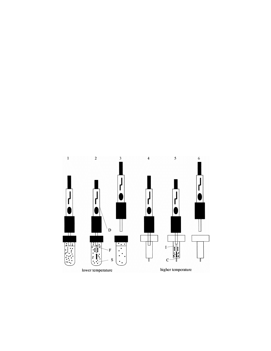
Fig. 1. The principle of SPME: 15introduction of syringe needle of the SPME device (D) into the sample vial and close to the sample (S),
25moving the fiber (F) into the position outside the syringe and into the sample (extraction), 35moving the fiber back into the syringe
needle and subsequent transfer of the device to the GC injector port (I) and capillary head (C), 45penetration of the septum with syringe
needle, 55moving the fiber into the position outside the syringe (desorption), 65moving the fiber back into the syringe needle and
withdrawing the syringe needle.
S
. Ulrich / J. Chromatogr. A 902 (2000) 167 –194
171
directly into the aqueous sample and HS-SPME with
extraction of the analyte from the HS of the sample.
Minor variants are derived from whether or not
derivatization is applied and in which phase, the type
of sample agitation as well as the option of cooling
of the fiber, for example.
3. Theory of solid-phase microextraction
3.1. Thermodynamics
Because of the physicochemical properties of, for
example,
polydimethylsiloxane
(PDMS,
melting
point: 2508C, glass transition temperature: 21268C),
which is most often applied in SPME, the extraction
obeys the rules of liquid–liquid equilibrium:
fw
K
analyte á analyte
w
fiber
fw
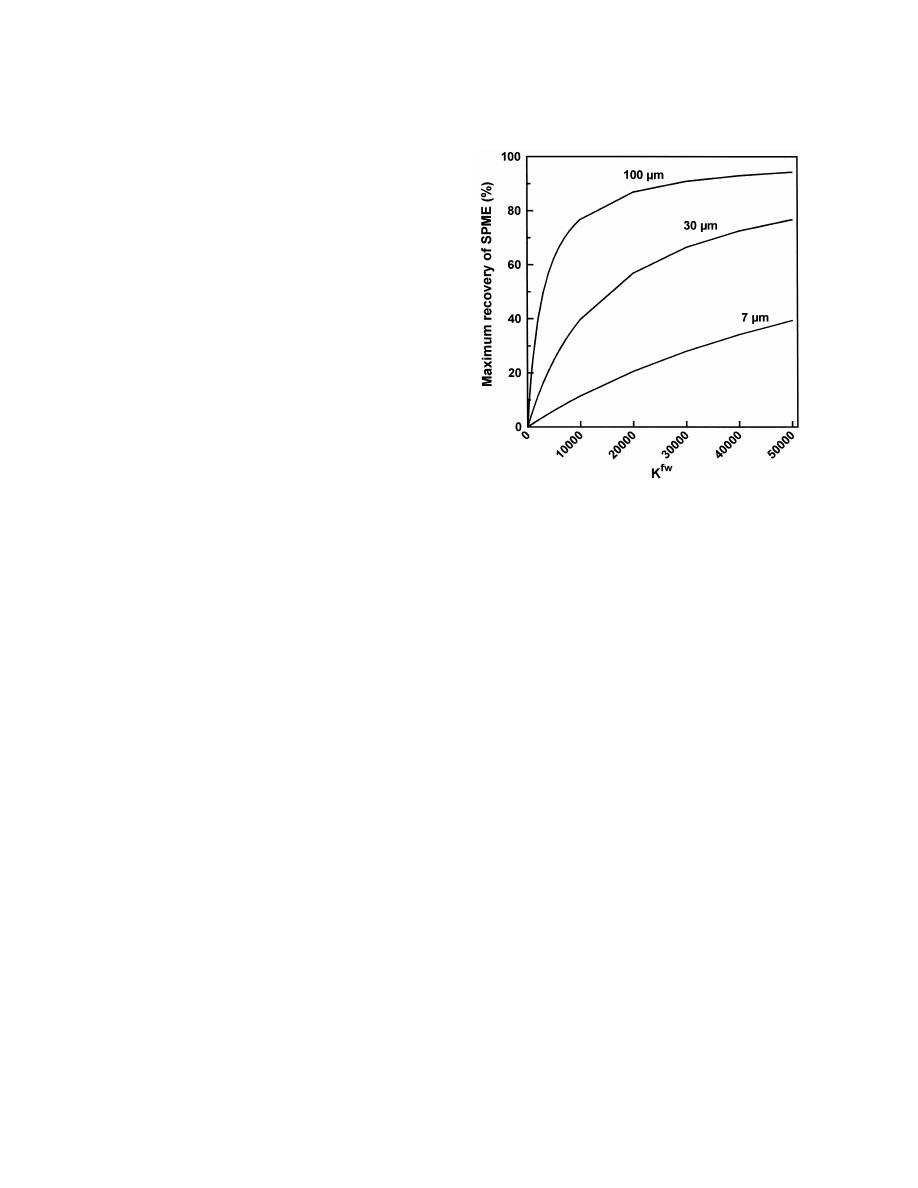
Fig. 2. Dependence of maximum recovery by SPME on K
c
according to Eq. (4) for three fibers with a length of 1 cm and
f
fw
]
K
5
(1)
25
24
coatings of 7 mm (V 52.6?10
ml), 30 mm (V 51.3?10
ml)
c
f
f
w
24
and 100 mm (V 56.6?10
ml) with V 52 ml.
f
w
fw
where K
is the equilibrium constant of liquid–
liquid equilibrium, c
is the equilibrium concen-
f
tration of the analyte in the coating and c
is the
w
(Eq. (4)). Thus, it is also evident that SPME will
equilibrium concentration of the analyte in the
mainly have a low or very low recovery (Fig. 2)
aqueous matrix. Eq. (1) can also be written as:
fw
because K
is in the range of 100 to 10 000 for
fw
fw
n V
many analytes, e.g., K
(benzene)5125, K
( p-
f w
fw
]]
K
5
(2)
fw
fw
n V
xylene)5831 [44], K
(clozapine)5226 and K
w f
fw
(loxapine)52671 [45]. The values K
of polychlori-
and because n 5n 1n
a rearrangement is possible
0
f
w
nated biphenyls (PCBs) were found between 250 and
to:
11 000 [46]. Octanol–water partitioning coefficients
ow
fw
fw
(K
) can be a good estimate of K , however, this
K V n
f
0
]]]]
n 5
(3)
f
fw
has to be confirmed for a special group of sub-
K V 1 V
s
d
f
w
fw
ow
stances. The K
of PCBs did not correlate with K
where n is the number of molecules in the fiber in
[46].
f
equilibrium, n
is the number of molecules in the
w
aqueous phase in equilibrium, n is the number of
0
fw
n
K V
molecules in the aqueous phase prior to SPME, V is
f
f
w
]
]]]]
Maximum recovery 5
5
(4)
fw
n
the volume of aqueous phase and V is the volume of
K V 1 V
s
d
0
f
f
w
the coating. It is evident from Eq. (3) that the basis
fw
The values of K
are influenced by temperature,
for a quantitative method is given because of the
salt, pH and organic solvents. The dependence of
linear relationship between n
and n . However,
f
0
fw
K
on temperature is expressed by Eq. (5) where
SPME is an equilibrium extraction but not an
fw
fw
exhaustive extraction. A simple rearrangement of Eq.
K
is the equilibrium constant at T and DG
is the
0
0
(3) gives an expression for the recovery of SPME in
free enthalpy of the transfer of analyte between the
equilibrium which is also the maximum recovery
two phases:
172
S
. Ulrich / J. Chromatogr. A 902 (2000) 167 –194
fw
2 DG
1
1
fw
fw
]]]
]
]
K
5 K
exp
?
2
(5)
S
D
0
R
T
T
0
fw
f
w
DG
5 G 2 G
(6)
fw
fw
K
2 DG
1
1
]
]]]
]
]
ln
5
?
2
(7)
S
D
fw
R
T
T
K
0
0
Because of the interference of organic molecules
with the intermolecular interactions of water the free
w
enthalpy in water (G ) is always higher than in
f
fw
PDMS (G ). Thus, according to Eq. (6) DG
should
be negative except for, perhaps, rare cases with a
fw
high entropy term. It can be concluded that K
decreases with increased temperature and, therefore,
also the amount of analyte extracted and the recovery
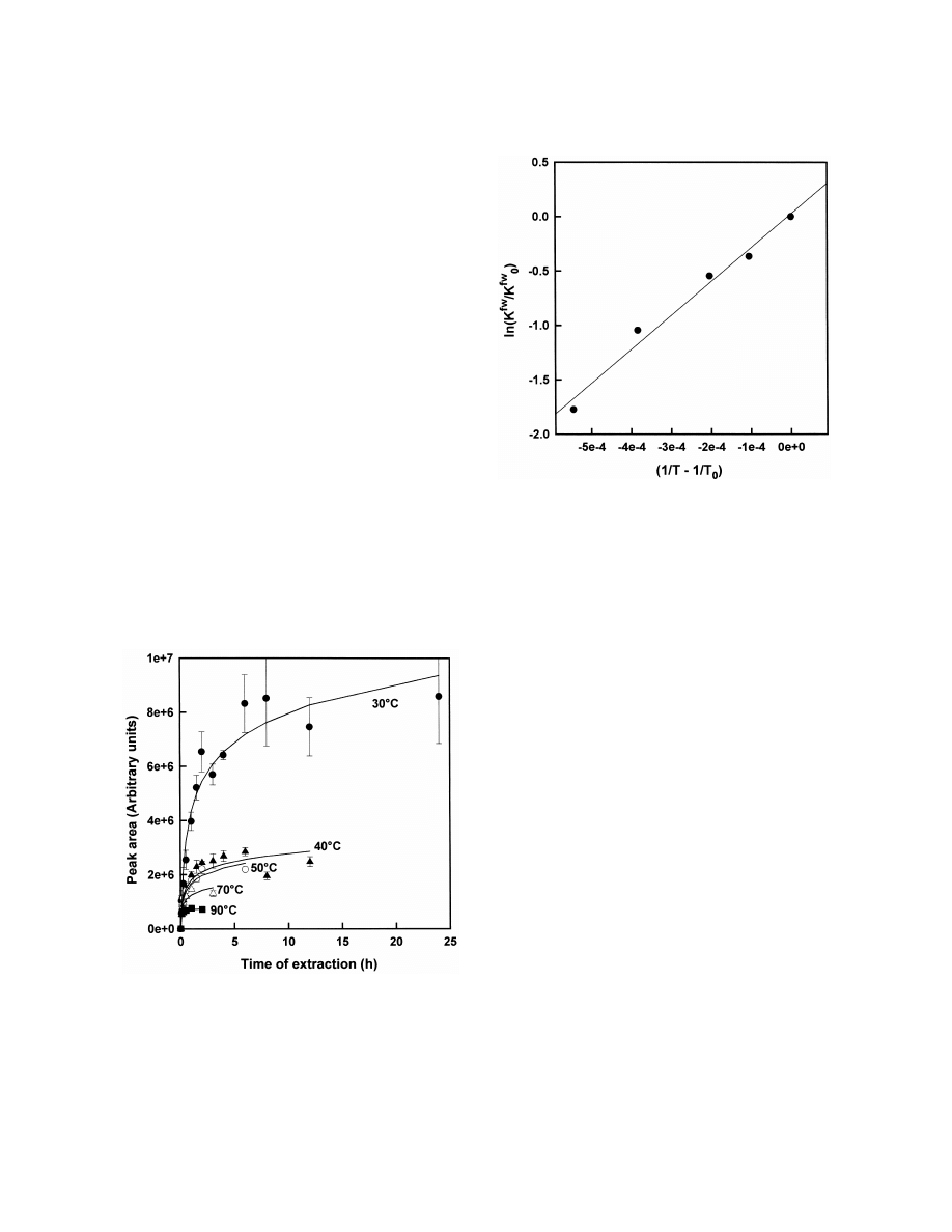
of SPME decrease. This is shown for the antipsy-
chotic drug clozapine in Fig. 3. According to Eq. (7)
fw
which can be received after rearrangement of Eq. (5)
Fig. 4. Linear relationship of K
and temperature according to
fw
Eq. (7) for the SPME of clozapine by a 100-mm PDMS fiber,
a linear relationship was found (Fig. 4) and DG
5
c 5500 ng / ml, V 51.5 ml, 100-mm PDMS fiber, pH 12.
0
w
225.9 kJ / mol was calculated [47]. The relationship
fw
between K
and concentration of salt (c ) can be
s
fw
fw
expressed with Eq. (8) where K
is K
at c 50
amount of analyte extracted [49]. However, this was
0
s
and k is a specific constant [48]. The higher the
not always confirmed in real samples [50,51]. The
s
fw
fw
concentration of salt the higher is K
and the
relationship between K
and pH can be described
fw
with Eq. (9) if only the acid is extracted where K
0
fw
is K
of the undissociated form. This was confirmed
for short-chain fatty acids [52]. The analyte is better
extracted at low pH. Eq. (10) can be used if only the
basic form is extracted. The analyte can be better
extracted at high pH. Finally, the presence of an
organic solvent in the aqueous sample usually de-
fw
creases K
[53]:
fw
K
]
ln
5 k c
(8)
fw
s
s
K
0
fw
K
0
]
log
2 1 5 pH 2 pK
(9)
S
D
fw
a
K
fw
K
0
]
log
2 1 5 pH 1 pK 2 14
(10)
S
D
fw
a
K
In HS-SPME Eq. (3) is extended to Eq. (13)
hw
where K
is the equilibrium constant of HS and
fh
aqueous sample (Eq. (11)), K
is the equilibrium
Fig. 3. SPME of clozapine in aqueous solution at various tem-
constant of fiber and HS (Eq. (12)), c
is the
h
peratures (filled circles 308C, filled triangles 408C, circles 508C,
equilibrium concentration of the analyte in HS and V
triangles 708C, filled squares 908C), c 5500 ng / ml, V 51.5 ml,
h
0
w
100-mm PDMS fiber, pH 12.
is the volume of HS:

S
. Ulrich / J. Chromatogr. A 902 (2000) 167 –194
173
3.2. Kinetics
c
h
hw
]
K
5
(11)
c
w
The relationship of the SPME with time as shown
c
f
in Fig. 3, for example, was mathematically described
fh
]
K 5
(12)
c
in a model which used several prerequisites with
h
regard to geometry, size of sample and access of
fh
hw
K K
V n
f
0
analyte molecules to the fiber [42,44]. If all analyte
]]]]]]]
n 5
(13)
f
fh
hw
hw
K K
V 1 K
V 1 V
s
d
molecules have access to the coating, i.e., the
f
h
w
perfectly agitated model, the time to equilibrium (t )
hw
fh
e
K
and K
can be calculated with the Henry’s
is given by Eq. (18) with r the outer radius of the
o
Law constants of the analyte in water (H ) and in the
w
coating, r the inner radius of the coating and D the
i
f
coating (H ), respectively (Eqs. (14a) and (14b)). The
f
diffusion coefficient of the analyte in the coating.
vapor pressures in aqueous sample ( p ) and coating
w
Taking into account the experimental error it can be
( p ) are given in Eqs. (15a) and (15b):
f
assumed that t is reached when 95% (t
) of the
e
95%
H
maximal amount was extracted. Otherwise, the theo-
w
hw
]
K
5
(14a)
retical t is infinitely long according to the model
RT
e
used:
RT
fh
]
K 5
(14b)
2
H
f
(r 2 r )
o
i
]]]
t 5 t
5
(18)
e
95%
2D
p 5 H c
(15a)
f
w
w
w
Not all analyte molecules have simultaneous ac-
p 5 H c
(15b)
f
f
f
cess to the coating in a more real approach. This is
described in a model using a hypothetical boundary
Eq. (16) and an alternative expression for the
layer of radius d with no agitation. Perfect agitation
amount extracted (Eq. (17)) can be derived from
occurs only in the sample outside the boundary layer.
Eqs. (1), (14a), (14b), (15a) and (15b) because the
The radius d of this static layer depends on the rate
equation p 5p is valid in equilibrium. A similar
w
f
of agitation. The higher the rate of agitation the
rearrangement as shown in Eqs. (3) and (4) provides
lower is d and vice versa. The time to maximal
the recovery of HS-SPME (Eq. (17a)). Accordingly,
extraction can be calculated with Eq. (19) where D
w
the recovery of HS-SPME should be lower than that
is the diffusion coefficient of the analyte in water:
of direct SMPE (Eq. (17b)):
fw
H
dK (r 2 r )
w
fw
hw
fh
o
i
]
K
5
5 K
K
(16)
]]]]
t 5 t
5 3 ?
(19)
e
95%
H
D
f
w
fw
K V n
It is concluded that the time of extraction is
f
0
]]]]]]
n 5
(17)
fw
f
fw
hw
increased with increased K , a higher fiber thickness
K V 1 K
V 1 V
s
d
f
h
w
(r 2r ) and lower diffusion coefficients of the
o
i
n
f
analyte molecule in the sample (D ). The time of
]
Maximum recovery (HS-SPME) 5
w
n
0
extraction may be decreased with an improved
fw
agitation method, thus by decreasing d. In the case of
K V
f
]]]]]]
5
(17a)
fw
hw
perfect agitation the minimal time of extraction is
K V 1 K
V 1 V
s
d
f
h
w
reached and t only depends on the geometry of the
e
Maximum recovery (HS-SPME)
fiber and the analyte’s diffusion coefficient in the
]]]]]]]]]]
Maximum recovery (direct SPME)
fiber (Eq. (18)). However, it is emphasized that
equilibrium is not a prerequisite for a quantitative
1
]]]]]
5
(17b)
method. The time of extraction t is independent of
hw
e
K
V
h
the concentration of analyte in the sample. The
]]]
1 1
fw
K V 1 V
f
w
relative number of molecules extracted at a distinct

174
S
. Ulrich / J. Chromatogr. A 902 (2000) 167 –194
t
time (n /n ) is also independent of the concentration
and quantitative analysis in plasma arise from (1)
f
f
of analyte. Finally, the absolute number of molecules
problems of selectivity because of interferences of
t
extracted at a distinct time (n ) is linearly propor-
endogenous substances and (2) problems of quantita-
f
tional to the concentration of analyte [44].
tion because of binding of the analyte with biopoly-
In HS-SPME Eq. (18) is also valid for the
mers. A short discussion of the impairment of
estimation of t if the aqueous phase and the HS are
quantitation by protein binding of the analyte is
e
perfectly agitated. Several variables have to be taken
presented.
into account for the estimation of t in the case of
The binding of the target analyte, a drug for
e
practical agitation (Eq. (20)): thickness of coating,
example, to proteins can be described by a chemical
HS and aqueous phase (L , L and L , respectively),
equilibrium reaction as shown in Eqs. (21) and (22),
f
h
w
revolution rate of the stir bar (N ), radius of the stir
where c
is the free concentration of drug in plasma
w
bar (R), D
and diffusion coefficients of analyte in
water in equilibrium, c
is the concentration of
w
pr
hw
fw
HS (D ) as well as K
and K . A simple model
binding sites of protein in equilibrium, c
is the
h
b
pr
was applied with the assumptions of only one-dimen-
concentration of bound drug in equilibrium and K
sional diffusion and R only slightly smaller than the
is the equilibrium constant:
radius of the vial [42]:
pr
K
c 1 c ác
(21)
w
pr
b
t 5 t
e
95%
c
b
pr
]]
L
K 5
(22)
h
c c
]]]]]]]
5 1.8 ?
w
pr
S
hw
25
2
K
? (D 1 2 ? 10
NR )
h
0
Eq. (23) can be derived with c
the concentration
pr
L
w
fw
of binding sites prior to equilibrium. Eq. (24) is
]]]]]]
1
? K L
(20)
2
D
f
0
1.6 ? (D 1 0.03NR )
w
obtained with the assumption c , ,c , which
b
pr
should be valid for trace analysis. Substitution of
3.3. Solid-phase microextraction in biological
concentrations yields Eq. (25) where n
is the
b
fluids
amount of bound drug, n is the amount of free drug
w
0
and n
is the amount of binding sites of protein (in
pr
The analysis of biological fluids is hampered by
moles):
the presence of dissolved biopolymers. For example,
c
b
pr
human plasma consists of about 7 to 8% of proteins.
]]]]
K 5
(23)
0
c ? (c 2 c )
w
pr
b
The main portion is albumin (about 55%). Immuno-
globulins account for about 20% and lipoproteins for
c
b
pr
]]
K 5
(24)
about 11% of proteins. Serum is formed from
0
c c
w
pr
nonstabilized plasma after coagulation. Thus, fibrino-
gen (about 3.5% of plasma proteins) is not present in
n V
b w
pr
]]
K 5
(25)
0
serum. Other components are triglycerides and elec-
n n
w
pr
trolytes as well as a huge number of trace com-
ponents such as hormones, transmitters and metabo-
With Eq. (26) and introduction of Eq. (2) an
lites. The composition of plasma can be subject to
expression is obtained for the amount of analyte
considerable differences due to pathological and
extracted by SPME (n ) in the ternary system fiber–
f
nonpathological influences. For example, plasma
plasma water–protein (Eq. (27)). A considerably
albumin can be decreased to about 50% of normal in
more complex result was described without the
hepatic diseases and the concentration of tri-
assumption made in Eqs. (23) and (24) [54]. The
glycerides depends on dietary status. However, in a
main problem of analysis by SPME in matrices
more general view plasma can also be considered as
containing protein can be concluded from Eq. (27),
a relatively fixed and well-described matrix in com-
i.e., a decrease of sensitivity. The factor of decrease
parison to real samples in some areas of environmen-
of sensitivity ( f
) can by calculated by the combi-
sens
tal analysis, for example. Problems of the qualitative
nation of Eqs. (3) and (27), where n is the amount
f

S
. Ulrich / J. Chromatogr. A 902 (2000) 167 –194
175
9
extracted in the absence of proteins and n
is in
equilibrium extraction but not an exhaustive ex-
f
presence of proteins (Eq. (28)). Accordingly, the
traction.
However,
the
experimental
conditions
sensitivity of SPME is decreased for a high capacity
needed imply a very low recovery and sensitivity of
pr
0
of protein binding, i.e., high K n . In addition, the
SPME and, therefore, this approach may be limited
pr
sensitivity of SPME may be decreased in the pres-
to selected problems. This was first shown and
ence of proteins if the coating is changed by the
experimentally confirmed for the SPME of organic
fw
irreversible adsorption of proteins, i.e., lower K
pollutants in waste water which was enriched with
due to protein fouling:
DOM [55,56]. The free concentration of analytes
(c ) was analyzed directly by external calibration as
w
n 5 n 1 n 1 n
(26)
0
f
w
b
discussed above. The total concentration (c 1c )
w
b
fw
was analyzed by internal calibration with isotopically
K V n
f
0
]]]]]]
n 5
(27)
f
fw
pr
0
labeled spikes. The total concentration can also be
K V 1 K n 1 V
f
pr
w
assessed by LLE, for example. Thus, the portion of
pr
0
freely dissolved analyte x 5c /(c 1c ) and the
w
w
w
b
K n
n
pr
f
pr
0
]
]]]
f
5
5 1 1
(28)
product K n
are available. The knowledge of x of
sens
fw
pr
w
9
n
K V 1 V
f
f
w
drugs is important in pharmacology, for example,
because only the free concentration is the pharmaco-
The amount of analyte in plasma water is given by
logically active portion in plasma.
9
Eq. (29) (n , with SPME) and by Eq. (30) (n ,
w
w
If the matrix is diluted by a dilution factor D 5
without SPME):
0
0
n
/n
(D 50 to 1) and with x 5c /(c 1c ) Eq.
pr,D
pr
w
w
w
b
0
n V
0 w
(33) can be derived where n
and n
are the
f,D
pr,D
]]]]]]
9
n 5
(29)
w
fw
pr
0
K V 1 K n 1 V
amount of analyte extracted and the amount of
f
pr
w
binding sites after dilution, respectively:
n V
0 w
]]]]
n 5
(30)
w
pr
0
n
V
V
1
K n 1 V
0
w
w
pr
w
]
]]
]]
]
5 1 1
1 D
?
2 1
(33)
S
D
fw
fw
n
x
K V
K V
f,D
w
w
f
9
The ratio of n and n provides a criterion for the
w
w
interference of SPME with the equilibrium between
Eq. (34) is obtained after rearrangement of Eq. (3)
fw
bound and free analyte (Eq. (31)). If the experimen-
with an expression for the ratio V /K V in buffer
w
f
tal conditions are chosen according to Eq. (32) the
solution and introduction in Eq. (33). Thus, x
can
w
interference of SPME with the equilibrium between
be calculated from a linear plot according to Eq.
bound and free analyte can be neglected because the
(34). If no linear relationship is found the assump-
0
amount in plasma water is changed by less than
tions made in the model are not valid, i.e., c , ,c ,
b
pr
10%. Thus, the free concentration of analyte can be
preformed binding sites, linear dependence of num-
measured:
ber of binding sites on the concentration of proteins,
fw
only one type of binding sites of proteins:
n
K V
w
f
]
]]]]
5 1 1
(31)
pr
0
Plasma
n
9
n
0
K n 1 V
w
pr
w
] 2 1
S
D
n
1
f,D
fw
]]]]]
]
5 1 1
2 1 ? D
(34)
S
D
K V
Buffer
n
f
x
0
w
]]]] , 0.111
(32)
] 2 1
pr
0
S
D
n
K n 1 V
f
pr
w
This is a unique advantage of SPME over other
In contrast to Eq. (21) an alternative approach is
sample preparation methods. A direct assay of free
possible. The protein is regarded as a third phase and
concentration can be performed without the sepa-
binding of analyte is regarded as an extraction but
ration of phases. This is possible because the binding
not a chemical reaction. Thus, Eqs. (35) and (36) are
pr9
of analyte to proteins is not impaired by the SPME,
used for further calculations where K
is the
i.e., no loosening occurs of the protein–analyte
equilibrium constant of extraction between the aque-
bonding as in LLE, and because SPME is an
ous phase and the protein phase and c
is the
b9

176
S
. Ulrich / J. Chromatogr. A 902 (2000) 167 –194
concentration of analyte in the protein phase. The
1998. A survey of these methods and approaches is
results of this approach are similar to the equations
presented. Some methods are presented in more
presented above:
detail to provide the reader with deeper insight into
the practice of SPME and to compare different
pr9
K
c á c
(35)
approaches.
w
b9
c
b9
pr9
]
4.1. Direct solid-phase microextraction
K
5
(36)
c
w
The vapor pressure of many important analytes is
Apart from the changes of equilibrium, the ex-
low because of a molecular mass between 150 to 450
traction profile of SPME is influenced by proteins or
g / mol and the presence of hydrophilic groups in
other DOM in the sample, too. This can be explained
their molecule. Thus, according to Eqs. (13), (14a),
if the equilibrium between free and bound analyte of
(15a) and (20) the concentration of the analyte in the
Eq. (21) is written kinetically with k
the rate
b
HS is low and the transfer to the fiber is slow at
constant of association with protein and k
the rate
2b
ambient temperature. The application of increased
constant of the dissociation of the protein–analyte
temperatures appears to be problematic because of
binding (Eqs. (37) and (38)). The rapidity of ex-
denaturation of proteins and decomposition of ana-
traction is determined by the rate of dissociation
lytes. An advantage of low vapor pressure is the
(r
5k
c ) if the rate of dissociation is slower than
2b
2b
b
option of storage of fibers after extraction and prior
the diffusion of analyte from the aqueous phase to
to desorption and GC analysis. Thus, field analysis is
the coating. This may occur for some analytes.
possible without the need of transport of the sample.
Furthermore, the viscosity (h) of plasma and blood
Furthermore, several fibers can be processed simul-
in vitro is about 2- and 4.5-times higher, respective-
taneously in the extraction and analyzed subsequent-
ly, than the viscosity of water. Because the diffusion
ly by GC thereafter as it is well-known in LLE.
coefficients are inversely related to h [D 5f(1 /h)]
Direct SPME was studied in several methods for the
diffusion coefficients of the analyte in the aqueous
assay of drugs and other analytes in plasma and
phase are about 2- and 4.5-times decreased in plasma
urine. Methods without derivatization and methods
and blood, respectively. Thus, t is increased accord-
e
with derivatization were described.
ing to Eq. (19) by factors of about 2 and 4.5 in
plasma and blood, respectively, in comparison to
4.1.1. Without derivatization
water. Finally, the formation of a diffusion barrier by
A method for the assay of eight barbiturates in
polymer molecules is supposed close to the surface
urine was described [57]. A 65-mm Carbowax–di-
of the coating which diminishes the transfer of
vinylbenzene (DVB) fiber was found to have the
analyte into the coating. However, this mechanism is
highest extraction efficiency in comparison with 100-
only little understood. In conclusion, the SPME in
mm PDMS and 85-mm polyacrylate (PA) fibers. The
biomedical samples may be substantially impaired
time of extraction as indicated by t was about 5 to
with respect to sensitivity and rapidity:
e
15 min with agitation by a stir-bar. The Carbowax–
k
b
DVB
coating
stripped
off
the
fused-silica
at
c 1 c á c
(37)
w
pr
b
k
2b
temperatures.2658C during desorption, therefore, a
desorption temperature of 2508C was used. However,
k
b
pr
]
K 5
(38)
a considerable carryover effect was found and a
k
2b
special clean-up procedure of the coating was neces-
sary after the 12-min desorption. For this purpose,
4. Application of solid-phase microextraction in
after each analytical run the fiber was cooled for 3
biomedical analysis
min, exposed to methanol–water (20:80) solution for
3 min and again desorbed in the hot injector for a
The number of published data of the application of
period of 4 min. The carryover was decreased to 2%.
SPME in biomedical analysis has increased since
The GC separation was performed with a PTE-5

S
. Ulrich / J. Chromatogr. A 902 (2000) 167 –194
177
column (30 m30.25 mm I.D., 0.25 mm film thick-
piperidine-1-oxide (TEMPO) and the metabolite
ness) with helium as the carrier gas and a tempera-
2,2,6,6-tetramethylpiperidine
were
measured
in
ture program starting at 608C, a 408C / min ramp to
human cell cultures with thymol as the internal
1108C and a 108C / min ramp to the final temperature
standard [59]. A 100-mm PDMS fiber was used and
of 2508C. Ion-trap mass spectrometric detection (IT-
a time of 5 min was sufficient for extraction (t
¯5
e
MS) in the electron ionization mode (EI) was
min). The recovery from the cell culture was about
applied and selected ions were used for quantitation.
10 to 20% for TEMPO and about 40% for the amine.
2
Calibration was linear (correlation coefficient r 5
Clotting of proteins on the surface of the fiber and
0.990), precision was between 1.4 and 12.0% (rela-
formation of a diffusion barrier was discussed.
tive standard deviation, RSD) and limit of detection
Desorption at 2508C was for 1 min and a HP-5
(LOD) was 1 to 5 ng / ml. The recovery as calculated
capillary (30 m30.25 mm I.D., 0.25 mm film
by Eq. (4) was considerably lower than the value
thickness) was used for the GC separation with
given by the authors (93–104%). It is emphasized
helium as the carrier gas and flame ionization
that some authors currently reporting on SPME
detection (FID). Peak areas increased with increasing
‘‘recovery’’ seem to be using the term interchangeab-
temperatures from about 5 to 258C but decreased at
ly to mean both: absolute recovery as given in Eq.
higher temperatures.
(4) and relative recoveries, for example, the recovery
The analysis of eight antidepressant drugs in
in a complex matrix relative to that from water. It is
human plasma and serum was described by direct
strongly recommended to provide absolute recoveries
SPME with a 100-mm PDMS fiber [54]. Aqueous
for a comparison with other methods. Fibers were
NaOH was added to the plasma and an internal
used for at least 100 extractions.
standard was used as usual also in the LLE of
Chlorophenols were analyzed in urine with a 85-
antidepressants. After 10 min of SPME the fiber was
mm PA fiber [58]. A time t 550 min was found and
successively washed for about 20 s in a 50% aqueous
e
used for the extraction at N 51000 of a magnetic
methanol solution and in water. This step was found
stir-bar. Desorption in the injector port was at 2908C
to be important to prevent burning-in of proteins
for 2 min. The GC separation was performed with a
adsorbed on the surface of the fiber during desorp-
DB-5.625 capillary (30 m30.25 mm I.D., 0.5 mm
tion. After 1 min of desorption at 2608C the GC
film thickness, J&W Scientific, Folsom, CA, USA),
separation was performed with a DB-17 capillary (30
with helium as the carrier gas and a temperature
m30.25 mm I.D., 0.25 mm film thickness) and
program starting at 608C, with a ramp of 308C / min
nitrogen at 0.7 ml / min as the carrier gas. The
to 1908C, a second ramp of 108C / min and a final
temperature program started at 1408C with a steep
temperature of 3108C. EI as well as negative chemi-
ramp of 208C / min to 2208C and a second ramp of
cal ionization (NCI) with selected ion monitoring
only 28C / min to 2708C. Nitrogen–phosphorus selec-
(SIM) MS was used for detection. The LODs were
tive detection (NPD) and MS detection (SIM) were
between 1 and 41 pg / ml. They were lower than with
used. Calibration was linear between 125 and 1000
sample preparation by LLE, however, with full-scan
ng / ml with r from 0.989 to 0.999. Precision was 6.1
fw
MS detection. The values for K
were between 8
to 39.6% at 125 ng / ml and 1.9 to 11.8% at 250
fw
and 212 at pH 6.2. Low pH (pH 1) increased K
by
ng / ml, for example. The limit of quantitation (LOQ)
factors of 1.2 to 9.2. Addition of salt (NaCl, KCl)
was 90 to 200 ng / ml. The assay provided good
fw
increased K , too, but the combined effect of salt
agreement with a standard method which was based
and pH was not better, even poorer than the single
on LLE. The time of SPME applied was chosen
effects. The method was linear over a range of three
because of an attempt to optimize the method with
orders of amounts with r 50.999 and the precision
respect to a minimum time. In fact, equilibrium was
was 5 to 10% (RSD is always used for precision) at a
not reached even after 300 min of extraction. The
concentration of 25 ng / ml. The authors estimated the
method was not sensitive for the assay of antidepres-
SPME method was better than SPE and LLE for the
sant drugs in patients taking therapeutic doses, i.e.,
assay of chlorophenols in urine.
for TDM. The LOQ required for example for ami-
The stable nitroxide radical 2,2,6,6-tetramethyl-
triptyline and its active metabolite nortriptyline
178
S
. Ulrich / J. Chromatogr. A 902 (2000) 167 –194
should be at least 10 to 40 ng / ml because the
binding of antidepressant drugs (90 to 99%) to
therapeutic window is at about 80 to 250 ng / ml for
proteins (Fig. 5).
the sum of both substances and the ratio of nor-
A method for the assay of the antipsychotic drug
triptyline and amitriptyline concentrations is about
clozapine was described by the same authors [45,60].
0.5 to 1.5. Thus, only the assay of increased con-
In contrast to the method for antidepressants the
centrations is possible with this method as usually
plasma was diluted with water 1:7 (v / v) and the time
encountered in intoxications. A case of a suicidal
of extraction was increased to 30 min. Loxapine
intoxication was presented. It was discussed that the
which has a similar chemical structure was chosen as
sensitivity could be considerably improved by in-
the internal standard. Desorption was carried out at
creasing the time of extraction, however, then the
2608C for 1 min. The time of desorption was shown
goal of SPME to present a fast method is abandoned.
to be sufficient in a desorption–postdesorption graph,
It was emphasized that the selection of a well-suited
i.e., no carryover effect was found. A BPX-5 mega-
internal standard is crucial for the direct SPME in
bore capillary (SGE, Weiterstadt, Germany) with the
plasma. Of course, internal standard calibration with
dimensions 30 m30.53 mm I.D. and 1.0 mm film
isotopically labeled spikes would be ideally. The
thickness was used for the separation with nitrogen
chemical structure of the internal standard should be
as the carrier gas (20 ml / min) and a temperature
very similar to the analyte. The internal standard
programme (T 51608C, T 52608C, T 52888C,
1
2
3
used (chloramitriptyline) may not have met this aim
ramp 5408C / min, ramp 548C / min). A linear cali-
1
2
for the secondary amine antidepressants and, there-
bration curve was found for the peak-area ratio of
fore, the poor precision of some antidepressants can
clozapine and loxapine from 100 to 1000 ng / ml of
be explained. Finally, it was shown that the peak
clozapine (r 50.987). The within-day precision was
area increased with decreased concentration of pro-
between 7.9 and 14.5% at concentrations of 100 to
teins if the concentration of analytes was held
1000 ng / ml. The between-day precision was 7.9 to
constant. This was explained by the considerable
12.7% at 200 to 1000 ng / ml. The between-day
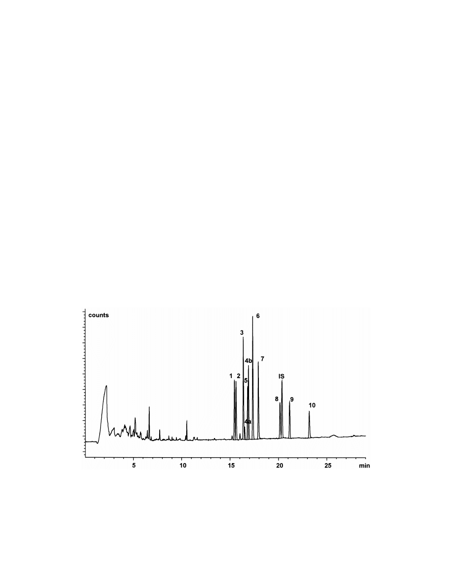
Fig. 5. Typical SPME–GC–NPD chromatogram of antidepressant drugs and metabolites in human plasma [15amitriptyline, 25
trimipramine, 35imipramine, 4a5cis-doxepin, 4b5trans-doxepin, 55nortriptylin, 65mianserine, 75desipramine, 85maprotiline, IS5
internal standard (chloramitriptyline), 95clomipramine, 105desmethylclomipramine, 375 ng / ml each, 30 min time of extraction].
S
. Ulrich / J. Chromatogr. A 902 (2000) 167 –194
179
precision of 22% at 100 ng / ml was an indication of
was used to estimate the protein binding of the local
the LOQ. The LOD was 30 ng / ml. The method was
anesthetic drug lidocaine in plasma. However, the
compared with two standard methods [three-step
pH 9.5 indicates nonphysiological conditions [68].
LLE–GC–NPD and on-line-SPE–LC–ultraviolet de-
tection (UV)] and good agreement was demonstra-
4.1.2. With derivatization
ted. Thus, the method may be applied in TDM
As in the case of LLE and SPE derivatization can
because the therapeutic window of clozapine is 350
be used also in SPME for the chemical transforma-
to 600 ng / ml. As in the case of antidepressants the
tion of the analyte into a more suitable form for GC,
LOD may be improved by increasing the extraction
i.e., polar groups should be eliminated or masked.
time. Maximal peak areas were obtained after about
Derivatization can be performed in situ or after
12 h in plasma and 2 h in water only. Increased
transfer of the analyte into the coating. The second
concentration of triglycerides decreased the peak
approach, however, is more time-consuming than
areas of clozapine and loxapine, however, the effect
simply adding the derivatization agent to the sample
was negligible for the peak-area ratio. This also
because, in fact, a second extraction is needed.
applied for the effect of salt, however, in contrast to
Therefore, in situ derivatization may be preferred in
theoretical expectation the peak areas remained
SPME. For this purpose, only a limited number of
constant over a wide range of salt concentration and
agents can be used because many derivatization
even decreased at high concentrations of salt.
agents are unstable in aqueous matrix.
Methadone and amphetamines were analyzed in
Benzodiazepines in urine were analyzed after acid
urine with a 100-mm PDMS fiber and extraction for
hydrolysis of glucuronides for 30 min at a tempera-
20 min at a temperature of 408C. The desorption
ture of 1008C. An 85-mm PA fiber was found
time was also 20 min at a temperature of 2508C. A
superior to a PDMS fiber for some benzodiazepines.
three-ramp temperature program with an initial tem-
The conditions of GC separation were as described
perature of 708C and final temperature of 3008C was
above. However, no more data were presented [61].
used. Helium was the carrier gas, a HP-5 capillary
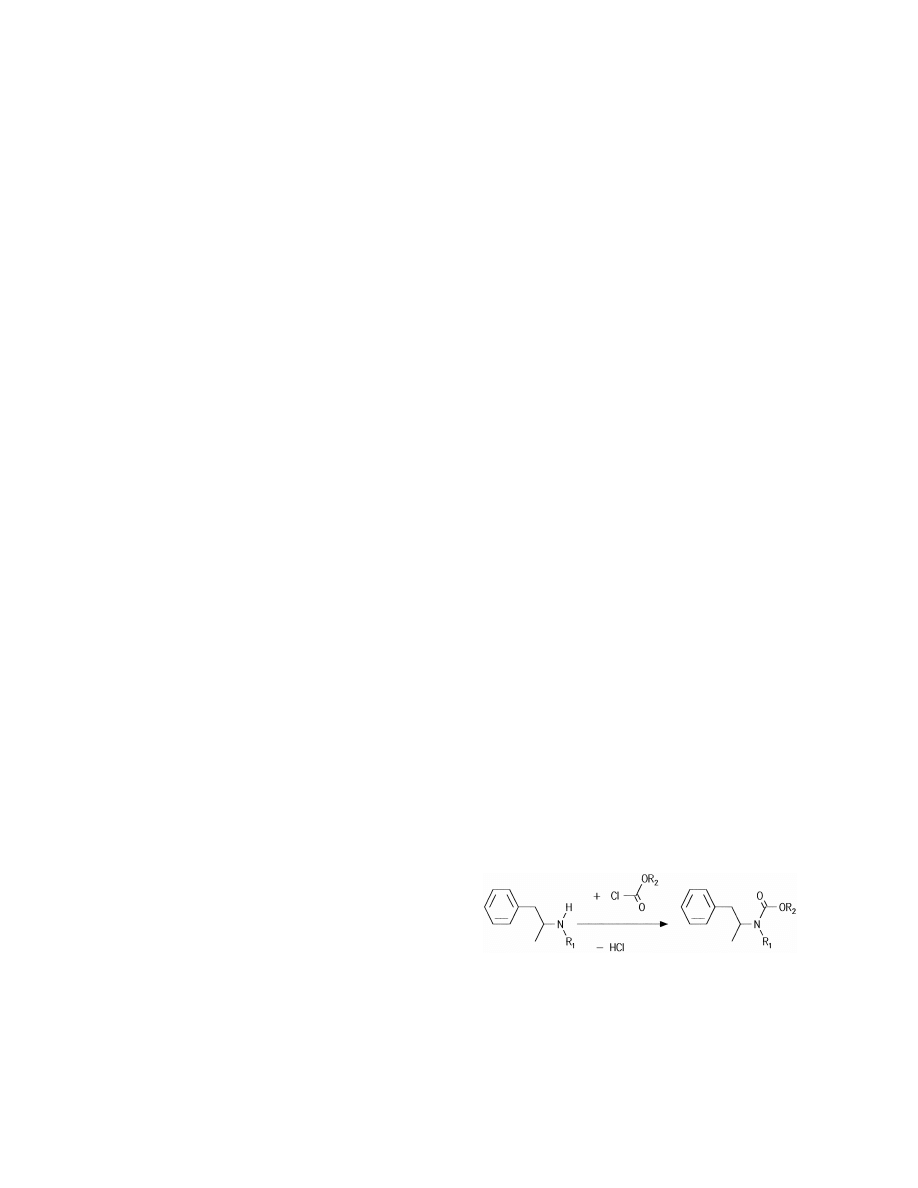
Amphetamine and methamphetamine were ana-
(30 m30.32 mm I.D., 0.33 mm film thickness) was
lyzed in urine by direct SPME with a 100-mm PDMS
used for separation and MS (SIM) for detection.
fiber after in situ derivatization with methyl-, propyl-
Calibration of methadone was linear between 10 and
and butylchloroformate at pH 10.8 for 1 min (Fig.
100 ng / ml but nonlinear at higher concentrations.
6). Methoxyphenamine was used as internal stan-
Precision was between 3 and 6%. It was claimed that
dard. Water–stable carbamates were formed during
the recovery of SPME of methadone was higher than
the reaction. The SPME of carbamates was found to
of LLE with dichloromethane–isopropanol (4:1, v / v)
be complete after 14 min. The desorption needed 1
[61].
min at 3008C. GC separation and detection were
Pethidine and methadone were analyzed in human
performed with a SPB-1 capillary (30 m30.25 mm
urine by SPME–GC–NPD with LODs below 1 ng /
I.D., 0.25 mm film thickness), with helium (1 ml /
ml [62]. A deuterated internal standard was used for
min) as carrier gas and NPD. The temperature
the assay of methadone and the main metabolite
program was 1808C for the initial temperature and a
2-ethylene-1,5-dimethyl-3,3-diphenylpyrrolidine (E-
ramp of 208C / min to a temperature of 3008C. A
DDP) in saliva [63]. Methadone and EDDP in hair
and in plasma were assayed by SPME–GC–MS
[64,65]. An interesting approach is the degradation
of proteins by hydrolases prior to SPME [64]. PCBs
in human blood were analyzed by SPME–GC–elec-
tron-capture detection (ECD). Precision was con-
siderably improved by enzymatic proteolysis [66].
Cannabinoids in hair were analyzed by SPME–GC–
Fig. 6. In situ derivatization of amphetamine (R 5H) and
1
MS and enzymatic proteolysis was also tested [67].
methamphetamine (R 5methyl) with alkylchlorformates (R 5
1
2
A similar approach as shown in Eqs. (33) and (34)
methyl, propyl, butyl) for the assay in urine by SPME.
180
S
. Ulrich / J. Chromatogr. A 902 (2000) 167 –194
SPME autosampler (Varian 8200 CX; Varian, Walnut
Creek, CA, USA) was used and one sample needed
15 min for analysis. Alternatively, GC–MS analysis
with a HP-1 capillary (12 m30.2 mm I.D., 0.33 mm
film thickness) was applied. The calibration was
linear (r 50.999) with an LOD of 50 ng / ml. Preci-
sion was 2.1 to 20.3%. The recovery was 2 to 7%.
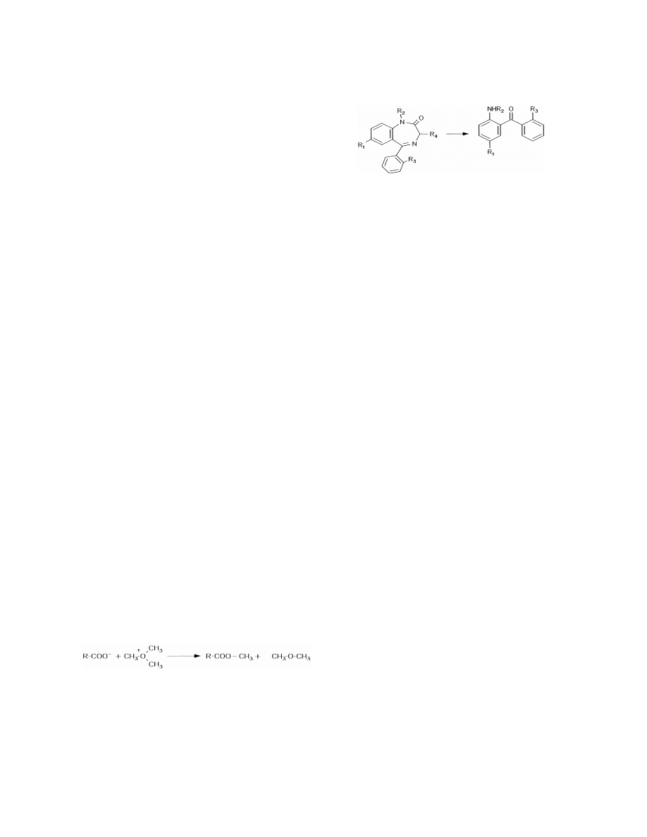
Fig. 8. In situ derivatization of benzodiazepines with formation of
benzophenones for the assay of benzodiazepines in urine.
The PDMS fiber was found to be more efficient and
robust than PA, PDMS–DVB and Carbowax–DVB.
The fiber had to be replaced by a new fiber after 100
analyses. The authors concluded that the method was
sufficient for bioanalysis [69].
however, the PDMS fiber was selected for further
Derivatization with trimethyloxonium tetrafluoro-
method evaluation because of a lower extraction of
borate and SPME with an 85-mm PA fiber for 20 min
interfering substances. This was explained by the
of the resulting methyl esters (Fig. 7) was described
lower affinity of PDMS to polar endogenous sub-
for the analysis of 29 organic acids in urine [70]. The
stances in urine. The time of derivatization was 40
fiber was conditioned for 2 h at 3008C in the
min and after neutralization and cooling to ambient
injection port of the gas chromatograph to get no
temperature the SPME was conducted for 30 min.
peaks in the blank analysis. This procedure was
Maximum peak areas were found after 20 to 40 min
repeated for 5 min after every analysis to avoid
of SPME. The recovery of each benzophenone was
carryover effects. Desorption was performed at
not affected by the pH of SPME in a range of pH
2808C for 4 min. The GC separation was completed
7.7–10.4. Desorption was performed at a tempera-
with a capillary (25 m30.25 mm I.D., film thickness
ture of 2708C for 1 min. A DB-17 capillary (30
not given) and an OV-1701 coating. FID and MS
m30.32 mm I.D., 0.25 mm film thickness) was used
were used alternatively for detection. No validation
with helium as the carrier gas. The temperature
data of the method were presented and, indeed, the
program was similar to other methods presented
derivatization was rather complicated because five
above and ECD was used. Calibrations were linear in
steps of successively adding the derivatization agent
two separate ranges of 10 to 100 ng / ml and 50 to
and sodium hydrogencarbonate for neutralization
500 ng / ml with values of r from 0.981 to 0.998.
were needed at a temperature of 1008C.
LODs were between 2 and 80 ng / ml. The within-day
A well-known derivatization method for benzo-
precision was from 2.1 to 14% and the between-day
diazepines was adapted to SPME (Fig. 8). Thus, a
precision was from 4.2 to 17%. The recoveries
method was described for the assay of 10 benzo-
ranged from 1 to 25%. This was lower than the
diazepines in urine by acid hydrolysis to the corre-
recoveries of a LLE standard method. However, the
sponding benzophenones with 8 M HCl at a tempera-
authors discussed that the amount of analyte on
ture of 1008C and with direct SPME of the ben-
column is higher in SPME than in LLE because only
zophenone derivatives [71]. A 100-mm PDMS fiber
1 ml of 100 ml of the LLE extract was injected to the
and an 85-mm PA fiber were tested. Both coatings
chromatograph. The transfer ratio is 100% in SPME.
gave nearly the same recovery for benzodiazepines,
Finally, it should be taken into account that the
derivatization method via benzophenones is not
selective for all benzodiazepines, i.e., some benzo-
diazepines form the same benzophenone.
SPME–GC–IT-MS after derivatization with hex-
ylchloroformate was used for the assay of benzoylec-
gonine in urine with an LOD of 30 ng / ml, linear
2
calibration between 100 ng / ml and 20 mg / ml (r 5
Fig. 7. In situ derivatization of organic acids with trimethylox-
onium tetrafluoroborate for the assay in urine by SPME.
0.999) as well as precision below 9% [72].

S
. Ulrich / J. Chromatogr. A 902 (2000) 167 –194
181
Thioglycol methylate derivatization with SPME–
The LOD was 1 mg / ml and precision was 1.3 to 5%
GC–IT-MS was used for the assay of arsenic species
[76].
in human urine [73].
Lidocaine was analyzed in human plasma after
protein precipitation with trichloroacetic acid. The
4.1.3. Other methods
calibration was linear in a range of 25 to 2000 ng / ml
A solvent-modified SPME for the assay of
(r 50.998) with an LOD of 5 ng / ml [68].
diazepam in plasma was described [74]. Thus, a
100-mm PDMS fiber and an 85-mm PA fiber were
4.2. Headspace solid-phase microextraction
soaked in 1-octanol and 2-octanone for 2 min and
these modified coatings were used for SPME instead
The outstanding advantage of HS-SPME in bio-
of the original coatings. Plasma was pretreated by
medical analysis is the prevention of direct contact of
adding methanol and precipitation of proteins with
the fiber with the sample and, therefore, prevention
trichloroacetic acid. The t was lower than 10 min in
of contamination of the surface of the fiber with
e
buffer solution and in the pretreated plasma. The
organic polymers. No diffusion barrier of clotted
enrichment with the solvent modified fibers was
proteins is formed, no burning-in of adsorbed or-
about two- to three-times improved in comparison
ganic material is possible during desorption in the
fw
with the original coatings. PA was superior to
hot injector, the risk of decreased K
due to changes
PDMS. Desorption was at a temperature of 3008C
of the coating is decreased and the life-time of fibers
for 1 min. A DB-1 capillary (30 m30.2 mm I.D.,
is considerably increased. The advantages of SPME
0.25 mm film thickness) was used for GC with either
can be completely and easily exploited in HS-SPME.
FID or NPD. The LOD was 30 ng / ml and the
The enrichment of analyte from the HS by SPME is
precision was between 3.2 and 6.5%. The method
unique in comparison to other HS sample preparation
was extended to other benzodiazepines, however, it
methods. It is considerably simpler than purge-and-
was recognized that the sensitivity was insufficient
trap techniques with cryofocusing of HS, for exam-
for low-dose benzodiazepines such as flunitrazepam
ple. It should be kept in mind that no enrichment
[75].
takes place in the sampling from the HS by gas-tight
Automated equilibrium dialysis was applied as a
syringes. On the other hand, HS-SPME is limited to
sample pretreatment for the assay of the free con-
special analytes because of the requirement of a high
centration of valproic acid in plasma with caprylic
vapor pressure of the analyte. Furthermore, the
acid as the internal standard. A 100-mm PDMS fiber
transfer of fibers to the gas chromatograph and
was used and a time of extraction of 3 min was
desorption should be performed immediately after
sufficient although equilibrium was not reached. No
extraction because of the high vapor pressure of
extraction occurred at pH 7.4, a partial extraction
analytes also in the coating and the risk of loss of
was found at pH 5 and optimum extraction was at
analytes during storage of the loaded fiber.
pH 2.5, i.e., below the pK of valproic acid of 5.0.
a
Thus, the results for other organic acids and the
4.2.1. Without derivatization
theoretical description of the influence of pH (Eq.
A method for the assay of inhalation anesthetics,
(9)) were confirmed [52]. The recovery of SPME
i.e., nitrous oxide, isoflurane and halothane, in
was about 4%. The analyte and internal standard
human urine was developed for the investigation of
were desorbed at a temperature of 2108C for 1 min.
occupational exposure of operating room personnel
Capillary GC–FID was used with a Nukol column
[77]. A 75-mm Carboxen–PDMS fiber and a 50 / 30-
(30 m30.2 mm I.D., 0.25 mm film thickness,
mm DVB–Carboxen–PDMS fiber were applied for
Supelco) with a temperature program beginning with
15 to 20 min at a distance of 2 cm above the
608C, a first ramp of 308C / min to 1508C and a
solution. Equilibrium was reached within this time.
second ramp of 108C / min to 1908C. The calibration
The recoveries were 0.3% for nitrous oxide, 20 to
of peak-area ratios of analyte and internal standard
60% for isoflurane and 30 to 80% for halothane.
was linear between 2 and 20 mg / ml with r 50.999.
Desorption was carried out at a temperature of 2408C

182
S
. Ulrich / J. Chromatogr. A 902 (2000) 167 –194
for 16 min. An RT-QPLOT capillary (30 m30.32
17 capillary (30 m30.53 mm I.D., 1 mm film
mm I.D., Restek, Bellafonte, PA, USA), i.e., a
thickness) and with helium as the carrier gas with a
capillary with a DVB porous homopolymer as the
flow of only 4 ml / min. The temperature program
stationary phase, was used for GC analysis with MS
was similar to other methods with an initial lower
detection (SIM). The temperature program was:
temperature of 1008C and a ramp of 108C / min to
408C initial temperature, a first ramp of 308C / min to
2208C. The final temperature was held for 3 min.
1308C and a second ramp of 108C / min to 1808C.
The recovery was 48 to 62%. The calibration accord-
Calibrations were linear with r from 0.994 (nitrous
ing to an internal standard method was linear be-
oxide) to 0.999 (halothane). LODs were 75 pg / ml
tween 0.4 and 15 ng / mg (r 50.998, LOD 0.1 ng /
(nitrous oxide), 15 pg / ml (isoflurane) and 20 pg / ml
mg) for amphetamine and between 4 and 160 ng / mg
(halothane) with the Carboxen–PDMS fiber. Within-
(r 50.999, LOD 0.4 ng / mg) for methamphetamine.
day precision was 3.0 to 7.2% and between-day
Precision was below 5%. The peak-area ratios of
precision was 6.5 to 12.9% (Carboxen–PDMS fiber).
analyte and internal standard were not influenced by
Addition of 10% of salt (NaCl) increased the peak
the extraction time.
areas by about 30%, however, no further increase of
Dinitroaniline herbicides were analyzed by HS-
peak areas was found at higher salt concentrations.
SPME in water, urine and blood. A 100-mm PDMS
The influence of temperature was investigated and
fiber was superior to a 85-mm PA fiber. A time of
analyzed according to Eq. (7). Linear relationships
about 40 min was needed to reach equilibrium, thus,
emerged for isoflurane and halothane with decreased
30 min was chosen as the exposure time. Addition of
fw
K
at increased temperatures. No linear relationship
salt increased peak areas in water and urine, how-
fw
was found for nitrous oxide. Values of DG
¯ 220
ever, salt decreased peak areas in the analysis of
kJ / mol were calculated for isoflurane and halothane.
blood. No linear relationships emerged for the de-
The method described above for the assay of
pendence of peak areas on temperature. Maximum
TEMPO
and
the
metabolite
2,2,6,6-tetra-
peak areas in water and urine occurred at 708C. The
methylpiperidine by direct SPME was extended to
maximum peak areas in blood were found for 908C,
HS-SPME with a 7-mm PDMS fiber at a temperature
however, coagulation and decreased peak areas were
of 908C [59]. Equilibrium was reached earlier and
a problem in nondiluted blood at increased tempera-
the recovery was higher than in direct SPME, i.e.,
tures. Dilution of blood with water also exhibited a
about 90 to 100%. The lower recovery of direct
nonlinear relationship with peak areas. The maxi-
SPME was explained by the adverse effects of
mum peak area was at a dilution of 0.5 ml of blood
proteins which are more pronounced for the direct
with 0.5 ml of water. The recovery was 35 to 64%
contact of coating and proteins. The peak areas
from water and urine. A low recovery of only 3.2 to
increased with increased temperature. This is un-
7.2% was found for blood. A time of 1 min was
expected with regard to theory and other experimen-
sufficient for complete desorption at a temperature of
tal results, however, the authors did not study and
2708C. A good GC separation was obtained with a
discuss the effect in more detail. The calibration was
DB-1 capillary (30 m30.32 mm I.D., 0.25 mm film
linear between 5 and 500 mg / ml (LOQ 35 mg / ml)
thickness), helium as the carrier gas and a tempera-
and the precision was between 5 and 9%. The
ture program: T 51008C (hold for 1 min), ramp 5
1
authors claimed an improved recovery and sensitivity
208C / min to 1708C (7 min), ramp 5208C / min to
2
of HS-SPME in a comparison with SPE and LLE.
1908C (3 min) and ramp 5208C / min to 3008C (5
3
Amphetamine and methamphetamine were mea-
min). ECD was used for detection. Calibrations were
sured in hair by HS-SPME–GC–NPD after a pre-
linear according to an internal standard method with
treatment of the sample with 5 M aqueous NaOH for
values of r from 0.994 to 0.999 in blood, for
5 min at 758C [78]. The HS-SPME was performed
example. The LODs were about 0.1 ng / ml in water
with a 100-mm PDMS fiber at a temperature of 558C
and urine and 1 ng / ml in blood. Precision was below
for 20 min. A temperature of 2208C was chosen for
14%. The authors valued the HS-SPME method as
the desorption and a time of 30 s was shown to be
being superior to a SPE standard method [79].
sufficient. GC separation was performed with a CBJ-
Five local anesthetics were analyzed in blood
S
. Ulrich / J. Chromatogr. A 902 (2000) 167 –194
183
using HS-SPME–GC–EI-MS-SIM [80]. After addi-
therapeutic concentrations of the drugs [81]. The
tion of 5 M aqueous NaOH a 100-mm PDMS fiber
within-day precision was between 1.3 and 6.7%. The
was exposed to the HS of a sample at a temperature
between-day precision was between 1.4 and 8.3%. In
of 1208C for a time of 45 min. Two compounds
contrast, the validation appeared insufficient for
exhibited an unusual extraction profile with time.
dibucaine. It was discussed that ester-type local
The amount extracted reached a maximum after 60
anesthetics such as procaine, tetracaine, benoxinate
min and decreased thereafter. This was explained by
and T-cain cannot by analyzed with this method
a retarded heating of the fiber in comparison to the
because of hydrolysis in the strong alkalic solution
fw
sample, i.e., the fiber had an increased K
during
and at increased temperatures. Thus, HS-SPME
the first period of the experiment. Nonlinear relation-
methods with drastic conditions as in the present
ships were found between peak area and tempera-
case have the disadvantage of a limitation to only
ture. The recovery was low, i.e., only 0.6 to 8.5%.
very stable analytes.
GC separation was performed with a DB-1 capillary
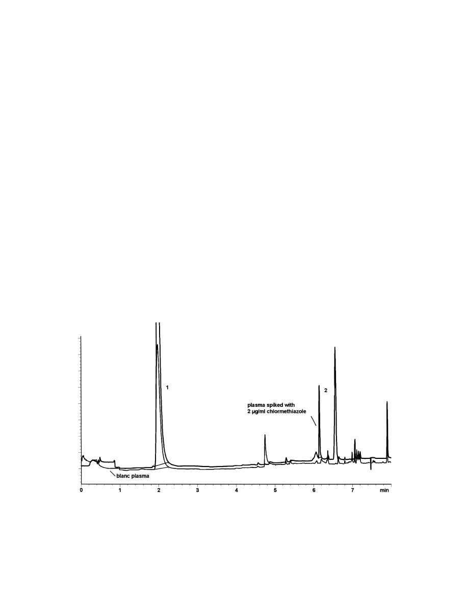
The sedative drug chlormethiazole was analyzed
(30 m30.32 mm I.D., 0.25 mm film thickness) with
in plasma by HS-SPME–GC–NPD. SPME was
helium as the carrier gas (flow-rate 1.8 ml / min) and
carried out with a 100-mm PDMS fiber at ambient
a temperature program beginning with a temperature
temperature [82]. After a 30-min extraction time the
of 1008C (for 5 min) and a ramp of 208C / min to
recovery was only 0.5%. However, calibration was
2808C. Desorption was performed at a temperature
linear between 0.5 and 5 mg / ml with an LOD of
of 2508C for 5 min. Calibrations were linear between
0.15 mg / ml and precision,10%. A HP-5 megabore
0.1 and 20 mg / ml for lidocaine (LOD 0.05 mg / ml)
capillary (30 m30.53 mm I.D., 0.88 mm film
and mepivacaine (LOD 0.05 mg / ml), between 0.5
thickness) was used for GC separation with nitrogen
and 20 mg / ml for bupivacaine (LOD 0.01 mg / ml)
as the carrier gas (Fig. 9).
and between 1 and 20 mg / ml for prilocaine (LOD
A less detailed survey of other HS-SPME methods
0.25 mg / ml) with values of r .0.999 in each case.
without derivatization should be added: trimethyl-
This is sufficient when taking into account the
amine was analyzed in urine by GC–MS to detect
Fig. 9. HS-SPME–GC–NPD chromatogram of chlormethiazole in plasma [155-methylthiazole, t 52.01 min (internal standard), 25
R
chlormethiazole, t 56.16 min, c 52.0 mg / ml].
R
0
184
S
. Ulrich / J. Chromatogr. A 902 (2000) 167 –194
trimethylaminuria [83]. Nereitoxin and metabolites
were exposed to the HS of the reaction mixture at a
in human serum were analyzed with GC–MS and
temperature of 1008C for 30 min. However, in
benzylacetone as internal standard. Ingestion of
contrast to direct SPME only five of the 11 drugs
herbicides was confirmed [84]. Parathion, dichloro-
were extracted and the HS method was not investi-
benzene isomers and VOCs in blood were assayed
gated in more detail.
by HS-SPME in combination with GC–MS, too
On-fiber
derivatization
with
1-pyrenyldiazo-
[85–87]. The GC–FID combination with HS-SPME
methane was developed for the assay of 12 short-
was sufficient for the assay of chloroform and
chain fatty acids in feces (Fig. 10) [93]. The fibers
methylene chloride in human blood and urine [88].
were loaded with the reagent by placing them into a
Approaches were described for the assay of nicotine,
solution of 1-pyrenyldiazomethane in n-hexane (5
amphetamine
derivatives,
local
anesthetics,
mg / ml) for 15 min. The HS-SPME was performed
phencyclidine, ketamine, methadone, diphenhydra-
with a 85-mm PA fiber at a temperature of 508C for
mine, tramadol, tricyclic antidepressants, phenothi-
30 min. A desorption time of 4 min at a temperature
azines and chlormethiazole by HS-SPME–GC–MS
of 2608C was needed with no carryover between
in hair [89]. HS-SPME–GC–ECD was used for the
samples. GC separation was obtained with a BPX-5
assay of tetrachloroethylene and trichloroethylene in
capillary (30 m30.22 mm I.D., 0.25 mm film
tissues [90]. Finally, an interesting approach was the
thickness) and helium as the carrier gas (flow-rate 1
assay of residues of ignitable liquids in the HS of
ml / min). The temperature program was as follows:
human skin [91].
1008C for 2 min, a first ramp of 208C / min to 2808C
(1 min) and a second ramp of 28C / min to 3108C (10
4.2.2. With derivatization
min). An internal standard method with deuterated
The assay of formic acid in urine and blood was
standards and MS detection was used for quantita-
performed by in situ derivatization with methanol
tion. The effect of temperature on the extraction was
and sulfuric acid to methyl formate at a temperature
dependent on the chain length of acids. A plateau of
of 358C for a reaction time of 5 min. A 75-mm
peak areas was reached at a temperature of 408C for
Carboxen–PDMS fiber was exposed to the HS at
C to C acids. Peak areas of acids.C continued to
1
3
4
358C for 10 min. Equilibrium was reached within
increase at higher temperatures. No equilibrium of
this time. It was found that several alternative fibers
extraction was reached within 60 min of extraction.
displayed a lower performance for the SPME of
The addition of lithium salts to the sample and low
methyl formate. Various salts increased the peak
pH caused damage of the PA coating. Thus, sodium
areas by a factor of about 2 to 4. Desorption was
chloride was added and acidification of the sample
carried out at a temperature of 2808C for 1.5 min. A
was not applied. It was demonstrated that silaniza-
Supelcowax capillary (30 m30.25 mm I.D., 0.25
tion of glassware increased peak areas. Recoveries
mm film thickness) was used for separation with
were between about 60 and 100%. Calibrations of C
2
helium as the carrier gas (0.7 ml / min flow-rate) and
to C acids were linear between 1.9 to 32.4 mmol
6
with a temperature program as follows: 3 min hold at
(acetic acid) and 0.004 to 0.07 mmol (n-hexanoic
308C, a ramp of 258C / min to 1058C and a second
acid), for example, with values of r of 0.987 to
ramp of 108C / min to 1458C. In spite of a low
0.993. In contrast, linearity of the calibration of
recovery of only 0.4 to 0.8% the calibration was
formic acid was poor with a value of r 50.939
linear between 3 and 1000 mg / ml (r 50.999) and the
within-day precision was 1.3 to 3.3% according to an
internal standard method. The LOD was 1.2 mg / ml
(FID) and the method was, therefore, described as
excellently for the detection of intoxications, for
example [92].
The method described above for the assay of
benzodiazepines by in situ hydrolysis to ben-
Fig. 10. SPME on-fiber derivatization of fatty acids in feces with
zophenones was extended to HS-SPME [71]. Fibers
1-pyrenyldiazomethane.

S
. Ulrich / J. Chromatogr. A 902 (2000) 167 –194
185
between 2.2 and 7.2 mmol. Within-day precision was
and serum were above 84% and 71%, respectively
pr
estimated to 2.4 to 12.7%, however, the value of
[97,98]. The binding constants K
between VOCs
formic acid was 15.3%. The authors concluded that
and bovine serum albumin were studied by SPME. It
HS-SPME–GC–MS is an exciting new approach to
was demonstrated that SPME is a tool to measure
pr
the analysis of fatty acids in feces. They further
K
and the freely dissolved analyte concentration in
concluded that the method is superior to previous
biomedical samples [99].
methods because it is easier to perform, sensitive and
capable of accurate quantitation.
Heptafluorobutyric anhydride as an derivatization
5. Optimization of solid-phase microextraction
agent was injected to the injector port immediately
prior to desorption of the fiber for the assay of
A discussion on the theory of SPME and a survey
amphetamine and fenfluramine in blood by HS-
of SPME methods for biomedical analysis were
SPME–GC–MS. Good linearity between 10 and
presented in the previous paragraphs. Some theoret-
1000 ng / g with LODs of 5 ng / g for fenfluramine
ical insight and practical experiences are available.
and 10 ng / g for amphetamine were obtained using a
Thus, a discussion should be possible of how to
calibration method with a deuterated internal stan-
establish a new SPME method for biomedical analy-
dard [94]. Another principle of derivatization was
sis and how to perform an optimization of a method.
applied for the assay of four amphetamine deriva-
Several variables have to be taken into account for
tives in urine by HS-SPME–GC–MS. After HS-
this purpose, for example temperature, agitation, pH,
SPME of the analytes with PDMS for 10 min the
addition of salt. These variables are discussed suc-
derivatization agent trifluoroacetic anhydride was
cessively and in detail below.
applied to the analyte in the HS of a separate vial for
20 min. Calibration was linear between 50 and 1000
5.1. Coating
ng / ml (r 50.995 to 0.999) with LOQs of 10 to 20
ng / ml [95]. Finally, lead was assayed in blood and
Several types of coatings are commercially avail-
urine after derivatization with sodium tetraethylbo-
able now. They consist of one or two polymers:
rate [96].
PDMS,
PA,
Carboxen–PDMS,
PDMS–polydi-
vinylbenzene and Carbowax–DVB, for example. The
4.3. Miscellaneous
coatings with a phase of DVB consist of porous
particles of DVB which are held together either by
An alternative geometry of SPME is described as
PDMS or Carbowax as a glue. Alternatively, the
in-tube SPME. Thus, the SPME coating is applied to
DVB phase is a template resin in another coating.
an open tubular capillary column which is well-
Recently, coatings prepared with three polymers
known from GC separation. Agitation is performed
have also become available, e.g., DVB–Carboxen–
by repeated aspiration and dispension of the sample
PDMS. The thicknesses of the usual coatings are 7
in the capillary. An aqueous solvent is used for
mm, 30 mm and 100 mm for PDMS, 85 mm for PA,
desorption and, therefore, on-line coupling with LC
75 mm for Carboxen–PDMS, 65 mm for PDMS–
can be used. The H -antihistaminic drug ranitidine
DVB and Carbowax–DVB. The 7-mm PDMS coat-
2
and nine beta-blockers were analyzed in urine and
ing is a bonded phase and the 30-mm and 100-mm
serum by in-tube SPME–LC–electrospray ionization
PDMS coatings are nonbonded phases. It should be
MS. Ten to 15 aspiration–dispension cycles of a
recognized that some coatings cannot be regarded as
sample volume of 30 ml were used. The calibration
liquids as presumed in Section 3.1, i.e., Carbowax–
of ranitidine was linear in a range of 5 to 1000
DVB and PDMS–DVB are solids and the mecha-
ng / ml (r 50.999) and LOD of 1.4 ng / ml. Within-day
nism of analyte enrichment is sorption instead of
and between-day precisions were 2.5% and 6.2%
extraction.
(n 55), respectively. Calibrations of beta-blockers
Two simple rules should be applied for the
were linear in a range of 2 to 100 ng / ml (r .0.998)
selection of the coating in a first attempt for a new
with LODs of 0.1 to 1.2 ng / ml. Recoveries in urine
SPME method. The polarity of the coating should

186
S
. Ulrich / J. Chromatogr. A 902 (2000) 167 –194
match the polarity of the analyte, i.e., according to
lower concern. A 7-mm coating should be preferred
fw
‘‘similar attracts similar’’ the value of K
is ex-
if the method should be fast and sensitivity is of
pected to be high for a nonpolar coating and a
lower concern because the concentrations of analytes
nonpolar analyte. The number of groups forming
are sufficiently high. It is concluded that the higher is
fw
hydrogen bonds is a special property of analytes, i.e.,
the affinity of an analyte to the coating (high K )
the number of NRH groups, NH
groups and OH
the lower should be r 2r . For biomedical analysis,
2
o
i
groups. These groups mainly determine the hydro-
because of lower diffusion coefficients of the analyte
philicity of an analyte and, therefore, also the affinity
molecule in plasma, for example, the decision for a
fw
to the coating. As a second rule the coating should
7-mm coating may be drawn already at lower K
be resistant to extreme chemical (pH, salts, additives)
than in water. However, if it was decided to stop
and physical (high temperature) conditions. For the
extraction prior to equilibrium the validity of these
main part, these requirements are met by PDMS
simple calculations is limited and the fiber thickness
coatings. Many organic analytes investigated in
may play a minor role in method optimization.
bioanalysis are nonpolar molecules and they have,
Finally, it should be taken into account that also the
therefore, a good affinity to the nonpolar PDMS.
time of desorption is increased with fiber thickness.
PDMS is the most resistant coating in SPME and the
performance of fibers is high also after many re-
5.2. Extraction method and sample pretreatment
peated extractions and desorptions. PDMS should be
for solid-phase microextraction
first tried for a new method. The PA coating can be
superior in the case of slightly more polar analytes
A HS method should be applied whenever pos-
such as chlorophenols [58] and benzodiazepines
sible in SPME of body fluids. The burden of the fiber
[61]. The affinity was equal for PDMS and PA if the
with proteins, for instance, is considerably decreased.
benzodiazepines were derivatized to the less polar
The lifetime of the fiber is increased because ir-
benzophenones. However, PA was worse because of
reversible damage is delayed. A reversible change of
the co-extraction of interfering substances. This is
extraction properties of the coating is also avoided
important for the analysis of complex matrices such
and, therefore, the precision of the method is im-
as body fluids. Furthermore, the PA coating is
proved. In addition, endogenous trace substances
damaged more easily than PDMS [93]. The selection
with molecular masses between about 200 and 450
of the other coatings is even more empirical. How-
g / mol are better separated from a volatile analyte in
ever, PDMS–DVB and Carbowax–DVB are re-
HS-SPME, too. As a rule of thumb analytes with a
garded to be suitable for more volatile analytes
molecular mass below 200 g / mol and (or) without
because of the adsorption to porous particles. The
groups forming hydrogen bonds, i.e., NRH groups,
Carbowax–DVB coating stripped off the fiber in
NH
groups and OH groups, are suitable for HS-
2
direct SPME at various temperatures and at various
SPME because they are likely to have a high vapor
pH [47]. The Carbowax additive has a special
pressure. Direct SPME should only be tried if HS-
affinity to alcohols. The linear range of Carbowax–
SPME failed to give sufficient peak areas. Deri-
DVB and PDMS–DVB is smaller than that of
vatization methods should be considered critically
PDMS.
because they are often laborious, time-consuming
The option of selecting a coating thickness is
and not easy to automate. Another sample prepara-
limited to the PDMS coating. Because it is advan-
tion without derivatization may be preferred in many
tageous to reach equilibrium of extraction and ac-
cases if SPME is possible only with derivatization.
cording to Eq. (4) and Eq. (19) the sensitivity is
For example, the assay of benzodiazepines by hy-
fw
increased with K
and r 2r but the rapidity of the
drolysis to benzophenones [71] can be easily per-
o
i
fw
method is decreased with K
and r 2r . A com-
formed with a simple one-step LLE. Furthermore,
o
i
promise is necessary with regard to coating thickness
laborious sample pretreatment for SPME should be
between these two criteria of performance of a
avoided if an alternative sample preparation method
method. A 100-mm coating can be used if sensitivity
is available without an extended sample pretreat-
should be maximum and the time of extraction is of
ment. For example, precipitation of proteins prior to

S
. Ulrich / J. Chromatogr. A 902 (2000) 167 –194
187
SPME is questionable for the assay of diazepam
5.4. Sample volume and volume of the headspace
because other methods are known devoid of this
additional step [74].
The amount of analyte extracted in equilibrium is
increased with the sample volume V
according to
w
Eq. (39), which was derived from Eq. (3), with c
0
5.3. Agitation
the concentration of analyte in the sample prior to
SPME. Therefore, V should be as large as possible.
w
The time to reach equilibrium is determined by the
However, because V
is present also in the de-
w
effectiveness of sample agitation. The radius d of the
nominator of Eq. (39) no further increase of V
is
w
fw
boundary layer of the practical agitation model is
needed over a limit of about V 510K V :
w
f
fw
decreased with increased revolution rate N of a
K V c V
f
0 w
]]]]
n 5
(39)
magnetic stirrer, for example. Thus, according to
f
fw
K V 1 V
s
d
f
w
Eqs. (19) and (20) the equilibration time t
is
e
Analogously, Eq. (40) can be derived from Eq.
decreased with an improved agitation. Apart from
(17) for HS-SPME. The situation is more compli-
magnetic stirring the following agitation methods can
cated than for direct SPME because of three terms in
be applied: vortex mixing (moving vial), fiber move-
the denominator of Eq. (40). However, V should be
ment, flow through agitation and sonication. Mag-
h
small for highly volatile analytes, i.e., for analytes
netic stirring was mainly applied for SPME in
hw
with high K
. The denominator is mainly deter-
biomedical analysis. Disadvantages of magnetic stir-
hw
hw
mined by the term K
V . In the case of lower K
ring are a more complicated automation, problems to
h
fw
the degree of the influence of V is modified by K
maintain a constant N and, perhaps most important,
h
and V :
the potential for carryover. Advantages are the good
w
effectiveness of agitation and the availability in
fw
K V c V
f
0 w
analytical laboratories. The moving vial approach is
]]]]]]
n 5
(40)
f
fw
hw
K V 1 K
V 1 V
s
d
f
h
w
mainly applied in the LLE of drugs in body fluids.
One apparatus can be applied to SPME with the
According to Eq. (19) the rapidity of extraction
simultaneous agitation of up to 24 1.5-ml vials and
can be regarded as independently of V
in direct
w
the options of setting a selected revolution rate of the
SPME. The linear model used for the description of
vials and temperature [45,60]. The fiber movement
extraction kinetics in HS-SPME (Eq. (20)) provides
agitation method, i.e., a vibration of the fiber, is
no explanation for the influence of sample volume
realized in a commercial autosampler (Varian). Both
and volume of the HS on the time of extraction.
methods provide good agitation similar to the effec-
However, it is discussed that the capacity of HS
tiveness of magnetic stirring. As an advantage, no
should exceed the capacity of the fiber for about 20
hw
fw
external object is needed in the sample. However,
times for a rapid extraction, i.e., K
V .20K V
h
f
HS-SPME is impossible with the moving vial meth-
[42]. Thus, a compromise between a rapid and a
od and it is less effective with the vibration of fiber.
sensitive method must be found in HS-SPME. Final-
The flow through method was rarely applied because
ly, it is emphasized that this discussion only applies
of several disadvantages. Sonication was expected to
to methods with equilibrium conditions of extraction.
provide a better agitation than magnetic stirring, for
This may not be accomplished in the analysis of
example. However, this was not confirmed for the
drugs in plasma [45], for example, because of
assay of clozapine in plasma [47]. Thus, it is
increased equilibration times. Finally, in practice, the
concluded that magnetic stirring at the highest N
sample volume is also determined by the available
should be applied for the sample agitation in direct
volume of sample and the available vials and equip-
and HS-SPME and, additionally, fiber movement and
ment of agitation.
moving vial can be applied for direct SPME. An
effective agitation is needed in biomedical analysis
5.5. Extraction time
because of a higher viscosity of samples and lower
diffusion coefficients.
A graph of the relationship of peak areas and time

188
S
. Ulrich / J. Chromatogr. A 902 (2000) 167 –194
of extraction is a prerequisite for method optimi-
Another important goal of the addition of salt in
zation. The time t can be obtained when no further
SPME is to compensate for a variable salt con-
e
increase of peak areas is detected with increased time
centration of the samples. This may be the only
of extraction. Care should be taken because the slope
reason of adding salts to plasma samples and for the
may decrease considerably without reaching t . An
assay of acid and basic analytes at low or high pH. It
e
overnight experiment may be necessary. It has to be
is recommended to add no more than about 10% of
decided with the known t
whether the method
NaCl, for example.
e
should work in equilibrium or in nonequilibrium. Of
The use of organic additives was recommended
course, an experimental time exceeding t is desir-
for matrices with polymer components, e.g., plasma.
e
able because experimental errors are decreased and
It is suggested that the binding of target analyte to
sensitivity is at maximum. However, the values of t
the proteins can be decreased and, therefore, the
e
in biomedical analysis can be very large (Fig. 3) and
sensitivity of the method can be substantially in-
the chosen times of extraction were considerably
creased. About 25 organic chemicals of different
shorter than t
in some methods [45] because of
classes were tested to improve the sensitivity of
e
practical reasons. This is possible because SPME is a
direct SPME of antidepressant drugs in plasma.
quantitative method at every time of the extraction
However, no substantial increase of peak areas was
time profile as already discussed above and because
found and even peak areas decreased for some
sensitivity may be sufficient prior to equilibrium.
additives. This also applied to the addition of drugs
Furthermore, internal standard calibration as usual in
with very similar structure or drugs which are known
drug analysis, for example, can compensate for
to decrease protein binding [47].
errors due to variable time of extraction and variable
agitation. Thus the use of internal standard methods
5.8. Temperature
is a general recommendation for SPME. An internal
fw
standard with very similar extraction time profile
As discussed above K
is decreased with in-
should be applied. This is best realized by isotopical-
creased temperatures. The sensitivity of the method
ly labeled spikes.
is decreased in equilibrium. Therefore, ambient
temperature is applied for the direct SPME. Because
5.6. pH
diffusion coefficients are increased with lower vis-
cosity and, therefore, also with increased temperature
The pH of the sample is crucial for the SPME of
the rapidity of the extraction may be improved
acids and bases. This is explained theoretically by
according to Eq. (19), however, at cost of a loss of
the coupled extraction equilibrium and acid–base
sensitivity. It may be tested whether the sensitivity of
equilibrium (Eqs. (9) and (19)) and was confirmed in
a method can be improved at a nonequilibrium
experiments. Thus, basic drugs such as antidepres-
extraction time and at increased temperatures due to
sants were analyzed in aqueous NaOH [45] and acid
this effect. This may be more important for plasma
analytes such as chlorophenols [58] and valproic acid
because of the higher viscosity than water or urine.
[76] were shown to be extracted better at low pH.
The extraction profiles at various temperatures
should cross one another. However, this was not
5.7. Salt and other additives
found in Fig. 3 [45] and no more data were available
fh
in the literature. In HS-SPME the values of K
and
hw
A salting-out effect is expected according to Eq.
K
are decreased and increased, respectively, with
(8)
and
was
described
in
several
methods
increased temperature. Thus, it is expected that the
[58,77,79,92]. However, the effect is less clearly
sensitivity of the method in equilibrium is also
than the effect of pH. No increase of peak areas was
decreased at higher temperatures according to Eqs.
found in plasma [48,79], the salting-out effect was
(13) and (17). On the other hand, the rapidity of
limited to low salt concentrations ( ,10%) [76] and
extraction is considerably increased in HS-SPME at
the effect of salt interfered with the effect of pH
increased temperatures as can be concluded from Eq.
hw
[58]. Lithium salts caused damage of the fiber [93].
(20). Increased K
can determine the improvement

S
. Ulrich / J. Chromatogr. A 902 (2000) 167 –194
189
of extraction by HS-SPME for instance for analytes
siderably lower after direct application in an aqueous
with low to moderate volatility. Therefore, increased
matrix.
temperatures were indeed recommended for HS-
After the needle was introduced into the insert the
SPME in many applications [59,71,80,93]. Further-
fiber should be exposed fast as possible because
more, peak areas increased at higher temperatures
partial desorption already within the needle can
[59] and nonlinear relationships between peak area
result in split peaks. This applies for instance for
and temperature were described [80,93]. In addition,
volatile analytes and the temperature of desorption
due to the slow transfer of heat in the HS the
may be decreased considerably below the maximum
temperature of the fiber was lower than the tempera-
temperature. Carryover of interferences of the matrix
ture of the sample for a considerable time of
must also be taken into account for instance in
extraction. Thus, the amount of analyte extracted can
biomedical analysis apart from the carryover of
be decreased with time because of the delayed
analytes only. Otherwise, accumulation of interfering
heating of the fiber [80]. Coagulation was a problem
substances can occur in the coating and, easily
at increased temperatures for nondiluted blood sam-
unnoticed, provide bias. Thus, the definition of the
ples [79]. In conclusion, the optimum temperature
desorption time to prevent carryover effects of
for HS-SPME of a distinct analyte is determined by
interferences from the matrix is also necessary.
several variables. This optimum temperature can
Blank samples should be analyzed repeatedly and the
only be found in a trial of various temperatures.
accumulation of trace interferences should be ob-
Nevertheless, for the main part an increased tempera-
served.
ture of about 50 to 1008C was used in the HS-SPME
methods described above and may be a guideline for
5.10. GC temperature program
other methods.
The temperature program of the GC oven after
5.9. Desorption
SPME sample preparation is determined by the need
for refocusing of the analyte on the head of the
The time of desorption should be short as possible
capillary. Otherwise, large peak widths, or even no
and carryover effects must be excluded. Thus, the
peaks, can be found in the chromatogram because of
highest temperature without damage of the selected
the very slow injection, i.e., the relative long period
coating and the smallest diameter of the injector
of desorption. Therefore, the temperature program is
fh
insert should be applied because K
is decreased
usually started at a temperature T
which is con-
1
with increased temperatures and the linear flow-rate
siderably lower than the desorption temperature and
is increased with a smaller diameter of the insert.
which is low enough for refocusing. T
may be
1
The maximum temperatures are about 3408C (7 mm)
maintained for 1 to 3 min. This is followed by a
and 2808C (30 and 100 mm) for PDMS, 2708C for
steep ramp of 10 to 308C / min until temperature T is
2
PDMS–DVB, 3208C for PA and Carboxen–PDMS,
reached as is usual for the GC separation of the
2658C for Carbowax–DVB and 2708C for DVB–
analyte. Then a less steep ramp of 1 to 58C / min to
Carboxen–PDMS. Special injector inserts are avail-
the final temperature T can be used or, alternatively,
3
able for SPME with I.D.s of 0.75 and 1.5 mm. The
the temperature can be maintained isothermally. This
diameter of the insert is less critical if a megabore
principle may be modified and even only 1 ramp was
capillary is used with flow-rates between 10 and 20
described as being convenient. However, the low T
1
ml / min. However, the optimum time of desorption
is crucial except in cases of very volatile analytes
has to be found experimentally in a desorption–
and a high film thickness of the analytical capillary.
postdesorption graph as described for clozapine [45],
for example. Apart from this theoretical considera-
5.11. GC capillary
tion, the maximum temperatures as provided by the
manufacturer were not applied in practice with the
SPME is a dirty extraction because it is only a
aim to increase the lifetime of fibers. Indeed, the
one-step extraction and because of the limited selec-
maximum temperatures of coatings may be con-
tivity of coatings. Therefore, endogenous trace sub-

190
S
. Ulrich / J. Chromatogr. A 902 (2000) 167 –194
stances of plasma, urine or other biological fluids are
5.13. Automation
easily co-extracted by SPME and they have to be
separated by the GC thereafter. A three-step LLE
Ease of automation can be regarded as an im-
provided considerably purer extracts than SPME and
portant advantage of SPME. This is caused by the
the risk of interferences in the chromatogram was
simple principle of SPME. Some complex processes
low in the analysis of drugs in plasma. The chro-
as known from SPE and LLE are not needed, i.e.,
matograms after SPME were similar to chromato-
transfer, separation and evaporation of liquid phases.
grams after a one-step LLE and the GC separation
The development of an instrumental periphery is
was considerably less comfortable than after three-
expected for the automation of SPME. An auto-
step LLE [45,47,60]. Furthermore, the production of
sampler for SPME–GC was developed using agita-
SPME coatings does not exclude an inherent con-
tion by vibration of the fiber. It can successively
tamination of fibers. Despite the usual conditioning
perform the SPME and desorption of several samples
of fibers prior to first use, i.e., desorption for several
with one fiber [100]. However, the agitation method
hours, interferences originating from the fiber were
is less effective in HS-SPME and the sample
found in the chromatograms. This effect prevented
throughput is low for methods with large extraction
the analysis of some antidepressants in plasma at
time and short GC separation time. Therefore, an
therapeutic concentrations [47]. Thus, the selection
equipment is needed which prepares several samples
of a GC capillary may be more critical in biomedical
in a batch by SPME and subsequently makes the
analysis for instance with direct SPME. More polar
desorption in a batch of fibers. This principle of
phases can be tried. The I.D. of the capillary, i.e., the
automation is known from LLE–GC, although it is
linear flow-rate of carrier gas, is a variable which
not a fully-automated system. It can be easily
determines the desorption time. A megabore capil-
calculated that the sample throughput of this ap-
lary should be tried to decreases the time of desorp-
proach is higher for methods with large time of
tion. Finally, the use of a retention gap may improve
extraction and short time of GC separation. This
the refocusing because of no temperature gradient in
applies to the analysis of drugs in plasma by direct
the head of the capillary.
SPME, for example.
5.12. GC detector
6. Advantages and disadvantages of solid-phase
microextraction
A more selective GC detector may separate inter-
ferences and analyte if the selectivity of sample
The advantages claimed for SPME were (1) no
preparation by SPME and subsequent GC separation
use of solvents, (2) easy handling, (3) little equip-
was insufficient. Thus, GC detection by MS with
ment necessary, (4) fast method, (5) ease of automa-
SIM or even IT-MS should be preferred for the
tion as well as (6) good linearity and high sensitivity
application of SPME in complex matrix. NPD as
[44]. However, taking into account the survey of
usual for the analysis of drugs in plasma is prone to
methods as provided above it is obvious that SPME
problems with interferences because the majority of
can display these advantages only in some areas of
endogenous substances also contain nitrogen atoms
biomedical analysis, i.e., the matrix and the volatility
in their molecule, i.e., they are also sensitively
of target analyte have to be taken into account. First
detected by NPD. This problem of chemical noise in
of all, the combination of low volatility of analyte
SPME–GC–NPD is expected to be considerably
and a complex matrix with polymer components,
decreased by the application of ECD. The application
e.g., proteins in plasma or cell cultures, considerably
of ECD for the analysis of benzodiazepines in urine
limits the application of SPME. The extraction is
[71] and dinitroaniline herbicides in water, urine and
very slow in contrast to LLE and SPE with packed
plasma [79] was described. ECD should be increas-
bed columns. Extraction times considerably lower
ingly tried in the SPME–GC analysis of biological
than t must be used because of practical require-
e
fluids.
ments. Thus, the recovery was very low and the

S
. Ulrich / J. Chromatogr. A 902 (2000) 167 –194
191
sensitivity was critical for some analytes within an
native sample preparation methods are available.
acceptable time of extraction [45,47]. In general,
Again, the labels fast, efficient and simple should not
recoveries reported for SPME were considerably
be abandoned.
lower in many cases than it is usually known for
The advantages of SPME can be used for both the
LLE and SPE. Another disadvantage of SPME is
assay of low volatile and highly volatile analytes in
uncovered if the sensitivity is low with regard to the
urine or other body fluids with no or only a low
target concentrations of analyte, the therapeutic
concentration of polymer biomolecules. The prob-
concentration of a drug in plasma for example: direct
lems of sensitivity and delayed t are considerably
e
SPME is a dirty sample preparation. The occurrence
decreased in comparison to plasma. A number of
of a huge number of interferences in the chromato-
methods with good precision, accuracy, sensitivity
gram which come from endogenous trace substances
and selectivity were demonstrated which were also
in biological fluids prevents the analysis of target
simple and fast [58,69,77–79,92].
analyte at low concentrations. This is not encoun-
Apart from the problems discussed above SPME
tered in three-step LLE or SPE. The values r of
has also some principle disadvantages. (1) Because
internal standard calibration of SPME were not
of desorption times of at least 1 min cryofocusing of
excellent and the precision was about 8 to 10%.
the analyte is needed. Thus, temperature programs
Attempts to by-pass this problem by special methods
with a very low initial temperature are needed for the
such as solvent-modified SPME [74], complicated
GC separation. The time of the GC program is,
derivatization methods [71] or equilibrium dialysis
therefore, longer than the time of an alternative
[76] can be regarded as misleading because the
sample preparation. (2) The desorption needs more
advantages of easy handling, little equipment and
time than the injection of liquid extracts after LLE
fast sample preparation are abandoned. A good
and SPE. Desorption times of 12 and 20 min [57,61]
SPME method should remain simple, otherwise,
waste the advantages of SPME and may be not
alternative sample preparation methods may be
accepted. (3) By nature, carryover effects occur very
superior. It was concluded that the application of
easily in SPME methods because of the repeated use
SPME for the assay of low volatile drugs and
of one fiber. Additional efforts are necessary to
metabolites in plasma may be limited to some drugs
handle this problem. Extended extra clean-up pro-
with high therapeutic concentrations in the range of 1
cedures for the fiber after every extraction may not
to 100 mg / ml. Because the analysis of drugs in
be accepted for routine methods. In fact, a carryover
plasma for pharmacokinetic studies and for TDM is
of 2% is very questionable [57]. (4) The condition-
extensively applied an important area of biomedical
ing of fibers prior to first use is not necessary in
analysis may be only little accessible for SPME.
LLE, for example. (5) By nature, SPME is a
The assay of volatile analytes in plasma and
relatively dirty extraction if it is compared with
similar matrices is more convenient because of the
multiple step LLE. (6) Because SPME is a
option of HS-SPME. SPME may clearly display
nonexhaustive sample preparation the methods can-
some advantages over other sample preparation
not equally compensate for changes of the com-
methods in this area. No solvent or only very little
position of the matrix as in the case of LLE.
solvent is needed, the handling of samples is easy
Quantitation is more prone to errors due to changes
with little equipment and the available SPME auto-
of the matrix also if internal standard methods are
sampler can be applied efficiently because the ex-
applied. Thus, matrix effects must be extensively
traction time is short. Thus, the SPME of one sample
investigated during method validation. (7) SPME is
is completed during the GC separation of another.
not an universal sample preparation method because
Methods with good linearity and sensitivity were
of the restrictions and limitations discussed.
described [77–80]. HS-SPME with derivatization
may also display a good performance with regard to
effectiveness, precision, accuracy, sensitivity and
7. Conclusions
selectivity [92]. Complicated and long-term deri-
vatization methods should be avoided [71] if alter-
SPME is an encouraging development for sample

192
S
. Ulrich / J. Chromatogr. A 902 (2000) 167 –194
preparation in biomedical analysis. The majority of
8. Nomenclature
applications were encountered in environmental
DOM
Dissolved polymer organic material
chemistry in the early developmental period. How-
DVB
Divinylbenzene
ever, an increased number of publications are now
ECD
Electron-capture detection
available on biomedical analysis. The special geome-
EI
Electron ionization mode
try of the extraction agent is an unique feature of
ELISA
Enzyme-linked immunoassay
SPME and, therefore, special advantages and dis-
EDDP
2-Ethylene-1,5-dimethyl-3,3-
advantages are involved as discussed above. The
diphenylpyrrolidine
most striking attribute of SPME is a low recovery as
FID
Flame ionization detection
reported for many methods. This is not unexpected
FPIA
Fluorescence polarization immunoassay
because SPME is an equilibrium extraction but not
GC
Capillary gas chromatography
an exhaustive extraction. However, several SPME
HS
Headspace
methods also presented with high recovery and the
I.D.
Inner diameter
performance of the majority of methods was high
IT
Ion-trap
with regard to sensitivity, linearity, precision and
LOD
Limit of detection
accuracy. It is concluded that SPME can be used as a
LOQ
Limit of quantitation
substitution and improvement of classical sample
LC
Liquid chromatography
preparation methods. Thus, it can be applied for the
LLE
Liquid–liquid extraction
analysis of drugs, metabolites, environmental pollu-
MS
Mass spectroscopy
tion and endogenous substances for instance in urine
MIP
Molecular imprinting material
and body fluids without or with low concentration of
NCI
Negative chemical ionization
biopolymers. It appears from the survey of published
NPD
Nitrogen–phosphorus selective detection
methods that HS-SPME–GC–MS, for instance, be-
PA
Polyacrylate
comes increasingly popular in forensic and environ-
PAH
Polynuclear aromatic hydrocarbon
mental toxicology. The need for an alternative
PCB
Polychlorinated biphenyl
method in the confirmatory analysis of positive tests
PDMS
Polydimethylsiloxane
by ELISA and FPIA may be a special requirement
RSD
Relative standard deviation
which promoted the application of SPME in forensic
RAM
Restricted access material
toxicology. The trace analysis in plasma may be
SFE
Supercritical fluid extraction
limited to highly volatile analytes. The SPME assay
SIM
Selected ion monitoring
of substances with low volatility is more complicated
SPE
Solid-phase extraction
in plasma and may be easily performed, therefore,
SPME
Solid-phase microextraction
only in samples with high concentrations. For this
TEMPO
2,2,6,6-Tetramethylpiperidine-1-oxide
reason, a wide application of SPME in TDM cannot
TDM
Therapeutic drug monitoring
be expected for the near future. On-line SPE–LC
UV
Ultraviolet detection
with column-switching techniques may be more
VOC
Volatile organic compound
recommended in this area. However, more studies
are necessary also by SPME in TDM applications.
Apart from this role of SPME as a substitution and
References
improvement of classical methods, SPME may lead
to new approaches in biomedical analysis. For
[1] J.H. Lin, A.Y. Lu, Pharmacol. Rev. 49 (1997) 403.
example, the potential of SPME to analyze directly
[2] A. Marzo, Pharmacol. Res. 36 (1997) 425.
the free concentration of drugs in plasma should be
[3] K.A. Jackson, S.E. Rosenbaum, Drug Dev. Ind. Pharm. 24
more investigated in future. The in-vivo analysis of
(1998) 1155.
[4] S. Ulrich, C. Wurthmann, M. Brosz, F.P. Meyer, Clin.
metabolites in animal experiments should be tried
Pharmacokinet. 34 (1998) 227.
using new geometries. New developments are ex-
[5] M.H.H. Ensom, G.A. Davis, C.D. Cropp, R.J. Ensom, Clin.
pected because the important role of the geometry of
Pharmacokinet. 34 (1998) 265.
extraction agent was first recognized in the invention
¨
[6] S. Ulrich, I. Schrodter, G. Partscht, P. Baumann, Psycho-
of SPME.
pharmakotherapie 7 (2000) 2.

S
. Ulrich / J. Chromatogr. A 902 (2000) 167 –194
193
[7] T.M. De Kok, P.J. Levels, A. Van Faassen, M. Hazen, F. Ten
[44] D. Louch, S. Motlagh, J. Pawliszyn, Anal. Chem. 64 (1992)
Hoor, J.C. Kleinjans, J. Chromatogr. 580 (1992) 135.
1187.
[8] V. Iyengar, J. Kumpulainen, K. Okamoto, M. Morita, S.
[45] S. Ulrich, S. Kruggel, H. Weigmann, C. Hiemke, J. Chroma-
Hirai, S. Nomoto, Prog. Clin. Biol. Res. 380 (1993) 329.
togr. B 731 (1999) 231.
[9] O.H. Drummer, J. Chromatogr. B 733 (1999) 27.
[46] Y. Yang, D.J. Miller, S.B. Hawthorne, J. Chromatogr. A 800
[10] A. Polettini, J. Chromatogr. B 733 (1999) 47.
(1998) 257.
[11] A. Astier, J. Chromatogr. 643 (1993) 389.
[47] S. Kruggel, S. Ulrich, unpublished results.
[12] D. Firestone, J. Assoc. Off. Anal. Chem. 74 (1991) 375.
[48] F.A. Long, W.F. McDevit, Chem. Rev. 51 (1952) 119.
[13] F.C. Cheng, J.S. Kuo, J. Chromatogr. B 665 (1995) 1.
¨
[49] L. Muller, E. Fattore, E. Benfenate, J. Chromatogr. A 791
[14] D. Tsikas, J. Chromatogr. B 717 (1998) 201.
(1997) 221.
[15] T. Toyo’oka, J. Chromatogr. B 671 (1995) 91.
[50] M.N. Sarrion, F.J. Santos, M.T. Galceran, J. Chromatogr. A
[16] R.N. Gupta, J. Chromatogr. B 576 (1992) 183.
859 (1999) 159.
¨
[17] M. Koel, A. Roth, P. Nebinger, Arztl. Lab. 35 (1989) 57.
[51] A. Penalver, E. Pocurull, B. Borull, R.M. Marce, J. Chroma-
[18] K.L. Sloan, V.M. Haver, A.J. Saxon, Am. J. Psychiatry 157
togr. A 839 (1999) 253.
(2000) 148.
[52] X. Yang, T. Peppard, LC?GC 13 (1995) 882.
[19] M.L. Rao, U. Staberock, P. Baumann, C. Hiemke, A. Deister,
[53] C.L. Arthur, L.M. Killam, K.D. Buchholz, J. Pawliszyn,
¨
C. Cuendet, M. Amey, S. Hartter, M. Kramer, Clin. Chem.
Anal. Chem. 64 (1992) 1960.
40 (1994) 929.
[54] S. Ulrich, J. Martens, J. Chromatogr. B 696 (1997) 217.
¨
[20] M. Banger, B. Hermes, S. Hartter, C. Hiemke, Phar-
¨
[55] J. Porschmann, Z. Zhang, F.D. Kopinke, J. Pawliszyn, Anal.
macopsychiatry 30 (1997) 128.
Chem. 69 (1997) 597.
[21] Applications Book, Probenvorbereitung Mittels Festphasen-
¨
[56] J. Porschmann, F.D. Kopinke, J. Pawliszyn, J. Chromatogr.
¨
extraktion, Machery–Nagel, Duren, 1997.
A 816 (1998) 159.
¨
¨
[22] S. Suss, W. Seiler, C. Hiemke, G. Schonhammer, H. Wetzel,
[57] B.J. Hall, J.S. Brodbelt, J. Chromatogr. A 777 (1997) 275.
A. Hillert, J. Chromatogr. B 565 (1991) 363.
[58] M.R. Lee, Y.C. Yeh, W.S. Hsiang, C.C. Chen, J. Chromatogr.
[23] S. Ulrich, F.P. Meyer, W. Knorr, S. Neuhof, J. Chromatogr. B
B 707 (1998) 91.
663 (1995) 289.
[59] C. Kroll, H.H. Borchert, Pharmazie 53 (1998) 172.
[24] E.J. Cone, W.D. Darwin, J. Chromatogr. 580 (1992) 43.
[60] S. Kruggel, S. Ulrich, Fresenius J. Anal. Chem. 364 (1999)
[25] K.R. Mallikaarjun, H.T. Karnes, J. Chromatogr. 531 (1990)
654.
407.
[61] K. Singer, B. Wenz, V. Seefeld, U. Speer, GIT Lab. Med. 2
[26] K.S. Boos, C.H. Grimm, Trends Anal. Chem. 18 (1999) 175.
(1995) 112.
[27] E.B. Asafu-Adjaye, S.Y. Su, G.K. Shiu, J. Chromatogr. B
[62] S.W. Myung, S. Kim, J.H. Park, M. Kim, J.C. Lee, T.J. Kim,
652 (1994) 35.
Analyst 124 (1999) 1283.
[28] A. Zlatkis, R.S. Brazell, C.F. Poole, Clin. Chem. 27 (1981)
[63] A.C. Lucas, A. Bermejo, P. Fernandez, M.J. Tabernero, J.
789.
Anal. Toxicol. 24 (2000) 93.
[29] S.H. Wong, Ther. Drug Monit. 15 (1993) 576.
[64] A.C. Lucas, A.M. Bermejo, M.J. Tabernero, P. Fernandez, S.
[30] U.A. Brinkman, J. Chromatogr. A 665 (1994) 217.
Strano-Rossi, Forensic Sci. Int. 107 (2000) 225.
¨
[31] H. Weigmann, J. Bierbrauer, S. Hartter, C. Hiemke, Ther.
[65] A.M. Bermejo, R. Seara, A.C. Lucas, M.J. Tabernero, P.
Drug Monit. 19 (1997) 480.
Fernandez, R. Marsili, J. Anal. Toxicol. 24 (2000) 66.
[32] H. Imai, T. Masujima, I. Morita-Wada, G. Tamai, Anal. Sci.
[66] K.F. Poon, P.K. Lam, M.H. Lam, Chemospere 39 (1999)
54 (1989) 389.
905.
[33] F. Munari, K. Grob, J. Chromatogr. Sci. 28 (1990) 61.
[67] S. Strano-Rossi, M. Chiarotti, J. Anal. Toxicol. 23 (1999) 7.
[34] J.J. Vreuls, A.J.H. Louter, U.A.T. Brinkman, J. Chromatogr.
[68] E.H. Koster, C. Wemes, J.B. Morsink, G.J. de Jong, J.
A 856 (1999) 279.
Chromatogr. B 739 (2000) 175.
[35] N.C. Van de Merbel, J.J. Hageman, U.A.T. Brinkman, J.
[69] H.G. Ugland, M. Krogh, K.E. Rasmussen, J. Chromatogr. B
Chromatogr. 634 (1993) 1.
701 (1997) 29.
[36] R.G. Belardi, J. Pawliszyn, Water Pollut. Res. J. Can. 24
¨
[70] H.M. Liebich, E. Gesele, J. Woll, J. Chromatogr. B 713
(1989) 179.
(1998) 427.
[37] C.L. Arthur, J. Pawliszyn, Anal. Chem. 62 (1990) 2145.
[71] F. Guan, H. Seno, A. Ishii, K. Watanabe, T. Kumazawa, H.
[38] J. Pawliszyn, Int. Pat. Publ. No. WO 91 / 15745.
Hattori, O. Suzuki, J. Anal. Toxicol. 23 (1999) 54.
[39] Z. Zhang, J. Pawliszyn, J. High Resolut. Chromatogr. 16
[72] B.J. Hall, A.R. Parikh, J.S. Brodbelt, J. Forensic Sci. 44
(1993) C689.
(1999) 527.
[40] C.L. Arthur, R. Belardi, K. Pratt, S. Motlagh, J. Pawliszyn, J.
[73] Z. Mester, J. Pawliszyn, J. Chromatogr. A 873 (2000) 129.
High Resolut. Chromatogr. 15 (1992) 741.
[74] M. Krogh, H. Grefslie, K.N. Rasmussen, J. Chromatogr. B
[41] C.L. Arthur, L. Killam, K. Buchholz, D. Potter, M. Chai, Z.
689 (1997) 357.
Zhang, J. Pawliszyn, Am. Environ. Lab. 11 (1992) 10.
[75] K.J. Reubsaet, H.R. Norli, P. Hemmersbach, K.E. Rasmus-
[42] J. Pawliszyn, Solid-phase Microextraction – Theory and
sen, J. Pharm. Biomed. Anal. 18 (1998) 667.
Practice, Wiley–VCH, New York, 1997.
[43] A.A. Boyd-Boland, J. Pawliszyn, Anal. Chem. 68 (1996)
[76] M. Krogh, K. Johansen, F. Tonnesen, K.E. Rasmussen, J.
1521.
Chromatogr. B 673 (1995) 299.

194
S
. Ulrich / J. Chromatogr. A 902 (2000) 167 –194
[77] D. Poli, E. Bergamaschi, P. Manini, R. Andreoli, A. Mutti, J.
[90] B. Dehon, L. Humbert, L. Devisme, M. Stievenart, D.
Chromatogr. B 732 (1999) 115.
Mathieu, N. Houdret, M. Lhermitte, J. Anal. Toxicol. 24
[78] I. Koide, O. Noguchi, K. Okada, A. Yokoyama, H. Oda, S.
(2000) 22.
Yamamoto, H. Kataoka, J. Chromatogr. B 707 (1998) 99.
[91] J.R. Almirall, J. Wang, K. Lothridge, K.G. Furton, J.
[79] F. Guan, K. Watanabe, A. Ishii, H. Seno, T. Kumazawa, H.
Forensic Sci. 45 (2000) 453.
Hattori, O. Suzuki, J. Chromatogr. B 714 (1998) 205.
[92] X.P. Lee, T. Kumazawa, K. Kondo, K. Sato, O. Suzuki, J.
[80] T. Watanabe, A. Namera, M. Yashiki, Y. Iwasaki, T. Kojima,
Chromatogr. B 734 (1999) 155.
J. Chromatogr. B 709 (1998) 225.
[93] G.A. Mills, V. Walker, H. Mughal, J. Chromatogr. B 730
[81] M. Schulz, A. Schmoldt, Pharmazie 52 (1997) 12.
(1999) 113.
[82] S. Kruggel, S. Ulrich, unpublished results.
[94] A. Namera, M. Yashiki, J. Liu, K. Okajima, K. Hara, T.
[83] G.A. Mills, V. Walker, H. Mughal, J. Chromatogr. B 723
Imamura, T. Kojima, Forensic Sci. Int. 109 (2000) 215.
(1999) 281.
[95] C. Jurado, M.P. Gimenez, T. Soriano, M. Mendenez, M.
[84] A. Namera, T. Watanabe, M. Yashiki, T. Kojima, T. Urabe, J.
Repetto, J. Anal. Toxicol. 24 (2000) 11.
Chromatogr. Sci. 37 (1999) 77.
[96] X. Yu, H. Yuan, T. Gorecki, J. Pawliszyn, Anal. Chem. 71
[85] F. Musshoff, H. Junker, B. Madea, Clin. Chem. Lab. Med.
(1999) 2998.
37 (1999) 639.
[97] H. Kataoka, H.L. Lord, J. Pawliszyn, J. Chromatogr. B 731
[86] J. Liu, K. Hara, S. Kashimura, T. Hamanaka, S. Tomojiri, K.
(1999) 353.
Tanaka, J. Chromatogr. B 731 (1999) 217.
[98] H. Kataoka, S. Narimatsu, H.L. Lord, J. Pawliszyn, Anal.
[87] F.L. Cardinali, D.L. Ashley, J.V. Wooten, J.M. McCraw, S.W.
Chem. 71 (1999) 4237.
Lemire, J. Chromatogr. Sci. 38 (2000) 49.
[99] H. Yuan, R. Ranatunga, P.W. Carr, J. Pawliszyn, Analyst 124
[88] H. Seno, A. Ishii, K. Watanabe, O. Suzuki, T. Kumazawa,
(1999) 1443.
Med. Sci. Law 39 (1999) 332.
[100] W.H. Koehler, H.G.H. Geppert, US Pat. 5693228 (1997).
[89] F. Sporkert, F. Pragst, Forensic Sci. Int. 107 (2000) 129.
Wyszukiwarka
Podobne podstrony:
Solid phase microextraction in pesticide residue analysis
Application of solid phase microextraction to the analysis o
Solid phase microextraction for the analysis of biological s
Solid Phase Microextraction Analyses of Flavor Compounds in
Vinyl chloride analysis with Solid Phase Microextraction
Solid phase microextraction for herbicide determination in
Comparison of Different Fibers in the Solid Phase Microextra
Trends in solid phase microextraction for
Application of Solid Phase Microextraction Gas Chromatograp
Headspace solid phase microextraction profiling of volatile
Solid phase microextraction as a clean up and preconcentrati
Solid phase microextraction as a tool for trace element spec
A Practical Guide to Quantitation with Solid Phase Microextr
Solid phase microextraction for the detection of termite cut
Solid phase microextraction to concentrate volatile products
Solid phase microextraction a promising technique for sample
Kinetics of solid phase extraction and solid phase microextr
Headspace solid phase microextraction for the determination
Optimisation of solid phase microextraction of volatiles
więcej podobnych podstron