
Journal of Chromatography B, 753 (2001) 259–268
www.elsevier.com / locate / chromb
Headspace solid-phase microextraction profiling of volatile
compounds in urine: application to metabolic investigations
a
b ,
*
Graham A. Mills , Valerie Walker
a
School of Pharmacy and Biomedical Sciences
, University of Portsmouth, Portsmouth PO1 2DT, UK
b
Department of Chemical Pathology
, Southampton General Hospital, Tremona Road, Southampton SO16 6YD, Hampshire, UK
Received 14 June 2000; received in revised form 8 September 2000; accepted 13 October 2000
Abstract
Volatile compounds contribute substantially to the metabolic pool in man. Their analysis in body fluids is problematic. We
investigated headspace solid-phase microextraction (HS-SPME) with Carboxen–polydimethylsiloxane fibres and gas
chromatography–mass spectrometry for profiling urinary volatile components. These fibres were more sensitive for very
volatile and sulfur compounds than three other phases tested. We detected a wide range of compounds in normal urine at acid
and alkaline pH. Profiles presented for five individuals with metabolic disturbances demonstrate abnormal accumulation of
sulfur compounds, fatty acids and plasticisers. HS-SPME can complement profiling of non-volatile compounds in metabolic
investigations and could be a useful extension of the diagnostic repertoire.
2001 Elsevier Science B.V. All rights
reserved.
Keywords
: Headspace solid-phase microextraction; Metabolism; Volatile compounds
1. Introduction
Multicomponent analysis (metabolic profiling) has
contributed considerably to our understanding of the
Volatile organic compounds have a boiling point
metabolism of non-volatile organic compounds in
below 3008C and generally less than 12 carbon
man, notably organic acids [7]. Urine has been the
atoms [1,2]. They make a substantial contribution to
preferred biological fluid since compounds are con-
the metabolic pool in man. Sources include food,
centrated by the kidney before excretion. Urine
food additives or contaminants, pollutants in air or in
profiling is much more difficult for volatile com-
medical devices introduced into the body, bacterial
pounds because of their volatility, structural diversity
fermentation in the large bowel, and metabolic
and differences in their polarity and concentrations
processes in the body [1,3–5]. They are chemically
[1]. Currently, capillary gas chromatography (GC)
very diverse. Those found in urine and plasma
provides the best resolution of urine components
include
alcohols,
aldehydes,
furans,
ketones,
with coupling to mass spectrometry (MS) for posi-
pyrroles, terpenes and other heterocyclic compounds
tive identification. Analytical methods have included
[1–3,6].
direct injection of urine [8] or headspace (HS)
vapour [1] onto a GC column and solvent extraction
[9,10]. However, these methods have poor sensitivi-
*Corresponding author. Tel.: 144-2380-796-433; fax: 144-
2380-796-339.
ty, and sample losses and contamination are risks
0378-4347 / 01 / $ – see front matter
2001 Elsevier Science B.V. All rights reserved.
P I I : S 0 3 7 8 - 4 3 4 7 ( 0 0 ) 0 0 5 5 4 - 5

260
G
.A. Mills, V. Walker / J. Chromatogr. B 753 (2001) 259 –268
with solvent procedures. Steam distillation is an
The aim of this study was to explore the potential
alternative [11]. Preferred procedures involve sparg-
of SPME for profiling volatile organic components
ing the urine with an inert gas and concentrating the
of normal urine and in illnesses associated with
stripped volatile compounds on a cryogenic trap or
disturbances of volatile compounds. We selected
by adsorption onto a porous polymer (usually Tenax)
Carboxen–PDMS fibres for the study, but have
before desorption into the GC system [1,2,4,6,12–
investigated other fibres. The examples shown indi-
15]. Transfer into the gas phase is favoured by
cate the versatility of the method, and highlight our
addition of salt, stirring and raising the temperature
ignorance of the metabolism of volatile compounds.
[1]. Transevaporator sampling combines HS or sol-
vent extraction with Tenax trapping [1,16].
No single analysis can provide a profile that
2. Experimental
represents the true concentrations of components in
urine because differences in their volatility and
2.1. Urine samples and materials
chemical properties influence extraction. However,
invaluable data has been obtained by research groups
Random urine samples were collected from 10
for healthy human subjects [4,6,12,13], patients with
healthy adult volunteers on a mixed diet. Urine
diabetes [1,12] and liver and kidney disease [6], and
specimens from patients with suspected metabolic
for
normal,
starved
and
diabetic
animals
disorders were random samples collected for diag-
[3,15,17,18]. Selected volatile components have been
nostic biochemical analyses. Urine used to compare
analysed in urine of patients with inherited disorders
different SPME fibres and incubation conditions was
of branched-chain amino acids [8,10,19,20]. To
obtained with informed consent from a 40-year-old
extend our incomplete knowledge of these com-
woman with cirrhosis and insulin dependent diabetes
pounds in health and disease we need a simpler
mellitus. All specimens were collected into 20 ml
analytical procedure that does not require dedicated
sterile PVC containers. Samples were analysed fresh,
equipment and which can be used in parallel with
or frozen immediately and stored at 2208C.
other diagnostic analyses in clinical laboratories.
Polydimethylsiloxane (PDMS) (100 mm film
Solid-phase microextraction (SPME) was intro-
thickness), polyacrylate (PA) (85 mm film thickness),
duced a decade ago by Arthur and Pawliszyn [21] as
Carbowax–divinylbenzene (CW–DVB) (65 mm film
a rapid extraction technique for the analysis of
thickness) and Carboxen–PDMS (75 mm film thick-
volatile and semi-volatile compounds from a variety
ness) SPME fibres and SPME fibre syringe holders
of matrices. The method uses a modified syringe
were from Supelco (Poole, UK). HS vials (22 ml)
assembly that houses a short fused-silica micro-fibre
with soft silicone rubber seals (20 mm diameter) and
externally coated with a stationary phase. A range of
aluminium caps were from Alltech Associates (Car-
phases is available for different applications [22,23].
nforth, Lancashire, UK). Analytical-grade chemicals
The technique involves either the equilibrium or
and reagents were from Sigma–Aldrich (Gillingham,
non-equilibrium partitioning of analytes between the
UK) or Fisons (Loughborough, UK). Water was
stationary phase and sample, followed by desorption
deionised by reverse osmosis.
of the analytes in the hot injector of a GC system.
SPME can be operated in two modes, either HS
2.2. Instrumentation
sampling or with immersion directly into the sample
[22]. HS sampling is preferred for biological fluids.
The bench-top GC–MS system was a 5890 series
SPME has been used to analyse blood and urine for a
2 GC instrument linked to a 5971A quadrupole MS
range of drugs and their metabolites, volatile anaes-
system (Hewlett-Packard, Bracknell, UK) fitted with
thetic gases and solvents [22–24]. So far its value for
a BP-20 fused-silica capillary column (30 m30.25
investigating metabolism has been barely explored.
mm I.D., film thickness 0.25 mm) from Scientific
Reports are limited to analysis of trimethylamine
Glass Engineering (Milton Keynes, UK). Helium
[25], organic acids [26] and steroids [27] in urine,
was used as the carrier gas at a flow-rate of 1
volatile compounds in blood [28], volatile fatty acids
ml / min. A narrow bore (0.75 mm) SPME injection
in faeces [29] and ketone bodies in breath [30].
liner was used (Supelco). The GC–MS system was

G
.A. Mills, V. Walker / J. Chromatogr. B 753 (2001) 259 –268
261
operated under the following conditions: no solvent
contents stirred continuously so as to release volatile
delay; injector 2508C; interface transfer line 2808C;
compounds into the HS. The septum of the sample
oven temperature programme 408C (5 min) then
vial was pierced with the SPME needle guide and the
108C / min to 2208C (10 min). The MS system was
SPME fibre exposed to the HS vapour for 30 min.
operated in the scan mode from 34 to 300 amu. The
The extracted compounds were then desorbed (2
detector signals were collected, integrated and re-
min) from the fibre in the GC injector port, split
corded using a HP Chemstation (Hewlett-Packard).
valve closed for 2 min. To ensure no carry over of
Compounds were identified with reference to authen-
extracted material during analyses, the SPME fibres
tic standards and / or the Wiley mass spectral library.
were further conditioned between runs for 6 min in a
hot injector of a separate GC (260 to 2908C depend-
2.3. Sample preparation and SPME procedure
ing on fibre chemistry) operating with a high split
flow of helium carrier gas. This additional step is
All SPME fibres were pre-conditioned by inserting
advised with the Carboxen–PDMS fibre because
them into the GC injector according to the manufac-
some larger-molecular-mass analytes may condense
turer’s instructions. To prevent surface adsorption of
deep inside the pores of the Carboxen 1006 phase,
analytes, all glassware, HS vials and magnetic
and can only be removed effectively by high desorp-
stirrers were silanised for 1 h in a solution of
tion temperatures [23].
dichlorodimethylsilane (approximately 10%, v / v, in
cyclohexane), thoroughly washed with methanol and
oven dried prior to use.
3. Results and discussion
Three different sample preparation procedures
were used. (1) No pH adjustment (pH 5.0–7.0): 4 ml
3.1. Instrumental conditions
of urine, 1 ml of water and approximately 3 g of
sodium chloride were added to the HS vial. (2) Acid
The polar (BP-20) GC stationary phase gave good
conditions (pH 1–2): 4 ml of urine, 1 ml of water,
peak shapes for a wide range of different analytes
approximately 3 g of sodium chloride and 100 ml of
extracted by SPME fibres. The GC conditions were
6 M HCl were added to the HS vial. (3) Alkali
adjusted so as to achieve good separation within a
conditions (pH 12–14): 4 ml of urine, 1 ml of water,
reasonable sample throughput time. At an initial
approximately 3 g of potassium carbonate and one
oven temperature of 408C, acceptable peak shapes of
pellet of KOH were added to the HS vial. A stirring
volatile early eluting analytes (which included gases
bar was added and each vial quickly crimp-sealed
at room temperature) were achieved and there was no
with a silicone rubber septum.
need for cryo-focusing. With a final oven tempera-
Urine from a child with the inherited metabolic
ture of 2208C all of the extracted compounds were
disorder medium-chain acyl-CoA dehydrogenase
eluted from the column. Consecutive analyses of
(MCAD) deficiency was subjected to a separate
samples and blanks confirmed that there was no
alkaline hydrolysis experiment in order to liberate
carry over of extracted material with any of the
fatty acids from glycine, glucuronide and carnitine
different fibres used.
conjugates [31,32]. A 4 ml volume of urine, approxi-
mately 3 g of sodium chloride, two pellets of KOH
3.2. Sample pH and choice of SPME fibres
and a stirring bar were added to the HS vial which
was sealed and placed in a heating block at 808C for
A range of fibre coatings is available for SPME,
1 h. The pH of the sample at the start of the
classed by polarity and film thickness. PDMS is
procedure was 14. After incubation, the vial was
non-polar and PA and Carbowax more polar. Blend-
cooled to room temperature. The hydrolysed sample
ing the phases with porous particles (DVB or
was adjusted back to pH 1–2 by the addition of 250
Carboxen 1006) improves sensitivity for some com-
˚
ml of 6 M HCl added directly through the septum
pounds. The pore size of Carboxen–PDMS (2–20 A)
using a 1 ml hypodermic syringe.
is ideal for trapping small molecules and its high
Vials were mounted on a magnetic stirrer sub-
porosity provides a large surface area. The pores
merged in a water bath maintained at 508C, and the
pass through the phase particles facilitating rapid

262
G
.A. Mills, V. Walker / J. Chromatogr. B 753 (2001) 259 –268
desorption [23]. These fibres have high sensitivity
was investigated. The volatile components were
for volatile acids and alcohols (,C ) (10 ppt–1
extracted under acidic (pH 1–2) conditions using
8
ppm), C –C
aldehydes (1–500 ppb) and C –C
only the Carboxen–PDMS fibre. Raising the tem-
2
8
3
9
ketones (5 ppb–1 ppm) [23,33] and have been used
perature progressively increased the number of peaks
to analyse sulfur compounds [34,35]. Many of the
in the profile. With the same integration parameters,
volatile components of urine fall into these chemical
13, 20, 29, 43 and 48 separate peaks were measured
groups.
at 30, 40, 50, 60 and 708C, respectively. This was
In order to assess the effects of different sample
attributed to larger peak areas (i.e., increased sen-
preparation and extraction procedures, aliquots of
sitivity) at higher temperature and not to the elution
one urine sample, from a patient with cirrhosis and
of additional compounds with higher boiling points.
diabetes mellitus, were analysed to compare the
Using the volatile sulfur compound dimethyldisulfide
range of compounds extracted by four fibre types at
as a marker, the largest peak area was obtained at
different pH: without pH adjustment (pH 6.8), acid
508C, with four times the signal obtained at 308C.
and alkaline pH. All analyses were carried out with
The peak area decreased at higher temperatures, and
30 min extraction times at 508C. Few compounds
at 708C was half that at 508C. The signal for acetic
were extracted by any of the fibres at pH 6.8 and
acid, on the other hand, did not change significantly
peak areas were small. Under acid conditions sharp,
over the range of temperatures tested. Although the
well resolved peaks were obtained with the Carbox-
greatest number of peaks was obtained at 708C, we
en–PDMS fibre for very volatile compounds such as
found previously that at temperatures above 508C
acetone (propanone), butanone and dimethylsulfide
with acidic conditions, stripping of the polymer fibre
but they were extracted poorly by the PDMS, PA and
coating from the inner core occurred sometimes [29].
CW–DVB fibres. These fibres performed better for
With higher extraction temperatures, the Carboxen–
less volatile compounds such as food flavourings and
PDMS fibre coating often powdered, swelled and
additives. The CW–DVB fibre was best for ex-
fragmented when it was withdrawn back into the
tracting medium-chain (C to C ) carboxylic acids.
needle guide. An extraction temperature of 508C was
8
12
A different range of compounds was extracted at
selected since good peak areas were obtained and
alkaline pH. Again the Carboxen–PDMS fibre was
there is a lower risk of damaging the fibre.
the most sensitive for the very volatile compounds
The effect of sampling time at 508C was investi-
such as acetone and trimethylamine.
gated for the Carboxen–PDMS fibre using the same
Choice of fibre type and pH of extraction will,
urine sample and extraction conditions as above.
therefore, influence the profiles obtained. The pH of
With the same integration parameters, 23, 27, 29 and
incubation and fibre type may be selected according
45 separate peaks were recorded for incubation times
to the urine constituents of interest. The most
of 10, 20, 30 and 60 min, respectively. The peak
representative profile of a range of components will
areas of dimethyldisulfide and acetic acid, used as
be obtained by sampling at acid and alkaline pH with
indicators, increased with time to 30 min. The signal
two or more fibres. This is feasible because of the
for dimethyldisulfide at 30 min was approximately
simplicity of the procedure and relatively small
twice that at 10 min and for acetic acid approximate-
sample requirement. For this study, we selected
ly three times greater. However, at 60 min the signal
Carboxen–PDMS fibres because of their broad spe-
for dimethyldisulfide was only 90% of that at 30
cificity and sensitivity.
min.
The objective was to profile a range of urinary
components with differing physicochemical prop-
3.3. SPME sample extraction temperature and
erties. It is impossible to achieve conditions which
sampling time
will extract them all optimally, simultaneously.
Increasing both extraction time and temperature can
Using an extraction time of 30 min with 4 ml
lead to desorption of some components from the
aliquots of the same urine sample, the effect of
fibre as new equilibrium conditions are achieved. A
extraction temperature at 30, 40, 50, 60 and 708C
compromise is necessary. For our clinical inves-
G
.A. Mills, V. Walker / J. Chromatogr. B 753 (2001) 259 –268
263
sulfur-containing molecules were extracted better at
acid pH. Alkaline pH favoured extraction of al-
cohols, amines, ketones and N-heterocyclic com-
pounds. Table 1 lists the compounds found at both
pH levels for all 10 samples. They are structurally
very heterogeneous.
The range of substances is comparable with those
reported for normal subjects using a variety of purge-
and-trap procedures [1,4,12] and for normal and
starved rats [15] and normal and diabetic mice [18].
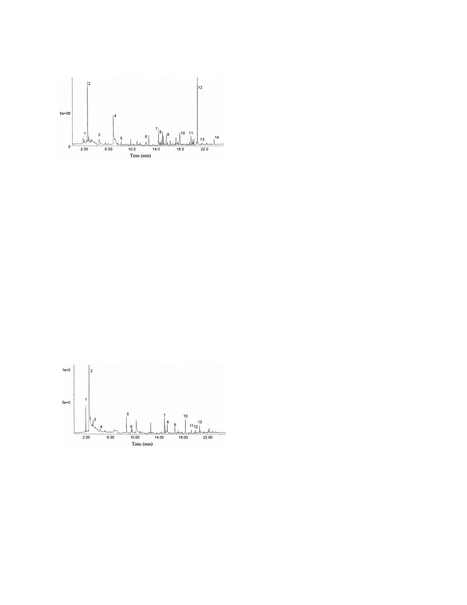
Fig. 1. Total ion current profile of volatile compounds extracted at
Like others [1,12], we noted variation in the range
acid pH from urine of a healthy adult on a normal mixed diet.
and concentrations of compounds excreted among
Urine was saturated with NaCl and acidified with HCl to pH 1–2.
individuals. Many components were food additives.
A 75 mm Carboxen–PDMS fibre was exposed to the HS vapour
for 30 min at 508C and desorbed for 2 min. Key: (1) methanethiol,
Some solvents may have been introduced as labora-
(2) acetone, (3) 2-pentanone, (4) dimethyldisulfide, (5) 4-hepta-
tory contaminants. As in the other reports, we found
none, (6) 2-methylmercaptofuran, (7) trans-linalol oxide, (8)
a large series of ketones which are probably decarb-
2-ethylhexanol, (9) vitispirane, (10) 1-a-terpineol, (11) p-cymen-
oxylation products of corresponding oxo-acids pro-
8-ol, (12) 2-ethylhexanoic acid, (13) phenol, (14) epoxy-butylated
duced in the urine or as analytical artefacts [3]. The
hydroxytoluene.
source of the precursor oxo-acids is uncertain. Some
are probably products of bacterial metabolism in the
tigations, we selected an extraction time of 30 min
colon, since the ketones were found in much higher
and temperature of 508C.
concentration in urine of conventional than germ-free
rats [14].
3.4. Profiles of volatile compounds in normal
One ketone, 4-heptanone, was found in low con-
urine samples
centration in most normal samples, often with a
small peak of 2-heptanone. Its origin is unknown, but
Ten normal urine samples were analysed at acid
it is probably from an exogenous source [6]. It has
and alkaline pH. Representative profiles are shown in
been reported as a metabolite of 2-ethylhexanol in
Figs. 1 and 2, respectively. The profiles differed
rats [36], a solvent used in the manufacture of
under acidic and alkaline conditions. Acids and
plasticisers and also released by hydrolysis from
bis(2-ethylhexyl)phthalate and bis(2-ethylhexyl)adi-
pate, plasticisers added to PVC to make it flexible
[36–38]. We have evidence (to be reported separ-
ately) that it is produced from the in vivo metabo-
lism of plasticisers in man. Sources for normal
subjects would include food contamination by PVC
contact films [39]. 2-Ethylhexanol and 2-ethylhex-
anoic acid were also found universally in our sam-
ples. We found that these were introduced as con-
taminants from the PVC containers used for urine
storage.
Fig. 2. Total ion current profile of volatile compounds extracted at
alkaline pH from urine of a healthy adult on a normal mixed diet.
3.5. Clinical studies
Urine was saturated with K CO and alkalinised with KOH to pH
2
3
12–14 and analysed using conditions as described in Fig. 1. Key:
We analysed urine from patients with metabolic
(1) trimethylamine, (2) acetone, (3) 2-butanone, (4) 2-pentanone,
disorders with predicted accumulation of volatile
(5) 1-butanol, (6) pyridine, (7) 2-ethylhexanol, (8) 1H-pyrrole,
compounds. Cases have been selected to show the
(9) seudenone, (10) car-2-en-4-one, (11) 1-methyl-2-piperidinone,
(12) benzylalcohol, (13) 2-ethylhexanoic acid.
versatility of the method.

264
G
.A
.
Mills
,
V
.
W
alker
/
J
.
Chromatogr
.
B
753
(2001
)
259
–
268
Table 1
a
Compounds identified in urine from 10 healthy adults on a normal mixed diet analysed at acid pH (1–2) and alkaline pH (12–14)
Acids
Aldehydes and
Amines
Food
Ketones
N-Hetero
O-Hetero
Solvents and
Sulfur compounds
alcohols
contaminants
Acetic
Benzaldehyde
Dimethylamine
Benzoic acid
Acetone
2-Cyano-2-butene
2-Acetylfuran
2-Butoxyethanol
Dimethyldisulfide
Acetic acid ethyl ester
Benzylalcohol
Trimethylamine
Car-2-en-4-one
2-Butanone
2,5-Dimethylpyrazine
2,5-Dimethylfuran
Chloroform
Dimethyltrisulfide
n-Butyric
1-Butanol
1-Carveol
3-Dimethyl-2-cyclo-
1-Me-2-piperidinone
2-Ethyl-5-mefuran
1-Ethyl-2,3-
1,3-Dithiacyclohexene
Butyric acid butyl ester
n-Hexanal
3-Carvo-menthenone
pentene-1-one
3-Mepyridine
2-Furanmethanol
dimethylbenzene
3-Isothiocyanato-1-propene
Formic
1-Hexanol
Isocineole
2-Heptanone
Me-1-pyrrole
2-Mefuran
2-Ethylhexanoic acid
5-Meisothiazole
Isovaleric / 2-mebutyric
3-Me-3-buten-1-ol
p-Cresol
4-Heptanone
Mepyrazine
2-Ethylhexanol
Me-2-propenyl-disulfide
n-Nonanoic
5-Me-3-hexanol
Cuminylalcohol
3-Hexanone
Nicotine
Styrene
Me-propyldisulfide
n-Octanoic
1-Octanol
Cymenene
3-Mecyclohexanone
Piperidine
Thymol
Methanethiol
Phenol
p-Cymene-8-ol
2-Me-2-cyclopentene-1-one
Pyrazine
Toluene
2-Methylmercaptofuran
b-Damascenone
3-Me-2-pentanone
Pyridine
Xylene
Dihydromyrcenol
4-Me-2-pentanone
1H-Pyrrole
trans-Edulan
4-Me-3-pentene-2-one
2-Vinylpyrazine
Epoxy-butylated
2-Pentanone
hydroxytoluene
Eucarvone
Furan
b-Ionone
a-Isophorone
Isopropyltoluene
Linalool
trans-Linalol oxide
Linaloyl oxide
Megastigmatrienone
Menthol
Phenol
Pinane
b-Pinene
Pulegone
Santene
Seudenone
a-Terpineol
1-a-Terpinene
g-Terpinene
Terpinene-1-ol
4-Terpinol
Vitispirane
a
Extraction and analytical conditions as described in Fig. 1 (acid) and Fig. 2 (alkaline). Key: me5methyl.
G
.A. Mills, V. Walker / J. Chromatogr. B 753 (2001) 259 –268
265
3.5.1. Case 1: Severe fasting ketoacidosis
A 5 year old boy was admitted acutely with
drowsiness, dehydration and severe ketoacidosis
after vomiting repeatedly for 24 h. Surgical and
likely inherited metabolic disorders were excluded.
Fig. 3 shows the profile of his alkalinised urine. The
major peak was acetone, the decarboxylation product
of acetoacetic acid. The origin of the unsaturated
ketones which were also present [3-pentene-2-one,
3-hexene-2-one (very large) and 3-heptene-2-one] is
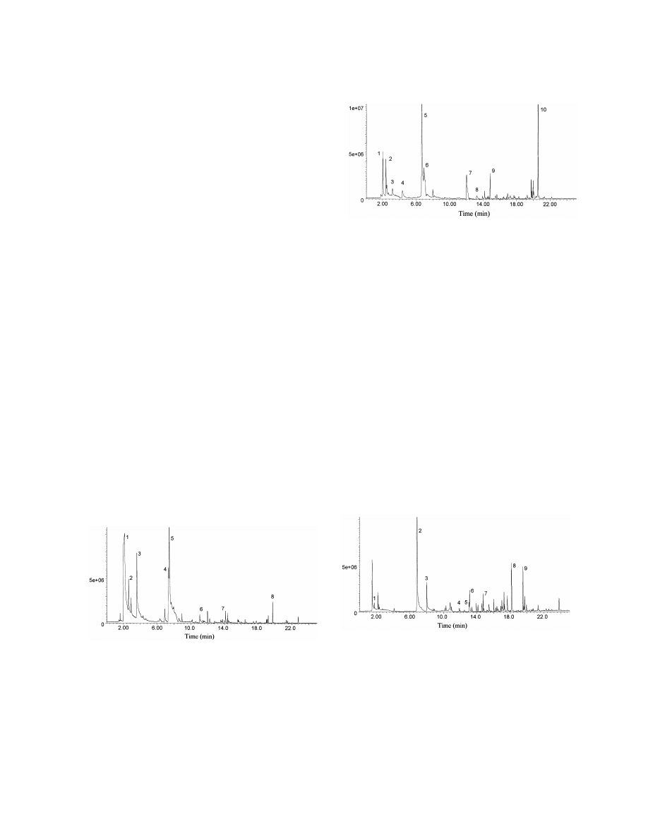
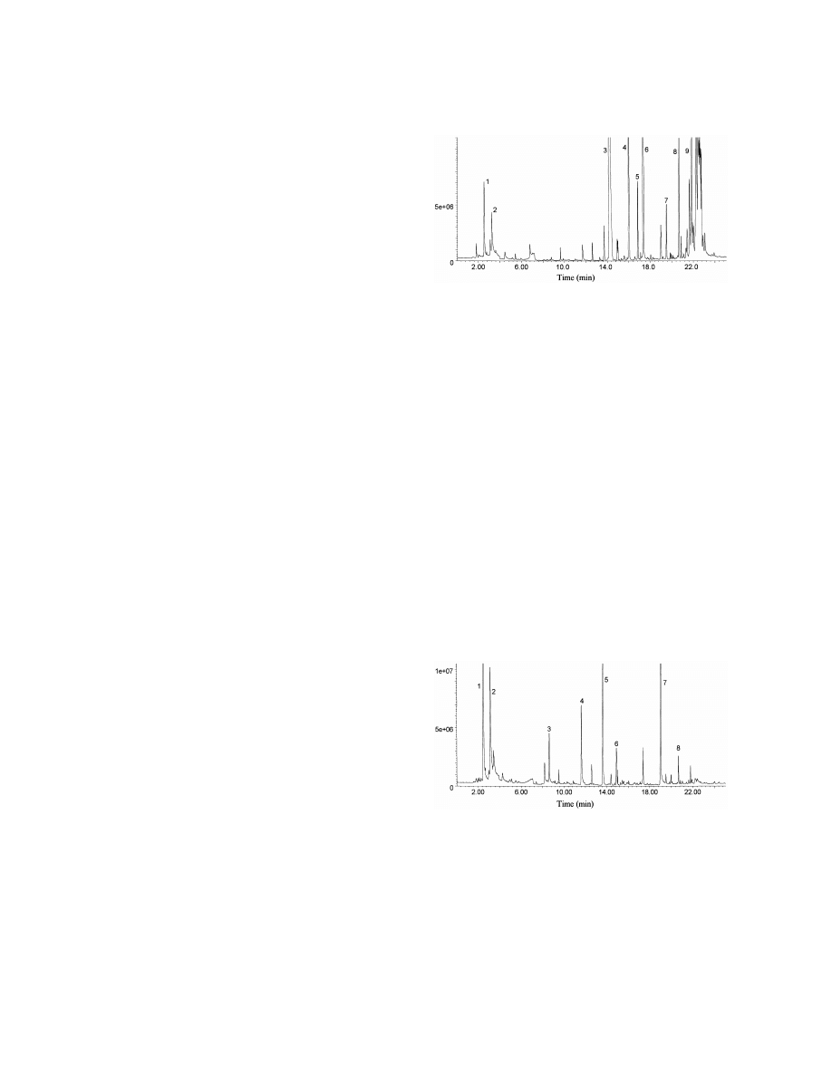
Fig. 4. Total ion current profile of volatile compounds extracted at
unknown. They are probably decarboxylation prod-
acid pH from urine of a 37 year old man with homocystinuria
ucts of the corresponding unsaturated oxo-acids. 3-
(case 2). Analytical conditions were as described in Fig. 1. Key:
Pentene-2-one was found in urine of starved [15] and
(1) methanethiol, (2) furan, (3) 2-butanone, (4) 2-pentanone, (5)
dimethyldisulfide, (6) hexanal, (7) 2-methylmercaptofuran, (8)
diabetic rats [3], but 3-hexene-2-one has not been
dimethyltrisulfide, (9) 2-ethylhexanol, (10) 2-ethylhexanoic acid.
reported to our knowledge. We have preliminary
evidence that it may be produced via condensation of
propanal with acetoacetate. Further studies to con-
a 37 year old man with homocystinuria, treated with
firm this are underway.
betaine, who was well. His plasma methionine was
1115 mmol / l (reference range,95 mmol / l). An
3.5.2. Case 2: Homocystinuria (McKusick 23620)
enormous peak of dimethyldisulfide is accompanied
In this disorder an inherited deficiency of the
by large peaks of methanethiol and 2-methylmercap-
enzyme cystathionine b-synthase [EC 4.2.1.22] pre-
tofuran and low concentrations of other sulfur com-
vents catabolism of methionine by trans-sulfuration.
pounds. A similar profile was obtained for his 34-
Homocysteine and methionine accumulate. Treat-
year-old brother, also treated with betaine for
ment includes betaine to reduce the plasma homo-
homocystinuria.
cysteine (by methylation to methionine) to prevent
thromboses [40]. The accumulating methionine is
3.5.3. Case 3: Decompensated alcoholic hepatitis
channelled through a minor transamination pathway
Fig. 5 is the profile of acidified urine from a 63
shown to produce methanethiol and other sulfides
year old woman with alcohol-induced liver failure.
[41]. Accumulation of these compounds would be
She
was
receiving
fluids
intravenously.
Di-
anticipated in homocystinuria during betaine treat-
methyldisulfide dominates the profile, and there are
ment. Fig. 4 shows the profile of acidified urine from
Fig. 5. Total ion current profile of volatile compounds extracted at
Fig. 3. Total ion current profile of volatile compounds extracted at
acid pH from urine of a 63 year old woman with liver failure (case
alkaline pH from urine of a 5 year old child with severe ketosis
3). Analytical conditions were as described in Fig. 1. Key: (1)
(case 1). Analytical conditions were as described in Fig. 1. Key:
methanethiol, (2) dimethyldisulfide, (3) 4-heptanone, (4) 2-
(1) acetone, (2) 2-butanone, (3) 2-pentanone, (4) 3-penten-2-one,
methylmercaptofuran, (5) dimethyltrisulfide, (6) 2-methyl-5-
(5) 3-hexene-2-one, (6) 3-heptene-2-one, (7) 2-ethylhexanol, (8)
methylthiofuran, (7) 2-ethylhexanol, (8) 3-carvomenthenone, (9)
2-ethylhexanoic acid.
p-cymen-8-ol.
266
G
.A. Mills, V. Walker / J. Chromatogr. B 753 (2001) 259 –268
smaller peaks of other sulfur compounds. These
increases were probably due partly to impaired
methionine catabolism through the trans-sulfuration
pathway because of the liver damage, with diversion
through the transamination pathway (see above). In
addition, decreased clearance of sulfur compounds
absorbed from the intestine from food [42] and
colonic bacterial metabolism [43–45] would be
contributory.
Analysis of volatile sulfur compounds is difficult.
Fig. 6. Total ion current profile of volatile compounds extracted at
In addition to their volatility, they adsorb to surfaces
acid pH from urine of a 4 day old baby with multiple acyl-CoA
readily, and may undergo partial oxidation or chemi-
dehydrogenase deficiency (case 4). Analytical conditions were as
cal changes during analysis, especially with lengthy
described in Fig. 1. Key: (1) acetone, (2) 2-butanone, (3) acetic
extraction procedures [46,47]. Mestres et al. used
acid, (4) isobutyric acid, (5) n-butyric acid, (6) isovaleric acid, (7)
n-hexanoic acid, (8) 2-ethylhexanoic acid, (9) a mixture of fatty
SPME with PDMS and PA [48] and Carboxen–
acid esters (probable artefacts).
PDMS [34] fibres to analyse sulfides, disulfides and
thiophene in the HS of wine. Carboxen–PDMS
fibres were best, with detection limits of 0.05–4.0
mg / l and recoveries close to 100%. We are unaware
formed during sample storage. The profile of alkalin-
of reported applications of SPME for sulfur com-
ised urine (Fig. 7) is strikingly different. Only small
pounds in biological fluids or breath.
amounts of short-chain acids were extracted and the
The large increase in urinary 4-heptanone found in
profile is dominated by very large peaks of the
case 3 could be explained by increased exposure to
plasticisers cyclohexanone, cyclohexanol and 2-(2-
2-ethylhexanol from the intravenous infusion [5,38].
butoxyethoxy)ethanol (butyl carbitol). Cyclohex-
anone is a solvent sealer for PVC used in many
3.5.4. Case 4: Multiple acyl-CoA dehydrogenase
medical devices, including dialysis tubing [5,50]. It
deficiency
(MADD) [Glutaric aciduria Type II
is reduced to cyclohexanol in vivo [51]. It is of
(McKusick 23168)]
interest that very little 4-heptanone was found in this
In this disorder, at least nine flavine dehydro-
baby’s urine. Because of his metabolic defect he
genases are inactivated because of an inherited
would be unable to produce this compound from
deficiency of electron transfer flavoprotein (ETF) or
2-ethylhexanol via the b-oxidation pathway [36].
ETF-coenzyme Q oxido-reductase [49]. A host of
metabolites accumulates, including volatile short-
chain carboxylic acids from branched-chain amino
acids and fatty acids. These contribute to metabolic
acidosis and cause an unpleasant body odour. Urine
was collected on the fourth (and last) day of life
from an acidotic term newborn baby with the most
severe form of the disorder. He received intensive
medical care which included parenteral nutrition,
peritoneal dialysis and haemofiltration and he was
therefore exposed to plasticisers from multiple
sources. Fig. 6 is the chromatogram of acidified
Fig. 7. Total ion current profile of volatile compounds extracted at
alkaline pH from urine of a 4 day old baby with multiple
urine. There are large peaks of short-chain fatty acids
acyl-CoA dehydrogenase deficiency (case 4). Analytical con-
and the profile is consistent with the diagnosis. A
ditions were as described in Fig. 1. Key: (1) acetone, (2) 2-
number of short-chain fatty acid esters eluted after
butanone, (3) 1-butanol, (4) cyclohexanone, (5) cyclohexanol, (6)
22 min which are not known endogenous metabo-
2-ethylhexanol, (7) 2-(2-butoxyethoxy)ethanol, (8) 2-ethylhex-
lites. They may have been analytical artefacts or
anoic acid.
G
.A. Mills, V. Walker / J. Chromatogr. B 753 (2001) 259 –268
267
3.5.5. Case 5: Medium-chain acyl-CoA
(isocaproic), 4-methylhexanoic and 5-methylhex-
dehydrogenase deficiency
(McKusick 201450)
anoic acids.
This is an inherited disorder of fatty acid oxidation
Volatile carboxylic acids accumulate abnormally
which may cause hypoglycaemia, brain swelling and
in inherited defects of branched-chain amino acids
death. C –C
fatty acids and their metabolites, and
and of short- and medium-chain fatty acid oxidation.
6
12
carnitine, glucuronide and glycine conjugates ac-
Some are detoxified in vivo by conjugation with
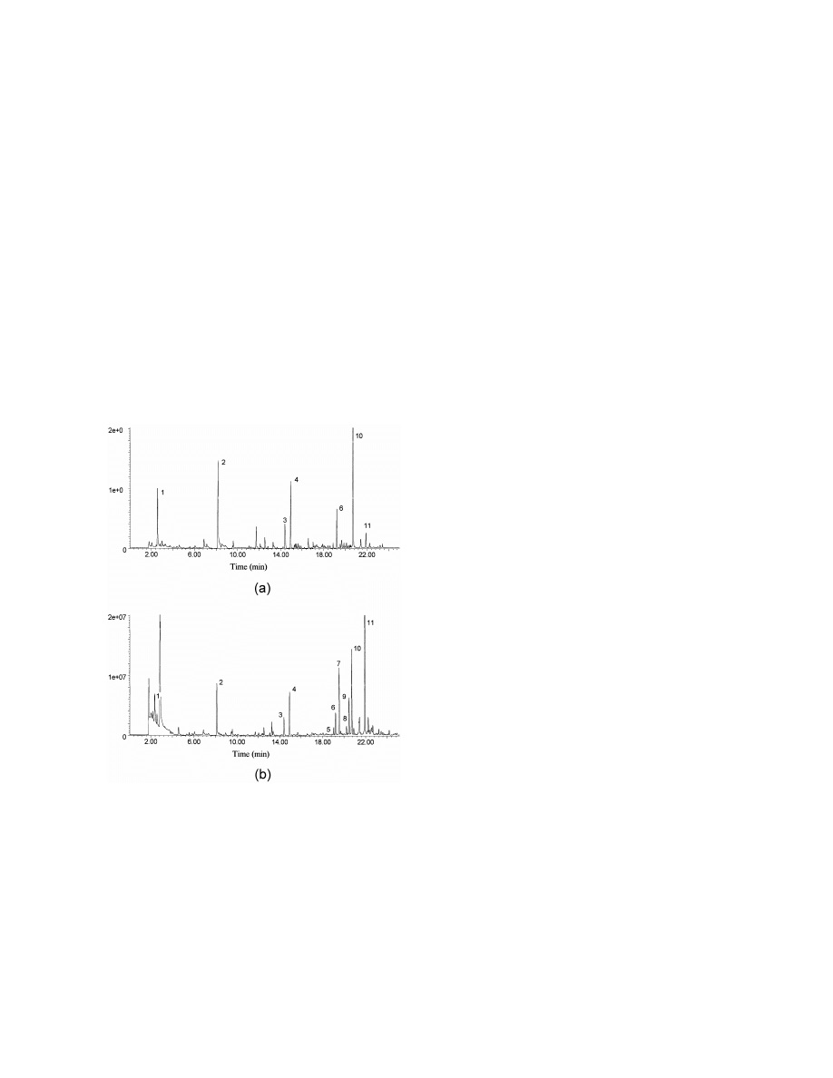
cumulate [52]. Fig. 8a shows the profile of acidified
carnitine, glucuronic acid or glycine. Metabolic
urine collected from a 4 year old boy on the day of
laboratories diagnose these disorders using GC–MS
admission following a respiratory arrest at home.
to detect the glycine conjugates and other, non-
There are small peaks of n-hexanoic acid, n-octanoic
volatile, organic acids increased in the diseases, and
acid and 5-hydroxyhexanoic lactone which we do not
tandem MS to detect acylcarnitines in body fluids.
find in urine of normal children. After alkaline
Few have facilities to analyse the free volatile acids,
incubation to hydrolyse glycine and carnitine conju-
yet these contribute to the clinical problems. There
gates (Fig. 8b), there were large increases in n-
are relatively few reports of volatile acid profiles in
hexanoic and n-octanoic acids and small peaks of a
these disorders [8,10,19,20]. With acidic extraction,
series of methyl acids appeared: 4-methylpentanoic
we found all the diagnostic volatile carboxylic acids
in urine from a baby with MADD (case 4). Medium-
chain carboxylic acids were found in acidified urine
from the patient with MCAD deficiency (case 5) but
were not diagnostic. However, the profile was clearly
abnormal after alkaline hydrolysis of conjugates. The
appearance of methylcarboxylic acids was interest-
ing.
4-Methylpentanoic
and
4-methylhexanoic
glycine conjugates were identified in urine in MCAD
deficiency [31]. Carnitine conjugates of these acids
and of 5-methylpentanoic acid have also been iden-
tified in urine after hydrolysis and derivatisation
[32]. They may be metabolites of branched-chain
fatty acids (e.g., phytanic acid) and are specific for
MCAD deficiency [31]. There are no reports yet of
their identification by tandem MS [32].
4. Conclusion
The five cases presented demonstrate that HS-
SPME analysis of urine can complement profiling of
non-volatile compounds (e.g., organic and amino
acids) in investigations of metabolic disturbances.
Furthermore, it can be used to profile other bio-
Fig. 8. Total ion current profiles of volatile compounds from urine
logical matrices (e.g., blood and faeces [29]) offering
of a 4 year old child with medium-chain acyl-CoA dehydrogenase
deficiency (case 5). Analytical conditions were as described in
a more comprehensive overview of intermediary
Fig. 1. (a) Extracted at pH 1–2, (b) after alkaline hydrolysis and
metabolism. HS-SPME is versatile, simple to carry
re-acidification to pH 1–2. Key: (1) acetone, (2) 4-heptanone, (3)
out and does not require special equipment other
acetic acid, (4) 2-ethylhexanol, (5) 4-methylpentanoic acid, (6)
than a GC–MS system, which is standard for meta-
5-hydroxyhexanoic lactone, (7) n-hexanoic acid, (8) 5-methylhex-
bolic laboratories. Its introduction into clinical analy-
anoic acid, (9) 4-methylhexanoic acid, (10) 2-ethylhexanoic acid,
(11) n-octanoic acid.
sis would expand our knowledge of the metabolism

268
G
.A. Mills, V. Walker / J. Chromatogr. B 753 (2001) 259 –268
¨
[26] H.M. Liebich, E. Gesele, J. Woll, J. Chromatogr. B 713
of volatile compounds, which is currently very
(1998) 427.
incomplete.
[27] D.A. Volmer, J.P. Hui, Rapid Commun. Mass Spectrom. 11
(1997) 1926.
[28] F.L. Cardinali, D.L. Ashley, J.V. Wooten, J.M. McCraw, S.W.
References
Lemire, J. Chromatogr. Sci. 38 (2000) 49.
[29] G.A. Mills, V. Walker, H. Mughal, J. Chromatogr. B 730
(1999) 113.
[1] A. Zlatkis, R.S. Brazell, C.F. Poole, Clin. Chem. 27 (1981)
[30] R. Hyspler, S. Crhova, J. Gasparic, Z. Zadak, M. Cizkova, V.
789.
Balasova, J. Chromatogr. B 739 (2000) 183.
[2] T. Niwa, J. Chromatogr. 379 (1986) 313.
[31] J.J. Pitt, J. Inherit. Metab. Dis. 16 (1993) 392.
[3] G. Rhodes, M.L. Holland, D. Wiesler, M. Novotny, S.A.
[32] R. Libert, F. Van Hoof, M. Thillaye, M.-F. Vincent, M.-C.
Moore, R.G. Peterson, D.L. Felten, J. Chromatogr. 228
Nassogne, V. Stroobant, E. de Hoffman, A. Schanck, J.
(1982) 33.
Inherit. Metab. Dis. 22 (1999) 9.
[4] Y. Ghoos, D. Claus, B. Geypens, M. Hiele, B. Maes, P.
[33] M. Abalos, J.M. Bayon, J. Pawliszyn, J. Chromatogr. A 873
Rutgeerts, J. Chromatogr. A 665 (1994) 333.
(2000) 107.
¨
[5] H.G. Wahl, A. Hoffmann, H.-U. Haring, H.M. Liebich, J.
[34] M. Mestres, M.P. Marti, O. Busto, J. Guasch, J. Chromatogr.
Chromatogr. A 847 (1999) 1.
A 849 (1999) 293.
[6] H.G. Wahl, A. Hoffmann, D. Luft, H.M. Liebich, J. Chroma-
[35] P.G. Hill, R.M. Smith, J. Chromatogr. A 872 (2000) 203.
togr. A 847 (1999) 117.
[36] P.J. Deisinger, R.J. Boatman, D. Guest, Xenobiotica 24
[7] R.A. Chalmers, in: J.B. Holton (Ed.), The Inherited Metabol-
(1994) 429.
ic Diseases, Churchill Livingstone, Edinburgh, 1987, p. 141.
[37] R.J. Rubin, P.M. Ness, Transfusion 29 (1989) 358.
[8] D.N. Buchanan, J.G. Thoene, Clin. Chim. Acta 145 (1985)
[38] S.L. Plonait, H. Nau, R.F. Maier, W. Wittfoht, M. Obladen,
183.
Transfusion 33 (1993) 598.
[9] M. Stafford, M.G. Horning, J. Chromatogr. 126 (1976) 495.
[39] N.J. Loftus, B.H. Woollen, G.T. Steel, M.F. Wilks, L. Castle,
[10] D.A. Maltby, D.S. Millington, Clin. Chim. Acta 155 (1986)
Food Chem. Toxicol. 32 (1994) 1.
167.
[40] S.H. Mudd, H.L. Levy, F. Skovby, in: C.R. Scriver, A.L.
[11] H.J. Bestmann, K. Haberkorn, O. Vostrowsky, R. Ferstl, F.
Beaudet, W.S. Sly, D. Valle (Eds.), The Metabolic and
Eggert, Z. Naturforsch. 51C (1996) 849.
Molecular Bases of Inherited Disease, 7th ed., McGraw Hill,
[12] A. Zlatkis, W. Bertsch, H.A. Lichtenstein, A. Tishbee, F.
New York, 1995, p. 1279.
Shunbo, H.M. Liebich, A.M. Coscia, N. Fleischer, Anal.
[41] H.J. Blom, J.P.A.M. van den Elzen, S.H. Yap, A. Tangerman,
Chem. 45 (1973) 763.
Biochim. Biophys. Acta 972 (1988) 131.
[13] I.R. Politzer, B.J. Dowty, J.L. Laseter, Clin. Chem. 22
[42] J.G. Moore, L.D. Jessop, D.N. Osborne, Gastroenterology 93
(1976) 1775.
(1987) 1321.
[14] M. Holland, G. Rhodes, M. DalleAve, D. Wiesler, M.
[43] S. Chen, L. Zieve, V. Mahadevan, J. Lab. Clin. Med. 75
Novotny, Life Sci. 32 (1983) 787.
(1970) 628.
[15] M. Yancey, M.L. Holland, R. Stuart, D. Wiesler, M.
¨
[44] P. Jappinen, J. Kangas, L. Silakoski, H. Savolainer, Arch.
Novotny, J. Chromatogr. 382 (1986) 3.
Toxicol. 67 (1993) 104.
[16] A. Zlatkis, C.F. Poole, R. Brazell, K.Y. Lee, F. Hsu, S.
[45] W.E.H. Roediger, A. Duncan, O. Kapaniris, S. Millard,
Singhawangcha, Analyst 106 (1981) 352.
Gastroenterology 104 (1993) 802.
[17] G. Rhodes, M.L. Holland, D. Wiesler, M. Novotny, S.A.
[46] R.H. Waring, S.C. Mitchell, G.R. Fenwick, Xenobiotica 17
Moore, R.G. Peterson, D.L. Felten, Experientia 38 (1982)
(1987) 1363.
75.
[47] S.-W. Myung, S. Huh, J. Kim, Y. Kim, M. Kim, Y. Kim, W.
[18] M.L. Holland, G.R. Rhodes, D. Wiesler, M. Novotny, J.
Kim, B. Kim, J. Chromatogr. A 791 (1997) 367.
Chromatogr. 306 (1984) 23.
[48] M. Mestres, O. Busto, J. Guasch, J. Chromatogr. A 808
[19] J.H. Menkes, J. Pediatr. 69 (1966) 413.
(1988) 211.
[20] D.G. Burke, B. Halpern, D. Malegan, E. McCairns, D.
[49] F.E. Frerman, S.I. Goodman, in: C.R. Scriver, A.L. Beaudet,
Danks, P. Schlesinger, B. Wilken, Clin. Chem. 29 (1983)
W.S. Sly, D. Valle (Eds.), The Metabolic and Molecular
1834.
Bases of Inherited Disease, 7th ed., McGraw Hill, New York,
[21] C. Arthur, J. Pawliszyn, Anal. Chem. 62 (1990) 2145.
1995, p. 1611.
[22] J. Pawliszyn, Solid Phase Microextraction – Theory and
[50] M. Sakata, J. Kikuchi, M. Haga, Clin. Toxicol. 27 (1989) 67.
Practice, Wiley–VCH, Chichester, 1997.
[23] S.C. Scheppers Wercinski (Ed.), Solid Phase Microextraction
[51] T.H. Elliott, D.V. Parke, R.T. Williams, Biochem. J. 72
– A Practical Guide, Marcel Dekker, New York, 1999.
(1959) 193.
[24] J. Pawliszyn (Ed.), Applications of Solid Phase Microextrac-
[52] C.R. Roe, P.M. Coates, in: C.R. Scriver, A.L. Beaudet, W.S.
tion, Royal Society of Chemistry, Cambridge, 1999.
Sly, D. Valle (Eds.), The Metabolic and Molecular Bases of
[25] G.A. Mills, V. Walker, H. Mughal, J. Chromatogr. B 723
Inherited Disease, 7th ed., McGraw Hill, New York, 1995, p.
(1999) 281.
1501.
Wyszukiwarka
Podobne podstrony:
Development of a headspace solid phase microextraction–gas c
Solid Phase Microextraction Analyses of Flavor Compounds in
Solid phase microextraction to concentrate volatile products
Headspace solid phase microextraction for the determination
Optimisation of solid phase microextraction of volatiles
Application of Solid Phase Microextraction Gas Chromatograp
Solid phase microextraction for the detection of termite cut
Kinetics of solid phase extraction and solid phase microextr
Applications of solid phase microextraction to
Comparison of Different Fibers in the Solid Phase Microextra
Application of solid phase microextraction to the analysis o
Solid phase microextraction for the analysis of biological s
Solid phase microextraction as a clean up and preconcentrati
Solid phase microextraction as a tool for trace element spec
A Practical Guide to Quantitation with Solid Phase Microextr
Vinyl chloride analysis with Solid Phase Microextraction
Solid phase microextration in biomedical analysis
Solid phase microextraction for herbicide determination in
Solid phase microextraction a promising technique for sample
więcej podobnych podstron