
Presenting Toxicology Results

CONTRIBUTING EDITORS
Graham Copping
Monique Y.Wells

Presenting
Toxicology Results:
How to evaluate data and write reports
EDITED BY GERHARD J.NOHYNEK

UK
Taylor & Francis Ltd, 1 Gunpowder Square, London EC4A 3DE
USA
Taylor & Francis Inc., 1900 Frost Road, Suite 101, Bristol, PA 19007
This edition published in the Taylor & Francis e-Library, 2002.
Copyright © Taylor & Francis Ltd 1996
All rights reserved. No part of this publication may be reproduced, stored in a
retrieval system, or transmitted, in any form or by any means, electronic,
electrostatic, magnetic tape, mechanical, photocopying, recording or otherwise,
without the prior permission of the copyright owner.
British Library Cataloguing in Publication Data
A catalogue record for this book is available from the British Library
ISBN 07484 0476 7 (Print Edition)
Library of Congress Cataloging Publication data are available
Cover design by Amanda Barragry
ISBN 0-203-48326-X Master e-book ISBN
ISBN 0-203-79150-9 (Glassbook Format)

This book is dedicated to my son Florian


vii
Contents
Contributors
page ix
Preface
xi
G.J.Nohynek
Foreword
xiii
W.Kluwe
Acknowledgments
xv
1.
Basic Biomedical English
1
D.Young
2.
The Structure of Toxicology Reports
17
G.J.Nohynek
3.
Writing the Report Summary
21
G.J.Nohynek
4.
General Principles of Regulatory Toxicology Report Writing
27
G.J.Nohynek and A.Lodola
5.
Plasma Drug Concentrations (Toxicokinetic Data)
41
R.J.Szot
6.
Reporting In-life Observations and Measurements
49
G.J.Nohynek
7.
Reporting Clinical Pathology Results
63
M.Y.Wells and S.Gosselin

Contents
viii
8.
Anatomic Pathology
75
T.Hodge and S.Gosselin
9.
Developmental and Reproductive Toxicology
89
R.L.Clark and G.Copping
10.
Example of a General Toxicology Report
99
G.J.Nohynek, M.Y.Wells, R.J.Szot and S.Gosselin
11.
Example of a Reproductive Toxicology Report
113
G.Copping and R.L.Clark
12.
Using References
133
13.
References and Recommended Reading
135
Index
137

ix
Contributors
Robert L.Clark
Associate Director of Toxicology, Head of Reproductive Toxicology,
Rhône-Poulenc Rorer Research and Development,
500 Arcola Road, PO Box 1200, Collegeville, PA 19426–0107, USA
G.Copping
Director of Operations, Drug Safety Department, Rhône-Poulenc Rorer,
Centre de Recherche de Vitry-Alfortville, 13 Quai Jules Guesde BP 14
F-94403 Vitry sur Seine Cedex, France
S.Gosselin
Director of Toxicology, ITR Laboratories Canada Inc.,
19601 Boulevard Clark Graham, Baie d’Urfé, Montreal, Quebec, Canada
T.Hodge
Senior Pathologist, Acting Director of Pathology,
Drug Safety Department, Rhône-Poulenc Rorer,
Centre de Recherche de Vitry-Alfortville, 13 Quai Jules Guesde BP 14
F-94403 Vity sur Seine Cedex, France
W.Kluwe
Director Drug Safety Department, Pfizer Central Research, Eastern Point Road,
Groton, CT 06340, USA
A.Lodola
Director of Toxicology, Pfizer Centre de Recherche,
Laboratoires Pfizer, BP 159–137401, Amboise Cedex, France
G.J.Nohynek
Principal Toxicologist, L’Oreal, Centre Charles Zviak, 90 rue du General Roguet,
F-92583 Clichy Cedex, France

Contributors
x
R.J.Szot
Consultant in Toxicology, 2 Rolling Lane, Flemington, NJ 08822, USA
Monique Y.Wells
Senior Pathologist, Drug Safety Department, Rhône-Poulenc Rorer,
13 Quai Jules Guesde, BP F-94403 Vitry sur Seine Cedex, France
D.Young
Scientific Writer, 66 Avenue de Generale de Gaulle, F-17690 Angoulines sur mer,
France

xi
Preface
In response to proliferating regulatory safety requirements for chemicals, pesticides,
food ingredients and drugs, thousands of regulatory toxicology studies are
performed and reported annually in laboratories of the industrial world. A
toxicology laboratory can be regarded as a production unit, and the toxicology
report as its final product. The end users are reviewers, who will use these reports
to make decisions regarding the safety of the test compounds. The reviewer may
be a regulator, a clinical investigator, the author of a texicological/pharmacological
expert report, or an attorney who is representing a complainant in litigation against
the company based on an alleged adverse effect of the compound. No reviewer
will be completely objective. The reviewer’s opinion regarding the safety of a
test compound will not be based solely on the results of the toxicology report;
the quality of the report, its readability and presentation may also bias the
reviewer’s evaluation.
Therefore it is of prime importance that the results of a toxicology study, and
their interpretation, are easy to follow. Consequently, writing a toxicology report
is a considerable challenge for the communication skills of the author. Reporting
and discussing the results of toxicology studies which may include a myriad of
numerical and diagnostic results is a difficult task and requires synthesis and
interpretation of multidisciplinary and complex results. However, toxicologists must
keep their reports concise, accurate and focused on the conclusion which is best
supported by the study data.
English has become the standard language used for reporting toxicological safety
evaluations. Given the location of many laboratories in non-English-speaking
countries, precise and comprehensive use of scientific English poses an additional
hurdle for toxicologists who do not have English as their first language and who
may have had no formal training in scientific writing.
To these ends we have compiled this Guide for the evaluation, reporting and
discussion of results of toxicology studies.
We have deliberately attempted to keep the Guide simple and practical and have
included numerous examples. All examples were modeled on actual toxicology

Preface
xii
reports which originated from established pharmaceutical or contract toxicology
laboratories and were written by native English speakers. However, the designation,
activity and therapeutic classes of the respective test compounds have been changed,
given the confidential and proprietary nature of the original data. Please note that
the sole criterion for selection of examples was not elegance of prose but clarity.
In addition, we have compiled two sample toxicology reports, which can be found
in Chapters 10 and 11 of the Guide. These examples are fictitious reports and do
not describe toxicity of existing developmental compounds.
This Guide covers only a narrow range of the wide field of toxicology. Given
the affiliation of all authors with the pharmaceutical industry, all examples refer
to drug toxicology evaluations; examples for studies on chemicals, agrochemicals
or food ingredients are not presented. However, regulatory studies performed on
chemicals and food ingredients are similar to those done for pharmaceutical
compounds and pose similar problems with regard to evaluation and reporting. In
addition, because of the limited scope of this book, many types of toxicity evaluations,
such as ecotoxicology studies, genetic toxicity studies, immunotoxicology studies
and exploratory toxicology studies, were not addressed.
The principal aim of the Guide is to aid scientists wishing to improve their report
writing and to propose a framework for reporting results of toxicology studies and
their discussion. We emphasize that this Guide is not designed as a Standard
Operating Procedure for writing of toxicology reports. It is not our intent to constrain
scientists to follow a rigid procedure, or to use a standardized vocabulary or structure.
Everyone familiar with toxicology studies will accept that each study is unique
and may require adjustment of the report’s structure, style or content to the methods
and results of the study—and this may become apparent to the critical reader who
may discover that the examples given in this Guide do not always religiously adhere
to the recommendations of the respective chapters.
Finally, we would like to encourage all readers to send us their comments or
proposals to make this Guide more comprehensive, complete and—of course—
more readable.
Gerhard J.Nohynek
Alfortville, 29 March 1996

xiii
Foreword
W.KLUWE,
Pfizer Central Research, Drug Safety Department, Groton, USA
Science is organized knowledge.
Herbert Spencer, 1861
Language is the dress of thought.
Samuel Johnson, 1759
How better to introduce a book providing guidance on organizing and writing technical
reports than to quote philosophers whose wisdom is ever more appreciated with the
passage of time? Generation of knowledge without benefit of presentation or
preservation is of little usefulness other than to its originators, and Spencer reminds
us that science progresses only as the knowledge obtained is organized for universal
understanding. In a similar manner, Johnson reminds us that our ideas and insight
are translated to others through the language we choose to communicate them.
Communication can be considered the critical capability differentiating live from
inanimate. From the most basic contact between individual cells to the complex
matrices of sight, sound, smell and touch achieved by the more highly developed
species, good communication describes that uncommon circumstance where the
message received bears close resemblance to the message sent. Too often the
message’s reception, or its higher stage of assimilation, perception, differs from
the message’s intent because unrecognized failures in communication erode clarity.
This invites speculation, a dreaded hazard in regulatory toxicology reports. More
than one “battle” has been lost, or a “war” begun, due to poor communication
amongst those writing toxicology reports and those assessing the significance of
the information provided.
This, then, is a book about communication, and the challenges we scientists
face in communicating highly technical biological information, with inferences
regarding human, in a clear and unambiguous manner. It is a formidable task for
those authors writing in their native language, with complete awareness of cultural
nuances and contemporary terminology. For neither a literary education nor detailed
scientific training provides much guidance on how to ensure that an audience
perceives a message in a manner consistent with the intent of the presenter. How
much greater a challenge, then, for those reporting in a language other than that
which they normally use for daily communication? Technical reports need to state

Foreword
xiv
facts and provide vital information in a format that facilitates universal and singular
understanding.
For better or for worse, the language most widely accepted for the exchange of
scientific information, including regulatory toxicology reports, is English. Favored
in many diverse countries, standard English is rife with idioms, colloquialisms,
slang, jargon and terms that fade in and out of use—in short, a more dynamic
language than one would have consciously chosen for scientific communication.
Yet, expression of the knowledge gained from experimentation, conclusions derived
therefrom, and extrapolation to the safety of clinical use of drugs is not only possible
in English, but quite widely accomplished.
As described in this book, the best approach for communicating toxicology results
is to first organize one’s data and thoughts, then list the important points to be
made, and, finally, write out the information in a format and style consistent with
logic and simple understanding. Recognizing that multiple toxicology reports often
follow in chronological sequence, the authors also must be wary in drawing
conclusions that could be superseded by subsequent studies. Now, with the proper
frame of mind, the report writer is ready to begin his task.
Readers will find general advice on how to organize the major sections of a
regulatory toxicology report in Chapters 2–8 of this book. Included are formats,
definitions and guidance on how to express numerical and more subjective findings
in a manner that supports the authors’ interpretations. The advice given is equally
valuable to readers of regulatory toxicology reports, such as reviewing bodies,
governmental representatives and even management from other parts of the authors’
organization. Chapter 1 (“Basic Biomedical English”) provides a concise and
extremely beneficial seminar on proper use of terminology. Even those with English
as a native language will benefit from a careful reading of this chapter. The very
specific types of information collected in reproductive and developmental toxicology
studies are addressed separately in Chapter 9, including comprehensive formats
for data presentation and definitive terminology. Finally, examples of complete
study reports are provided in Chapters 10 and 11, allowing this book’s authors to
show by example how to incorporate disparate data sets into a coherent report.
And now, let us proceed with the task at hand: regulatory toxicology report
writing.

xv
Acknowledgments
This Guide resulted from the exemplary co-operation of an independent scientific
writer with a group of toxicologists and pathologists from six different toxicology
laboratories in France, Canada, the USA and the UK. I thank all of them for their
dedication and effort.
Special thanks to Denise Munday for her help in reviewing the manuscript and
her astute and common-sense suggestions. Françoise Roquet, William Kluwe and
Peter MacAnulty, who performed the final review, deserve much credit for their
valuable advice and comments. I am particularly grateful to Peter for his clarification
of the term ‘reduction’ which I (and other authors of the Guide) may have abused
for many years. The contributors to the Guide, my colleagues and family also merit
special mention for their tolerance and good humor while enduring my co-ordination
and periods of frustration and bad temper. Many thanks to Graham Copping and
Monique Wells who came to my aid when I was ready to quit. Lastly, I acknowledge
the support of my son Florian (age 12) who made numerous suggestions—to his
disappointment, not all of them could be included —and contributed to the progress
of the manuscript by granting access, albeit limited, to his personal computer.


1
1
Basic Biomedical English
DAVID YOUNG
F-17690 Angoulines sur mer, France
1.1
Introduction
Writing toxicology reports is difficult in any language, as it involves condensing
weeks to months of work into a few thousand well-chosen words.
The need to write reports in English is both an obstacle and a challenge for the scientist
whose native language is not English. Yet after checking about five thousand manuscripts
for syntax and overall coherence I have come to the conclusion that grammar and vocabulary
are secondary problems in scientific writing. Basic English grammar is simple, and scientific
prose is one of the simplest forms in any language; the general vocabulary is that of a
10-year-old child, and there is no need for idioms, metaphors, etc.
From a grammatical point of view, all that is required to write a scientific report
in English is a good knowledge of tenses, comparatives and conditionals (would,
will, should, etc.), a non-technical vocabulary of a few hundred words, and an awareness
of words that are spelled the same way in English and one’s own language but have
different meanings.
All scientists who write in English read research papers in English-language
journals and thus have a basic knowledge of the language. So why is it that so
many scientists have trouble writing coherently and intelligibly? Clearly a large
part of the problem is that scientists are rarely trained to express their ideas on
paper. Often there is too little thought before putting pen to paper, and a lack —
or an excess—of confidence on the part of the writer.
One pitfall when writing reports in a foreign language is the notion that vague
ideas can be masked by vague word usage. Reviewers and editors are hypersensitive
to imprecision and incoherence; and, as Sir Ernest Groves remarked, “What appears
to be sloppy and meaningless use of words may well be a completely correct use
of words to express sloppy and meaningless ideas”. Data clearly speak louder than
words, so once you have obtained your results, relax. There is no need to mask
what you think in a cloud of conditionals, or to produce a literary work of art.

Presenting toxicology results
2
1.2
Myths
Certain myths hinder the simplification of scientific English. One is that American English
and British English differ fundamentally. In fact the only valid distinction is that between
good and bad style; good style is easy to understand and avoids word usage that distracts
the reader’s attention. Another myth is that English is better adapted than other languages
to scientific writing. A similar notion prevailed some eighty years ago about German
and, in the Middle Ages, about Latin. The only important difference, however, is that
an English paper is about 10 per cent shorter than a paper written in French or German,
for example. Otherwise a well-written paper in French or any other language may be
just as clear, concise and pleasant to read as a well-written paper in English.
Today, all important journals in the field of toxicology are written in English.
English has become such a standard that, for instance, a Swiss toxicology laboratory
employing predominantly German- or French-speaking personnel may charge
additional fees for reports written in these languages.
1.3
Brevity, Clarity and Coherence
One of the best ways to produce catastrophic sentences is to write in one’s own
language first and then translate into English. It is always preferable to formulate
your thoughts in English before transferring them to paper. Here are some examples
of incoherent thought I have come across:
•
“The problem of HIV-6 pathogenicity in NHL is a few arguments in consideration
of this hypothesis, which is in agreement with results of previous studies.”
•
“Extend experience and blood sampling should be incurred in this falling down
blood flow.”
•
“Only hair loss is spreading with no lethal cumulative effects.”
It is advisable to use one sentence for each result or idea, and the fewest possible
words. However, some ideas cannot be compressed without becoming
incomprehensible. This is particularly true with new methods. In this case, don’t
be afraid to use as many words as you feel necessary; you can always call on a
colleague to crystallize your ideas.
A scientific report must be coherent throughout. The report should not contradict
itself. This means verifying that sampling times, the number of animals, the incidence
of clinical signs, etc. do not differ between the Materials and Methods section and
the Results or Discussion. Toxicologists must not rely on the Quality Assurance
Unit to correct mistakes and inconsistencies. Coherence also means keeping to the
subject. This is imperative! For example, if the subject at hand is comparative ocular
toxicity, then other organs should hardly be mentioned.
1.4
Internal Review
Very few scientists, even those experienced in scientific writing, can formulate
their thoughts clearly and coherently on their own. All regulatory toxicology reports

Basic biomedical English
3
should be read by an independent reviewer before submission, preferably
someonewho was not directly involved in the study. If the reviewer is not a
toxicologist or pathologist, so much the better, although a minimum scientific
knowledge is required. Prior to issuing the draft report there should be an authors’
meeting involving all those (toxicologist, clinical pathologist, pharmacokineticist,
pathologist, statistician, etc.) who contributed to the work, and all should read
the entire report. The manuscript should be taken apart sentence by sentence to
remove vague, ambiguous and misleading word usage. It is amazing how many
inconsistencies can be found even in reports that, at first sight, are near-perfect.
1.5
Saying What You Mean
Only state what you are certain of. If you are not sure how to interpret your findings
then do not attempt to do so. If there are major discrepancies within your own
findings it is better to state that “the reasons for these discrepancies are unclear”.
Use conditional constructions judiciously—would, could, may, should, might and
can do not all mean the same thing! If you have obtained solid data with a sound
experimental protocol the study should be acceptable to regulators. Above all, don’t
speculate—this is one of the best ways of drawing criticism.
1.6
Construction of a Scientific Report
1.6.1
Introducing the Subject
This is fairly easy. Start with a sentence or two stating the reasons for performing
the study, before listing previous studies that provide the rationale for your
investigation. Avoid putting your results at the end of the Introduction. Most people
will find it annoying, since you already state your findings in the Summary, Results
and Discussion, not to mention the tables and figures. Also, avoid mentioning the
same information in the Introduction and the Discussion.
1.6.2
Describing the Materials and Methods
This is one of the easiest parts of any report. You may often adapt the text from
Materials and Methods sections from previous studies; indeed, your laboratory may
have standard templates for this section.
Remember that this entire section should be written in the simple past (“We
did this…”, “The drug was administered…”). One exception to this rule is the
description of someone else’s method, in which case the present tense and passive
form are used: “The catheter is introduced into the jugular vein…”. You can choose
between the passive (“the drug was given…”) and active form (“we gave the
drug…”). Traditionalists may prefer the passive voice, but people whose first
language is not English will find it easier to use the first person (“We incubated
the cells…”).
Remember that style has no place in Materials and Methods—simply state the
experimental materials and conditions as clearly and as thoroughly as necessaryto

Presenting toxicology results
4
reproduce the work. Some references may be necessary but they should simply
acknowledge the work on which your methods are based. Finally, scan the report
for silly terms and pleonasms like “killed by decapitation” (use “decapitated” alone;
most readers will guess that the animal died).
1.6.3
Reporting the Results
One of the main mistakes to avoid in the Results section is to repeat the
description of the methods and why they were used. When the reviewer has
read the Summary, Introduction and Materials and Methods and has at last arrived
at the Results, he or she wants to know what happened! There may be some
justification for repetition in particular cases but it is usually sufficient to state
that “10 of the 20 animals died 2 days after acute dosing with PP A1101, 50
mg/kg”, rather than “We administered PP A1101, 50 mg/kg, to 20 animals via
the indwelling catheter after an overnight fast and found that 10 died after 2
days”. If attentive readers are satisfied that the methods are sound they will
accept the results at face value. In addition, increasing the reading time between
the different results may lessen their flow, coherence and impact.
All results must be reported in the simple past (“5-HIAA levels increased…”).
This is a humble way of acknowledging that they may not be reproducible, but at
least they are what you observed. Don’t start sentences with phrases like “We
observed that 10 animals died…”; just state what you observed, i.e. “10 animals
died…”. It is important to note that the Anglo-Saxon reader generally wants to
know what happened before learning how or where it happened. For example: “10
animals died after acute dosing with PP A1101”, not “After acute dosing with PP
A1101, 10 animals died”. (Note too that the second construction requires a comma
(,), and that the use of commas should be minimized.)
1.6.4
Discussing Your Findings
There is no fixed pattern to follow when discussing your results. The only rule
is that you must end with a concluding sentence or two (not a paragraph or
two…). Remember to keep to the subject; a discussion is not a general review.
In my experience a good discussion in a toxicology report may take up about
half a page per page of results.
Keep in mind that scientific research is based on simple observation. If
you are sure of your findings then support and interpret them with firm
arguments. You may find that your interpretation of the results evolves as you
write the Discussion.
Do not try to cover up methodological weaknesses, as they compromise the
accuracy or relevance of the results and will generally be exposed in the long
term. Remember too that authors of scientific reports come under suspicion when
they defend their data too ardently before being criticized. If you have sound
results, state them clearly, along with your interpretation, and let others judge
for themselves.
Avoid starting every sentence with “In addition”, “Moreover”, “However”, “Yet”,
etc. If the Discussion is correctly structured each sentence should introduce the

Basic biomedical English
5
next in a natural flow of ideas. It is preferable to use words like “suggest” or
“indicate” rather than “demonstrate” and “prove”. Finally, do not qualify words
unnecessarily, as in “unequivocal demonstration”; “absolutely clear”, etc., as this
is less likely to persuade the reader than to raise suspicions.
1.6.5
Abstract/Summary
Together with the overall presentation of the report, it is the abstract that will
determine the reviewer’s initial impression of your work. Indeed, it is often the
only thing that some reviewers will read. Because of this, the abstract must be
perfect!
It is generally best to compose the abstract once the main body of the report is
complete, as you can usually simply import key sentences from the Materials and
Methods (“We tested the effects of PP 76543 on renal histology in rats”), Results
(“PP 76543 induced marked histologic lesions in a dose-dependent manner”) and
Discussion/Conclusion sections (“We conclude that PP 76543 damages the rat
kidney”). For consistency, the conclusion in the Abstract should be summarized
from the Conclusion in the main report.
1.7
Tenses
There are two forms of “simple” past tense in English: the simple past and the
present perfect. It is crucial to know the different uses of these forms.
Briefly, the simple past (e.g. “showed”) is used to describe actions that took
place during a defined (terminated) period, such as “in 1994”, “in our study” and
“in the 1960s”. It is also used with words like “when” and “ago” (“when we started
this work…”; “penicillin was discovered 50 years ago”).
In contrast, the present perfect (e.g. “has shown”) is used for actions and states
in an ongoing time frame. (Note that it is the time frame and not the action which
is ongoing.) Examples are “since 1994” and “for 10 years”.
What is most difficult to grasp for the person whose first language is not
English is that the time frame (terminated or ongoing) is often implied. For
example, all events in the study you are reporting are understood to have
taken place in a defined period, i.e. between the beginning and end of the
experiment (writing the report is not taken to be part of the study itself).
This explains why the methods and results are written almost exclusively
in the simple past (“we did”, “we observed”, “this rose”, “that fell”). In
contrast, the first sentence referring to another team’s work is usually written
in the present perfect (“Smith et al. have shown”; “It has been suggested
that…”). Words like “previously” and “before” are generally unnecessary
when using the present perfect, as it is understood that the time frame is
“since the beginning of scientific research”, or “since the beginning of work
on this subject”, neither of which is terminated.
Finally, once the present perfect has been used to introduce a subject you
can switch to the simple past. For example: “Smith et al . have reported
that…(ref.), but they used mice not rats. In a more recent study, Jones found
that… (ref.)”.

Presenting toxicology results
6
Note that the use of tenses must match in a given sentence:
•
‘Smith et al. have found that LPS activates neutrophils”;
•
‘Smith et al. found that LPS activated neutrophils”.
There follows a list of examples (singular/plural) of the tenses and modal verbs
most commonly used in scientific reports:
•
shows/show (simple present)
•
showed/showed (simple past)
•
has shown/have shown (present perfect)
•
had shown/had shown (past perfect)
•
will show/will show (simple future)
•
would show/would show (simple conditional)
•
can show/can show (=is able to)
•
must show/must show (=is obliged to).
Example 1.1 A fictional, ultra-simplified report to illustrate the use of the simpler
tenses in English
Title
PP 35824: A Mechanistic Study on the Relation Between D-fenfluramine-induced
Appetite Suppression and Striatal Serotonin Levels in Rats
Abstract
We treated rats with PP 35824 (D-fenfluramine) and monitored changes in food
consumption and striatal serotonin levels. We observed a fall in food consumption,
which correlated with an increase in striatal serotonin levels. We conclude that
PP 35824 suppresses appetite by increasing striatal serotonin levels.
Introduction
PP 35824 (D-fenfluramine or d-fen) suppresses appetite through an unknown
mechanism. Jones et al. have reported that d-fen increases rat striatal serotonin
(5-HT) levels (ref.), while Smith et al. have reported a reduction (ref). We investigated
whether appetite suppression by PP 35824 is due to changes in striatal 5-HT levels.
Materials and Methods
We treated rats with PP 35824 and measured changes in food consumption.
We measured striatal 5-HT levels by means of high-performance liquid
chromatography.

Basic biomedical English
7
continued
Results
The fall in food consumption during PP 35824 treatment correlated with an
increase in striatal 5-HT levels.
Discussion
We found that the fall in food consumption induced by PP 35824 correlated
with an increase in striatal 5-HT levels. This increase in 5-HT levels confirms a
report by Jones et al. (ref.), who used identical experimental conditions. In
contrast, Smith et al. (ref.) reported/have reported a fall in striatal 5-HT levels,
but they used far higher d-fen doses.
We conclude that PP 35824 suppresses appetite by increasing striatal serotonin
levels.
1.8
Spelling Mistakes and Other Typographic Errors
1.8.1
General Rules
•
Decimals take a period (.): 3.47 not 3,47.
•
Thousands take a comma (,) or a space: 200,000 or 200 000, not 200.000.
•
Concentrations are placed before the compound: 1 mM EDTA, 5% FCS, 2 gl
–
1
penicillin.
•
There is no space before semicolons (;) or colons (:), but there is a space after
—except where a colon is used to separate two numbers in a ratio, in which
case there is normally no space on either side.
•
Numbers beginning sentences are generally given in letters (e.g. “Fifteen”).
Some consider it more elegant to write numbers from one to nine in letters
(except in the Results section).
•
When writing temperatures, the space should be between the digits and units,
e.g. 10 °C and not 10 °C.
•
Only use metric units.
•
Be consistent in your spelling; do not mix British and American spelling within
the text.
•
Be aware that qualitative terms are used in the singular, i.e. increase in body
weight. The plural “mortalities” does not exist in English.
1.8.2
Common Spelling Mistakes and Other Simple Errors
The following exercise includes a list of common spelling mistakes and other errors.
Cover the right-hand column and try to spot the error:
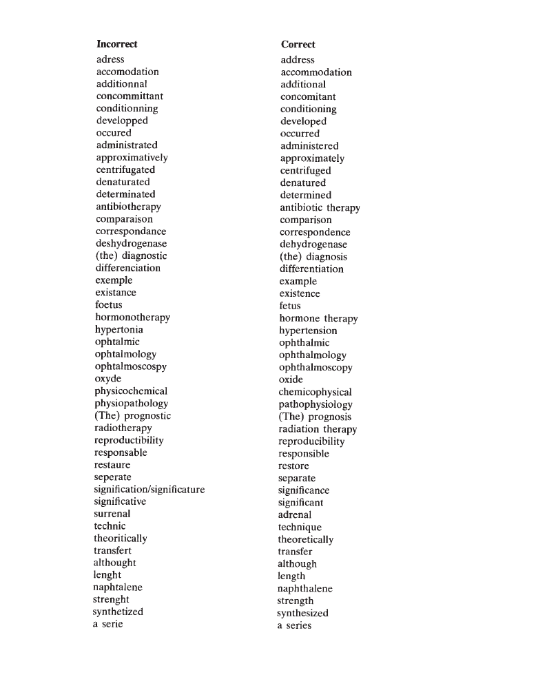
Presenting toxicology results
8
Basic biomedical English
9
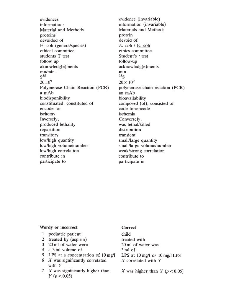
1.9
Verbosity and Incoherence
(See appendix to this chapter for explanations.)
Again cover the right-hand column and try to find the correct or simplified form.
The aim is not only to help you avoid making the same mistakes, but also to develop
your language awareness.
Presenting toxicology results
10
Basic biomedical English
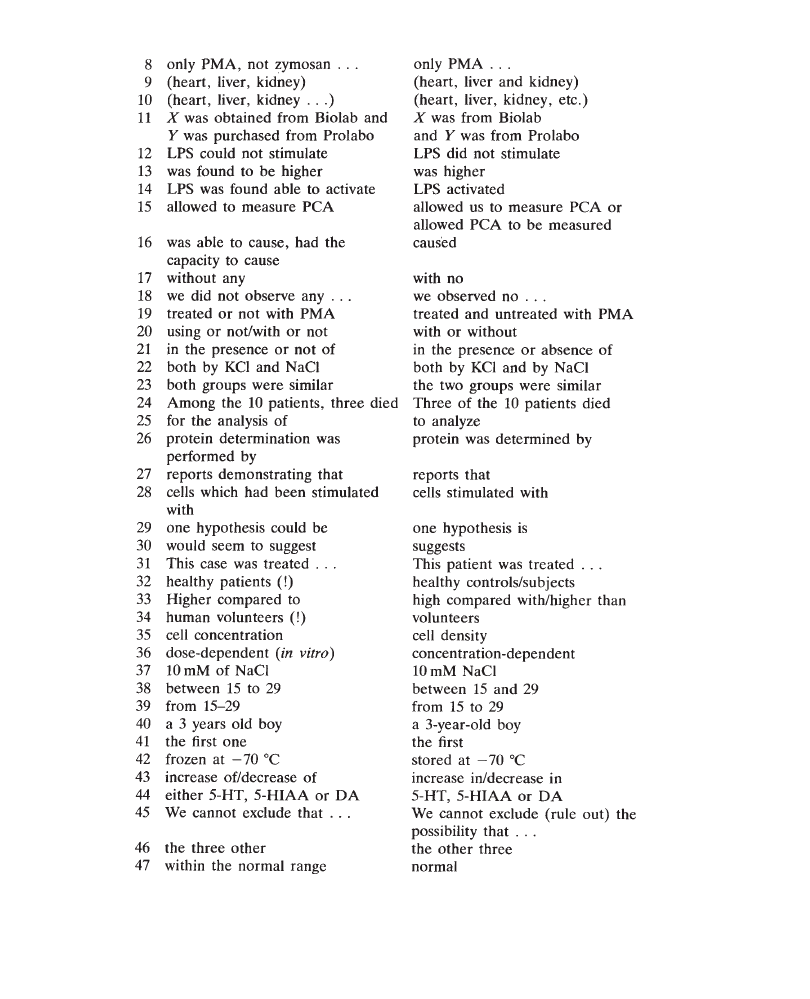
11
1.10
Commonly Misused Words and Expressions
(See appendix to this chapter for explanations.)

Presenting toxicology results
12
APPENDIX:
Explanations
Section 1.9:
Verbosity and Incoherence
1 Why not “immature human creature”!
2 A patient is treated by a doctor with a drug.
3 20ml is a volume, and is thus singular in English. 4 3 ml is a volume; there is
no need to underline the fact.
5 See point 4.
6 In scientific English a correlation is assumed to be statistically significant, by
definition. Also, a significant difference is assumed to be a statistically significant
difference.
7 If you have stated that the threshold of significance is p‹ 0.05, “significantly”
is redundant.
8 Or “PMA but not zymosan”.
9 Lists separated by commas (,) take “and” before the last item (this may or
may not be preceded by a comma).
10 Suspension marks are rarely used in English (and never to mean “etc”.). Note
the period (.) after “etc”. Note too that no extra period is required when sentences
end in “etc”.
11 Be careful: it is important to distinguish gifts from purchases. This raises a
general rule in scientific English: if you have found an appropriate word it is
perfectly acceptable to repeat it.
12 “Could” is an interpretation in this phrase; what the reader wants is observations.
13 An example of unnecessary words: just state what happened.
14 You added LPS and LPS did something. Get yourself out of the picture!
15 This is difficult, even for native English speakers. Try to avoid “allow”/ “permit”
completely (“We used XX to measure PCA”).
16, 17, 18
No comment.
19, 20, 21
Avoid “or not”. It is simplest to write “Animals were treated with
X”; “untreated animals served as controls”.
22 “Both by Y and by X”; or “by both Y and X”. This is not important, but it is
more elegant.
23 English-speaking authors often make this mistake. If you are making an indirect
comparison, use “the two”: “The two animals had similar lesions” but “Both
animals died”.

Basic biomedical English
13
24 This is a very common mistake when describing the results. Another example
is “In the control group, 3 of the animals died”, which can be simplified to “Three
of the control animals died”. Avoid starting sentences with “In” and “Among”.
25 This is a general rule (“for the determination of”, “for the treatment of”, etc.)
26 One of the most frequent examples of verbosity. Try to start each sentence
with the subject: “Cell viability was determined by…” (or “We determined
cell viability by…”) instead of “For the determination of cell viability, we ”.
27 A report itself doesn’t demonstrate anything.
28 Self-explanatory.
29 Don’t surround postulates with conditionals. And be careful with the word
hypothesis; it should mean a series of events possibly explaining a phenomenon.
“LPS might damage the brain” is not a hypothesis. Try “One possible explanation
is that LPS damages the brain…” or “We postulate that LPS damages the brain…”.
30 A suggestion is not a statement; it is not an explosive word; don’t surround it
with conditionals. If your evidence is indirect, you can qualify the word “suggest”
like this: “These results suggest that LPS might activate neutrophils”.
31 Be human!
32 This is one of my favorites! But is anyone perfectly healthy, after all? Avoid
“normal subjects”; they may be normal for the purposes of the study but bizarre
in other respects. Prefer “healthy controls”.
33 If something is “higher/increased compared to”, then it is simply “higher than”.
34 I’ve never seen an animal volunteer for an experiment.
35 “Concentration” is for solutions and “density” for suspensions.
36 A dose can be given to an animal or human, but not to a culture.
37 Molar is an adjective.
38, 39.
A question of coherence.
40 “A boy of 3 years” or “a boy aged 3 years”, but “a 3-year-old boy”. In this
construction, “3-year-old” is a compound adjective of “boy”.
41 “There are two possibilities: the first is that…”.
42 It’s difficult to store something at –70 °C without freezing it….
43 No comment.
44 “Either” is reserved for no more than two possibilities.
45 No comment.
46 “the first three”, “the best three”, etc.
47 If values are within the normal range they are normal; this is why we use
normal ranges….

Presenting toxicology results
14
Section 1.10
Commonly Misused Words and Expressions
1 To need is human; things require. Note the difference between “require” and
“necessitate” (“The patient required treatment”; “the patient’s condition
necessitated treatment”), although few will criticize indiscriminate usage.
2 “On the opposite” does not exist. Be careful when using “On the contrary”
and “In contrast”:
•
“Creatinine levels did not rise. On the contrary, they fell.”
•
“Creatinine levels rose. In contrast, urea levels fell.”
3 This is not important, but “Based on previous findings, we…” implies that
“we” were “based on”.
4 Unless you are talking about a mixture of identified proteins, use the generic
term “protein”.
5 In theory, a risk is always potential; only add “potential” if the existence of a
risk has not been proven.
6 “Unique” means the only existing example; it is very rarely used. Use “only”
instead of “uniquely”.
7 Never use “having”; it’s more trouble than it’s worth.
8 No comment.
9 “Progressive” generally means “deteriorating” (“progressive histological
changes”).
10 No comment.
11 In scientific English “raise” is almost always used as a verb (synonymous with
“increase”). “A rise” is synonymous with “an increase”.
12 “Important” has only one meaning in English: “of considerable significance
or consequence”; it never means “large”.
13 Always use the simplest word.
14 “Besides” is used only in spoken English.
15 “Throughout” is a very useful word: “throughout the colon”; “throughout the
study period”.
16 No comment.
17 “Digestive tract” is OK, but “digestive infection” is not.
18 “Dramatic” is too dramatic.
19 “Germ” is a popular term that should not be used in scientific text. Use “fungus”,
“bacterium”, “virus”, “parasite”, “pathogen”, “microorganism” or “organism”.
Note that “organism” does not mean “body”.
20 “To occasion” is too formal for scientific usage. Use “cause” or “induce”.
21 “Liberation” has political connotations. Nelson Mandela was liberated.
22 To localize is to confine. A localized tumor is restricted to a precise site. To
locate is to find.
23 No comment.

Basic biomedical English
15
24 A patient can present (to a doctor) with symptoms. After that he either has or
develops new manifestations. Animals never present.
25, 26
Be very careful when using “prove” and “proof”; these are absolute terms:
proof is unequivocal and cannot be challenged. Evidence is only an element
of proof, and is what most scientists obtain.
27 “Punctual” means “at the right time”.
28 “Sensible” is never used in scientific English (a “sensible person” is someone
who makes coherent decisions).
29 “Systematic” is rarely used, and means “in absolutely every case”. Note: “routine
vaccination”, “routine screening”, etc.
30 See point 17.
31, 32, 33
Use the noun as an adjective when possible. The general rule in English
is to use the simplest word.
34 Don’t use abbreviations like this in formal text (perfectly acceptable in most
correspondence).
35 Evolution generally has Darwinian connotations.
36, 37
It can be dangerous to mistake the two. Don’t use “etc”, in the same phrase
as “e.g.:” they are mutually redundant.
38 An efficient motor yields a given amount of work for a relatively small energy
input. An effective motor drives a vehicle, for example, regardless of energy
input.
39 To implicate generally means to accuse. For example, “Trimethoprim has been
implicated in skin reactions”.
40 “The train was supposed to leave at 8 pm. In effect, it left at 9 pm.” You probably
mean “In fact…”.
41 An index is a “quantitative marker”: blood creatinine levels are an index of
kidney failure. A marker is used to detect, not to quantify.
42 To control is generally to have control of. You probably mean to check or verify.
Note that “control” can also be used in the sense of “quality control”.
43 “Represent” may imply a presentation of reality or imagined reality (the painting
represents a spring scene). Be simple.
44 “Different” is different from “various”; “varying” means “unstable”. “Various”
means “several”. “Different times” means times different from those used before;
it does not mean “several times”.
45 A surface can be rough or smooth. If you are referring to the area (m
2
, etc.)
you must use area or surface area.
46, 47
Whenever the word “of” has been omitted, the possessive apostrophe is
required, i.e. 14 days’ treatment, or 14 days of treatment.


17
2
The Structure of Toxicology Reports
G.J.NOHYNEK
Rhône-Poulenc Rorer, Vitry sur Seine, France
Safety evaluation of pharmaceuticals, medical devices, food additives and chemicals
is performed with a single goal in mind—to provide safe and effective products.
Toxicology reports are written to supply regulatory authorities with the information
required to make sound decisions regarding the risks and benefits of allowing these
products to be marketed. Those employed to review these reports are not always
toxicologists or pathologists. They may be unfamiliar with the type of investigation,
the nature of the test compound or the types of adverse effects identified in the
study. Therefore, it is of paramount importance that the results of a toxicology
study and their interpretation are easy to follow. A “user-unfriendly” report is likely
to irritate a reviewer, and an irritated reviewer is unlikely to develop a favorable
opinion of the testing laboratory or the test compound.
A well-structured report will help the reviewer to understand and accept the
authors conclusions regarding the safety of a test compound. The structure of
scientific documents is discussed below.
2.1
The ‘IMRAD’ Structure
Almost all scientific publications and reports use the “IMRAD” structure. IMRAD
is the acronym for Introduction, Methods, Results, and Discussion/Conclusion,
and is the most common labeling of the components of a scientific report. This
structure was first prescribed as a standard by the American National Standards
Institute in 1972 (American National Standards Institute, Inc., 1979a). A scientific
report following IMRAD guidelines consists of the following components:
1
Title
2
Abstract/Synopsis:
Summarizes the principal elements of the study.
3
Introduction:
Why did you perform the investigation—what was the
objective?

Presenting toxicology results
18
4
Materials/Methods:
How did you perform the investigation?
5
Results:
What did you find?
6
Discussion:
What do your results mean?
7
Conclusion:
What is your conclusion in terms of what you set out to
investigate?
8
References
Most toxicologists would agree that all toxicology reports should contain a complete
introduction, an adequate description of the methods used, a presentation of results
and a discussion of their meaning and, finally, an overall conclusion. These sections
form the core of a toxicology report and should thus be provided before any detailed
data, i.e. summary tables and individual data tables. The organization of the report
should start with concentrated information and then move on to specifics. All this
is simple enough.
However, the description of the results and their discussion poses a more intricate
problem. Most toxicology reports include several distinct sections, i.e. in-life
observations and measurements, toxicokinetic data, clinical pathology data, and
post-mortem evaluations such as necropsy, organ weights and histopathology. The
results of these sections may include hundreds of findings and thousands of numerical
and diagnostic results. Only a few of these data are essential for the overall
interpretation of the study, whereas the great majority of them may be of no
importance whatsoever. While stringent application of the classical IMRAD structure
to the data accumulated in toxicology studies would necessitate a lengthy (and
undesirable) discussion of all results, relevant and irrelevant data alike, the structure
may be modified to improve its suitability for toxicology reports. In the following
sections, two such modifications are proposed. Each has its advantages and
disadvantages, but both may be considered to be equally appropriate for toxicology
reports.
2.2
Modified IMRAD Structure
A stringent adherence to IMRAD would require discussion of all results in an overall
Discussion section. In modified IMRAD, the Results section is followed by an overall
discussion which addresses the principal findings of the study only. Spurious or
unimportant results are addressed within the individual Results sections to prevent
overload of the discussion. An example of a toxicology report using the modified
IMRAD structure can be found in Chapter 10.
The advantages of this structure are clarity, consistency with IMRAD, and a
discussion which puts all significant results into perspective and correlates the
findings of different sections. In addition, this structure prevents the mixing of
results and their interpretation and represents the most familiar structure for a
reviewing scientist.
The disadvantage of the structure, particularly for lengthy reports, is that reviewers
may already have forgotten many of the results when they arrive at the Discussion
section. Thus, they may be obliged to continually flip back and forth between the

The structure of toxicology reports
19
Discussion and the Results. In addition, the report author who finds it difficult to
distinguish between significant and unimportant results may either overload the
Discussion section with spurious results or may raise the impression that significant
results are being “hidden” in the Results section. Clarity of presentation of results
and discussion (to be addressed in subsequent chapters) is the only way these
disadvantages may be overcome.
2.3
RDRD (Results-Discussion-Results-Discussion) Structure
Using this format, the results of each section of a study are discussed within
each respective section. Thus the description of in-life observations is followed
immediately by their discussion, toxicokinetic data are presented and then
discussed, etc. An example of a report using the RDRD structure can be found
in Chapter 11.
The RDRD structure facilitates the interpretation of results as they come along
and permits their immediate qualification according to their toxicologic significance.
However, correlating results from different report sections—e.g. linking in-life
findings to associated toxicokinetic data, or changes in clinical pathology to tissue
changes identified in histopathology—becomes more difficult. The conclusion of
the report then becomes the overall discussion section described in the modified
IMRAD structure. A final phrase (or two) placed at the end of the section serves
as the true conclusion of the report.
The distinction between results and their interpretation may be less clear with
the RDRD structure. A simple and efficient way to eliminate this confusion is to
use a distinct typeface, e.g. italic script, for the interpretive text. This method of
visual distinction lends considerable help to reviewers and has recently been
suggested for pharmaceutical expert reports (Matthews et al., 1994).
Whatever the structure chosen for your report, a discussion among all the divisions
contributing to the toxicology reports in your laboratory should determine which
of these (or any other existing) formats is best suited for your needs.


21
3
Writing the Report Summary
G.J.NOHYNEK
Rhône-Poulenc Rorer, Vitry sur Seine, France
The summary is the most important section of a toxicology report, and as such
we will treat it separately in this book. Many reviewers may read only the summary,
provided it is clear and contains all important aspects of the study and its results.
In addition, the summary may be used on its own or may be copied directly into
databases or summary documents, technical data summaries, new drug investigators’
manuals or product safety summaries. Therefore, the summary of a toxicology report
must be able to stand alone.
The report summary should be viewed as a miniature version of the complete
report. It should provide a brief summary of each of the main sections of the report
—the introduction, methods, results and discussion—and should end with one or
two short concluding sentences. “A well-prepared summary/abstract enables readers
to identify the basic content of a document quickly and accurately, to determine
its relevance to their interest, and thus to decide whether they need to read the
document in its entirety” (American National Standards Institute, Inc., 1979b).
3.1
The Introduction of the Summary
The introduction sets the scene. In it, the author has to explain the objective of the
study. A suitable approach is to describe the test compound and the reason the study
was performed, e.g. “The study was performed to investigate the repeated dose toxicity
of PP 27567, an ACAT inhibitor, in Sprague-Dawley rats”. Ideally, the introduction
should not exceed a single sentence. Therefore great detail should be avoided.
3.2
The Materials and Methods Section of the Summary
The materials and methods section of the summary should provide a brief review
of the study design and include information on the key aspects of the study such

Presenting toxicology results
22
as strain, total animal number, number of animals per group, dose levels,
administration route and mode, days and sampling intervals of plasma drug evaluation
samples, study and recovery period duration. In standard regulatory toxicology
reports, the methods section may be limited to the description of these key parameters.
It should be brief and avoid all unnecessary detail. However, any non-standard
parameter evaluated or uncommon feature of the study must be described here.
The materials and methods section of the summary of a standard 1-month toxicity
study might appear as shown in Example 3.1.
Example 3.1 Materials and methods section of the summary of a standard 1-month
toxicity study
Groups of 10 male and 10 female rats received single daily oral doses of 20,
60 or 200mg/kg PP 27567 for 28 to 31 days. Five additional animals per sex
in the control and high-dose groups were kept for a subsequent 3-week recovery
period. Four satellite groups of 5 animals/sex/dose were used for plasma drug
determination on days 1 and 28, at 1, 3, 5 and 24 hours after administration.
Control groups received the vehicle, Labrafil™/ethanol (95/5%). The dosing
volume was 20 ml/kg/day. Parameters evaluated in this study included in-life
observations and measurements, interim (day 16), post-treatment and post-recovery
clinical chemistry, urinalysis and hematology, and post-treatment and post-
recovery organ weights, necropsy and histopathology.
3.3
The Results Section of the Summary
Only those results which are biologically/toxicologically significant should be
described in the summary. If in doubt, describe the effect. In general, results should
preferably be described in descending dose group order, though this is not a hard-
and-fast rule. Major changes should be identified first, and the relationship to dose
and no-effect dose levels should be given for each individual finding or effect.
Results should always be reported with reference to the numerical dose level, e.g.
“The following clinical signs were observed in the group receiving 50mg/kg PP
27567”. Avoid terms such as “high-”, “mid-”, “intermediate-” or “low” dose. Any
changes should preferably be reported in numerical terms, such as percentages,
e.g.: “Lower body weight gain was observed in the group receiving 150mg/kg/day,
resulting in lower body weights (males: –15%; females: –8%), compared with control
mean values on day 28 of the study”. However, please note that percentages should
only be used if group size allows them not to be meaningless!
Negative results should be mentioned only if relevant, unexpected or if respective
changes which are present in a high- and/or mid-dose level are absent at lower
levels, e.g. “PP 27567 produced no mortality or clinical signs at 20 mg/kg/day”.
Any effect (including mortality) considered not to be related to the test compound
and which has been discussed and rejected in the Results/Discussion section of
the report should not be described in the summary.

Writing the report summary
23
Results may be summarized in the following order:
(A)
Single-dose (acute) and repeated-dose toxicity studies
1
Plasma drug analysis data
2
Mortality
3
Clinical observations
4
Body weight and food consumption data, other in-life observations/evaluations
5
ECG, cardiovascular parameters, ophthalmology findings (if applicable)
6
Hematology, clinical chemistry, urinalysis data
7
Necropsy observations, organ weights, microscopic evaluation
Plasma drug data may be reported at the beginning or at the end of the results
section. However, since the degree of systemic exposure frequently determines the
incidence and severity of adverse effects, it is preferable to report plasma drug
data “up front”.
(B)
Reproductive toxicity studies
1 Plasma drug analysis data
2 Mortality
3 Clinical observations
4 Maternal in-life data
5 Clinical pathology measurements (if applicable)
6 Maternal necropsy observations
7 Maternal organ weights
8 Histopathological observations
9 Litter data
10 Fetal observations
3.4
The Discussion Section of the Summary
The following questions may be addressed in the discussion section of the summary
of a regulatory study. They will be raised repeatedly throughout the remainder of
the book as they pertain to individual sections of the toxicology report.
1
What are the principal effects observed? Is there a target organ(s) for
toxicological effects of the test compound? What are the principal markers
for toxicity in this study? Which dose levels are to be considered toxic; which
are safe? Are the changes sex-specific?
2
Is there a correlation between multiple effects observed in the study?
3
Are the observed effects consistent with the results of earlier studies or effects
described in the literature, e.g. a known “class effect” for this type of compound?
Can the effects be attributed to (exaggerated) pharmacological activity of the
compound? Do the observed effects resemble toxicological effects described
for a different class of compounds?

Presenting toxicology results
24
4
Are there vehicle-related effects or effects related to methodology, e.g. the
mode of administration or other experimental conditions? Have compound-
related effects been exacerbated by vehicle effects?
5
Are there compound-related effects which are considered to be of no biological
or toxicological significance? Why?
3.5
The Conclusion of the Summary
When writing this part of the summary, always keep the objective of the study
in mind (see Chapter 4, Section 4.6). The conclusion should identify, whenever
possible, a dose level producing no changes or effects consistent with the
pharmacological activity of the compound. Key effects and target organs for toxicity
should be identified. If appropriate, toxicological effects should be put into
perspective (non-toxic, mild-, moderate-, severe toxicity, etc.). Generally, studies
on chemicals, pesticides or food ingredients will attempt to define a “No-observable
effect level (NOEL)”, “No-effect level (NEL)” or “No observable adverse effect
level” (NOAEL). Generally, toxicity studies on developmental drugs will avoid
these terms. Given the pharmacological and exaggerated pharmacological effects
commonly observed in drug safety studies, the use of the term “no-effect” level
may frequently be inappropriate, particularly if pharmacological effects are evident
at all dose levels. In reproductive toxicology (e.g. embryofetal toxicity) studies,
the conclusion should attempt, if possible, to relate maternal toxicity to fetal
observations and identify the dose level at which no fetal or maternal effects
were observed. The presence or absence of fetotoxic effects should always be
stated.
Note: In the conclusion of studies performed to select suitable dose levels for
subsequent studies, i.e. range-finding studies, maternal toxicity studies and 3-month
(pre-carcinogenicity) studies, be cautious when recommending numerical dose levels
for subsequent toxicology studies; new events may arise to change the perception
of the data and hence the dose proposal. However, the MTD (maximal tolerated
dose) should always be clearly identified.
Example 3.2 Summary of a single-dose toxicity study
This study was performed to investigate the single-dose toxicity of PP 45678,
an agonist of kappa opioid-receptors. Groups of 10 Sprague-Dawley rats (5/
sex/dose) received single oral doses of 50,100 or 200 mg/kg PP 45678. Parameters
evaluated included survival, clinical observations, body weight, food consumption
and necropsy findings in surviving animals after a 14-day observation period.
Mortality was 4/5 and 3/5 at 200 mg/kg and 1/5 and 0/5 at 100 mg/kg in males
and females, respectively. No mortality occurred at 50 mg/kg. Death occurred
within 1 hour of compound administration and was preceded by dyspnea (100
and 200 mg/kg) and convulsions (200 mg/kg only). Clinical signs (prostration,
dyspnea and ataxia) appeared approximately 30 minutes following treatment, lasted
approximately 2 hours and were no longer observed after 3 hours. These signs

Writing the report summary
25
continued
occurred with dose-related severity in all animals at 100 and 200 mg/kg. A mild,
transient reduction of motor activity was noted in the groups receiving 50 mg/kg.
Body weight gain and food consumption in all groups were comparable to control
means. There were no treatment-related findings at necropsy.
In conclusion, a single oral dose of 200 mg/kg PP 45678 was lethal, 100 mg/kg was
the approximate minimal lethal oral dose, and effects observed at 50 mg/kg were limited
to mild, transient clinical signs which were considered consistent with the
pharmacological activity of PP 45678.
Example 3.3 Summary of a repeated-dose (28-day) toxicology study
This study was performed to investigate the toxicity of PP 45678, an agonist
of kappa opioid receptors, following repeated daily oral administration. Groups
of 10 male and 10 female Sprague-Dawley rats received single daily doses of
15, 45 or 200 mg/kg PP 45678 by esophageal intubation for 28 days. Two
groups of 10 male and 10 female rats were kept as controls and treated with
the vehicle. For each dose level, 6 additional animals were included for
determination of plasma drug levels on days 1 and 28, at 1, 3, 7 and 24 hours
after administration. Blood samples were taken on days 15 and 28 for
hematology and clinical chemistry analysis. The animals were sacrificed and
necropsied on day 29, and principal organs were weighed and prepared for
microscopic examination.
After 15, 45 and 200 mg/kg/day, PP 45678 plasma levels were directly proportional
to the administered dose. No deaths occurred. Treatment-related clinical signs,
considered to be consistent with the pharmacological activity of this compound,
included ataxia and reduced motor activity. These were observed in both males and
females; they were marked at 200 mg/kg/day and mild at 45 mg/kg/day. Administration
of 200 mg/kg/day resulted in a lower food consumption (males: –5%; females: –8%),
associated with lower terminal mean body weight (males: –13%; females: –16%),
relative to controls. Terminal biochemical and hematologic parameters were similar
in all groups. An increase in liver weights (males: +28%; females: +18%, versus
controls), associated with centrilobular hypertrophy (more marked in males) was
observed in all animals at 200 mg/kg/day. In conclusion, 200 mg/kg/day PP 45678
was mildly toxic, causing reduced body weight gain, reduced food consumption and
adaptive changes in the liver. Administration of 45 mg/kg/day resulted in clinical
signs consistent with the pharmacological activity of PP 45678, while 15 mg/kg/day
had no apparent adverse effect.
Example 3.4 Summary of a reproductive toxicology study
This study was performed to investigate the potential embryofetal toxicity of PP
27567, a systemic inhibitor of ACAT. Groups of 26 mated female Sprague-Dawley

Presenting toxicology results
26
continued
rats received single daily oral doses of 0, 10, 45 or 200 mg/kg/day PP 27567
from day 6 to day 17 of gestation. Additional groups of 6 mated females per
dose level were included for determination of plasma drug levels on day 13 of
gestation, at 1, 3, 7 and 24 hours after compound administration. Clinical signs,
body weights and food consumption were recorded regularly. On day 20 of
gestation the animals were sacrificed and necropsied for examination of uterine
contents, which included a detailed external, visceral and skeletal evaluation
of fetuses.
A dose level of 200 mg/kg/day PP 27567 resulted in mild maternal toxicity consisting
of reduced motor activity, salivation, lower food consumption associated with lower
mean body weight gain (–9% lower mean body weight on day 16, when compared
with control values), a smaller litter size and a lower mean fetal weight (–11%, compared
with control means). In the groups receiving 45 and 10 mg/kg/day, all maternal and
fetal parameters were comparable to control values. In conclusion, a dose level of 200
mg/kg produced mild fetal toxicity secondary to maternal toxicity whereas no evidence
for adverse maternal or fetal effects was noted up to a dose level of 45 mg/kg/day.

27
4
General Principles of Regulatory
Toxicology Report Writing
G.J.NOHYNEK
Rhône-Poulenc Rorer, Vitry sur Seine, France
and A.LODOLA
Pfizer Centre de Recherche, Amboise, France
4.1
Titles of Toxicology Reports
Imagine you are a reviewer of toxicology reports, that you are unfamiliar with
different companies’ jargon, and that you are not a toxicologist. How are you
supposed to recognize that report titles such as “Subacute Oral Study”, “Subchronic
Oral Gavage Study”, “Subchronic Gavage Study”, “Subchronic Gavage Study by
the Oral Route”, “One-Month Oral Study”, “Study on the Subchronic Oral Toxicity”,
“28-Day Study”, “One-Month Repeated-dose Oral Toxicity Study”, “Four-Week
Study Per Os” all refer to the same type of investigation which was, incidentally,
a 1-month oral toxicity study? What would you make of a “dietary study”, an “in-
feed study”, an “oral in-feed study”, an “oral study by dietary ad-mix”, or, if the
study is to select dose levels for a subsequent carcinogenicity study, an “oral pre-
carcinogenicity study by dietary ad-mix”, or an “in-feed range-finder study”?
Strangely enough, these titles again refer to the same study, which was a 3-month
dietary toxicity study.
Using a different scenario, suppose now that you have been asked to search for
the report of a certain study in a computer printout containing 1500 different
toxicology studies done by your company between 1986 and 1995. The study in
question was carried out years ago on a developmental compound bearing your
company’s code number. The drug was originally a racemic mixture, was
subsequently developed as a pure enantiomer, later received a common name, and
was finally marketed under several different trademarks. Consequently, your computer
printout contains references such as PP 31234, then PP 127567, as well as D,L-
phenyletenelol, L-phenyletenelol, Carditon
®
, Carditex
®
, and Cardol
®
. In addition,
the compound designation appears at the beginning, in the middle and at the end
of the titles of the individual studies. Have you ever been in this infuriating situation?
The above examples are meant to illustrate that the best title for the report of a
scientific investigation contains the fewest possible words that adequately describe
the contents. Toxicology studies are defined by the test compound, the nature or

Presenting toxicology results
28
endpoint of the investigation, the route of administration, the duration of the study,
and the test animal species.
For general toxicology reports (single-dose, repeated-dose or carcinogenicity
studies), terms should be listed, in our view, in the following order:
1
Test compound
2
Study duration
3
Administration route
4
Study type
5
Test animal species
For reproductive toxicity reports (fertility, multigeneration, embryofetal toxicity
or peri-/postnatal toxicity studies), the duration of compound administration (e.g.
during the organogenesis period of gestation) and endpoints (e.g. fetotoxicity) are
defined in terms of reproductive parameters. Taking this into account, title terms
should be listed in the following order:
1
Test compound
2
Administration route
3
Study type
4
Test animal species
For a developmental compound, use only the code of your company/laboratory, if
available. This makes it easier to list studies chronologically in order of increasing
compound code number. Common names should be used for non-proprietary
compounds. As a rule, trademarks should be avoided. Within the title, the test
compound designation is followed by a colon or a hyphen.
Avoid poorly defined terms such as “subacute”, “subchronic” and “chronic”.
The duration of the study should be described in terms of days, weeks or months.
Since repeated-dose studies with large animal numbers often have staggered dates
of terminal sacrifice, it may be complicated to describe their duration in terms of
a defined number of days. Therefore, the duration of studies using more than 4
weeks of treatment is best described in terms of months, whereas the duration of
short studies should be described in days: 5-day, 14-day, 1-month, 3-month, 6-
month, 12-month, 24-month. Be consistent when describing the study duration (note
that 1 month is not identical to 28 days!). If necessary, fractions of months may
be expressed in weeks, e.g. a recovery period subsequent to the treatment period
(see examples at the end of this section).
We recommend the following terms to describe the route of administration:
Oral
Intramuscular
Intravenous infusion
Dietary
Intraarterial
Intranasal
Dermal
Intraperitoneal
Intravaginal
Intravenous
Subcutaneous
Inhalation
Ocular
Intradermal
Paravenous
Note that “dietary oral” is a pleonasm!
The type of study is determined by the objective of the study and its duration.

General principles of regulatory toxicology report writing
29
Avoid company jargon such as “pre-carcinogenicity study”, “dietary ad-mix study”
or “pilot study”. We recommend the following terms:
Single-dose toxicity
Repeated-dose toxicity
Toxicokinetic
Range-finding toxicity
Toxicity
Carcinogenicity
Exploratory toxicity
Tolerance
Intermittent dose
Exploratory
Rising dose
Fertility (male or female)
Embryofetal toxicity
Peri-/Postnatal toxicity
Neurotoxicity
The test animal species should always be in the plural, e.g. “in Sprague-Dawley
rats”, “in CD
®
-1 mice”. Avoid generic terms such as “monkey” but be specific,
e.g. “in Rhesus monkeys (Macaca mulatta)”, “in Cynomolgus monkeys (Macaca
fascicularis). With rodents or dogs, the strain or breed should be mentioned, e.g.
“Sprague-Dawley rats”, “Fischer 344 rats”, “CD
®
-1 mice”, “beagle dogs”.
When the above principles are applied, study titles take the following form:
•
PP 27567:1-Month intravenous toxicity study in CD
®
-1 mice followed by a
2-week recovery period
•
PP 27567:3-Month dietary range-finding toxicity study in CD
®
-1 mice
•
PP 27567: Single-dose oral toxicokinetic study in Sprague-Dawley rats
•
PP 27567:6-Month oral repeated-dose toxicity in beagle dogs
•
PP 27567:24-Month dietary carcinogenicity study in CD
®
rats
•
PP 27567:21-Day intravenous infusion study in Cynomolgus monkeys (Macaca
fascicularis)
•
PP 27567:3-Cycle intravenous intermittent-dose toxicity study in Sprague-
Dawley rats
•
PP 27567: Oral range-finding toxicity study in pregnant Sprague-Dawley rats
•
PP 27567: Oral embryofetal toxicity study in Sprague-Dawley rats
•
PP 27567: Oral embryofetal toxicity study in New Zealand white rabbits
•
PP 27567: Oral embryofetal and postnatal developmental toxicity study in
Sprague-Dawley rats
•
PP 27567: Oral fertility and early embryonic developmental toxicity study in
Sprague-Dawley rats.
4.2
The Introduction
The Introduction sets the scene for the report and should cover the following points:
•
A description of the test compound, e.g. its proposed use, the therapeutic class
or pharmacological activity
•
Location of the laboratory (for contract research organizations or companies
with more than a single toxicology laboratory)
•
Why the study was performed

Presenting toxicology results
30
•
The rationale for dose selection
•
The choice of test species.
In general the most contentious point in a study is the justification for dose selection.
Generally, the dose selection should be based on the results of previous, e.g. range-
finding studies, and not the results of the study under consideration. As an example,
if hepatic toxicity was not found in previous studies but was the main finding in
the present study you cannot say in the dose justification that hepatic toxicity was
the justifying factor for the choice of doses. In our view this is self serving and
unconvincing. We believe it is better to refer to principal results of previous studies
or to remain non-specific: for example, “Some adverse effects were expected at…”
or “Moderate toxicity was expected…”. This describes both the expected result
and the uncertainty inherent in any study. Examples 4.1 and 4.2 are of the
Introduction and justification of dose level selection.
Example 4.1 Introduction of a single-dose toxicity study
PP 24626, a metabolite of the anticancer drug PP 16569, has been identified
in the plasma of rodents, dogs and man. The current study was performed to
determine the acute toxicity of PP 24626 in mice following administration of a
single intravenous dose. The results of the present study will be compared with
the results of a previous acute toxicity study performed in mice on the parent
compound, PP 16569. Mice were chosen as test animals because this species
is commonly used for acute toxicity testing of anticancer drugs. The high dose
level, 100 mg/kg, was limited by the solubility of the test compound in the
vehicle (5% aqueous glucose solution containing 12% Tween® 80).
Example 4.2 Introduction of a carcinogenicity study
PP 83211 is an antiarhythmic agent under development for use in the prevention
of ventricular fibrillation. The present study was performed to assess the chronic
toxicity and carcinogenic potential of PP 83211 when administered orally as
an aqueous suspension to male and female Sprague-Dawley rats for 24 months.
The dose levels were selected in the light of data from a 3-month oral study
in Sprague-Dawley rats using dose levels of 3, 10 and 30 mg/kg. A dose of
30 mg/kg/day produced mortality (3/20 males), a lower mean body weight (–
12% to –18%) and food consumption associated with moderate clinical signs
(ptosis, peripheral vasodilatation), increased urinary output, reduced plasma
potassium levels, a marked increase in liver weight, and centrilobular
hypertrophy. At 10 mg/kg/day, findings were limited to a lower mean body
weight (–6% to –9%), a mild increase in liver weight and minimal to mild
centrilobular hypertrophy.
On the basis of these results, 10 mg/kg/day was selected as the highest dose level for
the present study; 3 and 1 mg/kg/day are multiples of the probable therapeutic dose
and were selected to establish a dose-relationship of potential adverse effects.

General principles of regulatory toxicology report writing
31
4.3
The Materials and Methods Section
This section should follow the introduction. The author may choose between a
detailed presentation of the methods or an abbreviated one (if abbreviated, the text
should be sufficiently detailed to allow the reader to understand how the study
was conducted). When using abbreviated materials and methods, the study protocol
should be supplied in an appendix to the report.
A common misconception is that the Materials and Methods section and the
study protocol are one and the same thing. The protocol is a “route-map” written
to ensure that the study is performed according to Good Laboratory Practice and
Standard Operating Procedures, whereas the Materials and Methods section is written
to ensure that the reviewer understands the study design. Therefore, this section
should not be a copy of the study protocol.
Whereas the Materials and Methods section can be abbreviated, all unusual
study methods should be described if the information is considered useful for
comprehension of the study objective or the study design. Any non-standard
parameters or features evaluated must always be described in this section. Such
information may include details of the vehicle, the administration volume and
rate (key information for intravenous studies), the number of satellite animals/
groups included for plasma drug analysis or recovery, particular aspects of the
statistical evaluation, specific in-life observations, ophthalmology, or cardiovascular
parameters, special clinical pathology measurements, details of euthanasia and
necropsy procedures, organ weight evaluation, histopathology or plasma drug
analysis.
Since most regulatory toxicology studies follow international regulatory
guidelines which specify most of the details of the study, this part of the report
can be prepared as a template which can be applied to each study of the same
type. Examples for Materials and Methods sections are given below. In some
studies, e.g. those in which the test compound is given in the diet or in the drinking
water, the calculated (nominal) dose levels are not identical to the actual doses
administered. For such studies, the actual and nominal doses should be reviewed
in the Materials and Methods section or at the beginning of the Results section
(see Example 4.4).
We recommend that the Materials and Methods section of any regulatory
toxicology study should not exceed one full page—even for studies having a
complicated design. As a rule, half a page should be sufficient for a standard
regulatory toxicology report.
Example 4.3 Materials and Methods section of a peri-/postnatal toxicity study in
rats
PP 27567 was administered by oral gavage as an aqueous suspension containing
0.5% methylcellulose and 0.1% polysorbate 80 to groups of 20 inseminated
Sprague-Dawley rats, from day 15 post-insemination (p.i.) until parturition and
throughout the entire lactation period at daily dose levels of 15, 30 and 60
mg/kg. A control group of 20 inseminated rats received the vehicle over the
same administration period.
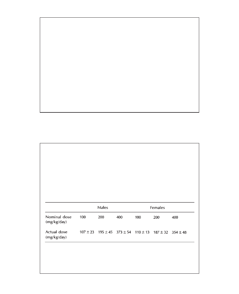
Presenting toxicology results
32
continued
The animals were weighed and examined daily for clinical signs. Food
consumption was determined daily. The number and body weight of pups were
recorded and their physical and functional development and behavior were evaluated
on standardized litters. On day 21 post-partum, maternal animals were sacrificed
and necropsied. Developmental parameters evaluated in the pups of the F1
generation included pinna detachment (from day 4 post-partum), incisor eruption
(from day 11), eye opening (from day 15), testes descent (day 25) and vaginal opening
(day 37). Functional parameters evaluated included the surface righting reflex (from
day 4), negative geotaxis (from day 4), swimming development (day 10), forelimb
support (from day 11), auditory startle reflex (from day 12), pupillary reflex (day 19)
and rotarod performance (day 25). Behavioral parameters evaluated included homing
behavior on a level surface (day 21), and learning/memory via the water maze test
(days 42 and 49) and exploratory behavior using the open field test (day 48). On
day 51 post-partum, all pups of the F1 generation were sacrificed and necropsied.
The study protocol containing detailed materials and methods is found in Appendix
1 of this report.
Example 4.4 Nominal versus actual dose levels
Dietary concentrations of PP 27567 were adjusted on a weekly basis;
appropriate concentrations were calculated prospectively on the basis of the
group mean food consumption expected to result in a nominal daily intake
of 100, 200 and 400 mg/kg/day. Actual daily intake data calculated on the
basis of the weekly food consumption are shown in Table 6, Appendix VI.
Throughout the study, the mean actual daily intake of PP 27567 is summarized
in Table 4.1.
Table 4.1 PP 27567:3-Month dietary toxicity study in rats—nominal and actual
dose levels.
The actual intake of PP 27567 was within approximately±10% of the calculated
intake. A variation of this magnitude is considered acceptable for studies in
which the compound is dosed in-diet.

General principles of regulatory toxicology report writing
33
Example 4.5 Materials and Methods section of a carcinogenicity study
Groups of 60 male and 60 female Sprague-Dawley (Charles River) rats were
treated daily by gavage with 1, 5 or 25 mg/kg PP 27567 for 24 months. At
the start of the study, the animals were approximately 6 weeks old and weighed
185 to 205 grams (males) and 155 to 173 grams (females). The compound
was suspended in an aqueous solution of methylcellulose (1%) and
administered at a constant volume of 5 ml/kg. Two control groups of 60 rats/
sex, received 5 ml/kg of the vehicle alone for the same treatment period.
Satellite groups of 5 animals/sex/dose level were included to determine plasma
drug levels.
All animals were observed daily for clinical signs. Their body weight was recorded
weekly. Food consumption was measured weekly during the first 3 months of the study,
then every 2 weeks for the remainder of the study. Water consumption was measured
over 24 hours every 2 months. An ophthalmologic examination was performed before
the start of the study and at 6, 12 and 18 months. Plasma drug levels were determined
from blood samples collected 4 hours after compound administration after 6, 12 and
18 months. After 24 months of treatment, animals were sacrificed, necropsied, and
organ weights determined. Hematology and clinical chemistry evaluations were
performed on blood samples collected at sacrifice. A histopathologic evaluation was
performed on a range of tissues.
The two control groups were combined for statistical treatment of findings whenever
there was no significant difference between them. Groups were compared using a
one-way analysis of variance. For some parameters, when a large heterogeneity of
variances was associated with a large variation of animal number per group, a non-
parametric analysis of variance was performed. Survival data were analyzed using the
log-rank test. Peto’s analysis was used to compare the tumor incidence in treated
groups with that in control groups. The study protocol containing detailed materials
and methods can be found in Appendix 1 of this report.
4.4
Describing Results
In our view a well-balanced toxicology report must describe the data in terms of
toxicological significance; this after all is the objective of the exercise. The description
of results should be as succinct as possible to avoid boring the reader. Therefore,
the use of short sentences is strongly advised. Condensing complex data into a few
(complex) phrases is to be avoided, particularly if English is not your first language.
Remember that the first consideration in any report is clarity, and that
incomprehensibility is the hallmark of a bad report.
For studies carried out with a single specific objective, such as toxicokinetic
studies, or investigative studies on a specific target organ, the Results section should
focus on those results which address the objective of the study. Thus the results
of a toxicokinetic study should primarily focus on plasma drug data; other results,
such as in-life effects, are of secondary interest in such a study, provided there
are no changes which may affect plasma drug levels (body weight, food consumption
changes, emesis, diarrhea, etc.).

Presenting toxicology results
34
In the Results section of a standard general toxicity or reproductive toxicity
study, the author should (a) describe all effects that are induced by the test compound
and (b) distinguish compound-related effects from those which are of uncertain
relationship to the compound, those which are related to the vehicle or the test
procedure, and those which are incidental. Given that “treatment-related” effects
may include changes caused by the procedure or the vehicle, refer to all effects
caused by a test article as “compound-related” effects!
Each subsection (clinical signs, cardiovascular parameters, clinical pathology,
etc.) should begin with a few sentences which clearly identify the key findings.
This focuses the attention of the reader and the author on the key points which
will be raised in the discussion. Describe data in terms of incidence, time of onset,
duration and/or severity. Subdivide findings by sex, dose level, or duration of
treatment to help reduce the complexity of your text. The results for males should
precede those for females. Although this is not politically correct, it is general
practice and expected by reviewers. Results may be described in descending or
ascending dose order. The preferred option is that which most clearly identifies
compound-related changes and the no-effect dose level. Within a given section the
same convention should be followed throughout. Identification of treated groups
by their numerical dose level is unambiguous and a reminder to the reader of the
actual dose level. However, the occasional use of “high-, mid- and low-dose”, while
less informative, lightens the text. If an individual animal is discussed, always indicate
the animal number and dose (“Male 11 (20mg/kg/day)” or “one 20mg/kg/day male
animal (M11)”). Also, when referring to animals other than those of the main study
groups, the dose and group descriptor should be given (satellite groups, sentinel
groups or recovery groups).
For quantitative data always report numerical changes, e.g., “Treatment with
150 mg/kg/day PP 27567 for 4 weeks produced a lower mean body weight gain in
males and females (Males: –15%; Females: –8%, relative to controls)” or “A 12%
lower mean body weight gain was observed”. However, be cautious when using
percentages to quantify changes, particularly when changes greater than 100% are
reported (see also Chapter 7). There may be different interpretations of what a
descriptor such as “a 125% increase in liver weight” means. An unambiguous manner
in which to address quantitative differences from controls is to express increases
or decreases in a parameter as a direct multiple of the control value. For example,
“absolute liver weight at 24 mg/kg/day was 1.85×those of the controls, WBC count
in the 25 mg/kg/day females was 0.45×that of the controls, body weights in the
mid- and high-dose groups were 0.95×and 0.85 ×that of the controls, respectively.”
A qualitative description of data in the Results section (for example, “a slightly
lower mean body weight gain”) is inappropriate since it does not allow the reader
to form a judgment as to the amplitude of the change. What represents a “small
increase” to you may be a “large increase” to someone else! However, the use of
qualitative descriptors is appropriate in summaries or abstracts when the intention
is to provide an overview of findings rather than details.
Be aware of the meaning of the term “reduction” —it implies a decrease from
a known initial value. Parameters such as weight gain, organ or fetal weights can
only be “reduced” if they are known to have been higher at some earlier stage.
Strictly speaking, body weights can be reduced when compared with pretest values,
but not when compared with control values. Similarly a “reduction in testes weight”
implies that the testes were heavier at a previous time point. However, whereas
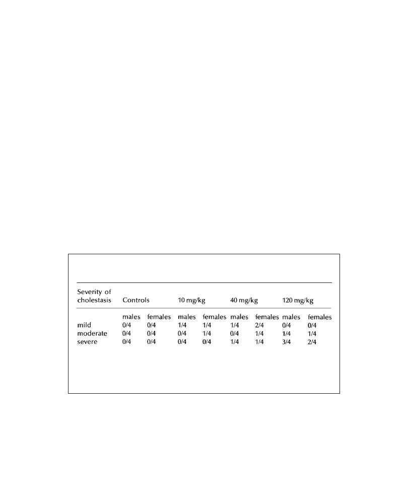
General principles of regulatory toxicology report writing
35
terms such as “a reduction (decrease) in mean body weight when compared with
control means” may not be accurate, they are commonly used in toxicology reports
and are acceptable.
Findings should be graded when they are difficult to quantify or when grading
may improve the reader’s comprehension of complex changes. The grading scheme
should be defined in the text or as a footnote to the data. A three-grade (mild —
moderate—severe) or five-grade system (minimal—mild—moderate—marked —
severe) is generally used (see Table 4.2 below).
The statistical significance of change should be described in the text, e.g. “A
statistically significant decrease in mean WBC count occurred in males treated
with 100mg/kg/day”. Alternatively, p-values may be used to indicate statistical
significance. For example: “There was a decrease (p<0.01) in mean WBC count
in males treated with 100mg/kg/day”. While there is no set rule, inclusion of p-
values lightens the text and facilitates reading.
Often, the simplest way of describing complex results is by using a table. Tables
should be numbered and preceded by a title. The title relates the purpose and the
contents of the table. The title, the footnotes and the column headings together
should form a complete unit that is independent of the text. Any abbreviation must
be explained in a footnote. The order of data presentation should always be from
control to high dose. An example is given in Example 4.6.
Example 4.6 Tabular presentation of results
Table 4.2 PP 27567:1-Month oral toxicity in dogs—incidence and severity of
intra-canalicular cholestasis.
In-life observations or other data from satellite groups (e.g. for plasma drug
determination) should not be described within the Results section, unless specified
in the protocol. Exceptions are pregnancy data in fertility study satellite groups
and compound-related mortality data. Note that mortality in rodent satellite groups
which may be attributed to anesthesia and/or blood sampling procedure and which
occur in absence of mortality in the main groups should be described as procedure-
related mortality, but not as compound-related mortality! Again, avoid the potentially
confusing term “treatment-related” mortality!
Grading system: mild: focal centrilobular change, comprising up to 5 bile plugs per
liver section; moderate: multifocal centrilobular change, comprising more than 5 but
less than 20 bile plugs per liver section; severe: >20 bile plugs, presenting as a zonal
change affecting most centrilobular areas.

Presenting toxicology results
36
The absence of an effect on an individual parameter which was measured in
the study should always be mentioned, e.g.: “There was no effect on body weight
or food consumption”, or “No changes were detected in blood pressure or heart
rates”. If changes are evident at the high- and/or mid-dose level, but not at a lower
dose level, the absence of an effect should be mentioned with reference to the dose
level where the effect was absent. Always conclude any description of compound-
related effects with an appropriate phrase highlighting the no-effect dose level,
e.g. “A reduction in mean body weight was noted in the male group receiving 150
mg/kg (–9%, when compared with the control mean). The body weights of all other
treated groups were comparable to control values.” The same rule applies if effects
that were anticipated were not observed, e.g. on the basis of the results of previous
studies or on the pharmacological activity of the test compound.
The reasons for dismissing findings which might be considered to be compound-
related should be given in the Results section (this is our personal preference, as
it simplifies the discussion and focuses it on the issues) or in the Discussion. In
general, data are dismissed on the basis of the absence of a dose response, isolated
occurrence, and/or being within the limits of historical control data. A brief statement
such as “All other findings were those which routinely occur in our laboratory in
rats/dogs of this age, sex and strain” is also useful in assuring the reader that all
the data have been reviewed to identify treatment-related changes.
4.5
The Evaluation/Discussion Section
The extent to which the results of a regulatory toxicity study should be discussed
is a controversial topic. Some laboratories do not include a discussion in reports,
or keep the discussion of the results to a minimum. Their appraisal of toxic effects
is done in the Toxicological Expert Review or Synopsis where the results from all
studies are discussed; this approach avoids discussion of spurious effects in early
studies and speculation as to the pathogenesis of confirmed toxic effects.
Alternatively, the principal results of individual toxicity studies can be discussed
extensively in each report. Individual study reports are frequently reviewed
independently of the toxicological expert report, and discussion within each report
allays the concern that the findings of a given study may be misinterpreted by a
naive reviewer. While the extent of the discussion is debatable, in our view it is
preferable to discuss the results of each study within the report itself.
The principal themes that should be addressed in the discussion include the
following.
•
Study objective: Focus the discussion on the study objective. You don’t need
a detailed discussion of in-life effects in a toxicokinetic study (provided such
effects did not affect the plasma drug levels)!
•
Principal effects: What are the principal effects observed in this study? Are
there principal target organs for the test compound? What are the principal
markers for toxicity in this study? Which doses are toxic? Are effects sex-
specific?
•
Correlation of effects: Is there a correlation between effects observed in the
study?

General principles of regulatory toxicology report writing
37
•
Pathogenesis: Are the effects consistent with the results of earlier studies or
effects described in the literature? Is the pathogenesis of the effects known?
Is it a “class effect”? Are the effects caused by the exaggerated pharmacological
activity of the compound? Are the effects relevant to man?
Vehicle-related effects and effects related to the method of compound administration
should be discussed after these points have been addressed. Compound-related effects
which are considered to be biologically or texicologically insignificant (provided
they have not been dismissed in the Results section) should also be discussed at
this time.
There are no hard-and-fast rules governing the ordering of facts in the
Discussion. Findings can be discussed in increasing or decreasing order of
importance, or in the same order in which they occur in the Results. Whichever
approach is preferred, discuss your results, do not repeat them! Keep your language
and your ideas as simple as possible; remember to KISS (Keep It Short and Simple!)
the text. Also, remember that, as the pre-clinical safety program unfolds, what
appeared to be of major significance in early studies can be of little or no relevance
in later studies. Therefore, avoid speculation unless it is based on a solid scientific
foundation. It is preferable to leave a point open rather than to develop an
unfounded speculation.
Finally the discussion should focus on the toxicological significance of findings;
after all, the purpose of the study is to study the toxicity of the test compound.
No discussion at all is better than one which skirts around the issue of toxicity.
Who is better placed than the author of the report to put findings into context?
Always keep in mind that if you do not have the courage to put findings into a
toxicological context, then someone else will!
4.6
The Conclusion
The golden rule in writing the Conclusion is that YOU SHOULD NOT JUMP TO
CONCLUSIONS! The Conclusion of a toxicology report should put the results
into perspective with reference to the objective(s) of the study. Therefore, the
Conclusion of a toxicokinetic study should be restricted to toxicokinetic findings,
the Conclusion of a study measuring bioequivalence of a new formulation should
refer only to bioequivalence, the Conclusion of a study comparing target-organ
toxicity in a test species with that of a reference species should be restricted to
the principal results observed in the target organ investigated, etc.
Toxic dose levels as well as doses producing no toxic effects should always
be identified. However, in studies on pharmaceutical compounds, the use of the
terms “No-Effect Level”, “No Observable Adverse Effect Level” and “No-
Observable Effect Level” is often elusive, particularly if pharmacological effects
are evident at the lowest dose level. Although these terms are not applicable to
studies which reveal pharmacological effects at all dose levels, definition of a
No-Effect Level is required by some regulations. Labels such as “toxicological
no-effect level” or “dose level devoid of toxic effects” may be used. The target
organ(s) for toxicity and/or the principal markers for toxicity should always be
mentioned. The Conclusion of studies performed to determine suitable dose levels

Presenting toxicology results
38
for subsequent investigations (e.g. sighting, maternal toxicity studies, range-finding
or precarcinogenicity studies) should not include a dose level proposal for
subsequent studies; new events may arise to change the perception of the data
and hence dose proposal. However, if appropriate, the MTD (maximal tolerated
dose) should be clearly identified.
In reproductive toxicology (e.g. embryofetal toxicity) studies, the Conclusion
should attempt to relate maternal toxicity to fetal observations and identify the
dose level at which no fetal or maternal effects were observed. The presence or
absence of fetotoxic effects should always be stated. The Conclusion of maternal
toxicity studies performed as range-finding studies for subsequent fetal-/
embryotoxicity studies should principally address maternally toxic dose levels. Fetal
effects are of secondary interest in such studies and should therefore be interpreted
conservatively.
In the interest of brevity and clarity, toxicological effects should be described
qualitatively rather than quantitatively; this helps the reader to appreciate the
importance of findings. If you cannot fit your “conclusions” into a few lines then
you are probably rehashing your results. Sample Discussions and Conclusions are
given in Examples 4.7 to 4.10.
Example 4.7 Discussion/Conclusion of a 1-month study in dogs
The maximal plasma drug concentrations of PP 27456 on day 3 were dose-
related. Rapid clearance was observed at 20 mg/kg/day; the relatively lower
clearance at 40 and 80 mg/kg/day resulted in a four- to ninefold increase in
systemic drug exposure at these dose levels (when compared with the values
noted at 20 mg/kg/day). This was most likely caused by saturation of metabolism
and/or excretion. Consequently, PP 27456 accumulated at 40 and 80 mg/kg/
day, resulting in markedly increased plasma drug concentrations on day 28,
with corresponding AUC values which were four- and sevenfold higher than
the respective values on day 3. This accumulation of the test compound may
be responsible for the progressive appearance of adverse clinical signs observed
at 40 and 80 mg/kg/day during the course of this study (progressive decrease
in body weight gain and food consumption at 40 mg/kg/day, body weight loss
and the progressive deterioration of clinical condition at 80 mg/kg/day).
A clear compound-related effect was evident at 80 mg/kg/day as shown by
marked clinical signs, marked changes in clinical chemistry and hematology
parameters, and histopathological changes in liver and bone marrow. Histopathologic
evidence of hepatic centrilobular necrosis was associated with a marked increase
in liver weight and accompanied by increased clinical chemistry markers (seven-
to tenfold increases in alkaline phosphatase, ASAT and ALAT activities and
plasma bilirubin values), and a four- to fivefold increase in hepatic microsomal
cytochrome P450 content. Histopathologic evidence of hypocellular bone
marrow was associated with a marked decrease in the principal RBC parameters.
Additional histopathologic findings in the 80 mg/kg/day group, such as testicular
atrophy, thymic and salivary gland atrophy and hypertrophy of the adrenal
z o n a f a s c i c u l a t a , w e r e p o s s i b l y r e l a t e d t o t h e p o o r c o n d i t i o n o f t h e s e

General principles of regulatory toxicology report writing
39
continued
animals, secondary to liver toxicity. The mild reductions in plasma electrolyte
levels (chloride, calcium, potassium) at this dose level are likely to be related
to PP 27567. The mechanism for these changes is unclear.
In contrast, administration of 40 mg/kg/day of PP 27456 produced no
microscopically discernible changes in the liver. Induction (two- to threefold) of
cytochrome P450, minimal to moderate increase in alkaline phosphatase and
moderate increase in liver weights at this dose may be regarded as adaptive
changes of the liver (Schulte-Hermann, 1972; Balazs et al., 1978; Gopinath et
al., 1987).
At 20 mg/kg/day, a minimal (1.0– to 1.3-fold, compared with control mean values)
increase of cytochrome P450 did not correlate with any other clinical chemistry
variation or histopathologic changes. While the increase in cytochrome P450 may be
regarded as a minimal adaptive effect of treatment, the values are well within our
historical control range for this parameter, and were not considered texicologically
significant.
In conclusion, treatment of dogs for 30 days with oral doses of 80 mg/kg/day of PP
27456 produced toxicity of the liver and bone marrow. Findings at a dose level of 40
mg/kg/day were limited to adaptive changes in the liver. No evidence for adverse effects
was observed at a dose level of 20 mg/kg/day of PP 27456.
Example 4.8 Discussion/Conclusion of a maternal toxicity study in rats
Signs of mild to moderate maternal toxicity (stained and soft feces, reduction
in food intake and progressive body weight loss) were evident at 100 and 200
mg/kg. Therefore, the reduction in fetal and placental weights at these dose
levels was considered to be related to the maternal effects at these doses. The
delay in skeletal ossification observed at 200 mg/kg (increase in incidence of
fetuses with three or four incomplete or unossified sternebral bones) was consistent
with the lower fetal weight recorded at this dose level and was considered to
be secondary to the maternal toxicity.
In conclusion, oral administration of PP 45678 to pregnant rats during the period of
organogenesis at dose levels of 100 and 200 mg/kg/day produced maternal toxicity
which was associated with reduced fetal and placental weights, and a reduction in
skeletal ossification at 200 mg/kg/day. No evidence for adverse maternal or
developmental effects was observed at a dose level of 50 mg/kg/day.
Example 4.9 Conclusion of a 1-month study in rats
In conclusion, single daily oral administration of PP 83211 to Sprague-Dawley rats
for one month at dose levels of 0, 5, 25 and 125 mg/kg/day did not affect survival

Presenting toxicology results
40
continued
or hematologic parameters. A dose level of 125 mg/kg/day produced a mild
reduction in mean body weight (–8% to –12%) and food consumption (–5% to
–8%), moderate clinical signs (transient ataxia), a mild increase in ALAT and
ASAT activity and a moderate increase in liver weight associated with centrilobular
hypertrophy. Changes at 25 mg/kg were limited to a minimal increase in liver
weight. No evidence of adverse effects was observed at 5 mg/kg/day.
Example 4.10 Conclusion of a carcinogenicity study in rats
The oral administration of PP 27567 to Sprague-Dawley rats at daily dose levels
of 0, 2, 8 and 30 mg/kg/day for 24 months did not induce neoplastic or non-
neoplastic histopathological changes. PP 27567 produced mainly effects consistent
with its pharmacological activity. In addition, an increase in mean body weight
as well as a decrease in mean survival in males treated at 8 and 30 mg/kg/day
and in females receiving 30 mg/kg/day, were noted. In conclusion, no evidence
of a carcinogenic potential was observed in this study and no indication for
adverse effects was noted at a dose level of 2 mg/kg/day.
4.7
Final Thoughts
Writing a toxicology report is like all other human endeavors. Few, if any of us,
are born with the innate skill to write clearly and concisely. These are skills which
must be learned and honed through constant use, practice and self-criticism. Given
the importance of the discovery of new and effective drugs for the well-being of
mankind we have to ensure that potentially valuable new drugs are not lost through
careless presentation of toxicology data. The most rigorously performed study is
of little value if badly reported.
The golden rule for report writing is that there is no golden rule. It is the
responsibility of the author(s) to do what is necessary to present data in the clearest
and most comprehensible manner. If this requires going against established orthodoxy,
then so be it!

41
5
Plasma Drug Concentrations
(Toxicokinetic Data)
ROBERT J.SZOT
Consultant in Toxicology, Flemington, NJ, USA
The degree of systemic drug exposure determined by plasma drug concentrations
provides a critical link for evaluating differences in toxicology between species
and therefore in assessing the risk of human exposure. Exposure data are usually
presented in a report as the toxicokinetic profile characterizing the concentration
of the drug and/or its major metabolites in blood (plasma or serum) or other tissue
over time. The toxicokinetic data serve as a surrogate of the dosage given to the
animal and constitute a more accurate reference to a biological response than dosage.
Thus the focus of the narrative of toxicokinetic data should be on its usefulness
in evaluating toxicity data and not on simply describing the rate of change of drug
concentration over time.
Toxicokinetic data should be reported as narrative and tables with minimal
duplication of information between these two formats. The primary focus of the
narrative is to provide qualitative information on systemic exposure relative to
dosage and goals of the toxicity study. The tabular data should present the
toxicokinetic data in a format in which details can be easily recognized and related
to the narrative. The tabulated data should also help the writer to minimize the
length of the narrative by avoiding sentences with long runs of numeric data.
The use of graphic data should also be considered. Graphs can simplify complex
relations or numerous data points between drug concentrations over time or between
dosage groups.
The following reporting objectives should be considered in describing systemic
drug exposure.
1
The degree of absorption of the compound using quantifying terms relative to
expectations (e.g. poorly or well absorbed, if appropriate), dosage given (linear
or saturation kinetics) and human blood concentrations (if known) should be
described.
2
Differences in blood concentrations relative to sex and duration of dosing (evidence
of accumulation or signs of metabolic induction) should be pointed out.
3
Exposure levels should be correlated with toxicity.

Presenting toxicology results
42
4
Always keep in mind any differences in dosing regimen (method, frequency,
formulation) used in the toxicity study compared with that in humans.
5
Briefly describe blood collection methods, storage of samples prior to analysis
and method of analysis.
When evaluating the degree of absorption, consideration should be given to drug
effects that might have affected absorption. Emesis following oral dosing or severe
local irritation following subcutaneous or intramuscular dosing could potentially
alter the degree of systemic exposure. Take care that descriptions of absorption
are appropriate to study conditions. Do not indicate that a compound given
intravenously was “well absorbed”.
Toxicokinetic data are usually reported in units of concentration (amount/volume)
or area under the curve (AUC, amount.time/volume). When reporting concentrations,
the same units should be used throughout the report and in all reports on the same
compound. Consistency in units minimizes the chance of errors and maximizes
readability and understanding of the report.
There is no correct degree of detail or format for writing any report. What is
right depends on the laboratory’s culture, history and study needs. However, the
report narrative must always be reader-friendly and provide the necessary information
to cover the study goals and to evaluate the data. Details can be tabulated in the
appendix. Several reporting situations are provided in Examples 5.1–5.4.
Example 5.1
Methods
Concentrations of PP 27567 were measured in plasma collected from each dog
at all dosage levels at 0.5,1, 3 and 12 hours following dosing on dose days 1
and 89. Blood (approximately 1.5ml) was collected from the jugular vein into
a heparinized container. The plasma was stored at –10 °C prior to assay. The
assay method consisted of extraction of drug from plasma into ethanol and analysis
by high-pressure liquid chromatography (HPLC).
Results
PP 27567 was well absorbed following oral administration. Plasma concentrations
were similar in males and were females and were generally dose-proportional.
There was no evidence of self-induction of metabolism. Mean plasma
concentrations at 1 hour (C
max
) following the initial dose were 60, 120 and 600/
µg/ml for males and 75, 135 and 550/µg/ml for females in the 3, 6 and 30 mg/
kg/day dosage groups, respectively. Similar concentrations were observed on
dose day 89. Mean AUCs (0–12 hours, combined data from both sexes) following
the initial dose were 150, 240 and 845 µg.h/ml in the 3, 6 and 30 mg/kg/day
dosage groups, respectively.
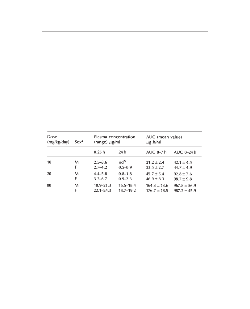
Plasma drug concentrations
43
Example 5.2
Methods
Blood was collected from each monkey at 5, 15 and 30 minutes and 1, 3, 12
and 24 hours following intravenous dosing on dose days 1 and 28 for measurement
of PP 27567 concentrations in plasma. At each interval approximately 0.5 ml
of blood was collected into an EDTA-coated container. Following centrifugation,
plasma samples were extracted on AASP C2 cartridges prior to separation by
high-pressure liquid chromatography (HPLC).
Results
Plasma concentrations on dose day 28 are summarized in Table 5.1. Plasma
concentrations and AUCs were dose-proportional on the first day of dosing and
there were no apparent sex-related differences.
Table 5.1 PP 27567:1-Month oral toxicity study in Cynomolgus monkeys-plasma
concentrations on day 28.
a
Each value is the range or mean of 3 monkeys/sex.
b
Concentration was below the limit of detection.
PP 27567 was slowly, but completely eliminated by 24 hours following dosing with
10 or 20 mg/kg/day. Concentrations on day 1 were similar to those on day 28. However,
at the 80 mg/kg/day dosage level, an approximate 4-fold increase in mean plasma
concentration occurred on day 28. This datum indicates significant accumulation
occurred at a dosage of 80 mg/kg/day. Systemic exposure in monkeys to daily dosages
of 10 mg/kg was approximately twice that expected in humans given the therapeutic
dosing regimen.
Discussion
Severe and persistent tremors with occasional convulsions were noted in all
monkeys dosed with 80 mg/kg/day after the 20th dose day. These effects were
probably associated with drug accumulation at this dosage level. Similar signs

Presenting toxicology results
44
continued
were not seen at lower dosages where accumulation was slight or did not occur.
Accumulation probably resulted from a reduced rate of renal excretion resulting
from the nephrotoxic effects of PP 27567 at this dosage level but not at lower
levels. Reductions in urine excretion, increased urea nitrogen levels and tubular
necrosis occurred at this dosage level. These observations are probably of no
clinical significance since plasma concentrations at the proposed maximum
clinical dose are approximately half those observed in monkeys dosed with 10
mg/kg/day.
Example 5.3
Methods
The systemic exposure of PP 27567 was measured in three satellite groups
of 12 rats each. On dose days 1, 30 and 90, blood samples were collected
from one rat/sex/group approximately 2, 6,10 and 24 hours following
subcutaneous dosing. Approximately 1.0 ml of blood was collected via the
retro-orbital sinus (under CO
2
anesthesia) into glass tubes using no
anticoagulants. Rats bled at each collection interval were euthanatized by
CO
2
asphyxiation and discarded. PP 27567 concentrations were determined
by a radioimmunoassay procedure.
Results
Subcutaneously administered PP 27567 was well absorbed in a dose-proportional
manner with no apparent difference between sexes. Peak plasma concentrations
occurred approximately 6 hours post-dosing. At 24 hours post-dosing on dose
day 1, PP 27567 was detected only in rats dosed with 10 and 30 mg/kg/day.
On dose day 1, peak plasma concentrations were 4, 10 and 27 µg/ml as opposed
to concentrations at 24 hours of 0, 4 and 13 µg/ml for the 3, 10 and 30 mg/kg/
day dosage groups, respectively. The graphs below show that, as the study
progressed, plasma concentrations of PP 27567 decreased. At the end of the
study (day 90), plasma concentrations of PP 27567 in rats dosed with 30 mg/
kg were approximately one-third of those measured on the first dose day. (See
Figures 5.1–5.3.)
Discussion
The decrease in plasma concentrations of PP 27567 over the course of dosing
suggests that self-induction of hepatic metabolism had occurred. This is consistent
with the dose-proportional increases in liver weight and proliferation of the
smooth endoplasmic reticulum observed by electronmicroscopy of liver tissues.
The reduction in plasma concentrations over time accounts for the decrease in
clinical signs observed during this study.
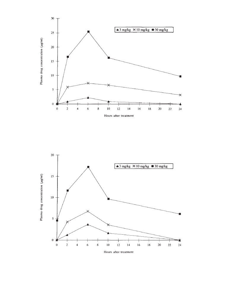
Plasma drug concentrations
45
Figure 5.1 PP 27567:90-Day subcutaneous toxicity study in rats. Mean plasma drug
concentrations (µg/ml) on day 1 (male and female values combined)
Figure 5.2 PP 27567:90-Day subcutaneous toxicity study in rats. Mean plasma drug
concentrations (µg/ml) on day 30 (male and female values combined)
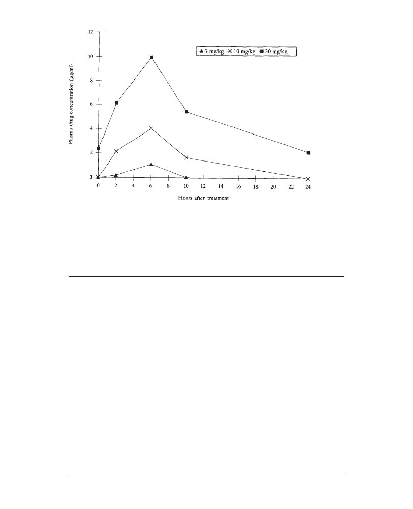
Presenting toxicology results
46
Example 5.4
Methods
Blood samples were collected approximately 2 hours after dosing from the first
6 rabbits per group that survived on gestation days 7 and 19 (dose days 1 and
13). The blood was collected from the medial artery in the ear and immediately
transferred into heparinized tubes. On gestation day 19, one dam per dosage
group underwent caesarean section approximately 0.5 hours following dosing,
and fetuses were removed. Blood was collected by decapitation under CO
2
anesthesia and pooled for each litter. Plasma from the dams and fetuses was
stored at –20°C. Concentrations of parent drug (PP 27567) and its major metabolite
(PP 39876) were determined by reversed phase HPLC. Two other metabolites
(PP 39877 and PP 39878) of PP 27567 were not measured, because they were
present at less than 2% of the total level of parent drug and metabolites that
could be detected.
Results
At 2 hours post-dosing, plasma concentrations of PP 27567 and its major
metabolite (PP 39876) in pregnant rabbits appeared to increase in a dose-
dependent manner on both gestation days 7 and 19. Marginal increases in
concentrations of PP 27567 occurred during the dosing period. Plasma levels
Figure 5.3 PP 27567:90-Day subcutaneous toxicity study in rats. Mean plasma drug
concentrations (µg/ml) on day 90 (male and female values combined)
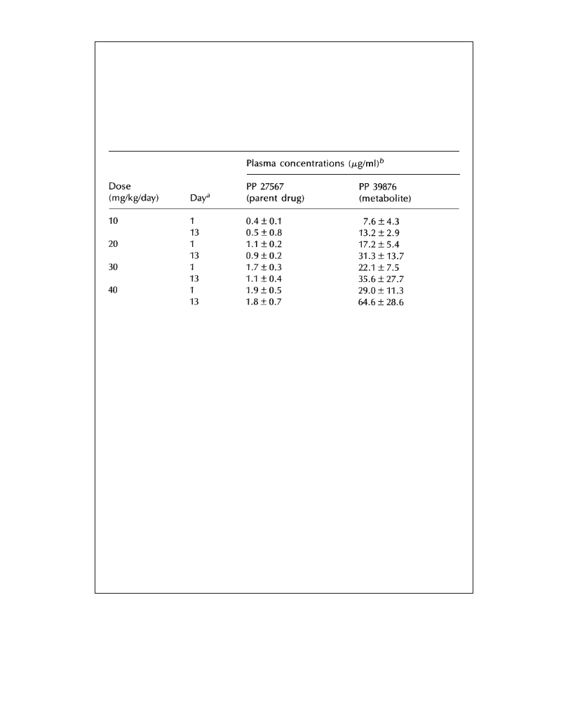
Plasma drug concentrations
47
continued
of the parent drug and its metabolite in the pregnant animals are given in
Table 5.2.
Table 5.2. PP 27567: Maternal toxicity study in pregnant rabbits—plasma drug
concentrations 2 hours after treatment.
a
Correspond to gestation days 7 and 19.
b
Mean±standard deviation of 6 rabbits/dose group.
Fetal exposure at the end of the dosing period was limited to the metabolite PP
39876 at the 30 (2.5±1.7 µg/ml) and 40 (4.7±2.6 µg/ml) mg/kg dosage levels only. No
metabolite was detected at lower dosages. Parent drug was not detected in fetal plasma
at any dosage level.
Discussion
Exposure to PP 27567 and PP 39876 in pregnant rabbits was dose dependent
and ranged from approximately one-half to twice that expected in humans given
the therapeutic dosage. This is in contrast to exposure to metabolite, which appears
to range from two- to fourfold greater than that expected in humans. The data
suggest that the rate of metabolism of parent drug may be greater in the rabbit
than human. The relatively small difference in maternal concentrations between
the 30 and 40 mg/kg dosages and the significant degree in maternal toxicity
observed at these dosages suggest that the metabolite may be more toxic than
the parent drug. Similarly, fetal deaths were observed only at those dosage levels
(30 and 40 mg/kg/day) at which the metabolite was detected in the fetuses.


49
6
Reporting In-life Observations and
Measurements
G.J.NOHYNEK
Rhône-Poulenc Rorer, Vitry sur Seine, France
The results of in-life observations/measurements of in vivo toxicity studies are
commonly described in the following order:
1
mortality
2
clinical signs and observations
3
body weight evaluations
4
food consumption data
5
other in-life evaluations, e.g. cardiovascular measurements.
6.1
Mortality
Mortality is the most serious result of toxicity and must be described with great care.
Describe mortality with reference to animals and/or dose identification, time of death
and clinical observations associated with or prior to death. Whenever mortality occurs
in a toxicology study, it must be clearly distinguished as compound-related, procedure-
related or caused by other factors. The incidence of mortality and dose identification
should always be numerical, e.g. “5/20 males died at 150mg/kg”. Necropsy observations
should not be included in this section except to explain non-compound-related death,
e.g. unscheduled or accidental death resulting from gavage error.
A description of mortality which occurred in satellite groups which were used
for plasma drug determination may be included in this section if death is considered
to be related to the compound and not to blood sampling procedures. Mortality caused
by accidents (gavage accidents, accidents during handling or restraining of animals)
should be described separately, including clinical signs preceding death and/or the
evidence (e.g. necropsy findings) for classifying these deaths as accidental. Example:
“Two animals [male No. 304 (40mg/kg) and female No. 605 (80 mg/kg)] were found
dead on days 15 and 22, respectively. Necropsy findings in these animals (congested
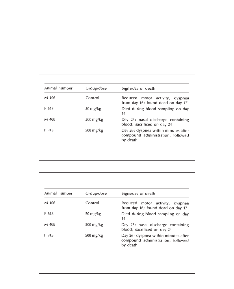
Presenting toxicology results
50
lungs, traces of white particles in the bronchi) suggest that their deaths were due to
gavage accident”. If a high incidence of mortality occurs in a study, and particularly
if compound-related mortality occurs concurrently with mortality not related to the
test compound, use a table to improve comprehensibility (see Examples 6.1 and 6.2).
Sacrifice for ethical reasons should be addressed in this section; include a brief
description of the rationale for sacrifice, e.g. “Male 41 (24 mg/kg) was found in a
moribund condition on day 21 and was sacrificed”, or “Two males at 50 mg/kg
displayed severe clinical signs (severe ataxia, convulsions) and were sacrificed for
Example 6.1 Use of table to report mortality details in a single-dose study
Table 6.1 PP 27567: Single-dose oral study in mice—incidence of mortality.
a
attributed to accidental death of female F 55 (gavage error—see necropsy
findings).
Example 6.2 Use of table to report mortality details in a repeated-dose study
Four animals died during the course of the study as described in Table 6.2.
Table 6.2 PP 27567:1-Month repeated-dose oral toxicity in rats—mortality.
Based on the circumstances of these deaths and/or necropsy findings (e.g. congested
lungs, traces of white particles in the bronchi) they were considered to be accidental
(due to gavage error for animals M106, M408 and F915, or due to the anesthesia and/
or blood-sampling procedures for F613).
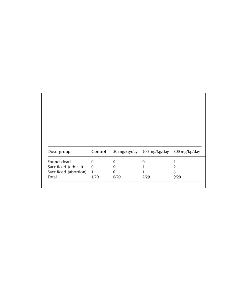
Reporting in-life observations and measurements
51
ethical reasons”. However, avoid referring to mortality of animals sacrificed for
ethical reasons as “compound-related” mortality! However, you may say: “Mortality
(including sacrifice of moribund animals for ethical reasons) was 0/10, 2/10 and
6/10 at 2, 8 and 32mg/kg, respectively”.
In those reproductive toxicology studies which had a high incidence of mortality
and sacrifice due to ethical reasons, a table explaining the incidence of unscheduled
death, animals sacrificed following abortion or sacrificed for ethical reasons may
be included (see Example 6.3).
Example 6.3 Use of table to report mortality details in a reproductive toxicity
study
One animal at 300 mg/kg/day was found dead on day 16 of the study. On days
18 and 19, 6/19, 1/20 and 1/20 animals in the 300 mg/kg/day, 100 mg/kg/day
and control groups, respectively, were sacrificed following abortion. In addition,
2 animals at 300 mg/kg/day and 1 animal at 100 mg/kg/day were found moribund
and sacrificed on days 17 and 18. No mortality occurred at 30 mg/kg/day. The
incidence of mortality is shown in Table 6.3.
Table 6.3 PP 27567: Maternal toxicity study in rabbits—mortality.
6.2
Clinical Signs and Observations
This section should first describe compound-induced symptomatic or behavioral
changes (ataxia, emesis, mydriasis, absence of motor activity, etc.), followed by
observations or signs related to general appearance (alopecia, stained fur, etc.).
The incidence of clinical signs should be numerical, if possible. The description
of clinical signs should include (numerical) data about their onset, duration, incidence
and intensity. The severity of a clinical sign may be graded mild, moderate, or
marked/severe. However, grading scales of clinical signs should only be used in
exceptional cases. Avoid using more than three severity grades. If grading scales
are used, the grading should be defined in the text or using a footnote of a table.
The terminology used for an individual sign should correspond to a clinical sign
dictionary or the respective Standard Operation Procedure. Signs and symptoms
should preferably be descriptive, using simple terminology and avoiding diagnostic
terms, terms which are poorly defined or not commonly used in toxicology (see
attached list). Rare clinical signs which require use of the appropriate medical or

Presenting toxicology results
52
veterinarian terminology should be defined and, if necessary, explained in the text
when used for the first time. Examples:
•
“Paresis (weakness) of the hind limbs was observed on day 12 in one animal
(F707, 80mg/kg). This sign became increasingly severe during days 13 to 15,
warranting sacrifice for ethical reasons on day 16.”
•
“Opisthotonos (prolonged and severe muscle spasm of the back causing arching
of the back, backbending of the head with rigid, extended limbs) was noted
on day 2 in two male dogs (M205, F707) in the 80mg/kg group.”
Sporadic clinical signs which are not related to the compound but which appear
in the tables of the individual clinical observations in the appendix of the report
should be addressed and dismissed (if not compound-related) in a final phrase of
this section. A table may be used to aid comprehension of multiple clinical signs
(see Example 6.5). In reproductive toxicology reports, the incidence of abortion
may be described in this section.
6.3 A
Clinical Signs Glossary
The following is an example of a simple list of clinical signs which is by no means
complete. However, it is more practical to use a simple list which includes only
the most common clinical signs. This prevents overloading your laboratory’s glossary
with diagnostic terms and potential misuse of terminology. Diagnostic terms for
rare clinical signs should be defined on a case-by-case basis after a careful
examination of the affected animal(s).
General appearance and condition
Abortion
Nasal discharge
Agalactorrhea
Pale skin (site)
Alopecia
Piloerection
Arched back
Reduced elasticity of the skin
Broken teeth
Rough hair coat
Brown stains on muzzle
Scab (site, size, appearance)
Cold to the touch
Skin flakes
Cough
Soiled ano-genital area
Discoloration of mucous membranes
Soiled fur
Discoloration of the skin (site, color)
Stained teeth
Discolored haircoat (color)
Swelling (site)
Distended abdomen
Tail loss (partial/complete)
Excessive salivation
Thin body
Galactorrhea
Thin hair coat
Hunched back
Umbilical hernia
Increased firmness (site)
Vulvar discharge
Loss of teeth
Wet fur
Mass (site)
Wound (site, size, appearance)

Reporting in-life observations and measurements
53
Eyes
Destruction of eye
Pinpoint pupils
Dry eye
Protruding eye
Eye opacity
Protrusion of nictitating membrane
Eyelids (partially) closed
Red area around eye
Lacrimation
Red conjunctiva
Mydriasis
Sunken eye
Functions
Absence of urine
Liquid feces
Absent/reduced feces
Noisy respiration
Discolored feces (color)
Reduced/increased urine volume
Discolored urine (color)
Regurgitation
Hiccup
Retching
Increased/decreased respiration rate
Soft feces
Increased/decreased water intake
Vomiting
Labored respiration
Behavior
Aggression
Hyporeactive to external stimuli
Agitation
Licking
Chewing
Lying on the cage floor
Circling movements
Unresponsive to external stimuli
Head tilt
Vocalization
Hyperreactive to external stimuli
Activity/Movements
Absence of motor activity
Lameness of limbs
Convulsions
Loss of righting reflex
Decreased motor activity
Loss of use of limbs
Flaccidity of limbs
Rigidity of limbs
Incoordination
Tremors
Increased motor activity
Example 6.4 Clinical signs
Treatment-related signs occurred from week 2 through study termination: all
animals treated with 150 mg/kg/day and 3/4 male animals at 50 mg/kg/day
displayed dryness of the nose and redness of the bulbar conjunctivae. In addition,
all animals at 50 and 150 mg/kg/day, and 3/4 males at 15 mg/kg/day had mydriasis.
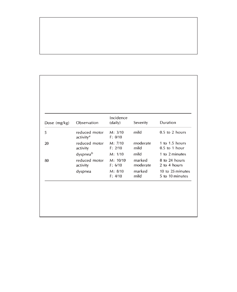
Presenting toxicology results
54
continued
All signs appeared within 15 minutes after administration of PP 27456 and
persisted approximately 6 to 8 hours. All other clinical signs recorded during
this study are commonly observed in our laboratory and were not considered
to be related to PP 27456.
Example 6.5 Table of clinical signs
Clinical signs (reduced motor activity and dyspnea) were observed throughout
the study with dose-related incidence, severity and duration, and appeared within
10 minutes following treatment. Their incidence, severity and duration are
summarized in Table 6.4.
Table 6.4 PP 27567: Single oral toxicity in rats—clinical signs.
a
Reduced motor activity: mild—sluggishness of animals; moderate—sleepy
animals/reduced reaction to external stimuli; marked—absence of reaction to
external stimuli, animals asleep for>2 hours following treatment.
b
Dyspnea: mild—occasional gasping; moderate—1–2 times per minute; marked –
episodes of continuous gasping lasting more than 1 minute, followed by brief recovery
and reoccurrence.
6.4
Abortions (Rabbit Segment II/Embryofetal Development
Toxicity Studies)
Strictly speaking, abortions represent a clinical sign and should be reported under
this section. On the other hand, since animals having aborted are normally sacrificed
for ethical reasons, the incidence of abortions may also be reported within the
mortality section, or within a separate section. However, in my view, abortions
are preferably reported within the Clinical Signs and Observations section of rabbit
Segment II studies.

Reporting in-life observations and measurements
55
Example 6.6 Compound-related abortions
Six females in the 300 mg/kg/day group, one female in the 100 mg/kg/day
group and one control female aborted during the treatment period. The
pregnancy losses of five of the 300 mg/kg/day females were preceded by body
weight loss, reduced food intake and reduced fecal output; the sixth female
had no significant history. It is known that adverse treatment, including dietary
deprivation (Matsuzawa et al., 1983), can lead to abortion in rabbits. Thus,
all of the abortions observed in this study can be considered to be related to
changes in maternal status, with PP 27567-related maternal toxicity contributing
to the increased incidence of abortion when the drug was administered at
300 mg/kg/day.
Example 6.7 Abortions not related to the test compound
One control female, two females in the 30 mg/kg/day group and one female in
the 300 mg/kg/day group aborted during the study. In view of the group
distribution and the absence of a treatment-relationship, involvement of PP 27567
was considered unlikely.
6.5
Body Weight Data
Describe body weight changes relative to dose and sex. As a rule, descriptions
of body weight changes refer to group mean changes for rodents and rabbits,
and individual animal change for large animals, i.e. dogs and monkeys. With
two different control groups, e.g. in intravenous studies which include a vehicle
control and a saline control group, group mean body weight changes in treated
groups should always be related to vehicle control group means. Body weight
changes in large animals may be related to the control group or to individual
body weights at study initiation. Be clear of your meaning when expressing
changes in body weight versus changes in body weight gain. Generally, it is
simpler not to attempt to describe a lower mean body weight gain, but instead
to refer to lower group mean body weights in treated groups at study termination
or at interim weighing points. Whenever there are multiple changes in body
weight across treated groups, they may be described using a table (see Example
6.9).
Please note that the term “body weight” is generally used in the singular unless
one refers to several groups or specifically to several animals, e.g. “Body weight
in the group receiving 74 mg/kg/day was unaffected. There was no compound-
related change in body weight in the groups receiving 75 or 150 mg/kg PP 46567.”
But: “5/10 animals receiving 55 mg/kg/day had lower body weights”.
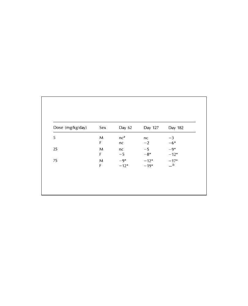
Presenting toxicology results
56
Example 6.8 Body weight data
PP 27567 produced dose- and compound-related effects on body weight: on
day 29, males treated with 100 and 300 mg/kg/day had a lower mean body
weight (–8% and –12%, when compared with control values); in females, the
respective mean body weight differences on day 29 at 100 and 300 mg/kg/day
were –11% and –16%, respectively. The difference in body weight was statistically
significant at 300 mg/kg/day for both male and female groups. No effect on
body weight was observed in the groups receiving 30 mg/kg/day.
Example 6.9 Body weight table
Table 6.5 PP 27567:6-Month repeated dose toxicity in rats—mean body
weight on days 62, 127 and 182 of the study (percentage change compared
with control mean values).
*p<0.05
a
no change
b
treatment discontinued on day 128
6.6
Food Consumption
Describe food consumption changes according to sex and dose. Whenever multiple
changes across several treated groups are observed, they should be described in a
table. Minor or elementary changes may be described in the text. Changes may
be expressed as percentage change compared with control means for rodents and
rabbits. For dogs or primates, food consumption in treated groups may be compared
with control means with sufficient group size and body weight homogeneity.
However, food consumption in dogs is generally described semi-quantitatively. In
short-term studies (<1 month) in large animals, food consumption may be compared
with pre-study mean and/or individual values. Correlate changes in food consumption
with corresponding changes in body weight. Please note that the term “food
consumption” is always used in the singular!
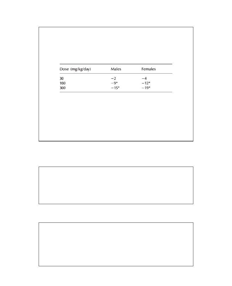
Reporting in-life observations and measurements
57
Example 6.10 Food consumption—rodents
In all treated groups, PP 27567 produced a compound- and dose-related decrease
in food consumption as shown in Table 6.6.
Table 6.6 PP 27567:28-Day repeated dose oral toxicity in rats–
food consumption during week 4 (percentage change compared
with control mean values).
*p<0.05 (comparison of means with respective control means)
The decreased food consumption in males at 100 and 300 mg/kg/day was
statistically significant and correlated to the lower body weight gain observed
at these dose levels. While a relation to treatment of the lower food consumption
at 30 mg/kg/day cannot be excluded, this change was minimal, within the range
of the common variation of this parameter, and is therefore considered to be
of no toxicological significance.
Example 6.11 Food consumption—dogs
Food consumption of dogs receiving 100 mg/kg/day was severely decreased
(approximately –75% in females and –50% in males). At 30 mg/kg/day, a sporadic
decrease in food consumption was noted with a slightly higher incidence in
females than in males, while at 10 mg/kg/day food consumption was unaffected
by treatment. The lower food consumption at 100 mg/kg/day correlated with
the observed body weight loss in males and females.
Example 6.12 Food consumption—rabbits
1
Food intake of 45 and 150 mg/kg/day females was decreased during the
second half of the treatment period and correlated with the lower body
weight gain in these groups. Food consumption of females in the 15 mg/
kg/day group was unaffected by treatment.
2
A dose-related and statistically significant decrease in food consumption
was recorded in all treated groups for the dosing period. Following the
recovery period, food intake was similar in all groups.

Presenting toxicology results
58
6.7
Cardiovascular Parameters
6.7.1
Electrocardiography
Cardiovascular parameters are of particular interest as potential markers in clinical
trials. However, care should be taken with respect to the meaning of the individual
parameters in the human clinical situation when addressing changes in canine ECGs
(e.g. “sinus arrhythmia” is pathologic in man but “respiratory sinus arrhythmia”
is physiologic in dogs). Always report the quantitative change (are there any
modifications in parameters or values?) before the qualitative ones (are there any
rhythm abnormalities?). For quantitative analysis, describe the changes numerically.
Complex and multiple changes should be reported using a table.
Example 6.13 Cardiovascular parameters not modified
Heart rate values, PR, QRS and QT duration were not modified by PP 27567.
No treatment-related changes in rhythm were noted.
Example 6.14 Increase in heart rate
Two hours following administration of PP 27567, animals treated at 200 mg/
kg had increased heart rates (20–30% when compared with pre-test values) and
a concomitant reduction in QT interval (–12% to –15%, when compared with
pre-test values). These changes were no longer present 24 hours after treatment.
PQ and QRS interval were not modified by the treatment.
Example 6.15 Changes in ECG parameters
In week 4, monomorph interpolated ventricular premature beats and a transient
bundle branch block were noted in the ECG of one male dog (M31) treated at
100 mg/kg/day. These changes were neither observed in ECGs performed 24
hours later, nor in those performed at week 13. Ventricular premature beats
and bundle branch block may occur spontaneously in dogs (Patterson et al.,
1961); since these changes were observed only in a single examination in one
animal, they were considered to be of spontaneous origin and not to be related
to administration of PP 27567.
Example 6.16 Increase in heart rates—table
Heart rates measured during the pre-test period and during the study before the
administration of PP 27567 were comparable in all groups (range: 85–134/min).
During the treatment period, a dose-related increase in heart rate was noted in
all treated groups 1 to 2 hours after treatment. This is shown in Table 6.7.
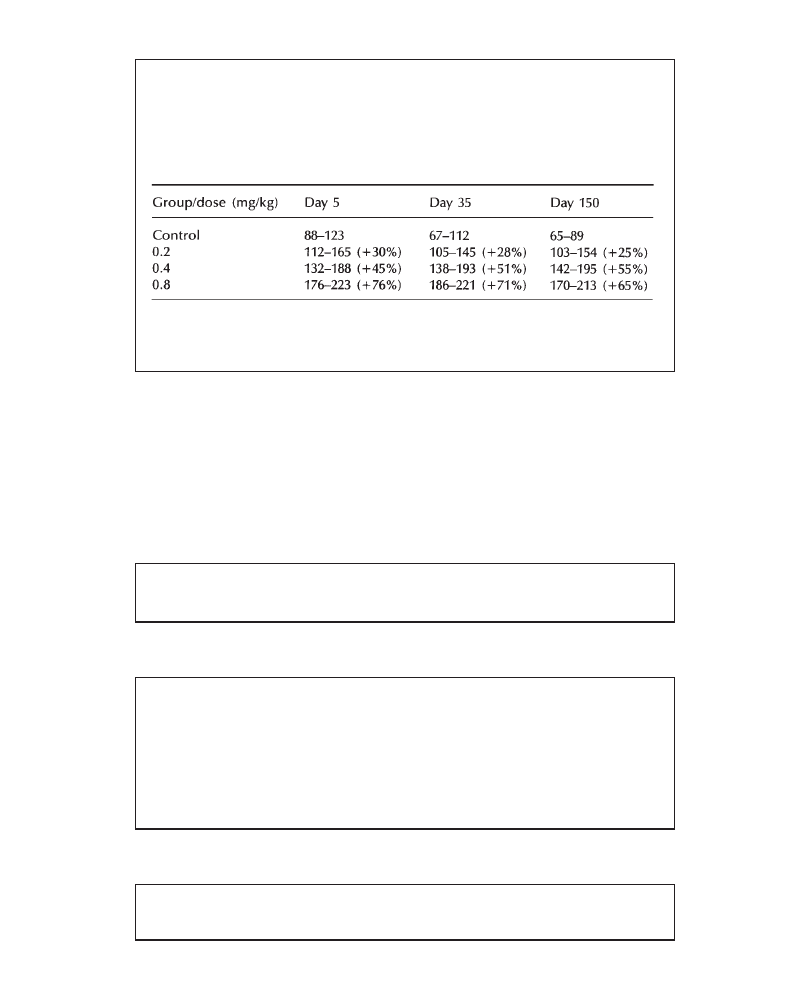
Reporting in-life observations and measurements
59
continued
Table 6.7 PP 27567:1-Month oral toxicity study in dogs. Range of heart rates
(and percentage mean increase, compared with pre-administration group mean
values) 1 to 2 hours after administration of PP 27567 on days 5, 35, and 150
of the study.
Heart rates in all groups taken 24 hours after treatment with PP 27567 were
comparable to control or pre-test values.
6.7.2
Systolic Blood Pressure
In standard toxicology studies on non-rodents, both systolic and diastolic pressure
are generally measured, although the evaluation is principally performed using the
systolic arterial pressure.
Example 6.17 Blood pressure not affected by treatment
The values of systolic and diastolic blood pressure were not affected by
treatment.
Example 6.18 Compound-related change in blood pressure
A compound-related increase in blood pressure was measured in the groups
receiving 60 and 120 mg/kg/day on day 21, approximately 2 h following
administration of PP 27567. In 60 mg/kg females, the mean systolic arterial
pressure increased by about 20%, when compared with pre-test values. In
the 120 mg/kg group, the corresponding increases were+20% in males
and+32% in females. No compound-related effect was measured at 30 mg/
kg. Twenty-four hours after treatment, arterial blood pressure was comparable
in all groups.
Example 6.19 Use of a table for blood pressure values
A dose-related reduction in mean systolic blood pressure was noted 1 to 2 hours
after treatment in all treated groups, relative to pre-treatment values. The extent
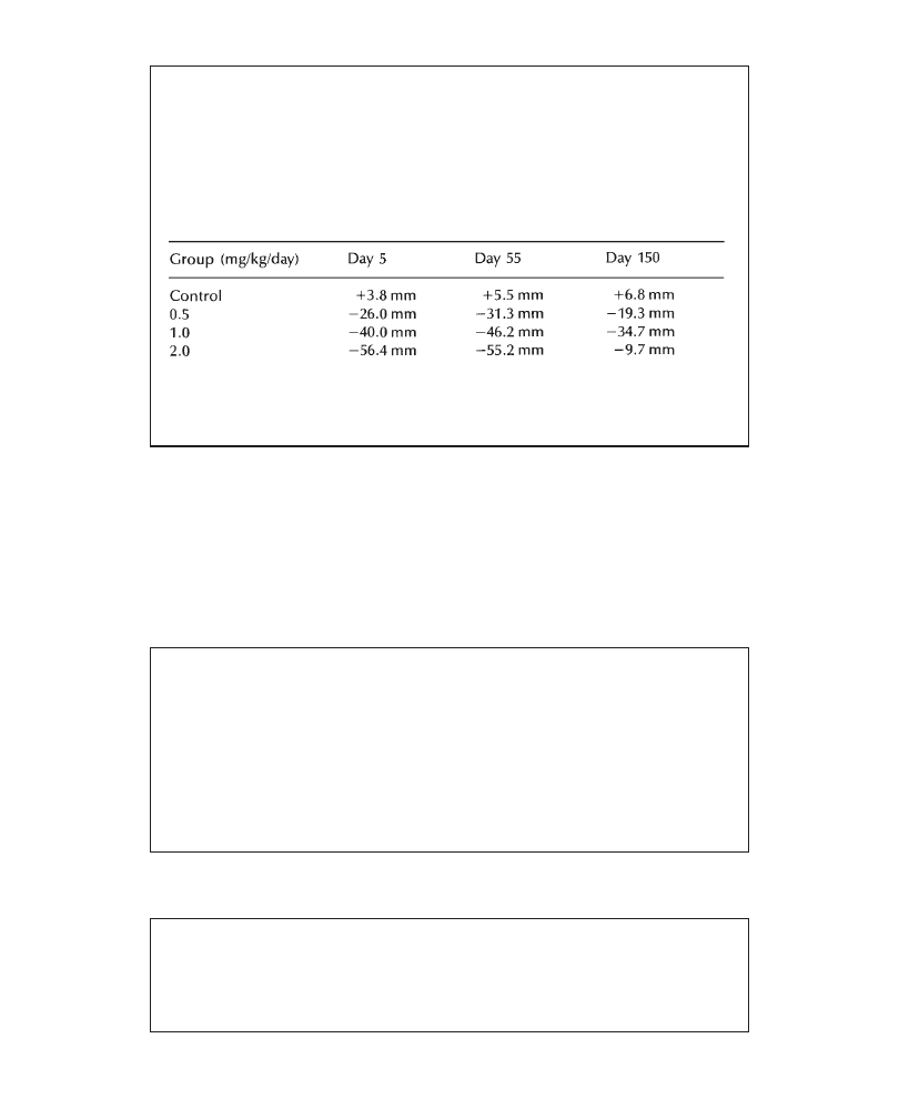
Presenting toxicology results
60
continued
of this effect was comparable on days 5, 55 and 150 of the study and is shown
in Table 6.8.
Table 6.8 PP 27567:6-Month oral toxicity in dogs—change of mean systolic
blood pressure, compared with pre-treatment group mean values. Values on study
days 5, 55 and 150, 1 to 2 hours after administration of PP 27567.
Systolic blood pressure values, taken 24 hours after administration of PP 27567
were comparable to control or pre-test values.
6.8
Ophthalmology
Describe all compound-related changes first and indicate the number of animals/
sex/dose with the change.
Example 6.20 Ophthalmic observations
All lesions observed were considered typical for the age and strain of animals
examined. There were no findings of toxicological significance.
A bilateral hypopigmentation of the fundus was observed in one control male at
all examinations. A slight unilateral focal posterior lens opacity was noted in one
100mg/kg/day female in week 13. The latter finding is commonly observed in dogs of
this strain and age and was therefore not considered to be related to treatment. The
main ocular findings were either of embryological origin (remnants of hyaloid vessels)
or variations on the aspects of the vessels of the bulbar conjunctiva. The latter
observation remained within physiological limits and is commonly observed in dogs
of this age and provenance.
Example 6.21 Ophthalmic observations
Remnants of hyaloid vessels or a noticeable suture line of the anterior cortex of
the lens were observed during the pre-study examination and on day 30 in some
control animals and in some animals at 125 mg/kg/day. These vestigial fetal
structures were no longer observed at the end of the study and were attributed
to the young age of the animals. Small opalescent vesicles were observed in the

Reporting in-life observations and measurements
61
continued
superficial part of the corneal stroma in some male animals of the control, the
125 mg/kg/day and 5 mg/kg/day groups. This lesion was diagnosed as corneal
dystrophy. Some minor changes were noted in the nucleus of the lens in animals
from the control, 5 and 25 mg/kg/day groups. A comparable incidence of these
lesions has been reported for Sprague-Dawley rats of this strain and age (Taradach
and Greaves, 1984). In absence of the dose-relationship and since the incidence
of these changes remained within our historical control values for incidence
for these lesions, these changes were considered to be of spontaneous origin.


63
7
Reporting Clinical Pathology Results
M.Y.WELLS
Rhône-Poulenc Rorer, Drug Safety Department, Vitry sur Seine, France
and S.GOSSELIN
ITR Laboratories Canada Inc., Montreal, Canada
7.1
A Few Words on Data Evaluation
Hematology, clinical chemistry and urinalysis data can be among the most difficult
data within a given study to evaluate, interpret and report. While the guidelines
on how to write this section should give you some ideas on how to evaluate data,
there is significant information on this subject that cannot be obtained herein. Also,
because complete and thoughtful assessment of these data can be a trying process
even for those specifically trained in clinical pathology, and because so many of
us who must work with it are not trained in this area, a few words on the philosophy
of data evaluation seem to be in order.
Methods of clinical pathology data evaluation include the use of statistics and
the comparison of data from treated animals with absolute control, vehicle control,
pretest, and/or historical control values. The method(s) chosen can vary with the
species used and the length of study performed. Correlation between clinical
pathology, in-life observations and anatomic pathology data is highly desirable
but often not possible, and thus there is often no additional support for the conclusions
drawn from analysis of clinical pathology data. Moreover, clinical pathology
parameters are presumably more sensitive than histopathology for the interpretation
of some lexicologically significant effects, which accentuates the importance of
their accurate interpretation.
Statistics are often used for the assessment of clinical pathology data; but reliance
on this type of evaluation alone often leads to the erroneous attribution of
toxicological significance to “flagged” data, as well as erroneous inattention to
data which are far more important for the interpretation of compound-related effects.
For example, a large standard deviation can cause a compound-treated group to
appear statistically similar to its control group, even when one or several individual
values in the treatment group do not fall within the control range. Closer evaluation
of these individual values may reveal a compound-related effect. On the other hand,

Presenting toxicology results
64
statistically significant values may be within the range of normal variation; these
should be minimized or dismissed when writing the clinical pathology report. Thus,
statistics should be neither the sole nor the primary criteria for determining the
toxicological significance of clinical pathology findings.
Among the species used in industrial toxicology safety assessment, large animals
(e.g. dogs, monkeys, rabbits) generate the data which are the most complicated to
evaluate. The number of animals per group is often less than five, meaning that
the power of statistical analysis is significantly reduced. The wide range of individual
variation in these animals requires more rigorous assessment of the data they generate,
which should include evaluation of raw data from each individual animal. In these
situations, individual animal data may take precedence over mean values.
All data should be evaluated and reported in relation to study controls. These
are generally animals given the vehicle only. When two control groups are used
(e.g. saline and vehicle control groups in intravenous studies), any differences
between the vehicle and absolute control groups should be reported. The values
from compound-treated groups should be compared with those of the vehicle control
group to identify potential compound-related effects. When treated animals have
laboratory values outside the study control range, historical control and pretest
data (when available) may be evaluated to gain perspective on the significance of
the observed variations. This does not mean that comparisons to pretest or historical
control data must be reported; these are mentioned in the report only if they cause
you to doubt or alter your interpretation of values in treated groups as compared
with study controls.
In large animal studies, pretest analyses are usually performed, while in small
animal (primarily rodent) studies, they are rarely performed. Pretest data should
be considered in the overall evaluation of data for large animal studies of a duration
of three months or less. In studies where animals are to be handled extensively
and/or repeatedly bled, two to three pretest bleeds provide a more reliable database.
In many, cases however, such an extensive base is not available. For longer studies,
the age difference in the animals between study onset and study termination makes
pretest data less useful in assessing compound-related changes (this may be less
true in monkey studies if the animals are mature at the time of study initiation).
In the pretest period, animals destined for compound administration should be
evaluated in relation to pretest control animals to discern whether or not any
differences among these groups are appreciable before study initiation. During the
study and at its termination, treatment values from individual animals in each group
should be compared with their respective values during the pretreatment phase,
so that each animal serves as its own control. Additionally, values from individual
animals in a specific group may be compared with the range of values observed
in that pretreatment group (e.g. values from high-dose animals should be compared
with those of pretest “high-dose” animals, and not those from pretest control animals).
Pretest values from all groups may be used to form one large pretest reference
range, but only if there are no differences observed among the pretest groups. This
is not usually necessary, but may be desirable if the number of animals per group
is very small.
Historical control data are another source of information which can be used to
evaluate clinical pathology parameters. Many clinical pathologists limit their use
of this database to the evaluation of animals during the acclimatization period because
they believe that historical control values are too broad and not representative enough

Reporting clinical pathology results
65
of the animals on study. For evaluation of changes occurring during studies using
large animals, many favor pretest data over historical control data.
When historical controls are used during a study, they must be matched with
the evaluated study parameters by age and by sex. Just as pretest data are not
especially useful for parameters evaluated 6 months after study initiation, a historical
control database for animals aged 6 months will not be very useful for interpreting
data obtained from animals aged 14 months.
Differences in mean or individual values between treated and control groups
which are determined to be compound-related may fall into the historical control
and/or pretest range. If you believe that the observed changes are not biologically
significant, then you may refer to these databases to support your position. However,
if you do believe the changes to be significant, the fact that the values fall within
reference ranges should not alter your conclusion. In other words, it is irresponsible
to summarily dismiss compound-related effects simply because their values fall
within reference range.
In rodent studies, the range of individual variation in clinical pathology parameters
is smaller, and concurrent study controls are likely to constitute a sufficient database
with which to compare and evaluate changes in treated animals. This is generally
true regardless of the length of the study performed. However, in studies longer
than 52 weeks, it is important to realize that data from all groups (including controls)
are increasingly subject to variations secondary to age-related changes. In the
combined chronic toxicity/carcinogenicity studies performed in the chemical industry
for example, clinical pathology parameters are evaluated periodically through the
entire study, which usually lasts 104 weeks. The data collected after 52 weeks
generally yield little useful information with regard to primary compound-related
effects, because age-related renal disease and spontaneous tumor generation become
more prevalent, beginning at this time. These pathological changes affect clinical
pathology parameters, and are likely to complicate the interpretation of any effects
resulting directly from compound administration. Thus, if you are forced to interpret
data in a study such as this, be fully aware of the “background noise” with which
you are dealing.
The end result of the data evaluation described above should be the determination
of all changes resulting from compound administration. In addition to the comparison
of data with one or more reference ranges, the presence of a dose-relationship and/
or the reversibility of an effect help to determine whether or not variations are
compound-related. These issues are further described in the next section of this chapter.
While this preface in no way pretends to offer the reader a crash course in the
evaluation of clinical pathology data, we hope that it highlights some general
principles and, perhaps more importantly, some pitfalls of data interpretation in
this domain. Before going on to the section addressing the actual writing of a clinical
pathology report, here is a final note of caution: avoid overinterpretation of data
but beware of under-reporting effects because of data which are overlooked!
7.2
Reporting the Data
As in all types of writing, clarity of expression is paramount in producing a
high-quality document. It is equally important to keep in mind the final audience
for whom you are writing (e.g. the regulatory authorities who will ultimately

Presenting toxicology results
66
pass judgment on the compound being assessed, the physicians who will use
your conclusions to plan clinical trials), and to prepare your report in a way
that will be the most comprehensible for that audience. Finally, make every
attempt to express your findings succinctly, without sacrificing necessary
information which could compromise the reader’s understanding of the
conclusions presented.
You want to focus the reader’s attention on what you believe to be important.
Therefore, limit the scope of your descriptions of secondary and incidental changes
so that you do not require paragraphs of text to explain them. The meat of the
report should address compound-related changes.
Regardless of the number of databases (study controls, historical controls, etc.)
used to evaluate studies, data are usually reported in relation to study control values.
Additional information regarding data reviewed in relation to pretest and/or historical
control values should be included when it helps clarify study results. An example
of such a situation would be the following: “Minimal increases in phosphorus
excretion were observed in males and females (+20% and +30% of mean control
values, respectively) at 120mg/kg/day. These values were increased over pretest
values, but were within the range of historical control data. Thus they were not
considered to be biologically significant”.
Whether one is reporting hematology, clinical chemistry, or urinalysis values,
answering the following questions will ensure complete reporting of the information
required in this (or any) section of a toxicology report.
7.2.1
Are There Compound-related Changes?
The most important information given in any section of a toxicology report is whether
or not there are effects related to compound administration. The first paragraph
of the section should address this question. If there are no compound-related changes,
simply state that none was observed; but if compound-related changes are present,
they must be specifically identified. State the numeric dose levels at which the
effects were noted, and whether one or both sexes were affected. If the number of
changes is small, you may indicate their magnitude and direction in this paragraph
as well. Identify any effects which have statistical significance (remember that
statistical significance alone does not mean that an effect is compound-related!).
See Example 7.1.
Example 7.1
The administration of PP 19875 was associated with a moderate, statistically
significant increase in ALAT (fivefold increase over mean control values) at 100
mg/kg in males only.
If there is only one effect, it can be fully described in the opening paragraph. If
there are several changes to describe, you may summarize them in the opening

Reporting clinical pathology results
67
statement and quantify them in subsequent paragraphs. Alternatively, you may choose
to provide a list of the changes in the opening paragraph (see Example 7.2).
Example 7.2
Compound-related clinical chemistry findings (as compared with mean vehicle
control values) consisted of:
•
a mild increase in ALAT at 100mg/kg (+80%)
•
a mild decrease in triglycerides at 50 and 100mg/kg (not less than –35%)
•
a moderate increase in cholesterol at 25, 50 and 100 mg/kg (not greater
than +45%)
•
a moderate dose-related increase in BUN at 25, 50 and 100 mg/kg (+10%,
+35% and+50%, respectively).
Large amounts of data or otherwise complicated data are best reported in the form
of tables, figures and/or graphs. When using tables, values which are statistically
significant should be indicated; these are more easily visualized in the tabular format
than in prose.
Once you have made your opening statement, elaborate the results supporting
that statement. Group related effects in a single paragraph (e.g. changes in liver
enzymes, cholesterol and triglycerides indicate an effect on the liver and should
be discussed together). Some clinical pathologists use absolute values, either in
text or in tables, to express variations in study parameters. Some quantify effects
by expressing the changes (increases or decreases) as compared with the appropriate
control values. Others qualify the effects using severity modifiers such as minimal,
marked, etc., with or without quantifying the changes. This communicates the
author’s interpretation of the changes, and requires her/his scientific expertise to
reflect what has occurred biologically. Still others prefer to accentuate the changes
seen by placing the control or pretest value in parentheses next to the effect described.
Using this approach assumes that the reader has sufficient knowledge to make her/
his own interpretation of the data, and additionally assumes that the interpretation
will be the same as your own! When deciding which approach to use, remember
your audience, and take into consideration its level of expertise in clinical pathology.
If your reader is not knowledgeable in this area, you are strongly advised to qualify
as well as quantify your results.
Using percentages to quantify changes is acceptable, but for changes greater
than 100%, this terminology can become confusing. The interpretation of your prose
can become the reader’s exercise in semantics. Consider the case where the control
mean value for alkaline phosphatase is 50 IU/liter and the high-dose value is 150
IU/liter. An increase reported as “+200% as compared with control values” may
be interpreted in at least two ways:
1
200% of the control value (200%×50mg/l), or 100 IU/1
2
200% greater than the control value [(200%×50 IU/l)+50 IU/l], or 150 IU/l
(the true high-dose value).

Presenting toxicology results
68
A reader may consider these or other options, and not know which one to
choose. This may force him/her to go to tables in a separate section to look
for the actual high-dose value, which means that you have not achieved the
clarity of expression that you wish to be the hallmark of your report. The
term “-fold” may be used as an alternative in this instance. If the above-
mentioned change is reported as a threefold increase over the control value,
the only conclusion that the reader can reach is that the high-dose value is
triple the control value.
Some clinical pathologists will use a combination of terminologies to express
data in a single report, e.g. using percentage change to describe some chemistry
parameters, -fold change to describe enzyme or hematology changes, and absolute
numbers to describe specific individual parameters. They make their choices based
on experience in dealing with the types of numbers generated by these parameters
(e.g. since doubling or tripling of serum sodium or chloride is biologically impossible,
one would not use the term “-fold” to describe increases in these parameters). Others
prefer to choose one method of quantification for all parameters. Still others do
not quantify results at all in the text, but provide tables for this purpose. The options
available to you will likely be dictated by your experience and your department’s
report-writing policy.
Remember that food consumption, body weight and other types of data in the
report will require quantification as well, and that other authors may report their
data differently. Thus, it may be advisable to standardize the terminology used
for quantifying data among the departments contributing to the toxicology report
so as not to confuse the reader. Again, this will depend on your laboratory’s report-
writing policy.
There may be compound-related changes which can be considered side-effects
of clinical signs or histopathologic changes resulting from compound
administration. These changes should be identified as such, along with the clinical
sign(s) to which the effects are attributed. For example, severe diarrhea can lead
to dehydration, which in turn can cause increased total red blood cell count,
hemoglobin and hematocrit as well as increased total protein and urea nitrogen.
Glucose levels could also increase on account of the stress experienced by the
affected animals. None of these clinical pathology findings is directly attributable
to administration of the compound, but they are all secondary to clinical signs
caused by the compound. They should be described after primary compound-
related effects are discussed.
Compound-related changes should not be confused with other “treatment-related”
changes. These encompass changes resulting from the vehicle used, the method
of administration of the product tested, and physiologic effects caused by study
procedure (e.g. decreased red blood cell counts resulting from repeated bleedings).
Treatment-related changes not directly attributed to the compound should be
discussed later in the Results section.
7.2.2
Are the Changes Dose-related? At What Dose Do They Appear?
The strongest indicator of a compound-related effect is the presence of a dose-
relationship. Stating that such a relationship exists accentuates the conclusion that
an effect is compound-related. Intimately tied to this are the dose levels at which
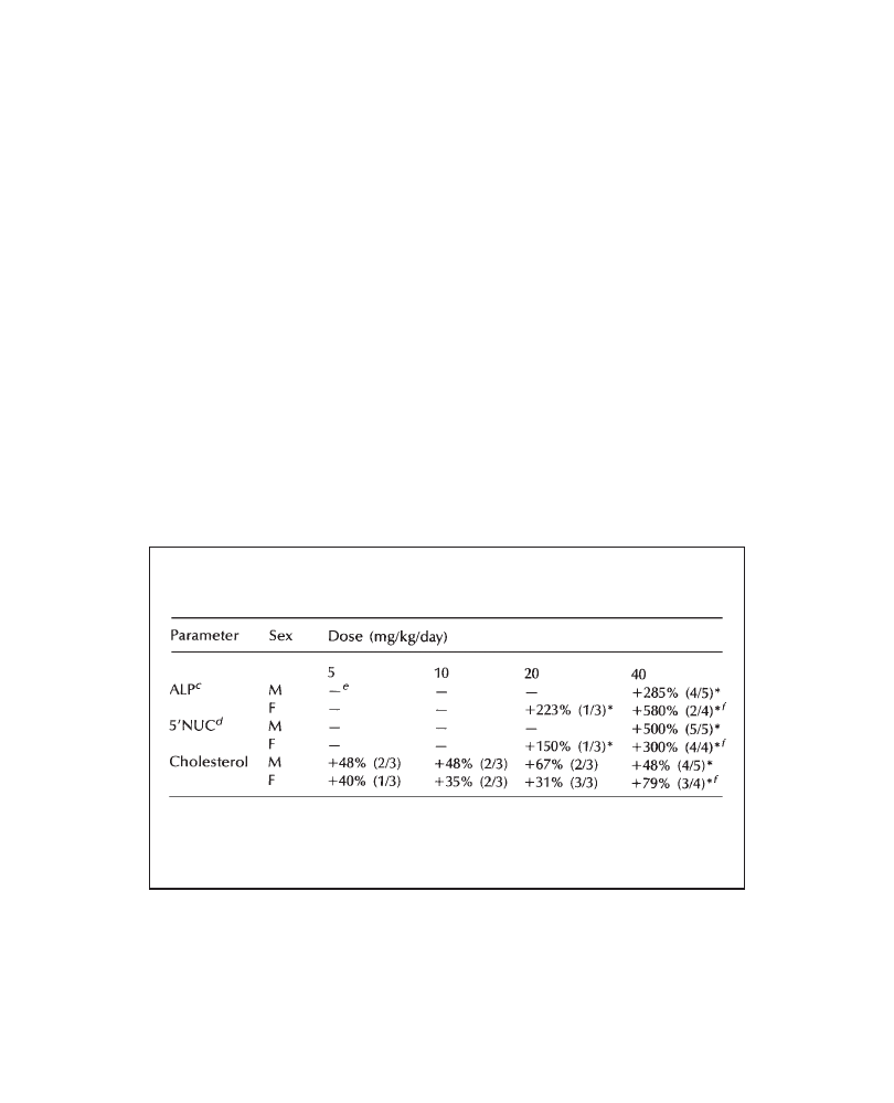
Reporting clinical pathology results
69
the effect is seen. Affected dose levels MUST be clearly stated, preferably using
the numerical values of doses instead of terms like “high-dose” or “mid-dose”.
7.2.3
What Is the Incidence of the Change?
The incidence of an effect reflects the number of animals with values outside the
defined reference range for a given parameter. It is generally reported as a fraction,
e.g. 2/4 or 9/10 animals affected. The incidence can often help to determine whether
or not an effect is related to compound administration and, more specifically, at
what dose level it begins to be manifested. For example, imagine that a change is
clearly identified in the mean of the high-dose group of a study. If the mean value
of the intermediate-dose group is slightly different from that of the control group,
yet several individual animal values within the group are outside the range of
individual control values, there is more evidence to suggest that animals at this
dose level are affected by the compound.
Incidence is reported only when it provides important additional information.
It can effectively be reported in text: “Compound-treated males had moderate to
marked bilirubin scores [2/5 at 5 mg/kg (moderate), 1/5 at 10 mg/kg (marked), 1/
5 at 20 mg/kg (marked)] which were not observed in the pretest urinalysis”, as
well as in tables (see Example 7.3).
Example 7.3 Tabular presentation of incidence
Table 7.1 PP 75167:1-Month oral toxicity study in beagle dogs—compound-
related clinical chemistry findings on day 28: percentage change
a
and
incidence
b
.
a
Percentage change is expressed in relation to mean control values.
b
Number
of animals with values outside the reference range.
c
Alkaline phosphatase.
d
5’-
Nucleotidase.
e
Values were within reference range.
f
One female died on day
15 of the study. *p<0.05.
Data from individual animals are not discussed unless they help to clarify a result.
Consider the following scenario: In a study in which anemia and thrombocytopenia
are expected compound-related changes, a dose-related effect is observed. The
incidence is one out of four at the lowest dose affected, two out of four at the
middle dose, and four out of four at the highest dose. The paragraph shown as
Example 7.4 was placed in the Results section. The information justifies asserting
that 0.1 mg/kg should be considered a no-effect dose.
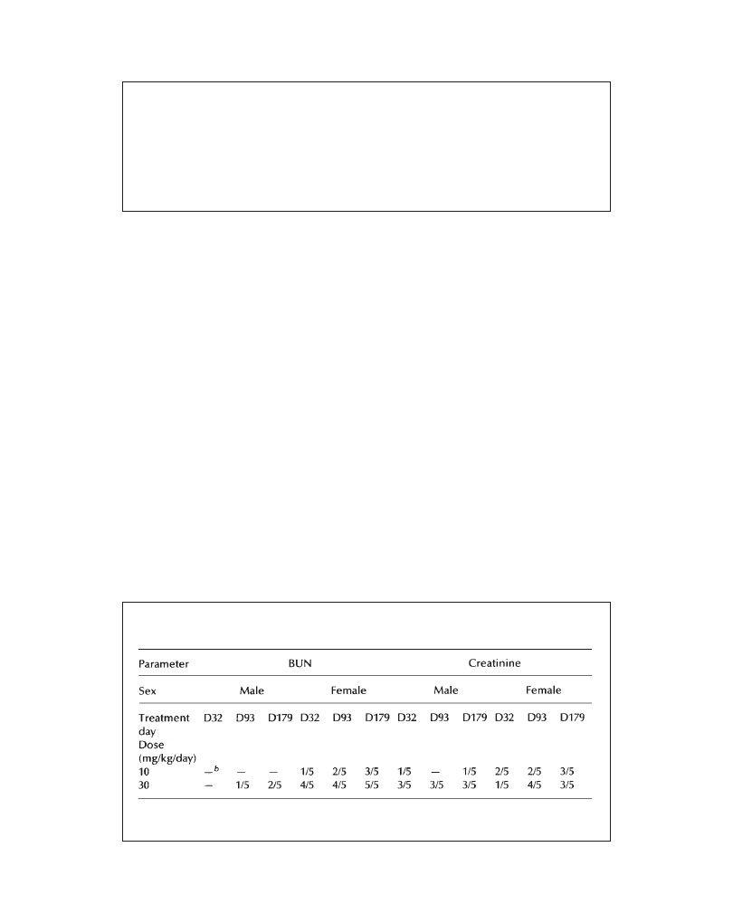
Presenting toxicology results
70
Example 7.4 Use of data from an individual animal to clarify a result
One 0.1 mg/kg male (No. 2003) was sacrificed during the study (day 83) because
of severe anemia and thrombocytopenia associated with increased prothrombin
time, reduced fibrinogen and neutrophilic leukocytosis. These changes were
compatible with disseminated intravascular coagulation, which was interpreted
to be secondary to the presence of a permanent intravenous indwelling catheter
(the route of compound administration), and a resulting bacterial septicemia
(confirmed by light microscopy). This effect was not considered to be related
to compound administration.
7.2.4
Is the Effect Noted in Both Sexes?
An effect does not need to be found in both sexes to be interpreted as a compound-
related effect. The differences in metabolism and hormone status between sexes
sometimes make one sex more susceptible or resistant to a given toxicologic effect.
These differences are very important to note, particularly in relation to eventual
clinical trials in the case of pharmaceutical compounds.
7.2.5
Is There a Progressive Increase in the Severity and/or Incidence of the
Change?
In studies of several months’ duration, the progression of the severity of effects
is important to note. When necessary, the specific sampling periods at which effects
are noted should be discussed. In large-animal studies, the number of animals
having progressing signs (the incidence) is equally important to track and discuss
in the Results section. Detailed results may be reported in a table (see Example
7.5).
Example 7.5 Tabular presentation of variation of incidence with time during a
lengthy study
Table 7.2 PP 34163:6-Month oral toxicity study in beagle dogs—incidence
a
of compound-related increases in blood urea nitrogen (BUN) and creatinine.
a
Number of animals with values outside the reference range.
b
Values were within
the reference range.

Reporting clinical pathology results
71
It is important to remember that certain clinical pathology parameters will
spontaneously change over time (e.g. decreasing alkaline phosphatase levels in
young dogs), and that these should not be interpreted as effects related to compound
administration. Indeed, knowledge of these normal biological shifts in parameters
will increase your ability to identify changes which should be attributed to the
compound, such as stable or increasing alkaline phosphatase levels in treated groups
while control group levels decrease as would be expected.
7.2.6
Is the Effect Reversible? Is the Recovery Partial or Complete? How Is
It Manifested?
In studies in which there are multiple sampling periods, changes noted on one
sampling date may not appear on subsequent sampling dates. In this instance, one
should carefully consider whether or not the change is transient but compound-
related, or whether it may be considered unrelated to compound administration.
Certain sporadic and transient changes merit discussion because they may be
important markers for clinical trials (e.g. transient increases in liver enzyme values,
decreases in red blood cell parameters or platelets). Thus, even a change in one
animal may be compound-related (see Example 7.6). Prior knowledge of how the
compound, or compounds of its class, is expected to behave can greatly help
determine the significance of such effects.
Example 7.6 Discussion of transient change
The creatinine in male dog No.15 (50 mg/kg/day) increased twofold from
day 3 (8 mg/l) to day 14 (16 mg/l), then decreased to reference range by
day 28 (9 mg/l). Though transient, this increase exceeded the concurrent study
control range and reference range values for dogs of this age and sex, and
was considered to be compound-related. Associated histopathologic changes
were observed in the kidneys. All other values were comparable between
treated and control groups and did not vary significantly during the course
of the study.
In studies in which there is a reversibility or recovery period, animals in which
changes were noted at the end of the treatment phase may be examined at the end
of an additional period during which treatment is withheld. Here, one looks for a
trend toward regression of the effects noted during the treatment period. For more
severe changes, the reversal may not be complete, because of the generally short
duration of the reversibility period as compared with the treatment period; but milder
changes may return completely to reference range values.
Compound-induced effects sometimes cause additional changes in related clinical
pathology parameters; these secondary changes may indicate a biological response
to the compound-induced effect. For example, decreased total red blood cells,
hemoglobin, and hematocrit (primary changes) noted during the treatment period

Presenting toxicology results
72
may increase by the end of the reversibility period, with an increase in reticulocytes
(secondary change) providing additional evidence of recovery. A distinction between
these two types of effect should be made in the text when possible.
Finally, data gathered at the end of the reversibility period may be used to confirm
or reject the hypothesis that questionable variations in clinical pathology analytes
observed during the treatment period are actually caused by compound administration.
Consider a case where mild elevations in liver enzymes are consistently found in
two out of five high-dose monkeys during the 28-day treatment period, and yet there
are no increases in the high-dose mean when compared with mean control values.
Could these individual variations be due to a direct effect of the compound
administered? If the enzyme levels in these two animals are found to be within reference
range at the end of the reversibility period, one can say with more confidence that
the elevations observed during treatment were compound-related. Again, prior
knowledge of the expected effect of the compound can help make this determination.
7.3
Putting Findings into Perspective
Depending on the style of report writing used in your company, you may discuss
the relevance of each group of findings in the same section in which you present
the results, or in a separate section altogether. Whatever the style used, the data
should be put into proper perspective and, when possible or appropriate, the following
issues regarding compound-related effects should be addressed.
1
Indicate whether compound-related findings are due to a direct effect of the
compound, or are considered to be secondary changes.
2
If there is an attempt to establish a no-effect dose, or to convince the reader
that compound-related effects present at the lowest dose are not texicologically
relevant, the potential significance of these effects must be clarified (e.g. severity,
incidence, relation to reference range values).
3
Make any possible correlations with in-life observations, anatomic pathologic
findings and other data.
4
When possible, address whether or not the effect is expected with the class of
compound. If so, briefly describe the mechanism (if known). Is it an exaggerated
pharmacological effect? Here, references to earlier studies with the same
compound, compounds with the same pharmacological activity, or scientific
literature clarifying the meaning of the effect may be used: for example,
“Increased cholesterol was previously observed in a 14-day toxicity study in
rats receiving 20mg/kg PP 23369, but to a lesser extent. Increases in calcium,
albumin and total protein associated with changes in hematology indicate
hemoconcentration which is probably secondary to the known diuretic action
of this compound (Author, year)”. Use of references, whether they be to studies
done in your facility, in contract labs, or to scientific literature, should be
harmonized throughout the report. It is strongly recommended that the actual
text of the reference be kept short (as in the example above), and to have a
full reference list at the end of the report.
5
The biological/toxicological significance (or lack thereof) of the effect should
be discussed: for example, “The minimal effect on PT and aPTT observed after

Reporting clinical pathology results
73
treatment with 15 mg/kg/day of PP 11506 was not considered to be biologically
significant, because all values noted in compound-treated animals were near
or within the range of values found in control animals”.
6
Discuss whether or not the effect in the test species is relevant to man. Certain
species have particular ways of reacting to the administration of xenobiotics
which have little or uncertain relevance in humans. For example, certain
regulatory agencies may require the evaluation of creatine phosphokinase (CPK)
and lactic dehydrogenase (LDH) as part of the biochemistry panel for toxicity
studies. The reference ranges for CPK and LDH are very wide in normal animals,
and levels are susceptible to variations resulting from animal handling and sample
collection. Increases in these enzymes can be of questionable significance in
animals, and are not likely to be predictive of toxic effects in humans.
Once all compound-related effects have been addressed, additional treatment-related
effects should be described. This section of the report should be brief. If the report-
writing policy at your company separates results from discussion, and if the observed
changes are not considered to be important, then describe these effects in the Results
section. However, if treatment-related changes are important in the overall
interpretation of study results, then they should be placed in the Discussion section.
Examples 7.7 to 7.9 show some ways of dealing with this subject.
Example 7.7 Vehicle-related effects
Variations in other clinical pathology parameters (urine volume and urine
osmolarity) were not considered to be related to treatment with PP 27567 since
they were also observed in the vehicle-treated control group.
Example 7.8 Pathological effects secondary to the route of compound
administration
Increases in RBC and WBC parameters and reductions in total protein, albumin,
globulin and A/G ratios in treated and control animals were attributed to
inflammation at injection sites.
Example 7.9 Physiological effects secondary to study procedure
Minimal reductions in RBC count (–2% to –6%, in males and –7% to –11%, in
females) in rats at all dose levels, including controls, were interpreted to be
the result of reduced blood volume following blood collection at 2-day intervals.
Because the compound caused a primary decrease in total RBCs, this
methodology-induced decrease interfered with the assessment of the severity
of the compound’s effect on red cell counts at all dose levels.

Presenting toxicology results
74
At the end of the Results section, variations which are not considered to be treatment-
related should be addressed as simply and as succinctly as possible. An example
of a short but all-encompassing paragraph which could be useful is the following:
“Other variations were observed among groups and at different sampling periods.
While some of these variations reached statistical significance, they were near or
within the limits of our reference range values (control values and/or pretest values
and/or historical data), and no consistent trends were observed that could be attributed
to treatment”.
In Conclusion
The Clinical Pathology section of the toxicology report should be written simply,
clearly and with the reader’s needs in mind. It should contain the fewest words
possible to explain your ideas, without sacrificing comprehensibility and clarity.
Lead your reader so that, with minimal effort, she/he can understand and accept
your conclusions with regard to data interpretation.

75
8
Anatomic Pathology
T.HODGE
Rhône-Poulenc Rorer, Drug Safety Department, Vitry sur Seine, France
and S.GOSSELIN
ITR Laboratories Canada Inc., Montreal, Canada
The Anatomic Pathology section of a regulatory toxicology report contains
information related to organ weight changes, necropsy findings and histopathologic
evaluation. Depending on the objectives of the study (single-dose study, reproductive
study, range-finding study, investigative study, etc.), some or all of these parameters
may be excluded. Depending on your laboratory or company report-writing policy,
the Anatomic Pathology section is either an integral part of the regulatory toxicology
report or a separate report written as an annex and included as an appendix of the
regulatory toxicology report. It may consist either of results with a discussion within
each respective subsection (organ weight, necropsy, histopathology) of the narrative,
or only the results—with an overall discussion of all results placed at the end of
the toxicology report. The writing philosophy of this Guide is based on clarity of
data presentation and interpretation, flow of ideas and a coherence of the logical
relation between information from different sources; therefore, only examples of
an Anatomic Pathology section which would immediately follow the Clinical
Pathology section are presented here.
In the Materials and Methods, in addition to the standard list of tissues to be
examined, the dose levels, organs evaluated during the treatment and recovery (if
applicable) periods and the special techniques used (histochemistry, electron
microscopy, immunocytochemistry, morphometry, etc.) should be mentioned. A
statement on peer review may be included.
In general, the order of presentation in the Results should begin with organ
weights, followed by necropsy, then by histopathology. Identify as clearly as possible
which effects are compound-induced and which ones are secondary to the vehicle
or the technical procedure. Avoid detailed description of spontaneous or spurious
events which may unnecessarily confuse the reader. Naturally occurring effects
should be mentioned if they interfere with the evaluation of compound-related
findings or if the administration of the compound exacerbates an effect/lesion of

Presenting toxicology results
76
a spontaneous nature. If the discussion of the results follows each subsection, detailed
significance of the associated results (correlations with in-life observations, clinical
pathology findings, organ weight changes, etc.) are included in the most appropriate
subsection, and only referred to in the remaining subsections.
8.1
Organ Weights
Organ weight changes are the differences, expressed in percentages, between treated
and non-treated groups in terms of relative and absolute organ weights. Since body
weight changes have already been addressed in the section on in-life measurements,
these should be mentioned only in terms of explaining changes in organ weights.
If body weight changes are substantial, it may be preferable to express the relative
organ weight values with respect to brain weight rather than to body weight. Because
of the wider variability between individual body weights in large animals compared
with small animals, relative organ weights are more useful than absolute organ
weights in these species. Therefore, if a table within the text is appropriate in large-
animal studies, only the relative organ weights should be tabulated. A text table
should be used when multiple or complex organ weight changes are present. The
organ weights of animals found dead during the course of a study should be taken,
since they may provide useful information on reasons for early deaths; but may
be excluded from the final calculation of means.
The narrative should begin by describing compound-related changes. If there
are no compound-related effects, simply state that no compound-related effects
were observed in the organ weights. If compound-related effects are present (whether
the changes are statistically significant or not), this should be stated in the first
sentence (Example 8.1). Subsequently, the organ weight changes should be qualified
in terms of whether the absolute weights, the relative weights, or both are altered;
these changes should be expressed as a mean percentage change (increase or decrease)
compared with the appropriate control mean values (usually the vehicle-control
values). In addition, the following points, if applicable, should be mentioned.
•
Is the change dose-related? If so, at what dose does it begin? Often, a slight
change in organ weights which is not statistically significant and would not
otherwise be worthy of mention, may indicate the beginning of a trend if the
organ weights at higher doses indicate a clear compound effect.
•
Is the change statistically significant? Often when there is a dose-response related
to an organ weight change, the changes observed at the lower dose/doses may
not be statistically significant. Therefore, do not rely on statistical significance
to indicate a compound effect.
•
Is it noted in both or only in one sex? If an organ weight change is present in
only one sex, try to relate the change to other parameters measured in the study,
i.e. clinical pathology changes or the presence or absence of histopathological
findings, which can substantiate whether or not the change is compound-related.
•
What is the incidence at each dose/sex affected? It is usually not included as
part of the report; however, it may help in interpreting the significance of the
results and establish a dose-relationship. If there is more than one organ affected
and if changes are complex, a text table may be appropriate.

Anatomic pathology
77
Example 8.1
Compound-related organ weight changes were noted for the liver in males at
10mg/kg/day and in both sexes at 25 mg/kg/day and for the testis at 25 mg/kg/
day.
When compared with the mean vehicle-control values, statistically significant
dose-related increases in mean absolute and relative liver weights were noted in
males treated at 10 and 25 mg/kg/day (+20 and+40%, respectively). In females, a
similar increase in liver weight (+30%) was limited to the 25 mg/kg/day group. In
addition, the 25 mg/kg/day males had lower (–25%) mean absolute and relative
testis weights. Liver and testicular changes correlated microscopically with
hepatocellular hypertrophy and bilateral degeneration of the seminiferous tubules,
respectively (see Histopathology). There were no other organ weight changes which
were considered related to treatment with compound PP 27567. Several other
differences in group mean absolute and/or relative organ weights between control
and treated groups achieved statistical significance, but these were considered
incidental and not compound-related as they were slight, not dose-related,
observed in only one sex and/or unaccompanied by correlated morphologic
findings.
•
Are organ weight changes mainly due to changes in body weight?
•
If a recovery group is used, is the recovery partial or complete?
•
Values that are clearly out of the range of the control values or statistically
flagged, but not considered compound-related and/or of pathologic
significance, should be addressed in one to two sentences with appropriate
qualifications as to their significance. State the reason for these differences
if known, or supply a statement similar to the following: “Some differences
in organ weights were observed among individuals but were considered
incidental since they were sporadic, unaccompanied by morphologic findings,
and/or without relation to dose or sex”. The organ weight changes may be
specifically cited if they are few in number and if the citation clarifies the
reader’s perspective.
•
The number of animals per group, normal population variation, tissue trimming
techniques and degree of exsanguination at necropsy may have an effect on
organ weights and should be addressed in the narrative if they are considered
to have affected the evaluation.
In the last paragraph of the organ weight section (RDRD structure) or in the overall
discussion (modified IMRAD structure), the data should be put into perspective
and, when possible, the following questions should be addressed.
•
Are there any correlations with clinical observations, clinical pathology, gross
or microscopic findings, etc.? These should be mentioned with reference to
the respective section.
•
Is there a no-effect level? If there is an attempt to establish a no-effect dose
level while dose-related findings are described at the lowest dose level, the
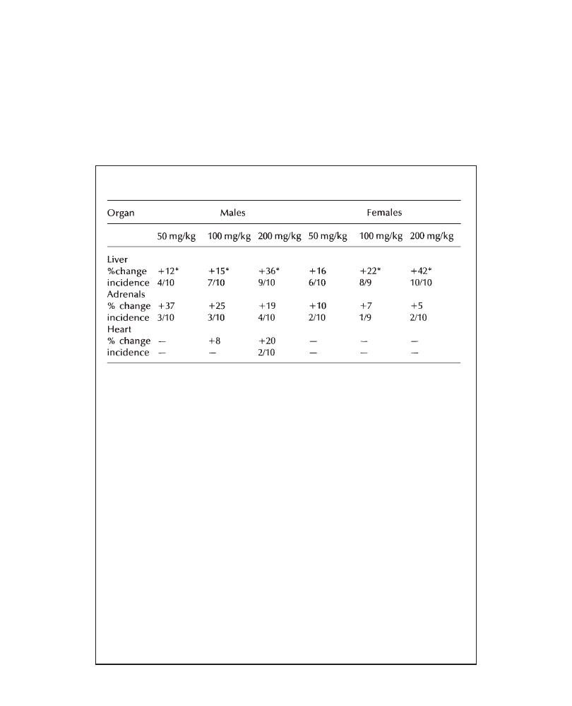
Presenting toxicology results
78
toxicological significance of the finding at that level should be clarified (minimal
incidence and/or severity, within normal historical range, etc.) (Example 8.3).
•
Is the change a consequence of the known pharmacological activity of the
compound? For example, a compound with enzyme-inducing activity might
be expected to cause increased liver weights (Example 8.2).
Example 8.2
Table 8.1 PP 27567:6-Month oral toxicity in rats—compound-related changes
in mean relative organ weights.
a
a
Mean compound-treated group values compared with mean vehicle-control
values
The administration of PP 27567 was associated with:
• dose-related increases (incidence and severity) in mean relative liver weight
at 50, 100 and 200 mg/kg/day in both sexes. This effect correlated with dose
-related increases in mean absolute liver weight in males (+15% to +35%
of mean control values) and in females (+16% to +42% of mean control values)
• increases in mean absolute and relative adrenal weight at 50, 100 and 200
mg/kg/day in males only
• increases in mean absolute and relative heart weights at 100 and 200
mg/kg/day in males only.
At all dose levels, higher liver weight correlated with hepatocellular
hypertrophy, while adrenal changes correlated with diffuse hypertrophy of
the zona fasciculata of the adrenal cortex. Individual heart weight increases
were within the range of values observed in controls and were therefore
considered to have little toxicological significance. Increased heart weight
may be secondary to the tachycardia observed at these dose levels which
is a known pharmacologic effect of the compound (see Cardiovascular
parameters in the Clinical Observation section) (Author, year).
*p<0.05
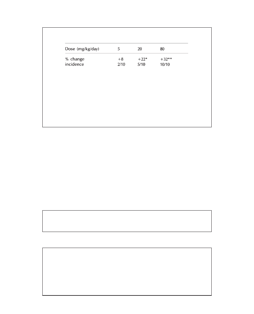
Anatomic pathology
79
Example 8.3
Table 8.2 Mean absolute liver weight increases in female rats
receiving PP 27567 for 6 months.
a
*p<0.05; **p<0.01
a
Mean compound-treated group values compared with mean
vehicle-control values
Note: a similar weight increase was observed with mean relative
liver weights
There were no microscopic findings associated with the liver weight changes.
Minimal increases in liver weight at 5 mg/kg/day noted in only 2/10 females
were considered to have little, if any, toxicological significance.
8.2
Necropsy
Observations at necropsy should be used primarily to identify the extent of the
compound-related lesions and any other lesions which are better described grossly,
such as skin lesions, injections site lesions, etc. (Examples 8.4 to 8.7). If there
are no compound-related findings, simply state that no compound-related findings
were observed at necropsy. If compound-related findings are present, indicate the
number of animals affected per sex and per group for each finding. Note whether
the gross findings correlate with clinical observations or microscopic findings.
Summarize all non-compound-related gross findings in one succinct sentence or
Example 8.4
Skin lesions at injection sites (thickening/swelling, erosions/ulcers, exudation,
dark firm, edema) were observed in all compound-treated groups and reflect
the irritant nature of the compound when administered subcutaneously.
Example 8.5
Compound-related gross findings were noted only in the testis and thymus of
the 10 mg/kg/day animals. Small testes and thymuses were observed in 10/10
males. These lesions correlated with microscopic findings of testicular
degeneration and lymphoid depletion of the thymus (see Histopathology).
Following the recovery period, small testes were still present in 4/5 males while
compound-related changes in the thymus were no longer present. All other gross
observations were incidental and spontaneous in nature and bore no relation
to treatment with PP 27567.

Presenting toxicology results
80
paragraph. Refrain from going deeply into details, since the final diagnoses will
be confirmed by light microscopy.
Following the description of any morphologic findings, a paragraph on mortality
and any associated gross findings should follow (Examples 8.8 to 8.11). Depending
on the philosophy of the company, this section could be part of Clinical Observations
and the result of a team-effort between Toxicology and Pathology. If there was no
mortality in the study, state that all animals survived until the end of the study. If
animals were sacrificed early or found dead during the study, state whether or not
these deaths were compound-related. Early deaths that are related to gavage accidents
can be mentioned here along with any histopathological findings confirming the
diagnosis of a gavage accident. These animals need not be referred to again. If
there were multiple deaths, describe the number of dead animals per group and
comment on the cause of death.
Example 8.6
Compound-related gross findings were characterized by:
• thin body at 200 mg/kg (2/5 males, 1/5 females)
• edematous pericardial and thymic fat at 200mg/kg/day (2/5 males, 1/5 females)
• small thymus at 200 mg/kg/day (4/5 males, 3/5 females)
• enlarged spleen at 200 (3/5 males) and 100 mg/kg/day (1/5 males)
These findings were attributed to anemia and weight loss (see Clinical
Observations and Clinical Pathology).
Example 8.7
Compound-related gross findings were noted in the liver and testis. Enlargement
of the liver was observed in 13/20 males and in 9/20 females treated at 25 mg/
kg/day. Small, soft testes were noted in 14/20 males treated at 25 mg/kg/day.
Gross observations in the liver and testis correlated with the microscopic
observations described in the Histopathology section. All other gross observations
were incidental findings typical of those routinely observed in Sprague-Dawley
rats of this age.
Example 8.8
Four animals (2/10 females at 50 mg/kg/day, 1/10 males at 100 mg/kg/day and
1/10 females at 200 mg/kg/day) did not survive until the scheduled necropsy.
The two 50 mg/kg/day females (No. 91: found dead; No. 100: sacrificed as
moribund) had gross observations compatible with gavage error (see Table X).
The cause of poor condition requiring sacrifice for the remaining animals was
undetermined from gross or microscopic examination.

Anatomic pathology
81
Example 8.9
One of the females (No. 38) at 200 mg/kg/day died on day 8 of the study because
of extensive hemorrhage into the gastric and intestinal lumen. The gastrointestinal
hemorrhages were the result of gastric mucosal ulcers, probably secondary to
severe repeated emesis and retching (Author, year). Emesis has been previously
reported in association with the oral administration of high doses of this
compound, where death was often preceded by sodium and chloride imbalance
(references).
Example 8.10
The only unscheduled death occurred in a 10 mg/kg/day male (No. 034) on
d ay 27. Necropsy observations on the kidney, lung, liver and spleen
corresponded microscopically with acute tubular necrosis, pulmonary edema
and congestion with hemorrhages in the liver and spleen. While these changes
probably contributed to the animal’s death, their cause could not be
determined. However, the changes are not considered to be related to
compound, because of their isolated occurrence and the absence of a dose
relationship.
Example 8.11
Gross pathologic findings observed in moribund animals were the following:
• red-brown staining of the fur around the nose in rats receiving 500 and 1000 mg/kg/
day
• yellow-green or red-brown staining of the fur of the abdomen in animals given 500
(females) and 1000 mg/kg/day (both sexes)
• erosion(s)/ulcer(s) of the stomach in animals given 500 and 1000 mg/kg/day (both
sexes)
• decreased size of the thymus in animals given 500 and 1000 mg/kg/day (both
sexes).
The lesions in the stomach were confirmed microscopically to be erosions. The
decreased size of the thymus was consistent with a decreased thymus weight
and lymphoid depletion observed microscopically. These non-specific findings
are frequently noted in rats in poor condition.
8.3
Histopathology
The major responsibility of the toxicologic pathologist is to identify and describe
microscopic changes induced by a test article in the tissues of laboratory animals.
These changes need to be correlated with clinical observations, alterations in clinical

Presenting toxicology results
82
pathology parameters and organ weights, and their significance must be interpreted
taking into account their relationship with the amount of compound to which the
animals have been exposed. The data should be put into proper perspective for a
reader who may have limited training or experience in toxicologic pathology.
Therefore, the histopathology results must be written clearly, succinctly, accurately
and be presented logically.
At the time of this writing, the Society of Toxicologic Pathologists in the
USA and the Registry of Industrial Toxicology Animal-data (RITA) in Germany
are undertaking the formidable task of standardizing the vocabulary used in
toxicologic pathology. Historically, the language of pathology has been complex,
confusing, sometimes ambiguous and often subjective. With pathologists
themselves often in disagreement over terminology, it is not surprising that
readers who are not pathologists are often confused when presented with pages
of text filled with unfamiliar and sometimes “frightening” terms. The reader,
who may be a biologist or a pharmacologist in a regulatory agency, is often
left to his own devices to put pathologic changes into perspective relative to
human risk assessment.
A well-defined glossary of diagnoses should be decided upon within an
organization in order to impose uniformity among pathologists. In addition to
ensuring that all pathologists in an organization use the same terminology to
describe the same histopathologic finding, a glossary is important both in the
production of uniform reports and for establishing historical databases within
an organization. The number of diagnoses should not be exhaustive, but rather,
an effort should be made to combine or “lump” similar pathologic processes
together. This is especially important for long-term studies where there are large
numbers of findings. Also, since the difference between hyperplasia and neoplasia
of a particular cell type is often vague, it is important to make a distinction between
the two. When a hyperplastic or neoplastic lesion is considered a test article effect
in a carcinogenicity study, it is desirable both to combine and separate the possible
stages of that lesion (hyperplasia, benign and malignant neoplasia) for statistical
purposes. To make these manipulations, strict definitions regarding the morphologic
boundaries between malignant and benign neoplasia and hyperplasia need to be
specified.
Standardization and definition also should be given to descriptive terminology,
such as severity degrees (e.g., minimal, mild, moderate and marked) and distribution
modifiers (e.g., focal, multifocal, locally extensive, diffuse, unilateral and bilateral).
This is of paramount importance in decreasing diagnostic subjectivity, in putting
the severity of findings into perspective regarding their overall toxicologic
importance, and ultimately in arriving at the best decisions related to risk assessment.
The histopathology section should begin by stating that Compound X either did
or did not produce an effect. If it did, state the change, organ, sex, and dose groups
affected. If numerous changes are present, list the target organs and describe the
changes in subsequent sentences/paragraphs. Text tables may be used when there
are multiple findings. The text should concentrate on the concise presentation of
compound-related findings, and long, detailed histopathologic descriptions should
be avoided.
In the first sentence/paragraph, identify the dose levels which are affected along
with the target organ, the number of animals per dose group and per sex, and its
associated change (Examples 8.12 and 8.13). If possible, list the organs in order

Anatomic pathology
83
Example 8.12
Compound-related histopathologic findings were noted at the end of the
treatment period in multiple organs in animals treated at 1 and 5 mg/kg/
day. These findings consisted of hypocellularity of the bone marrow at 1
and 5 mg/kg, focal or multifocal atrophy of the seminiferous tubules of
the testes with individual spermatocyte necrosis at 5 mg/kg, and single cell
necrosis and increased numbers of mitotic figures in salivary and lacrimal
glands at 1 and 5 mg/kg. All were considered to be related to the anti-
mitotic properties of this compound. After a 4-week recovery period,
treatment-related findings persisted only in the testis, epididymides and
lacrimal glands.
of importance: e.g. liver would be listed before harderian gland. If the study includes
a recovery period, mention whether or not the recovery occurs and, if so, whether
it is partial or complete (Example 8.12).
After the compound-related changes have been listed, a description of the
changes, organ by organ, should follow. Diagnostic terms should be defined
keeping in mind that most readers of the report will not be pathologists. During
the description of the lesions, one or two sentences can be used to describe
their distribution and severity. Distribution modifiers (focal, multifocal, diffuse,
locally extensive) and severity modifiers (minimal, mild, moderate, marked,
severe) are useful to put findings into perspective. Histopathologic lesions may
be compound-related because of their distribution or their severity, even if the
incidence of the finding is not increased. In other words, a dose response related
to severity or distribution must be considered as well as a dose response related
to incidence. If a special technique such as histochemistry or electron microscopy
helped confirm or supplement the initial histopathological diagnoses, it should
be mentioned.
For each histopathologic finding, briefly mention any possible correlation or
association with organ weight changes, gross findings and ante-mortem findings
(Clinical Observations, Clinical Pathology, Toxicokinetic, etc.).
If possible, explain findings based on the known pharmacological properties
of the compound. Instead of being part of the Results, this could be included in
the Discussion, depending on the philosophy of the organization. This can help to
put into perspective and often minimize the importance of certain findings. Explain
if the finding is expected with this class of compounds or is secondary to an
exaggerated pharmacological effect (Example 8.13).
Lesions related to route of administration, methodology, or secondary to the
vehicle should be grouped together and be kept to a minimum of one or two
sentences. Inflammatory lesions related to irritant compounds given subcutaneously
or intravenously or ocular lesions related to orbital sinus bleeding are examples
of this type of finding.
In some instances, certain histopathologic findings may have no or little relevance
to man, e.g. thyroid follicular cell hypertrophy/hyperplasia in rats secondary to
microsomal enzyme induction. Findings may also be limited to one species. Any
known information related to species specificity will help to minimize and put into

Presenting toxicology results
84
perspective a compound-related effect. Interpretation of such changes in this manner
should be accompanied by references.
Finally, group all non-compound-related findings into one sentence or one succinct
paragraph. State why these changes are not considered compound-related (similar
incidence in controls, common findings in this strain, incidental, spontaneous,
developmental, caused by animal handling, etc. —Example 8.14). If spontaneous
lesions interfere with the proper evaluation of possible compound-induced lesions,
this should be mentioned (Example 8.15).
Example 8.13
Compound-related histopathologic findings were noted in the liver and thyroid
at 25 mg/kg/day, and in the testis at 10 and 25 mg/kg/day. In the liver, centrilobular
hepatocellular hypertrophy was observed in 10/20 males and in 7/20 females
treated at 25 mg/kg/day, and was characterized by increased size and cytoplasmic
eosinophilia of centrilobular hepatocytes. Severity ranged from mild to moderate
in affected males and from minimal to mild in affected females and correlated
with the increased liver weight observed in these groups. Centrilobular
hepatocellular hypertrophy is most likely the result of increased hepatic
microsomal enzyme activity induced by PP 27567, as demonstrated in a previous
study (Reference).
In the thyroid, multifocal to diffuse hyperplasia was noted in 8/20 males and in 5/20
females treated at 25 mg/kg/day. It was characterized by small follicles which were
lined by columnar epithelial cells with finely vacuolated cytoplasm surrounding retracted,
basophilic colloid. This change was frequently associated with centrilobular
hepatocellular hypertrophy, and could be related to altered hepatic clearance of thyroid
hormones resulting from increased hepatic microsomal enzyme activity induced by PP
27567 (Reference).
Finally, in the testis, degeneration of the seminiferous tubules was observed in
13/20 males treated at 25 mg/kg/day and in 7/20 males treated at 10 mg/kg/day,
and was characterized by degeneration of spermatocytes in some tubules,
associated with a clear loss of germ cells and retention of Sertoli cells in others.
Severity and distribution of this change were dose-related (minimal to focal at 10
mg/kg/day and mild to multifocal at 25 mg/kg/day) and correlated with decreased
testicular weights and with the gross necropsy observation of small, soft testes at
25 mg/kg/day.
All other microscopic findings were incidental and spontaneous in nature and were
not attributed to the compound.
Example 8.14
There were no compound-related findings in this study. All microscopic
findings were either related to orbital sinus bleeding, to venipuncture, or
were incidental and of the type routinely observed in Sprague-Dawley rats
of this age.

Anatomic pathology
85
Example 8.15
The administration of PP 27567 was associated with histopathological findings
which consisted of moderate to marked tubular dilatation, basophilia and
degeneration in the kidneys at doses of 50 and 250 mg/kg/day (4/6 and 10/10
animals, respectively). At 10 mg/kg/day (2/6 animals), the findings were limited
to minimal to mild renal tubular changes. Spontaneous renal lesions (interstitial
inflammation) were noted in several animals, including controls; these
spontaneous lesions interfered with the interpretation of the renal findings,
particularly in the 10 mg/kg/day group.
8.4
Reporting the Results of Carcinogenicity Studies
When reporting the results of a carcinogenicity study the writer must keep in mind
that the primary purpose of the study is to identify compound-related neoplastic
and/or hyperplastic changes. If non-neoplastic changes have occurred during the
course of a carcinogenicity study, it is possible that these lesions have been identified
previously in studies of shorter duration. In this case, they should be discussed
briefly after the section(s) on neoplastic and hyperplastic findings, and appropriate
references given. Along these lines, it is not unusual to observe exaggerated
pharmacological effects of compounds after prolonged administration, and this should
also be discussed briefly if observed.
Carcinogenicity studies may or may not have clinical pathology or organ weight
data to analyze as part of the results. If these sections are present, however, they
should be dealt with as outlined previously. Often the only clinical pathology
parameters examined are those related to hematology, especially if the compound
is suspected to have either a suppressive or stimulatory effect on any of the blood
cell lines.
Cause of death is often dealt with in the histopathology section of a carcinogenicity
report (Example 8.16). State what the major causes of death were in the preterminal
animals, e.g. pituitary adenoma or spontaneous renal disease in rats, and express
the incidence as a percentage value between groups. State whether or not the number
of animals dying of undetermined causes was comparable between groups.
The histopathology section should begin by saying “up front” whether or not
compound-related neoplastic or hyperplastic changes were observed (Example
8.17). If neoplastic changes were observed, the neoplasm and its associated organ
should be listed in the first paragraph. Subsequently, try to devote a short but
complete paragraph to describing and explaining each neoplastic change, its
statistical significance, and the incidence of the change when coupled with any
associated hyperplastic changes in a given organ. A neoplastic change in any
given organ should be discussed in terms of the following possible combinations
of reporting:
1
the total number of malignant tumors per sex per group
2
the total number of benign tumors per sex per group
3
the total combined number of benign and malignant tumors

Presenting toxicology results
86
4
the total number of proliferative lesions, i.e. malignant tumors+benign tumors
+ associated hyperplastic lesions.
The significance or cause of a given neoplastic or hyperplastic change can be
discussed either in the histopathology Results or in an overall Discussion section
depending on the philosophy of the organization.
Other topics which should be mentioned in the Results section include the
following:
•
whether or not the compound increased the total number of primary tumors
•
if the compound affected the total number of clinically palpable or observable
masses and the correlation of palpable masses with the histopathologic findings
•
the number of systemic tumors, i.e. lymphomas, histiocytic sarcomas
•
the total number of malignant tumors
•
the tumor frequency in individual organs
•
whether or not the compound influenced the time of onset of any tumors.
Example 8.16
The number of animals which died on the study or were euthanatized early
in a moribund condition was comparable between the control and the treated
groups. A total of 217 (72.3%) males and 187 (62.3%) females died or were
euthanatized in a moribund condition during the 104-week treatment period.
The individual group percentages for animals which died or were euthanatized
early in a moribund condition were 65, 76.7, 75, 73.3 and 71.7% for males
and 55, 63.3, 56.8, 66.7 and 70% for females for control groups 1+2 and
the 5, 15 and 45 mg/kg/day groups, respectively. The major causes of death
or moribundity among the preterminal animals were related to conditions
frequently observed in Sprague-Dawley rats and included pituitary adenoma
(the most frequent cause of death or moribundity among all groups),
glomerulonephropathy, and mammary carcinoma in females. In addition, the
number of animals dying of undetermined cause was comparably distributed
among all groups, including controls.
Example 8.17
Treatment with PP27567 was associated with a slight but statistically significant
(p=0.043) increase in pheochromocytoma (a benign tumor of adrenal origin
frequently observed in this strain of rats) in females treated with 45 mg/kg/day.
The incidence of pheochromocytoma in this group (10.3%), although only slightly
increased over that of the controls (2.5%) and slightly greater than that reported
in most references for Sprague-Dawley rats (Authors, year), was considered a
compound-related effect because similar effects have been observed with
structurally related compounds (References).
Since the distinction between medullary hyperplasia and pheochromocytoma

Anatomic pathology
87
continued
is often unclear and since the two diagnoses are most likely part of a
continuum of the same lesion, the numerical values for these two findings
were merged to provide a value for total proliferative medullary lesions.
This combined value was also greater in the 45 mg/kg/day females than in
the control females. The incidence of total proliferative medullary lesions
in treated males was comparable to that of the control groups. In addition,
there was no increase in bilateral tumors or in the incidence of animals
which had both a pheochromocytoma and focal medullary hyperplasia. Focal
m e d u l l a r y hy p e r p l a s i a wa s c o m p o s e d o f m u l t i f o c a l c l u s t e r s o f s m a l l ,
medullary cells which were darker and had larger nuclei than surrounding
medullary cells. Focal medullary hyperplasia generally did not exhibit
compression of the adjacent adrenal cortex. Pheochromocytomas were
morphologically similar between all treatment groups, but their size ranged
from masses only slightly larger than focal medullary hyperplasia with some
compression of the adjacent medulla and cortex to large masses compressing
the cortex into a thin rim of cells visible often only at one pole of the tumor.
Vascular dilatation, congestion and hemorrhage were occasionally observed
in tumors of all sizes.
Although most pheochromocytomas occurring in the 45 mg/kg/day females were
found in animals that died or were euthanatized in a moribund condition, none
was thought to be a cause of death or reason for early sacrifice. None of the
pheochromocytomas displayed any evidence of malignancy and none of the animals
bearing a pheochromocytoma had multiple endocrine neoplasia, except with
pituitary adenoma, which was present in 93% of the female rats in this study and
was present in similar numbers among all groups, including the controls.
The occurrences of two other neoplasms (hepatocellular adenoma in males
and females, and testicular interstitial cell adenoma in males) were also
statistically significant, but were clearly without relation to treatment with PP
27567 and were considered incidental findings. In males, the occurrence of
hepatocellular adenoma was significant only by the trend analysis (p=0.048)
and represented only a slight increase in an extremely common tumor in this
strain of rat. In females, the incidence of hepatocellular adenoma was significant
(p=0.05, Fisher’s exact test) only in the 5 mg/kg/day animals and was not
accompanied by a dose-response in the 15 and 45 mg/kg/day groups. Testicular
interstitial cell adenoma was significant by the Fisher’s exact test (p=0.05) only
in the 15 mg/kg/day males and was considered a fortuitous increase in the
incidence of an infrequently occurring tumor. There were no occurrences of this
tumor in the 45 mg/kg/day males.
There were no other increases in the incidence of other neoplastic or non-
neoplastic findings which were considered to have a relationship to treatment with
PP 27567. The total number of primary neoplasms (benign, malignant, or combined)
and the total number of animals bearing a primary neoplasm among the compound-
treated groups were comparable to the two control groups (Groups 1 and 2). In
addition, PP 27567 had no effect on the number of clinically palpable masses, the
tumor frequency in individual organs or the time of onset of any tumor.

Presenting toxicology results
88
continued
All other histopathologic findings in this study were spontaneous or incidental
and were typical of those encountered in control male and female Sprague-
Dawley rats used in long-term studies (Authors, year). These findings were
observed with comparable frequency in both the control and compound-treated
groups and none of these lesions was considered to have a relationship with
the administration of PP 27567.

89
9
Developmental and Reproductive
Toxicology
R.L.CLARK
Rhône-Poulenc Rorer Research and Development, Collegeville, USA
and G.COPPING
Rhône-Poulenc Rorer, Vitry sur Seine, France
9.1
Introduction
The principles and guidelines for writing toxicology study reports described in
this book also apply to reports for developmental and reproductive toxicology studies.
As is true for other reports, a developmental or reproductive toxicology report should
be written to make it as easy as possible for the reader to understand the study
design, results and interpretations. The author should keep in mind that the report
will generally not be read cover to cover, but instead the reader will refer to the
report to answer specific questions. This is why each section of the report should
be as distinct and complete as possible. It should be recognized that, in most scientific
writing, authors should express their point of view. The report should reflect the
fact that the writer has carefully considered all the data and other information about
the test compound and related test compounds and has arrived at an interpretation.
The author’s job is to convince the reader of that interpretation.
9.2
Terminology and Definitions
Some information concerning the development and use of the test species is
prerequisite to understanding the terminology employed in developmental and
reproductive toxicity studies. In this section we discuss the terminology used in
reproduction toxicology reports. Terminology is summarized in Table 9.1.
9.2.1
Cohabitation and Mating
Pregnant rodents are usually obtained by cohabiting males and females at least
overnight and often continuously until evidence of mating is observed. Rodents
typically mate in the evening, and ovulation and fertilization occur in the morning
following mating. Finding sperm or a seminal plug in the vagina in the morning
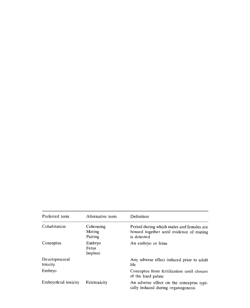
Presenting toxicology results
90
is considered conclusive evidence of mating but a seminal plug in the cage tray
may or may not be considered evidence of mating. The day that evidence of mating
is found is variably considered to be either day 0 or day 1 of gestation (this chapter
will consider this day to be day 0). Whether female rabbits are naturally mated
during a brief cohabitation with a male or artificially inseminated, almost all
laboratories consider the day of occurrence to be day 0 of gestation.
9.2.2
Embryonic Development
The period of development of the embryo (embryogenesis) begins with fertilization
and is typically considered to conclude with the closure of the secondary (hard)
palate (mouse—day 15; rat—day 16 to 17; rabbit—day 19) after which the conceptus
is referred to as a fetus. Embryogenesis includes the period of major organogenesis
which is considered to begin at implantation (approximately gestational day 5 to
6 in rodents and rabbits). When not wishing to specify either embryo or fetus, the
term “conceptus” can be used or, as the adjective, “embryofetal”. After delivery
(caesarean section or birth), the rodent fetus is referred to as a pup.
9.2.3
Effects on the Conceptus
There are various types of effects on the conceptus (developmental toxicity).
Treatment with the test compound may cause the death of the conceptus (resorption),
effects on fetal weight, or congenital anomalies. In some species, such as the rabbit,
conceptuses can be expelled during gestation (abortion) as a result of the death of
the conceptuses or excessive maternal toxicity.
Two categories of congenital anomalies (alterations) are malformations and
variations. Malformations tend to occur at low incidences, have a major impact
on the animal, and are irreversible whereas variations are not. There are congenital
anomalies for which there is no unanimous agreement on the classification as a
Table 9.1 Selected terminology for reproductive toxicity reports
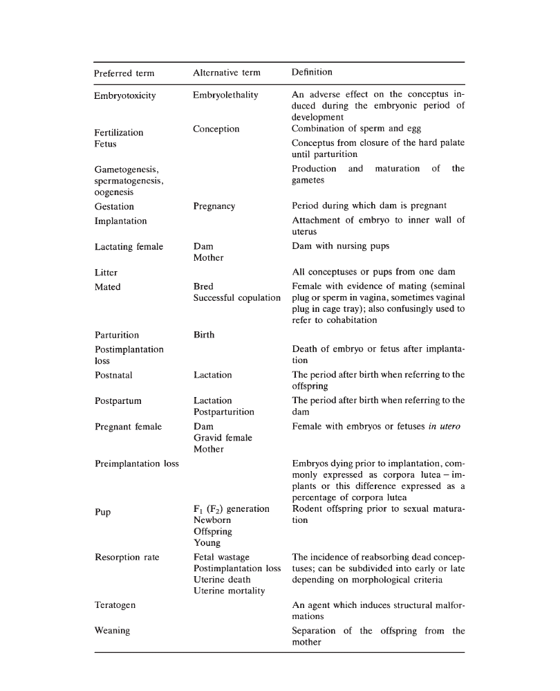
Developmental and reproductive toxicology
91
Table 9.1 continued

Presenting toxicology results
92
malformation or a variation (e.g. wavy rib and supernumerary rib). Furthermore,
there are sites of incomplete ossification which typically indicate delayed
development.
Early workers in the field then known as “teratology” focused on the study of
congenital malformations. Malformations had (and have) a tremendous clinical and
social impact, much more than, for example, abortions, which are frequently
undetected. This emphasis on malformations was reinforced by the thalidomide
tragedy in which thousands of children were born with severely debilitating drug-
induced congenital defects. Thus, embryofetal toxicity (Segment II) studies were
for decades referred to as “teratology studies” and the focus was on detecting
teratogens, i.e. agents which induce malformations.
More recently, there has been increased emphasis on other endpoints of
embryofetal toxicity studies in addition to malformations: fetal weight, embryofetal
death, and defects not considered to be malformations. This change reflects the
recognition of several points. The first is that embryofetal death is a severe outcome
which may have resulted from malformation. Since drug-induced developmental
effects can vary from species to species, this may indicate a potential for causing
malformation in other species. Secondly, it is now clear that malformations in animal
models can occur secondarily to maternal toxicity and are therefore not all
“teratogenic” responses necessarily indicating a severe hazard for humans. Thirdly,
the induction of, or the increase in, the incidence of “variations” by a test compound,
though not a “teratogenic” response, can nevertheless indicate a serious perturbation
of development. Finally, there is no universal agreement on the definition of
“teratogen”. Thus, to avoid confusion one should refer to embryofetal toxicity or
developmental toxicity rather than teratogenicity; however, a perspective on the
risk should be provided on the nature of a particular adverse developmental finding.
Treatment in an embryofetal toxicity study approximately coincides with the
period of major organogenesis, and examination of the uterine contents occurs at
the end of gestation. The timing of the induction of toxic effects observed at the
end of gestation can sometimes be inferred from the type of effect. For example,
malformations of the cardiovascular system or early resorptions can only be induced
during the embryonic period, and therefore the toxicity can be referred to as
embryotoxicity. Often, however, it cannot be inferred whether the toxic insult
occurred during the embryonic or fetal period. For example, it is usually not possible
to conclude when decreases in fetal weight were induced or whether late resorptions
occurred at the end of the embryonic period or the beginning of the fetal period.
Even if it is clear that fetuses died late, just before study termination, the lethal
insult may have occurred during the embryonic period. Thus, the term “fetotoxicity”
cannot correctly be used in reference to an embryofetal toxicity study unless
additional studies are conducted. For these reasons, it is common to refer to effects
on the conceptus observed at the end of gestation as “embryofetal toxicity” or
“developmental toxicity”.
9.2.4
Pre- and Postnatal Toxicity
Since development, particularly of the brain, continues during the early postnatal
period and, since effects induced during gestation and lactation can be expressed
in F
1
adults, adverse effects observed during the postnatal period can appropriately

Developmental and reproductive toxicology
93
be referred to as “developmental toxicity”. For example, retardation of growth,
delayed development of reflexes and developmental landmarks, and aberrations
in behavioral tests can all be considered developmental toxicity when induced in
a pre- and postnatal toxicity study in which treatment begins at implantation and
continues through the lactation period (see Study Definitions below). When
describing adverse developmental effects clearly induced during gestation in this
type of study (e.g. effects observed in newborns), one provides more specific
information by using the term “embryofetal toxicity” rather than “developmental
toxicity” to refer to them.
9.2.5
Multiple Phases and Generations
Writing reports for fertility studies and pre- and postnatal toxicity studies is
complicated (compared with general toxicity studies) by having multiple phases
and generations. For example, in the fertility study, there are precohabitation,
cohabitation, and gestation phases for the parental (F
0
) generation and two
generations: F
0
adults and F
1
fetuses. In the pre- and postnatal toxicity study, there
are the gestation and postpartum (lactation) phases for the F
0
females and the
preweaning, postweaning, cohabitation, gestation, and, in some cases, postpartum
(lactation) portions of the postnatal period for the F
1
generation. Many of the
parameters measured are specific to a particular phase and generation. In the fertility
study, one or both sexes may have been treated. When both males and females are
treated and cohabited in a ratio of 1:1, the results for fertility and mating performance
for the two sexes will obviously be identical.
9.2.6
Study Definitions
For detailed design requirements for reproductive toxicity studies, the relevant
regulatory guidelines should be consulted. In general, such studies should involve
the exposure of sexually mature adults and all developmental stages (gametogenesis
and conception to sexual maturity). The combined results from these studies give
an overview of the immediate and delayed effects following administration of a
compound throughout one complete life cycle (conception to conception in two
generations). For convenience, the reproductive cycle is broken down into multiple
phases, and studies designed to assess reproductive toxicity should ensure that
compound administration covers all the phases shown in Table 9.2, as recommended
in guidelines. Typically, these phases are combined into three or four studies based
on the guidelines of the International Conference on Harmonization. These guidelines
replaced similar guidelines which had specified three study types based on dosing
period, which were referred to as Segment I, Segment II and Segment III studies.
The new standard study types are briefly described below.
Fertility and early embryonic development (formerly “Segment I”)
This study assesses toxicity resulting from treatment of males and females
prior to and through mating, up to implantation. Estrus cycle, tubal transport,
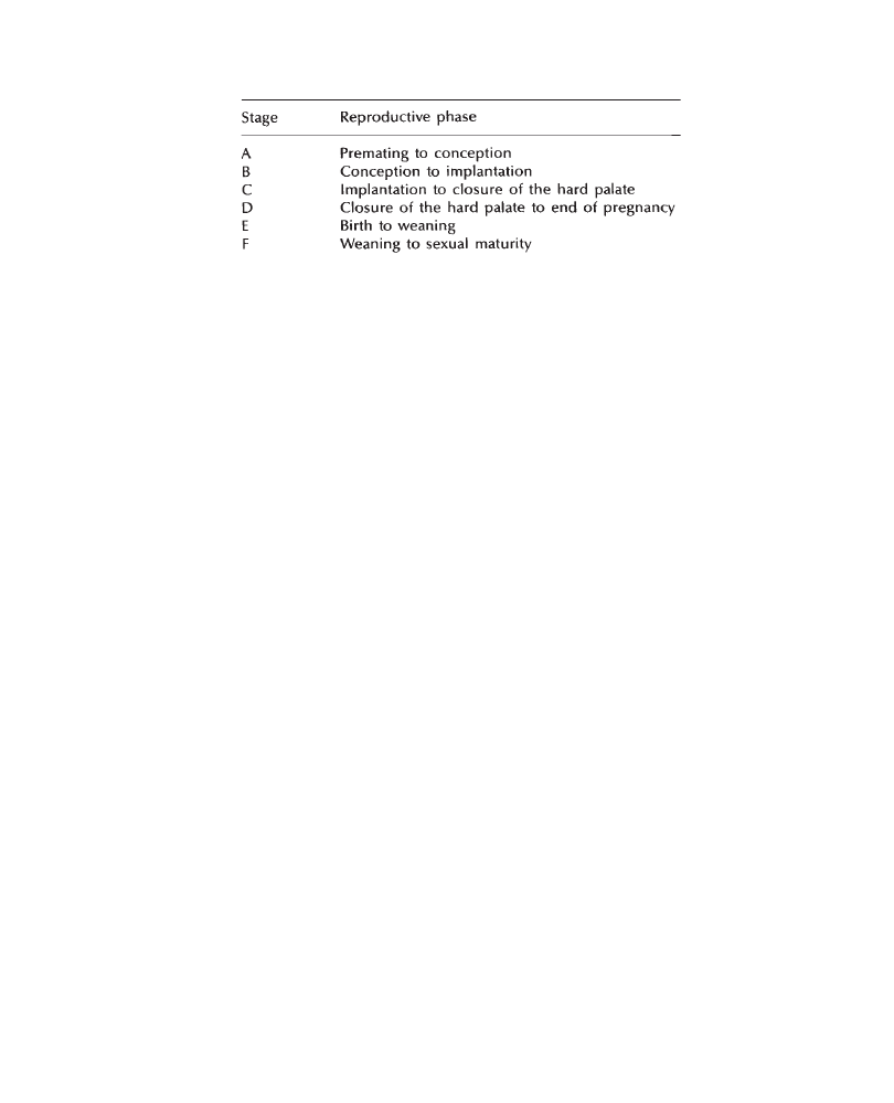
Presenting toxicology results
94
implantation and early preimplantation embryonic development are assessed in
females. Functional defects on male reproductive organs not normally detected by
histopathological examination are assessed in males.
Separate male and female fertility studies are often performed (cohabitation with
non-treated animals) to identify clearly sex-specific toxicity.
Embryofetal development (formerly “Segment II”)
The aim of this study is to detect adverse effects in the pregnant female and in
embryofetal development after administration of the compound to pregnant females
from implantation to closure of the hard palate. Endpoints include maternal toxicity
and effects on embryofetal mortality, growth and development.
Pre- and postnatal development (formerly “Segment III”)
This study is designed to detect the effects of treatment from implantation through
weaning on the pregnant and lactating female and on the conceptus and offspring
up to sexual maturity, including assessment of reproductive performance. The
treatment period is a combination of those used previously in the Segment II and
Segment III studies; however, the endpoints evaluated are only those included in
the former Segment III studies.
9.3
Report Outline
In general, it is clearest to have separate sections in both the Materials and Methods
and the Results to describe each phase within each generation and, in the fertility
study, to separate F
0
males and females when applicable. Thus, in a generalized
outline form applicable to any study design including treatment of both sexes, both
the Materials and Methods and Results sections would be organized as shown below.
I.
F
0
Generation
A. Mortality
B. Clinical signs
C. Body weight and food consumption
D. Precoital interval, mating and fertility indices and pregnancy rate
Table 9.2
Phases of the reproductive cycle

Developmental and reproductive toxicology
95
E.
Gestation length and observations at parturition
F.
Necropsy
1.
Males (including sperm motility and count)
2.
Non-mated females
3.
Caesarean section females
a.
Maternal observations
b.
Corpora lutea, implantations and pre-implantation loss
c.
Post-implantation loss and litter size
d.
Fetal weight and sex ratio
e.
Fetal examination
4.
Dams on Day 21 postpartum
a.
Maternal observations
b.
Implantations and post-implantation loss
II. F
1
Generation
A. Mortality
1.
Pup data at birth
2.
Preweaning
3.
Postweaning
B. Clinical signs
C. Body weights
1.
Pup data at birth and preweaning
2.
Postweaning
D. Developmental tests
1.
Physical development
2.
Functional development
3.
Behavioral development
E. Precoital interval, mating and fertility indices and pregnancy rate
F.
Gestation length and observations at parturition
G. Necropsy
1.
Day 56±3
2.
Males selected for reproductive performance
3.
Non-pregnant females
4.
Mated females which littered
a.
Maternal observation
b.
Implantation and post-implantation loss
III. F
2
Generation
A. Mortality
B. Clinical signs
C. Body weights
D. Terminal necropsy
Obviously, sections that do not apply to a particular study would be omitted.
9.4
Methods Sections
Methods sections should be kept as simple as possible. The sequence of topics
should reflect the order described in Section 9.3, which approximates the

Presenting toxicology results
96
chronological order of the study. Some writers may elect to use an outline form
to simplify reader reference to specific elements of the methods used.
9.5
Results Sections
9.5.1
General Issues
The sequence of topics should reflect the order described in Section 9.3, which
order will also be reproduced in the Methods section. The Results section should
focus on presenting the findings, classifying the relationship of the findings to
treatment (treatment-related, not treatment-related, or of uncertain relationship to
treatment) and then justifying those classifications. When treatment-related findings
or findings of uncertain relationship to treatment are not considered texicologically
significant, the reasons should be given.
Consistent with the policy of making it easy for the reader to find key information,
each paragraph should begin with a topical sentence summarizing the major
conclusions contained in the paragraph. This is different from writing a manuscript
for publication, for which one builds an argument by making several points and
then reaching a conclusion. In a report, the conclusions are given first, where they
are easiest to find by the reader, and then the supporting arguments are given. For
example: “There were statistically significant (p
ⱕ0.05), treatment-related lower
body weight gains in the 30 and 100mg/kg/day groups (–11% and –18%, respectively)
compared with the control during the first 2 weeks of the study. Thereafter body
weight gain was similar to control. There was no effect on body weight gain at
10mg/kg/day.”
In some more complicated cases, it may be advantageous to have a second
paragraph describing the findings for a particular parameter. For example, when
there is a treatment effect at the high dose and a finding of uncertain relationship
to treatment in the middle dose, the first paragraph can describe the findings at
the high dose and then a new paragraph could consider the middle-dose findings.
This second paragraph could begin with the topical sentence summarizing the position
concerning the middle-dose group and then elaborate on the arguments on both
sides: “Body weight gain at 30 mg/kg/day during Weeks 1 through 4 was 8% lower
compared with the controls and is considered to be of uncertain relationship to
treatment. While a part of an apparent dose response, this was largely attributable
to lower body weight gain in 2 animals and was not statistically significant (p>0.05)”.
Also, in some cases where questionable findings are not considered treatment-related,
the reasons to support the interpretation should be provided: these may include
an absence of a dose response, similar historical control values, or the restriction
of the findings to only a few animals.
An interpretation is most convincing if it is presented when the corresponding
data are first described. The immediate interpretation does not leave the reader
wondering what the writer’s interpretation of the data is and may prevent the reader
from reaching incorrect interpretations. Thus, when an observation is first mentioned,
it should be made clear whether the finding is considered to be treatment-related,
not treatment-related, or of uncertain relationship to treatment. Avoid reporting
increases or decreases in a particular parameter without making it immediately
clear whether the changes are considered to be treatment-related.

Developmental and reproductive toxicology
97
When describing findings, provide information about the severity of the effect.
Provide quantitative information whenever practical. For example, “12% decrease
in body weight gain” is preferable to “slight decrease in body weight gain”. When
percentages are not applicable, provide data such as “there was a 78 g body weight
loss in the 100 mg/kg/day group compared with a 220 g increase in the control
group”.
9.5.2
Litter Data
The litter is the unit of treatment (via the dam) and therefore must be considered
when discussing and interpret litter data. Statistical analyses on incidences of
congenital anomalies are typically conducted on the percentage of litters affected
(litter incidence) or on the proportion of fetuses affected (e.g. the percentage of
fetuses affected within each litter). These are referred to as the litter and fetal
incidences, respectively, and can be defined in the Methods section. The fetal
incidence is often a more sensitive indicator of toxic effects than the litter incidence.
Typical descriptions of findings are shown below:
•
“There were no effects of treatment observed during the external, visceral and
skeletal examinations of fetuses.”
•
“There was a treatment-related higher incidence of cleft palate in the 100 mg/
kg/day group (7 fetuses from 3 of 21 litters compared with 0 fetuses among
23 litters in the control group).”
•
“There was a dose-related higher incidence of supernumerary rib in the 30
and 100 mg/kg/day groups (fetal incidences of 17% and 28%, respectively)
compared with the control (11%), which was statistically significant (p
ⱕ0.05)
and considered treatment-related.”
•
“A low incidence of cleft palate in the 100 mg/kg/day group (2 fetuses from
2 of 21 litters) compared with control (0 fetuses from 23 litters) was not
considered treatment-related, since no fetuses with cleft palate were observed
in a previous study at that dosage level (PP 96–123) and equal or higher
incidences have commonly been observed in historical control groups.”
9.5.3
Relationship between Maternal and Developmental Toxicity
An agent which causes developmental toxicity at dosage levels which are not
maternally toxic is referred to as a selective developmental toxin. More often,
developmental toxicity occurs only at maternally toxic dosage levels. In this situation,
it is of interest to know if the developmental toxicity is a result of a direct effect
on the conceptus (direct developmental toxin) or is secondary to maternal toxicity
(indirect developmental toxin). This is usually very difficult to demonstrate
conclusively. However, it should be possible to determine if the developmental
toxicity occurred primarily in those animals with the most severe maternal toxicity,
i.e. when the developmental toxicity was correlated with the maternal toxicity.
Depending on the outcome of this analysis, the following sorts of statements could

Presenting toxicology results
98
be made to describe the relationship between the maternal and developmental
toxicities.
•
“The developmental toxicity occurred only in association with maternal toxicity.”
•
“The developmental toxicity occurred only at high, maternally toxic dosage
levels and was of the type typically induced by maternal toxicity. Therefore,
the test compound is not considered a developmental hazard.”
•
“The observation that the developmental toxicity occurred only in animals with
the most severe maternal toxicity suggests that the developmental toxicity was
secondary to the maternal toxicity.”
9.6
Summary, Discussion and Conclusion sections
In the Summary, Discussion and Conclusion portions of the reports, it is important
to separate the discussion of effects into the following categories, specifying the
no observed effect level (“NOEL”) for each:
1
F
0
males
a.
Effects on fertility and mating performance (with “reproductive” NOEL)
b.
Other toxic effects (with NOEL)
2
F
0
females
a.
Effects on fertility and mating performance (with “reproductive” NOEL)
b.
Other toxic effects (with NOEL)
3
Developmental toxicity (with NOEL).
Toxicologically significant findings are summarized in the Conclusion section and
placed in the appropriate perspective. For example, the observed toxicity may reflect
exaggerated pharmacological activity, may be expected for that class of compounds,
or the species used in the study may be particularly sensitive to the test compound
or the class of test compounds.
The Conclusion section should be as simple as possible. Avoid speculation. Refer
to other studies, other compounds and literature references only as essential to
support the arguments made. Raise issues only if pertinent to the safety assessment
of the test compound. Ideally, each report should stand alone. One exception is
when the selection of dosage levels has been based on a previous study in which
case the basis for dosage selection is mentioned in the report for the subsequent
study. Another exception is when studies have been repeated to confirm or refute
previous findings. In this case, the overall interpretations made in each report should
be based on all of the data with references between the studies as necessary. The
conclusions of the two studies should agree.
In the Summary, state the purpose of the study. Briefly describe the study design
in text form. Summarize the results in a manner similar to that in the Conclusion
section. Findings that are dismissed in the Results or Conclusion sections as being
not treatment-related or not texicologically significant need not be mentioned in
the Summary.

99
10
Example of a General Toxicology
Report
G.J.NOHYNEK
Rhône-Poulenc Rorer, Vitry sur Seine, France
M.Y.WELLS
Rhône-Poulenc Rorer, Drug Safety Department, Vitry sur Seine, France
R.J.SZOT
Consultant in Toxicology, Flemington, NJ, USA
and S.GOSSELIN
ITR Laboratories Canada Inc., Montreal, Canada
PP 27567:3-Month Oral Toxicity in Sprague-Dawley Rats
Dose levels: 5, 25, 125 mg/kg/day
Study Number: PP 94–0112
This document contains proprietary confidential information
of the sponsor, Poisonous Prose Inc. and may not be disclosed
verbally, abstracted or published without written consent of the sponsor.

Presenting toxicology results
100
Contents
Summary
000
Signatures
000
Introduction
000
Materials and Methods
000
Results
000
1
Plasma Drug Analysis
000
2
In-life Observations/Measurements
000
2.1 Mortality
000
2.2 Clinical signs
000
2.3 Body weight
000
2.4 Food and water consumption
000
2.5 Ophthalmology
000
3
Clinical Pathology
000
3.1 Hematology
000
3.2 Clinical chemistry
000
3.3 Urinalysis
000
4
Anatomic Pathology
000
4.1 Organ weights
000
4.2 Necropsy findings
000
4.3 Histopathology
000
Discussion
000
Conclusion
000
References
000
Quality Assurance Statement
000
Summary Tables
000
1
Plasma drug analysis
000
2
Body weight
000

Example of a general toxicology report
101
3
Food consumption
000
4
Water consumption
000
5
Hematology
000
6
Hematology, historical values
000
7
Clinical chemistry
000
8
Clinical chemistry, historical values
000
9
Urinalysis
000
10
Necropsy observations
000
11
Organ weights
000
12
Relative organ weights
000
13
Microscopic findings
000
14
Missing tissue list
000
Appendix I: Individual Data
000
1
Plasma drug analysis
000
2
Clinical signs
000
3
Body weight
000
4
Food consumption
000
5
Water consumption
000
6
Ophthalmology
000
7
Hematology
000
8
Clinical chemistry
000
9
Urinalysis
000
10
Organ weights
000
11
Relative organ weights
000
12
Necropsy observations
000
13
Microscopic findings
000
Appendix II: Experimental Procedure
000
1
Chronology of the study
000
2
Materials
000
Compound
000
Vehicle
000
Drug concentration testing
000
Stability of the formulation
000
Animals and housing
000
3
Methods
000
Preparation and administration of solutions
000
Plasma drug evaluations
000
In-life observations/measurements
000
Clinical pathology measurements
000
Sacrifice, organ weights, necropsy, histopathology
000
Statistical methods
000
Appendix III
000
Professional, supervisory and technical staff
000

Presenting toxicology results
102
PP 27567:3-Month Oral Toxicity in Sprague-Dawley Rats
Study Number: PP 94–0112
Summary
The purpose of this study was to assess the oral toxicity of PP 27567, a systemic
inhibitor of acetyl-CoA:cholesterol acyltransferase (ACAT). Four groups of 20 male
and 20 female Sprague-Dawley rats received daily oral doses of 0 (control), 5, 25
or 125 mg/kg PP 27567 for 3 months. Satellite groups of 6 males and 6 females
per dosage level were included for determination of plasma drug levels. Parameters
evaluated included clinical signs of toxicity, body weight and food consumption,
plasma drug levels on days 1 and 88 of dosing at 2, 6, 10 and 24 hours after
compound administration, mid-study and terminal hematology, clinical chemistry,
urinalysis, organ weights, necropsy observations and histopathologic examination
of principal tissues.
Plasma drug concentrations were dose-related, higher in females than in males,
and similar on days 1 and 88. Maximal values were observed at 2 (5 or 25 mg/kg/
day) or 6 (125 mg/kg/day) hours after administration (range of c
Max
values at 125
mg/kg/day on day 1 was 6.1 to 11.4 µg/ml in males, and 8.2 to 12.5 µg/ml in females).
No compound-related mortality occurred during the study. Clinical signs of dose-
related severity and incidence were limited to groups receiving 25 or 125 mg/kg/
day and consisted of reduced motor activity, chromodacryorrhea and ptosis. PP
27567 produced a dose-related decrease in mean body weight as compared with
control values, which was moderate (–7% and –10% in males and females,
respectively) at 25 mg/kg/day and marked (–16% and –19% in males and females,
respectively) at 125 mg/kg/day, and which correlated with a lower food consumption
at these dose levels. Compound-related clinical pathology changes at 125 mg/kg/
day included higher RBC parameters, changes in mean electrolyte values, higher
ALAT and ASAT (max. 3.2-fold and 3.0-fold, respectively) values, slightly higher
plasma cholesterol (females only) and mean urinary volume. Changes at 25 mg/
kg/day were limited to minimal increases in ALAT and ASAT. Clinical pathology
values at 5 mg/kg/day were comparable with those of control groups. At 125 mg/
kg/day the mean liver weight was higher than that in controls, adrenal and testis
weights were lower; these weight differences correlated histologically with periportal
hepatocellular hypertrophy, degeneration and atrophy of the adrenal cortex, and
atrophy of the seminiferous tubules of the testes. Changes at 25 mg/kg/day were
limited to slightly higher mean liver weights, associated with mild periportal
hepatocellular hypertrophy. Organ weights in the groups receiving 5 mg/kg/day
were unaffected, and no compound-related histopathologic changes were observed
in this group.
In conclusion, oral administration of 125 mg/kg/day PP 27567 for 3 months
affected the adrenals, liver and testis. A dose of 25 mg/kg/day produced mild changes
in the liver. No adverse effects were noted at 5 mg/kg/day.
Example of a general toxicology report
103
Report Signatures
1
Management Approval
Introduction
PP 27567 is a systemic inhibitor of acetyl-CoA:cholesterol acyltransferase (ACAT)
which is developed for treatment of hypercholesterolemia in man. The purpose of
this study was to assess the oral toxicity of PP 27567 when administered in daily
doses to Sprague-Dawley rats for 3 months.
In a previous 1-month oral study in rats which used dose levels of 25, 125 or
500mg/kg/day (Report PP 93–0063; 1993), a dose of 500mg/kg/day produced marked
clinical signs, a markedly lower body weight (–20% in females at study termination,
compared with control mean) associated with lower food consumption, and adverse
changes in the adrenal gland (marked atrophy of all layers of the cortex), testis
(reduced testis weights, diffuse testicular degeneration) and the liver (markedly
higher liver weight, fatty changes, periportal hypertrophy). Effects at 125 mg/kg/
day were limited to a minimally lower body weight, and mild to moderate changes

Presenting toxicology results
104
in the adrenals, testes and liver. A dose of 25 mg/kg/day produced a minimally
higher liver weight only.
Therefore, considering the longer duration of administration in the present study,
125 mg/kg/day was considered an appropriate high-dose level. The low and
intermediate doses of 5 and 25 mg/kg/day were selected to establish a dose-response
relationship for potential adverse effects of PP 27567.
Materials and Methods
Male and female Sprague-Dawley rats (Charles River France) were used in this
study. At the start of the study, the animals were 53–55 days old and their mean
body weights were 225 and 182 grams for males and females, respectively. Three
groups of 20 male and 20 female rats were treated orally (esophageal intubation)
with PP 27567 at daily dose levels of 5, 25 or 125 mg/kg for 90 or 91 consecutive
days. PP 27567 was suspended in a 0.5% aqueous solution of methylcellulose.
The volume of administration was 3 ml/kg. Groups of 20 male and 20 female
rats were kept as controls and received the vehicle. Satellite groups consisting
of 5 males and 5 females per dose level were included in this study for
determination of plasma drug levels on days 1 and 88, at 2, 6, 10 and 24 hours
after compound administration. After collection of the last blood sample, these
animals were sacrificed but not necropsied. During the treatment period, the animals
of the main groups were observed daily for clinical signs and weighed weekly.
Their food and water consumption was measured weekly. Blood samples were
collected from treated animals on day 44 (males) or 45 (females) for determination
of interim clinical chemistry or hematology values. Approximately 24 hours
following the last treatment, blood samples were collected for terminal clinical
chemistry and hematology analysis. On day 90 (males) or 91 (females), the animals
were sacrificed by carbon dioxide inhalation and necropsied. The weight of
principal organs was determined, and principal tissues were processed for
histopathologic examination. Details of the materials and methods used in this
study are provided in Appendix II of this report.
Results
1 Plasma Drug Analysis (see individual and summary values, pages 000 and
000)
The magnitude of the observed plasma concentrations indicated that PP 27567 was
well observed after oral administration. Plasma concentration of PP 27567 were
dose-related, approximately proportional to the administered dose, and higher in
females than in males. Values measured on day 88 of the study were comparable
with those observed after the first dose. Maximal values (c
Max
) which were recorded
in the 125 mg/kg/day group 2 or 6 hours after administration were in the range
shown in Table 1.
The concentration/time curves for the plasma levels of PP 27567 on day 1 and
day 88 are displayed in Figures 1 and 2.
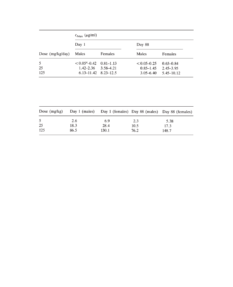
Example of a general toxicology report
105
At 24 hours after doses of 5 or 25 mg/kg/day, plasma concentrations of PP 27567
were generally below the limit of detection of the analytical method. At 125 mg/
kg/day, approximately 1.9 to 3.9 µg/ml of the compound was present in the plasma
for 24 hours after administration.
Mean AUC values were higher in females than in males. AUC values on Days
1 and 88 were comparable and increased in approximate proportion to the
administered dose as shown in Table 2.
2
In-life Observations/Measurements
2.1
Mortality (see page 000, necropsy observations)
No compound-related mortality was observed during this study. Two male animals
(No. 402, 125 mg/kg/day and No. 315, 25 mg/kg/day) and one female animal (No.
807, 125 mg/kg/day) were found dead on study days 28, 65 and 81, respectively.
Necropsy findings (congested lungs, traces of white particles in the bronchi)
suggested that these deaths were caused by dosing accidents. One female (No. 713,
25 mg/kg/day) was found prostrate on day 70, had a stained anogenital region and
lost weight on the two subsequent days. Because of its deteriorating clinical condition,
this animal was sacrificed for ethical reasons on day 72 of the study. Although
the cause of the poor condition of the animal was undetermined (see Pathology
section of this report, page 000), it was not considered to be related to PP 27567
since it was an isolated incident at the mid-dose level.
Table 1 PP 27567:3-Month oral toxicity study in rats. Range of maximal plasma
concentrations (c
Max
/µg/ml) on study days 1 and 88.
a
Limit of quantification of the analytical method
Table 2 PP 27567:3-Month oral toxicity in rats. Mean AUC
0–24h
values (µg.h/ml) on
days 1 and 88.
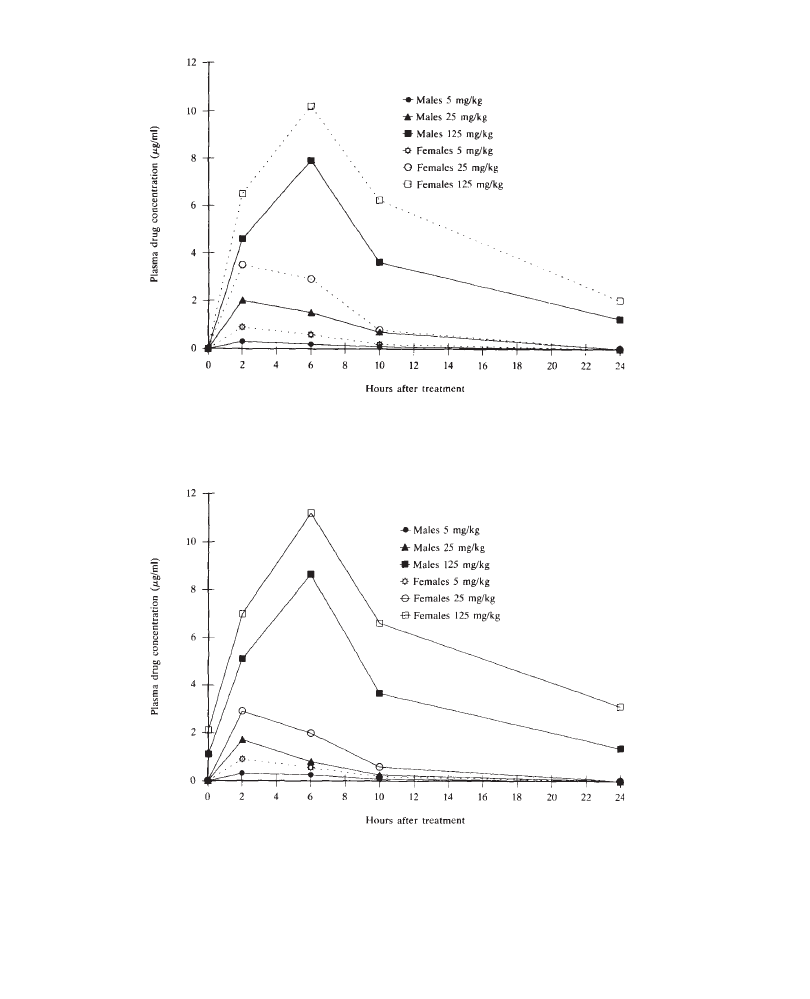
Presenting toxicology results
106
Figure 1 PP 27567:3-Month oral toxicity study in rats. Mean plasma drug
concentrations (µg/ml) at 5, 25 and 125 mg/kg/day. Values on day 1.
Figure 2 PP 27567:3-Month oral toxicity study in rats. Mean plasma drug
concentrations (µg/ml) at 5, 25 and 125 mg/kg/day. Values on day 88.
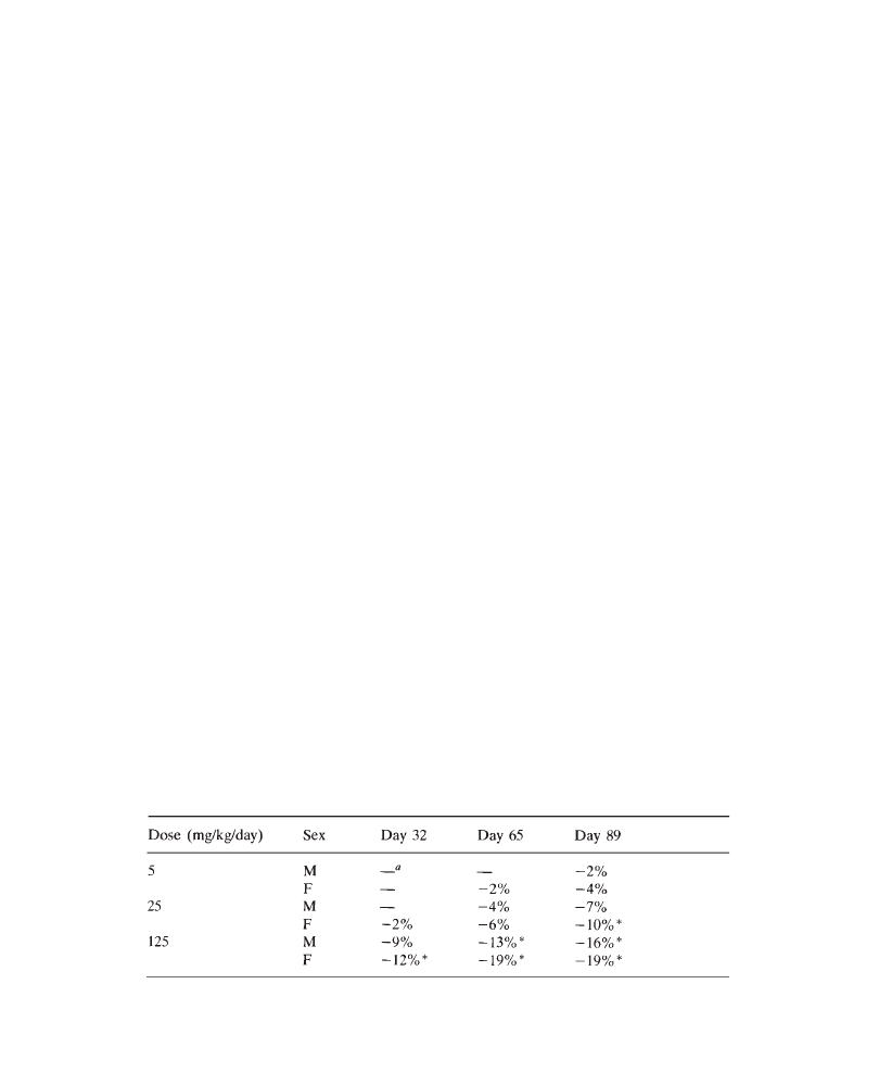
Example of a general toxicology report
107
2.2
Clinical signs (see pages 000–000)
Compound-related clinical signs were noted from week 2 through the end of the
study in all animals receiving 125 mg/kg/day and from week 15 to study termination
in some female animals receiving 25 mg/kg/day. The incidence and the severity
of these signs were dose-related and were generally more severe in females than
in males; they consisted of reduced motor activity and ptosis (all animals at 125
mg/kg/day and 3/20 to 6/20 females at 25 mg/kg/day) and chromodacryorrhea (3/
20 to 5/20 females at 125 mg/kg/day). Most animals at 125 mg/kg/day had a rough
hair coat during the last weeks of the study. No compound-related clinical signs
were noted at 5 mg/kg/day. With the exception of the prostration observed in female
No. 713, 25 mg/kg/day (see Subsection 2.1), all other signs/observations recorded
during this study are commonly observed in our laboratory and were not attributed
to PP 27567.
2.3
Body weight (see table on page 000, and individual results, pages
000–000)
Administration of PP 27567 affected the mean body weight in groups receiving
25 or 125 mg/kg/day as shown in Table 3.
At 125 mg/kg/day, lower mean body weights became apparent in male and
female groups after 3 weeks of treatment. The difference in body weight increased
progressively during the course of the study, reached statistical significance
after 4 weeks and resulted in markedly lower mean body weight at the end of
the study. Similar, but milder effects on body weight were noted in the groups
receiving 25 mg/kg/day; the difference reached statistical significance only during
the final 3 weeks of the study. At 5 mg/kg/day, minimal effects on body weight
were noted at the end of the study. Although this difference may be related to
the administration of PP 27567, it was slight (–2% and –4% compared with
control means in males and females, respectively) and within the range of the
common variation of this parameter, and was therefore considered to be of no
toxicological significance.
Table 3 PP 27567:3-Month oral toxicity in rats. Mean body weight on days 32, 65
and 89 of the study (percentage change compared with control mean values).
*P<0.05
a
No effect
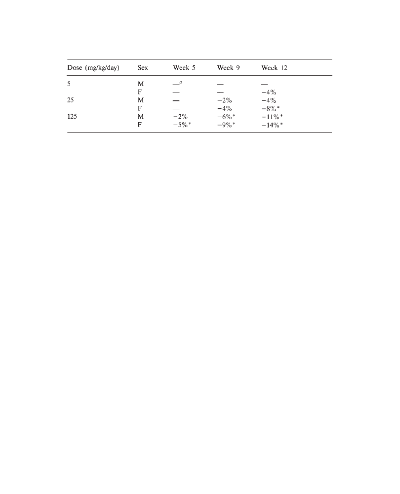
Presenting toxicology results
108
2.4
Food and water consumption (see tables, pages 000–000 and
individual results on pages 000–000)
A compound- and dose-related lower food consumption which was correlated with
the reduction in body weight was noted in all groups receiving PP 27567 at 25 or
125 mg/kg/day. Mean values are shown in Table 4.
Treatment with PP 27567 resulted in a dose-related lower mean food consumption
when compared with control means. Female rats appeared somewhat more affected
than males. At 125 mg/kg/day, lower food consumption became apparent in male
and female groups during the fifth week of treatment. The difference in food
consumption between the groups receiving 125 mg/kg/day and the control groups
became progressively more marked during the course of the study and reached
statistical significance from week 12 until the end of the study. Similar, but milder
effects on food consumption were noted in the groups receiving 25 mg/kg/day;
these reached statistical difference in females during the final weeks of the study.
At 5 mg/kg/day, minimally decreased food consumption, which occurred only in
females during the final weeks of the study, remained within the range of the common
variation of this parameter and was considered to be of no toxicological significance.
Water consumption was unaffected by treatment.
2.5
Ophthalmology (see individual results, pages 000–000)
Remnants of hyaloid vessels or a noticeable suture line of the anterior cortex of
the lens were observed during the pre-study examination and on day 30 in some
control animals and in some animals at 125 mg/kg/day. These vestigial fetal structures
were no longer noted at the end of the study and were attributed to the young age
of the animals. Small opalescent vesicles were observed in the superficial part of
the corneal stroma in some male animals of the control, the 125 mg/kg/day and 5
mg/kg/day groups. This lesion was diagnosed as corneal dystrophy. Some minor
changes were noted in the nucleus of the lens in a few animals of the control, 5
and 25 mg/kg/day groups. A similar incidence of these lesions has been reported
for Sprague-Dawley rats of this strain and age (Taradach and Greaves, 1984). Because
Table 4:
PP 27567:3-Month oral toxicity in rats. Mean weekly food consumption
in Weeks 5, 9 and 12 of the study (percentage change compared with control mean
values).
*p<0.05
a
No change, compared with control means
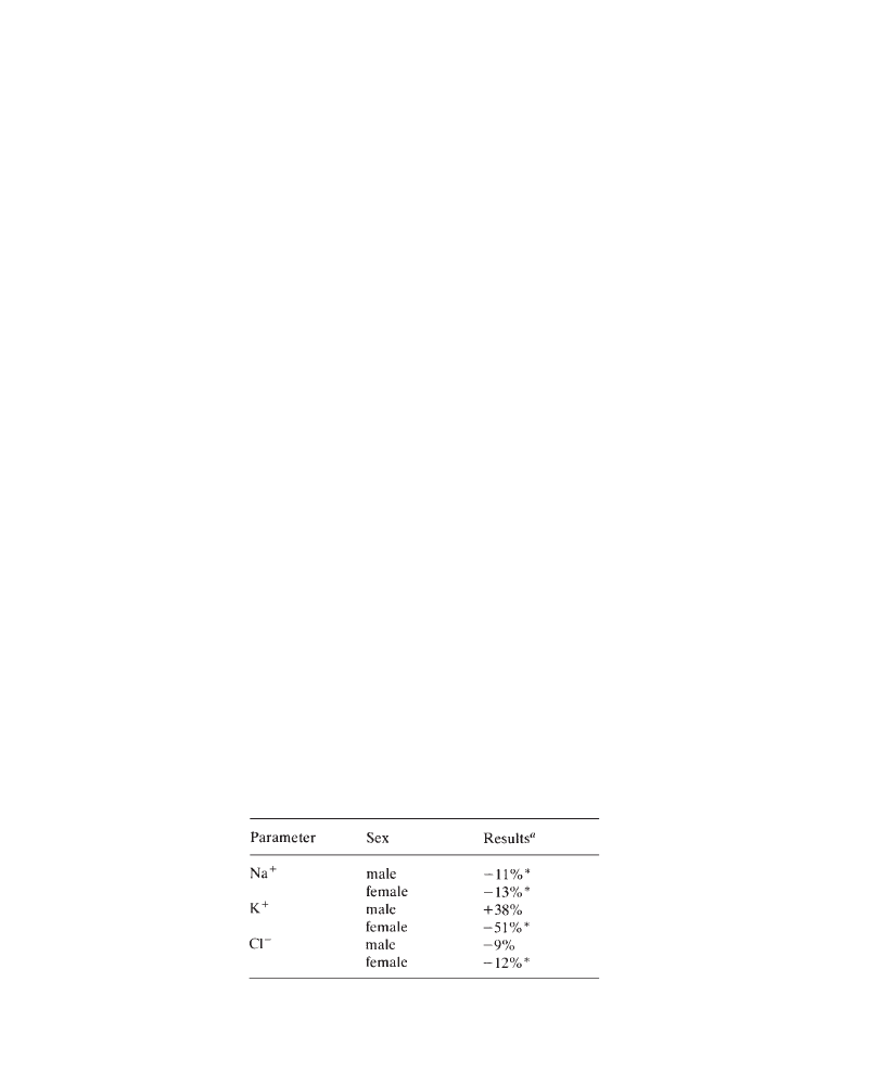
Example of a general toxicology report
109
these changes were not dose-related in incidence, and since the incidence of these
changes remained within our historical control range, they were considered to be
of spontaneous origin.
3
Clinical Pathology
3.1
Hematology (see summary and individual values, pages 000–000
and 000–000)
At 125 mg/kg/day in both sexes, the administration of PP 27567 was associated
with increases in RBC count, hemoglobin and hematocrit (+18%, when compared
with control mean values). This effect correlated with electrolyte imbalances and
increased urinary volume, and indicates hemoconcentration.
3.2
Clinical chemistry (see summary and individual values, pages
000–000 and 000–000)
Compound-related changes included electrolyte abnormalities, higher mean ALAT
and AS AT values in both sexes and increases in cholesterol in females only, as
compared with mean control values. Markedly higher serum K
+
and moderately
lower serum Na
+
and Cl– were observed at 125 mg/kg/day. Mean values are displayed
in Table 5.
Mild increases in ALAT (maximum 3.2-fold and 2.5-fold of control means in
males and females, respectively) and ASAT (maximum 3-fold and 2.2-fold in males
and females, respectively) were observed at 25 and 125 mg/kg/day. A slightly higher
mean cholesterol value (+36%) was observed in females only at 125 mg/kg/day.
Slightly higher urea and lower total protein, albumin and albumin/globulin ratio
in both sexes were associated with administration of 125 mg/kg/day PP 27567.
These changes may be secondary to the decreased food consumption observed at
this dose level.
Other compound-associated variations in clinical chemistry parameters were within
the limits of our reference range data, were slight in magnitude and/or displayed
no trends which were attributable to compound administration.
Table 5 PP 27567:3-Month oral toxicity study in
rats. Mean serum electrolyte changes on day 90
at 125 mg/kg/day.
*p
ⱕ0.05
a
Percentage change compared with control mean values
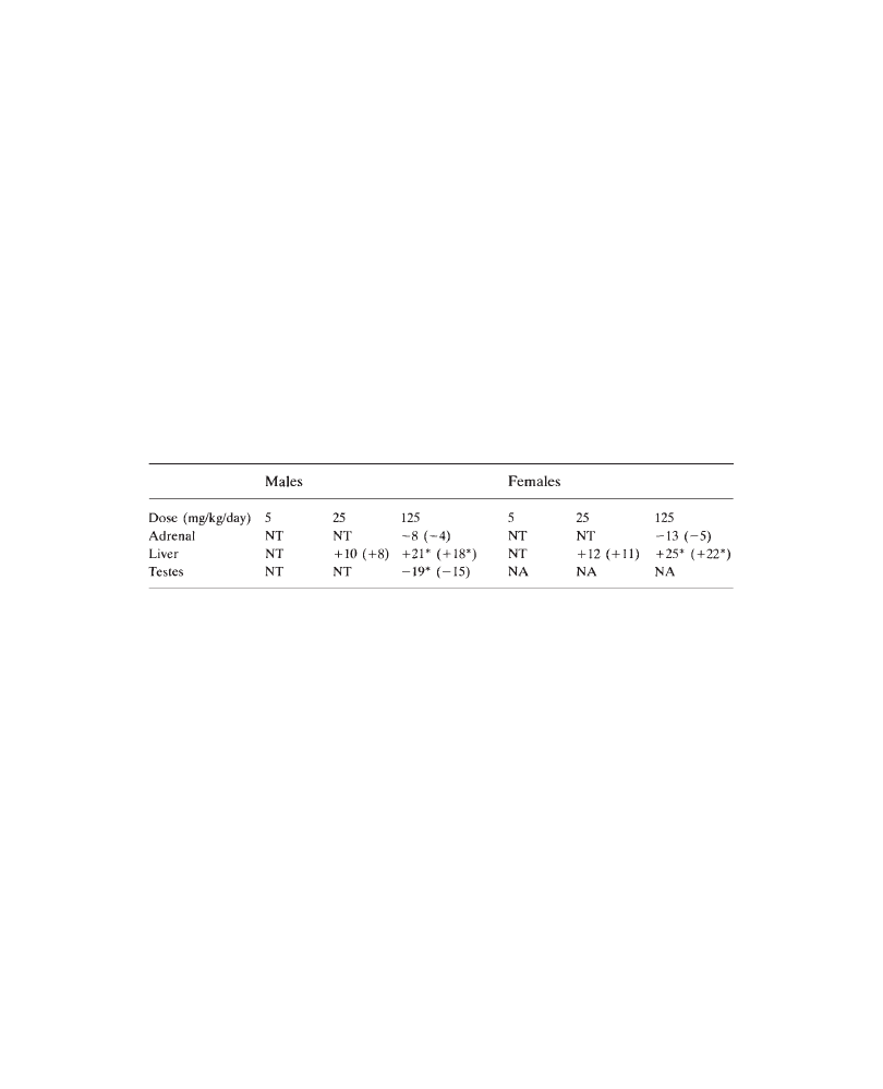
Presenting toxicology results
110
3.3
Urinalysis (see summary and individual values, pages 000 and 000)
Compound-related effects were limited to increased mean urinary volume in the
125mg/kg/day rats (+38% in males; +55% in females), when compared with control
means.
4
Anatomic Pathology
4.1
Organ weights (see summary and individual values, pages 000 and
000–000)
Treatment with PP 27567 was associated with slightly lower mean absolute and
relative adrenal weights at 125 mg/kg/day (both sexes), dose-related, slightly higher
mean absolute and relative liver weights at 25 and 125 mg/kg/day (both sexes),
and lower absolute and relative testis weights in males treated at 125 mg/kg/day.
These findings are summarized in Table 6.
There were no other compound-related organ weight findings. Slight variations
between individual animals in both the absolute and relative organ weights, which
had no relationship to dose or sex, and which sometimes achieved statistical significance,
were observed but were considered incidental and unrelated to treatment with PP 27567.
4.2
Necropsy findings (see summary and individual values, pages
000–000 and 000–000)
Compound-related macroscopic findings consisted of bilateral small and soft testes
in males treated at 125 mg/kg/day. All other macroscopic findings were incidental
and typical of Sprague-Dawley rats of similar age. Three animals died prior to the
termination of the study: two males and one female (M402, 125 mg/kg/day; M315,
25 mg/kg/day; F807, 125 mg/kg/day) which were found dead on study days 28, 65
and 81, respectively, and had gross and microscopic findings which suggested that
their deaths were related to gavage accidents (pulmonary congestion and hemorrhage,
foreign material in trachea, bronchi and alveoli). The cause of the deteriorating
condition of the female (F713, 25 mg/kg/day) which was killed for ethical reasons
(day 72) was not determined by either gross or microscopic examination. However,
Table 6. PP 27576:3-Month oral toxicity study in rats. Compound-related absolute
and relative organ weight changes (percentage) compared with controls.
NA=not applicable; NT=no compound-related changes; *p
ⱕ0.05

Example of a general toxicology report
111
its deteriorating condition was not considered related to treatment with PP 27567,
because of the lack of a dose relationship as well as its isolated incidence.
4.3
Histopathology (see summary and individual values, pages 000–000
and 000–000)
Treatment with PP 27567 was associated with degeneration and atrophy of the adrenal
cortex at 125 mg/kg/day (both sexes), periportal hepatocellular hypertrophy and
increased individual cell necrosis in the livers of rats (both sexes) at 25 and 125
mg/kg/day, and atrophy of the seminiferous tubules of the testes in males treated
at 125 mg/kg/day.
In the adrenal gland, 12/20 males and 20/20 females treated at 125 mg/kg/day
had mild to marked degeneration and necrosis of the zona fasciculata and zona
reticularis, and mild atrophy of the zona glomerulosa. Degenerate cortical cells
were characterized by decreased cell size, pyknotic nuclei and decreased lipid content.
Other adrenocortical lesions included thickening of the capsule, multifocal
mononuclear cell infiltrates, occasional cholesterol cleft formation, presence of
hemosiderin pigments, and multifocal mineralization.
In the liver, 15/20 males and 17/20 females treated at 125 mg/kg/day had minimal
to mild periportal hepatocellular hypertrophy. An increased incidence of individual
cell necrosis in the liver observed at 25 and 125 mg/kg/day was interpreted to be
an incidental finding since the lesions were minimal to mild in severity and were
also observed in control animals.
In the testes, 12/20 males treated at 125 mg/kg/day had bilateral, mild to moderate,
multifocal atrophy of the seminiferous tubules. This lesion was characterized by
multifocal areas of seminiferous tubules which were either devoid of spermatozoa
and/or immature sperm types or seminiferous tubules with germinal epithelium
undergoing various stages of degeneration. In all cases, Sertoli cells, spermatogonia
and the basement membrane in affected seminiferous tubules were intact and did
not show evidence of degenerative changes. There were no other histopathologic
findings related to treatment with PP 27567. All other microscopic findings were
incidental or related to orbital venous plexus blood sampling and were typical of
those lesions observed in Sprague-Dawley rats of similar age.
Discussion
The plasma drug levels, which were higher in females than in males, correlated
with the adverse in-life effects (clinical signs, lower mean body weight and food
consumption), which were generally more severe in females than in males. These
findings confirm the results of a previous 1-month study which indicated that females
are more affected by PP 27567 than males (Study PP/DS 93–0063). In the present
study, clinical pathology, organ weight and histopathologic changes occurred at
similar severity and incidence in both sexes. Moderately lower serum Na
+
and Cl–
and markedly elevated serum K
+
at 125 mg/kg/day correlated with atrophy in the
adrenal cortex (zona glomerulosa), and were consistent with adrenal insufficiency.
The higher mean urinary volume at this dose level is interpreted to be secondary
to the loss of serum Na
+
. Though the areas in the adrenal cortex which are responsible

Presenting toxicology results
112
for the production of both mineralocorticoids and glucocorticoids were affected,
no compound-related changes in serum glucose were noted in these animals. Lower
mean absolute and relative adrenal weights at 125 mg/kg/day correlated with adrenal
cortical degeneration (zona fascicularis and zona reticularis) and atrophy (zona
glomerulosa). The adrenal cortical lesions were interpreted to be a primary cytotoxic
effect of PP 27567, and were consistent with lesions reported with other ACAT
inhibitors in the literature (Dominick et al., 1993).
Mild increases in ALAT and ASAT at 25 and 125 mg/kg/day and a minimal
increase in cholesterol in females only at 125 mg/kg/day correlated with higher
absolute and relative liver weights at 25 and 125 mg/kg/day and periportal
hepatocellular hypertrophy and a higher incidence of individual cell necrosis at
125 mg/kg/day. No microscopic changes in the liver were observed at 25 mg/kg/
day. The periportal hypertrophy was interpreted to be an adaptive response of the
liver to PP 27567, and was consistent with lesions reported with other ACAT
inhibitors in the literature (Dominick et al., 1993). Because individual cell necrosis
was also found in control animals, and because it was minimal to mild in severity,
this lesion was considered to be an incidental finding.
Lower mean absolute and relative testis weights at 125 mg/kg/day correlated with
the small, soft testes seen at necropsy and the atrophy of the seminiferous tubules
observed histopathologically in this dose group. This effect may be due to decreased
testosterone production secondary to the perturbation of cholesterol metabolism in
the Leydig cells of the testis and the zona reticularis of the adrenal cortex. These
results are consistent with those found in an earlier reproduction study (Study No.
PP 92–037, 1992), in which a lower fertility was observed in male rats.
Conclusion
Adverse in-life effects of oral administration of 125 mg/kg/day PP 27567 for 3
months were more severe in females than in males and included clinical signs,
lower mean body weight and food consumption. The target organs were the adrenals,
liver and testis. Administration of 25 mg/kg/day produced mild in-life effects and
mild changes in the liver. No adverse effects were noted at 5 mg/kg/day.
References
Dominick M.A., McGuire E.J., Reindel J.F., Bobrowski W.F., Bocan T.M.A. and Gough
A.W. (1993) Subacute toxicity of a novel inhibitor of Acyl-CoA: cholesterol
acyltransferase in Beagle dogs. Fundamental and Applied Toxicology, 20; 217–224.
Report PP/DS 92–037 (1992) PP 27567: Oral Fertility Study in Male Sprague-Dawley
Rats.
Report PP/DS 93–0063 (1993) PP 27567:30-Day Oral Toxicity Study in Sprague-Dawley
Rats.
Taradach C. and Greaves P. (1984) Spontaneous eye lesions in laboratory animals:
incidence in relation to age. CRC Critical Reviews in Toxicology, 12; 121–147.

113
11
Example of a Reproductive
Toxicology Report
G.COPPING
Rhône-Poulenc Rorer, Drug Safety Development, Vitry sur Seine, France
and R.L.CLARK
Rhône-Poulenc Rorer Research and Development, Drug Safety Department, Collegeville, USA
PP 27567: Oral one-generation reproductive toxicity study
in rats including F
1
reproductive performance
Dose levels: 3, 10, 30 mg/kg/day
Study Number: PP 93–1234
This document contains proprietary confidential information
of the sponsor, Poisonous Prose Inc. and may not be disclosed
verbally, abstracted or published without written consent of the sponsor.

Presenting toxicology results
114
PP 27567: Oral one-generation reproductive toxicity study in rats including
F
1
reproductive performance
R.Clark and G.Copping
The purpose of this study was to assess the effect of oral administration of PP
27567 upon gonadal function, mating behavior and fertility of male and female
rats when treated in the F
0
generation, and to assess the subsequent development
of the F
1
generation up to sexual maturity including mating to produce an F
2
generation.
Thirty-six Sprague-Dawley rats/sex/group (Crl:CDr(SD)BR), approximately 7
weeks (males) and 9 weeks (females) old at the beginning of treatment, were used.
Animals in the control group received water, and animals in the treated groups
received PP 27567 as an aqueous solution at dose levels of 3, 10 or 30mg/kg/day
by the oral route in a dosing volume of 2 ml/kg/day.
Only F
0
rats were treated: males for 28 days pre-pairing, throughout pairing
and up to necropsy, and females for 14 days pre-pairing and throughout pairing,
gestation and lactation. Observations included mortality, clinical examination, food
consumption, body weight and macroscopic examination at necropsy. Twenty F
0
females per group were sacrificed on day 20 of gestation for examination of their
uterine contents, including external, visceral and skeletal examination of fetuses.
The remaining females were allowed to litter and rear their young to weaning on
day 21 postpartum. The physical and functional development and behavior of the
F
1
generation were evaluated on standardized litters. On postnatal day 58±2, 1 male
and 1 female per litter were selected for subsequent evaluation of reproductive
performance following pairing and the observation of the F
2
generation up to postnatal
day 7.
At 30 mg/kg/day, PP 27567 induced adverse parental effects including salivation,
reduced food intake and body weight gain, slight reductions in fertility index and
pregnancy rate, and a slight increase in gestation length. These effects were associated
with mild effects in the F1 generation, as indicated by lower mean fetal body weight,
delayed fetal ossification, a slight decrease in birth index, and reduced pup body
weight at birth and during the pre-weaning period.
At 10 mg/kg/day, with the exception of salivation in a few F
0
animals, no adverse
effects were observed in the F
0
, F
1
and F
2
generations.
At 3 mg/kg/day, no adverse effects were observed in the F
0
, F
1
and F
2
generations.
In conclusion, the reproductive and developmental no-effect level of PP 27567
under the conditions of this study is 10 mg/kg/day.
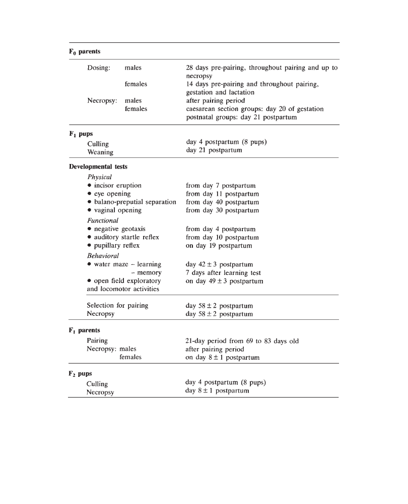
Example of a reproductive toxicology report
115
Study chronology

Presenting toxicology results
116
1
Introduction
The purpose of this study was to assess the effect of oral administration of PP
27567 upon gonadal function, mating behavior and fertility in male and female
rats, and to assess the subsequent development of the F
1
generation up to sexual
maturity including mating to produce an F
2
generation. The dose levels selected
were based on the results of an embryofetal developmental toxicity study in rats
(Report PP 674, 1993) which indicated lower maternal body weight gain at 30
mg/kg/day associated with lower fetal weight, and the results of a 6-month oral
toxicity study in rats (Report PP 711, 1993) which indicated that administration
of 30 mg/kg/day produced a lower mean body weight gain in both males and females
and a reduction in mean ovary weight and ovarian atrophy in females. The no-
effect level was 10 mg/kg/day in the embryofetal developmental toxicity study and
3 mg/kg/day in the 6-month toxicity study. Based on these results, dose levels of
3, 10 and 30 mg/kg/day PP 27567 were selected.
2
Materials and Methods
Aqueous solutions of PP 27567 were administered by oral gavage to groups of 36
male and 36 female rats at daily dose levels of 3, 10 or 30 mg/kg/day. Males were
treated for 28 days pre-pairing, throughout pairing and up to necropsy. Females
were treated for 14 days pre-pairing and throughout pairing, gestation and lactation.
A control group of 36 males and 36 females received the vehicle alone over the
same periods. Observations included mortality, clinical examination, food
consumption, body weight and macroscopic examination at necropsy.
Twenty F
0
females per group were euthanatized on day 20 of gestation for
examination of their uterine contents, including external, visceral and skeletal
examination of fetuses. The remaining females were allowed to litter and rear their
young to weaning on day 21 postpartum. The physical and functional development
and behavior of the F
1
generation were evaluated on standardized litters. Details
of study protocol materials and methods are given in Appendix 1 of this report.
On postnatal day 58±2, 1 male and 1 female per litter were selected for subsequent
evaluation of reproductive performance following pairing and the observation of
the F
2
generation up to postnatal day 7.
3
Results and Discussion
3.1
F
0
Generation
3.1.1
Mortality
No compound-related mortality was observed in the F
0
generation.
One male (No. 3109, 10 mg/kg/day) was euthanatized for ethical reasons on
day 36 of the pre-pairing period because of the presence of an abdominal and
subcutaneous mass which was observed from day 19.
Two females (No. 3307, 10 mg/kg/day and No. 3357, 30 mg/kg/day) were
euthanatized for procedural reasons (litter less than six pups) on days 4 and 6
postpartum. No macroscopic findings were noted in these animals at necropsy.

Example of a reproductive toxicology report
117
3.1.2
Clinical signs
At 30 and 10 mg/kg/day, compound-related clinical signs were limited to salivation
observed immediately after treatment. This sign was recorded from the first week
of treatment up to the end of the treatment period in both sexes. The incidence
and frequency of salivation were dose-related.
All other clinical signs monitored during the treatment period were not considered
to be related to PP 27567.
No compound-related clinical signs were noted in animals treated at 3 mg/kg/
day.
3.1.3
Body weight and food consumption
PP 27567 produced a treatment-related decrease in food intake and body weight
gain at 30 mg/kg/day in males and females. Mean food consumption was less than
that of controls at 30 mg/kg/day from the second week of treatment in males and
from the first week of treatment in females. As a consequence, mean body weight
gain during the dosing period (in males and females during pre-pairing and in females
during gestation and lactation periods) was lower in this group (approximately –
15% to –30% when compared with controls). These differences in food intake and
mean body weight gain reached statistical significance during the pre-pairing (males
and females) and gestation periods.
Slightly lower food consumption and body weight gain were noted at the end
of the pre-pairing period in 10 mg/kg/day males.
There were no differences in group mean body weight and food consumption
between the control and the 3 mg/kg/day groups.
3.1.4
Precoital interval, mating and fertility indices and pregnancy rate
Precoital interval and mating index were comparable in all groups.
Fertility indices and pregnancy rate were slightly lower at 30 mg/kg/day when
compared with controls (22% and 19%, respectively). These reductions did not
reach statistical significance.
No effects on fertility indices or pregnancy rate were observed at 10 and 3 mg/
kg/day.
3.1.5
Gestation length-and observations at parturition
Gestation length and parturition index were similar in all groups and there were
no signs of dystocia in any group.
At 30 mg/kg/day, gestation length was significantly but minimally higher (mean
22±0.5 days compared with mean 21.5±0.5 days) than that of controls, while birth
index was slightly and non-significantly lower (–11% when compared with controls).
At 10 and 3 mg/kg/day, gestation length and birth index were similar to those
of the controls.

Presenting toxicology results
118
3.1.6
Necropsy
3.1.6.1 Males
There were no treatment-related effects on mean sperm count, motility or vitality
of spermatozoa.
Group mean relative testicular weight was significantly higher at 30 mg/kg/day
when compared with controls. This increase was related to the significantly lower
mean body weight observed in this group. Group mean absolute epididymal weight
was significantly lower at 30 mg/kg/day when compared with controls. Group mean
absolute and relative testicular and epididymal weights were comparable in the
control, 10 and 3 mg/kg/day groups.
No compound-related macroscopic findings were noted at terminal necropsy.
3.1.6.2 Non-mated females
No compound-related macroscopic findings were noted at necropsy.
3.1.6.3 Caesarean section of mated females on day 20 of gestation
Maternal observations. No compound-related macroscopic findings were noted
at terminal necropsy.
Corpora lutea, implantations and pre-implantation loss. Mean numbers of
corpora lutea, implantations and pre-implantation loss were similar between the
control and treated groups.
Post-implantation loss and litter size. Mean numbers of early and late uterine
deaths and post-implantation loss were similar between the control and treated groups.
Fetal weight and sex ratio. Sex ratio was unaffected by treatment. Mean fetal
weight was significantly lower at 30 mg/kg/day (–14% when compared with controls).
This effect was considered likely to be secondary to decreased maternal food
consumption and body weight. At 10 and 3 mg/kg/day, mean fetal weight was
unaffected by treatment.
Fetal examination. External and internal observations revealed a range of
malformations and variations in all groups. There was no indication of a compound-
related trend in the type or incidence of these anomalies. At 30 mg/kg/day, there
was a slight delay in skeletal ossification characterized by higher incidence of fetuses
with incomplete or unossified supraoccipitals, exoccipitals, thoracic vertebral centra,
sternebrae, pubis bones and hindpaw distal phalanges. These observations were
consistent with the lower mean fetal weight secondary to lower maternal body weight
observed at this dose level. At 10 and 3 mg/kg/day, skeletal examination of fetuses
revealed a range of changes which were of a type or which occurred at an incidence
similar to that of controls. There was no evidence of a teratogenic effect.
3.1.6.4 Dams on day 21 postpartum
Maternal observations. No compound-related macroscopic findings were noted
at necropsy.
Implantations and post-implantation loss. At 30 mg/kg/day, the mean number
of implantations observed on day 21 postpartum was slightly lower than the control
value. As there was no equivalent effect observed in the caesarean section group,
this was not considered to be compound-related. At 10 and 3 mg/kg/day, the mean

Example of a reproductive toxicology report
119
number of implantations was comparable to that for controls. Mean post-implantation
loss was comparable in all groups.
3.2 F
1
Generation
3.2.1
Mortality
3.2.1.1 Pup data at birth
In the 30 mg/kg/day group, mean litter size was significantly lower than the control
value and was considered to be related to the lower number of implantations. Since
such an effect was not observed in the caesarean section group or in previous
embryofetal developmental toxicity studies (Reports PP 674 and PP 684, 1993),
this was not considered to be compound-related. In the 10 and 3 mg/kg/day groups,
mean litter size was similar to that for controls.
Sex ratio was unaffected by treatment.
In the 30 mg/kg/day group, there was a higher number of stillborn (male and
female) pups and dead (female) pups than in the controls. The difference reached
statistical significance in female pups. In the 10 and 3 mg/kg/day groups, the numbers
of stillborn and dead pups were similar to that for controls.
There were no malformed pups at birth in any group.
3.2.1.2 Pre-weaning period
Viability (on day 4 postpartum), survival (on days 7 and 14 postpartum) and lactation
(on day 21 postpartum) indices were comparable in all groups.
3.2.1.3 Post-weaning, gestation and lactation perbiods
No deaths occurred during the post-weaning and gestation periods.
One female (No. 1109, 3 mg/kg/day group) was sacrificed for procedural reasons
(litter less than six pups) on day 4 postpartum. At necropsy, absence of mammary
tissue development was noted.
3.2.2
Clinical signs
No compound-related clinical signs were observed in the F1 generation.
3.2.3
Body weight
3.2.3.1 Pup data at birth and pre-weaning period
In the 30 mg/kg/day group, mean body weight of male and female pups at birth
was lower (–8% and –5%, respectively, when compared with controls) and correlated
with lower mean fetal weight observed in the caesarean section group at this dose.
Mean body weight of male and female pups was slightly lower on days 7, 14 and
21 postpartum (–5% to –8% compared with respective control values) in the 30
mg/kg/day group. These differences did not reach statistical significance.
In the 10 and 3 mg/kg/day groups, mean body weight of pups at birth and during
the pre-weaning period was similar to control values.

Presenting toxicology results
120
3.2.3.2 Post-weaning period
Mean body weight was comparable between control and treated groups.
3.2.3.3 Gestation and lactation periods
Mean body weight was comparable between control and treated groups.
3.2.4
Developmental tests
Physical development
Physical development as assessed by the time of onset and completion of the incisor
eruption, eye opening, balano-preputial separation or vaginal opening showed no
effect that could be attributed to PP 27567.
Functional development
The results of functional development tests (negative geotaxis, auditory and visual
functions) were comparable in all groups.
Behavioral development
The results of behavioral tests (locomotor and exploratory activities, and learning
and memory abilities) were comparable in all groups.
3.2.5
Precoital interval, mating and fertility indices and pregnancy rate
Precoital interval, mating and fertility indices and pregnancy rate were comparable
in all groups. Therefore, maternal treatment with PP 27567 was considered to have
no influence on reproductive performance of the F
1
generation.
3.2.6
Gestation length and observations at parturition
Gestation length and parturition index were comparable in all groups and there
were no signs of dystocia in any group. Birth index was significantly lower in the
30mg/kg/day group (–15% when compared with controls). In the 10 and 3 mg/kg/
day groups, birth index was slightly lower when compared with controls (–5% and
–9%, respectively). However, as these differences did not reach statistical significance
and the values remain within the range of our historical control data, they were
not considered to be related to the compound.
3.2.7
Necropsy
3.2.7.1 Day 56±3 postpartum
No compound-related macroscopic findings were observed.
3.2.7.2 Males selected for reproductive performances
No macroscopic findings which could be attributed to the test substance
administration were observed.

Example of a reproductive toxicology report
121
3.2.7.3 Nonmated and non-pregnant females
No macroscopic findings were noted.
3.2.7.4 Mated females which littered
Maternal observation. No maternal macroscopic findings were noted.
Implantation and post-implantation loss. There was no difference between
the groups in the mean number of implantation sites. Mean post-implantation loss
was significantly higher in the 30mg/kg/day group. In the 10 and 3mg/kg/day groups,
mean post-implantation loss was slightly higher when compared with controls.
However, these differences did not reach statistical significance and the values
remained within the range of our historical control data and, therefore, they were
not considered to be related to the compound.
3.3 F
2
Generation
3.3.1
Mortality
There was no effect of treatment of the F
0
animals on the F
2
litter size. In the 30
mg/kg/day group, mean litter size was slightly lower, but this decrease did not
reach statistical significance and was considered to be spontaneous.
Sex ratio was unaffected by treatment of the F
0
animals.
There was no dose-related trend in the number of stillborn and dead pups.
One male pup in the 3 mg/kg/day group showed abnormal flexure of hindpaws.
This isolated observation was considered an incidental finding and was not attributed
to treatment of the F
0
parents.
Survival index on day 4 postpartum was similar in all groups. There was a slight
decrease in the survival index on day 7 postpartum for male pups in the 30 mg/
kg/day group which was not statistically significant and not considered treatment-
related. In the 10 and 3 mg/kg/day groups, survival index on day 7 postpartum
was similar to that for controls.
3.3.2
Clinical signs
There were no clinical signs attributable to treatment of the F
0
animals.
3.3.3
Body weight
Mean body weight at birth was similar in all groups. In the 30 mg/kg/day group,
mean body weight was slightly lower when compared with controls on days 4 and
7 postpartum (–4% to –9%, respectively). These differences were not considered
to be related to PP 27567. In the 10 and 3 mg/kg/day groups, mean body weights
on days 4 and 7 postpartum were similar to control values.
3.3.4
Terminal necropsy
No macroscopic findings which could be attributed to the test substance
administration were observed.

Presenting toxicology results
122
4
Conclusion
At 30 mg/kg/day, PP 27567 induced adverse parental effects including salivation,
lower food intake and body weight gain, slightly lower fertility index and pregnancy
rate, and a slight increase in gestation length. These effects were associated with
mild effects in the F
1
generation, as indicated by lower mean fetal body weight,
delayed fetal ossification, a slight decrease in birth index, and lower pup body
weight at birth and during the pre-weaning period.
At 10 mg/kg/day, with the exception of salivation in a few F
0
animals, no adverse
effects were observed in the F
0
, F
1
or F
2
generations.
At 3 mg/kg/day, no adverse effects were observed in the F
0
, F
1
or F
2
generations.
In conclusion, the reproductive and developmental no-effect level of PP 27567
under the conditions of this study is 10 mg/kg/day.
References
Altman J. and Sudarshan K. (1975) Postnatal development of locomotion in the laboratory
rat. Anim. Behav., 23; 896–920.
Butcher R.E., Wootten V. and Vorhees C.V. (1980) Standards in behavioral teratology testing:
test variability and sensitivity. Teratog. Carcinog. Mutag., 1; 49–61.
Dawson A.B. (1926) Note on the staining of the skeleton of cleared specimens with Alizarin
Red S.Stain. Tech., 1; 123–124.
EEC Council Directive (1975) 75/318/EEC, dated May 20, 1975 (O.J. No. L147 of 9.6.75).
EEC Council Directive (1983) 83/570/EEC, dated October 26, 1983 (O.J. No. L332 of
28.11.83).
EEC Council directive (1986) 86/609/EEC, dated November 24, 1986 (O.J. No. L358 of
18.12.86).
Gad S.C. and Weil C.S. (1989) Statistics for Toxicologists, Principles and Methods of
Toxicology, 2nd edition, 1989, A.Wallace Hayes, Raven Press, 435–462.
Report PP 674 (1993) Teratology study of PP 27567 in the rat by the oral route with plasma
level determination. Poisonous Prose, Inc.
Report PP 684 (1993) Teratology study of PP 27567 in the rabbit by the oral route with
plasma level determination. Poisonous Prose, Inc.
Report PP 711 (1993) 6-Month oral toxicity study in rats. Poisonous Prose, Inc.
Ryan P.C., Whelan C.A. and Fitzpatrick J.M. (1988) The vas deferens count: a new accurate
method for experimental measurement of testicular exocrine function. Eur. Urol., 14;
156–159.
Salewski E. (1964). Färbemethode zum makroskopischen Nachweis von Implantationsstellen
am Uterus der Ratte. Arch. Exp. Path. Pharmakol., 247; 367.
US Federal Guidelines (1985) Laboratory Animal Welfare Act (1966) (PL 89–544) as amended
in 1970 (PL 91–579), 1976 (PL 94–279) and 1985 (PL 99–198).
Vorhees C.V., Butcher R.E., Brunner R.L. and Sobotka T.J. (1979) A developmental
test battery for neurobehavioral toxicity in rats: a preliminary analysis using
monosodium glutamate calcium carrageenan, and hydroxyurea. Toxicol. Appl.
Pharmacol., 50; 267–282.
Weil C.S. (1970) Selection of the valid number of sampling units and a consideration of
their combination in toxicological studies involving reproduction, teratogenesis or
carcinogenesis. Fd. Cosmet. Toxicol., 8; 177–182.
Zbinden G. (1981) Experimental methods in behavioral teratology. Arch. Toxicol., 48;
69–88.

Example of a reproductive toxicology report
123
APPENDIX 1: Materials and Methods
A1.1
Test and Control Articles
The bulk compound, identified as PP 27567, batch CA 9128000, had a purity of
100%.
The titer of the test article was considered to be 91.6% as free base. The doses
referred to throughout this report are expressed in terms of PP 27567 as free base.
The test article was administered as an aqueous suspension containing 0.5%
methylcellulose and 0.1% polysorbate 80. Three concentrations for oral dosing
were prepared daily or weekly and were administered at a dosing volume of 2 ml/
kg/day to achieve dose levels of 3, 10 or 30mg/kg/day. The vehicle for the test
article and control article was water containing 0.5% methylcellulose and 0.1%
polysorbate 80. The control article was administered at a dosing volume of 2 ml/
kg/day.
The homogeneity and stability of the formulations were satisfactory for the range
of concentrations used in this study. Samples of formulations were taken during
the study and analyzed for achieved concentrations. The results were close to nominal
concentrations and never exceeded a 10% variation.
A1.2
Test System
Male and female Sprague-Dawley rats of the Crl:CDr(SD)BR strain approximately
7 weeks (males) and 9 weeks (females) old at the beginning of treatment were
used in this study. The animals were COBS (Cesarean Originated Barrier Sustained)
and VAF (Virus Antibody Free). The rat was selected because it is a recognized
animal model for this type of safety evaluation study. The Charles River CD strain
of rat was used because of the background data available on this strain in our
laboratory.
Animals were sequentially delivered, examined physically prior to acceptance
in the study, and allowed one week acclimatization. They were then randomly
allocated to a dose group where they were individually identified.
A1.3
Housing and Care of Animals
The animals used in this study were handled and maintained in accordance with
the requirements of the EEC Guideline (1986) and US Federal Guidelines (1985).
Compliance with the above legislation was ensured by adhering to the standards
set forth in the Guide for the Care and Use of Laboratory Animals, DHHS Publication
No. (NIH) 86–23, revised 1985.
Animals were housed in environmentally controlled rooms and assigned to
individual stainless-steel cages in order to minimize possible inter-group differences
due to environmental factors. Animals were housed in stainless-steel wire cages
throughout the F
0
pre-pairing, the pairing period and from day 35 postpartum to
the end of the pairing period for the F1 generation. Females allocated to the postnatal

Presenting toxicology results
124
phase were housed in Makrolon-bodied cages with stainless-steel wire lids and
sawdust for bedding.
The animals were allowed free access to a commercially available pelleted diet
DSC01 (batches 30804, 30917, 31011, 31215, 40120, 40217 and 40414) supplied
by Diet Supply Co., and to filtered tap water.
Copies of the relevant certificates of analysis for sawdust, diet and water have
been filed in the research center archives.
A1.4
Experimental Design
The design of this study was consistent with EEC Council Directives (1975 and 1983).
Two hundred and eighty-eight animals (144 males and 144 females) were randomly
divided into 4 groups of 36 males and 36 females.
Only F
0
rats were treated: males for 28 days pre-pairing, throughout pairing
and up to necropsy, and females for 14 days pre-pairing and throughout pairing,
gestation and lactation.
Observations included mortality, clinical examination, food consumption, body
weight and macroscopic examination at necropsy.
Twenty F
0
females per group were euthanatized on day 20 of gestation for
examination of their uterine contents, including external, visceral and skeletal
examination of fetuses. The remaining females were allowed to litter and rear their
young to weaning on day 21 postpartum. The physical and functional development
and behavior of the F
1
generation were evaluated on standardized litters. On day
58±2 postpartum, 1 male and 1 female per litter were selected for subsequent
evaluation of reproductive performance following pairing and the observation of
the F
2
generation up to day 7 postpartum.
A1.5
In-life Observations
A1.5.1 Mortality
All animals were checked daily.
All animals found dead or those euthanatized prior to scheduled euthanasia were
subjected to a thorough macroscopic examination of the visceral organs to identify
the cause of death or morbidity. For females the number of implantation sites in
the uterine horns was also recorded.
A1.5.2 Clinical examination
All animals were examined daily.
A1.5.3 Body weight
Males were weighed weekly. Females were weighed weekly until mating and then
on days 1, 3, 6, 9, 12, 15, 18 and 20 of gestation and on days 1, 4, 7, 14 and 21
postpartum for those allocated to the parturition phase. Pups were weighed on days
1, 4, 7, 14 and 21 postpartum and weekly thereafter.

Example of a reproductive toxicology report
125
A1.5.4 Food consumption
Food consumption was determined weekly for males and females (F
0
generation)
during the pre-pairing period and twice weekly for F
0
females from day 1 of gestation
to day 13 postpartum. Daily food consumption was calculated for each interval.
A1.5.5 Assessment of reproductive performance (F
0
)
After the pre-pairing period, each female was placed with a male for a maximum
of 21 consecutive days. Vaginal smears were carried out daily during the pairing
period up to mating for all females in order to establish estrus cycle and mating.
The day a copulation plug (in the vagina or under the cage) and/or spermatozoa
in the vaginal smear was detected was designated day 0 of gestation. The time
between initial pairing and detection of mating (precoital interval) was recorded.
If mating was not detected during this period, females were maintained for
approximately 2 weeks, at the end of which they were euthanatized. Uterus and
ovaries were sampled for histological examination and the presence of implantation
was checked by the Salewski staining method (Salewski, 1964).
At the end of the pairing period, sperm collection was performed (vas deferens
method) after anesthesia in 10 males/group having mated. Sperm count, motility
and vitality parameters were recorded (Ryan et al., 1988).
A1.5.6 Lactation examination
During the parturition period, females were observed at least twice daily. Gestation
length was recorded as well as any signs of dystocia.
A1.5.6.1 Litter size
The number and sex of live, dead, stillborn and malformed pups were recorded at
birth. Stillborn and dead pups were identified using a lung flotation test.
A1.5.6.2 Culling
Culling within sex was performed on day 4 postpartum to obtain 8 pups per litter
(4 males and 4 females, where possible). Litters containing less than 6 pups were
discarded.
A second cull was performed after the last post-weaning test (on day 58±2
postpartum) to obtain 2 pups per litter (1 male and 1 female, where possible), and
a minimum of 15 males and 15 females per group for assessment of reproductive
performance.
A1.5.6.3 Examinations on day 4 postpartum (F0)
F
1
pups were examined and the following recorded for each litter:
•
number of live and dead pups
•
individual sexes
•
examination of individual pups

Presenting toxicology results
126
A1.5.7 Developmental tests
The timing of pup development was assessed on a total litter basis by recording
the days on which the onset and completion of the parameter occurred or the
performance on a defined day (Altman and Sudershan, 1975; Vorhees et al., 1979;
Butcher et al., 1980; Zbinden, 1981).
Physical
•
Incisor eruption: eruption of the upper incisor(s) through the gum from day 7
postpartum
•
Eye opening: separation of the upper and lower eyelids from day 11 postpartum
•
Balano-preputial separation: from day 40 postpartum
•
Vaginal opening: separation of the vaginal edges from day 30 postpartum.
Functional
•
Negative geotaxis: assessment of a pup’s ability to turn and face uphill on an
inclined plane (15°) within 30 seconds from day 4 postpartum
•
Auditory function (startle reflex): assessment of a pup’s ability to respond to
a sudden sharp noise from day 10 postpartum
•
Visual function (pupillary reflex): assessed by examination of pupil closure
in response to a bright source of light on day 19 postpartum.
Behavioral
A water-filled M maze was used to evaluate learning ability on day 42±3 postpartum
and memory 7 days later under the same conditions (1 male and 1 female per litter).
The time taken by each pup to swim through the maze in six successive trials was
measured. A maximum of 60 seconds was allowed for each trial, and pups were
considered to have failed if they exceeded the time limit. Improvement in swimming
time was taken as an indication of learning ability.
Locomotor and exploratory activities were evaluated on day 49±3 postpartum
(1 male and 1 female per litter) during a 9-minute test using a Columbus Optovarimex
3. Output data included distance traveled, resting time, ambulatory time, stereotypic
time and number of rearings.
A1.5.8 Development and reproductive performance (F
1
)
Following completion of the final behavioral test, F
1
rats were selected from each
group for assessment of their reproductive performance. Rats were at least 10 weeks
old when pairing took place (see subsection A1.5.5), and sibling pairings were
avoided.
During the parturition period, females were observed at least twice daily. Gestation
length was recorded as well as any signs of dystocia.
A1.5.8.1 Litter size
The number and sex of live, dead, stillborn and malformed pups were recorded at
birth. Stillborn and dead pups were identified using a lung flotation test.

Example of a reproductive toxicology report
127
A1.5.8.2 Culling
Culling within sex was performed on day 4 postpartum to obtain 8 pups per litter
(4 males and 4 females, where possible). Litters containing less than 6 pups were
discarded.
A1.5.8.3 Examinations on day 4 postpartum
Pups were examined and the following recorded for each litter:
•
number of live and dead pups
•
individual sexes
•
examination of individual pups.
A1.6
Terminal Examinations
A1.6.1 Necropsy
A1.6.1.1 F
0
and F
1
males
After pairing, F
0
males and F
1
males selected for assessment of reproductive
performance were euthanatized by CO
2
inhalation and subjected to macroscopic
examination.
The testes and epididymides were weighed in pairs. These tissues were collected
and fixed for possible microscopic examination.
AT.6.1.2 Non-mated F
0
and F
1
females
Two weeks after the end of pairing, non-mated females were euthanatized. The
presence of implantation sites was checked by the Salewski staining method
(Salewski, 1964). Uterus and ovaries were collected and fixed for possible
microscopic examination.
A1.6.1.3 F
0
females of caesarean section phase (F
0
)
Rats were euthanatized on day 20 of gestation by CO
2
inhalation and were subjected
to macroscopic examination. Their uteri and ovaries were removed and the following
parameters were recorded:
•
number of corpora lutea in each ovary
•
number and distribution of implantation sites
•
number and position of early and late uterine deaths
•
number and distribution of viable fetuses in each uterine horn.
Uteri from apparently non-pregnant females or individual uterine horns without
visible implantations were checked for evidence of implantation sites using the
Salewski staining technique (Salewski, 1964). Uterus and ovaries were collected
and fixed from non-pregnant females for possible microscopic examination.
External examination of fetuses. The following parameters were recorded:
•
weight and sex of viable fetuses
•
external abnormalities of viable fetuses, their placentae and fetal envelopes.

Presenting toxicology results
128
Individual viable fetuses weighing less than 2g were classified as ‘small fetuses’.
Each viable fetus was euthanatized by a subcutaneous injection between the shoulder
blades of about 0.1 ml sodium pentobarbitone solution and examined in detail by
dissection. All viable fetuses were identified individually using numbered tags.
Internal and skeletal examination of fetuses. Approximately half of the fetuses
were examined by dissection immediately after necropsy. The head and heart, where
possible, were fixed in Bouin’s solution for examination by serial sectioning and
dissection. Following evisceration, the skeletons of the remaining fetuses were
examined after staining using a modified Dawson technique (Dawson, 1926).
A1.6.1.4 F
0
and F
1
females allowed to litter
Following weaning or total litter death, the females were euthanatized by CO
2
inhalation and subjected to macroscopic examination. The number of implantation
sites was recorded.
Any female failing to produce a litter on day 25 postcoitum was euthanatized
by CO
2
inhalation and subjected to macroscopic examination for the presence of
implantation sites. Uterus and ovaries were collected and fixed from non-pregnant
females for possible microscopic examination.
A1.6.1.5 F
1
pups
After the second cull, non-selected young were euthanatized by CO
2
inhalation
and subjected to macroscopic examination.
A1.6.1.6 F
2
pups
F
2
pups were euthanatized on day 8±1 postpartum and subjected to macroscopic
examination.
A1.6.2 Microscopic examination
No microscopic examination was performed as no macroscopic changes were
observed.
A 1.7
Calculations
Calculations (Weil, 1970) were performed using a SAS (Statistical Analysis System)
software package. Group mean values were calculated for each recorded parameter.
Mating performance and fertility
For each sex and group the following were calculated:
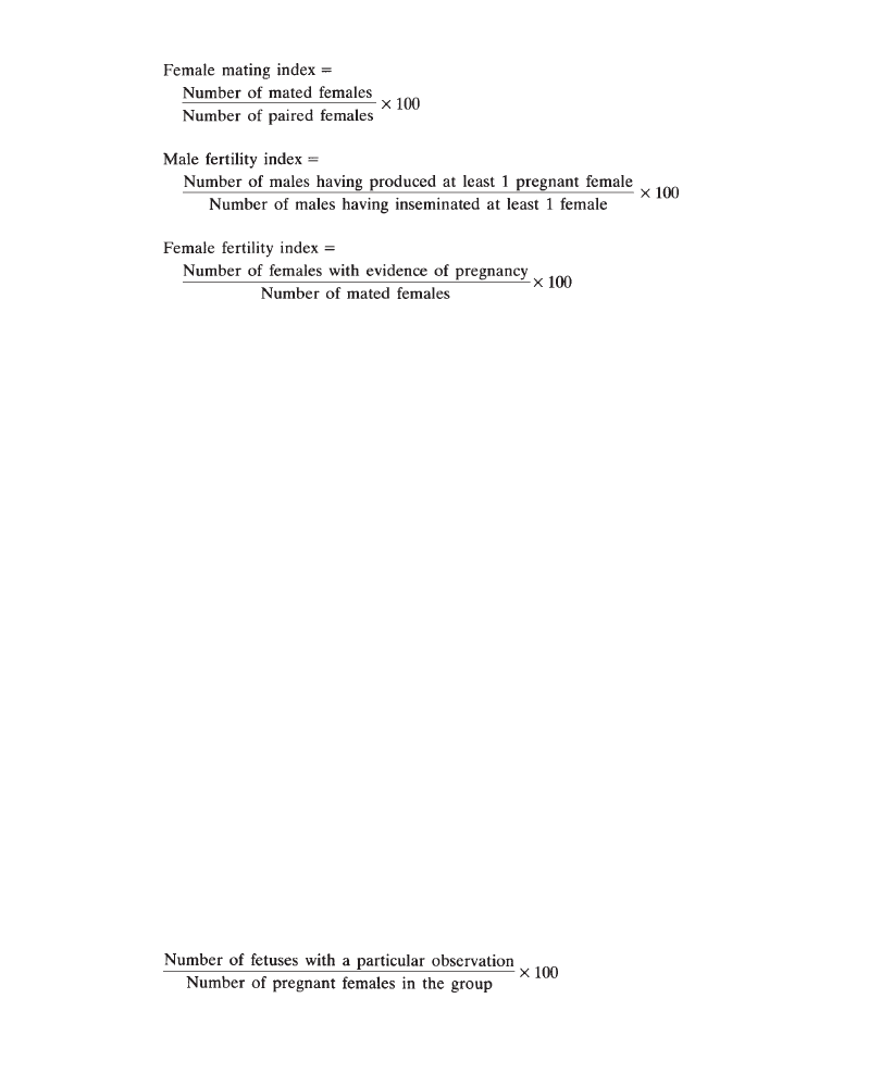
Example of a reproductive toxicology report
129
A1.7.1 Caesarean data
Group mean values with standard deviations were calculated for each litter and
each group.
Group mean values for the size of litters, uterine deaths, early and late resorptions
were calculated using either of the following means:
•
Mean 1: All pregnant females surviving to term bearing evidence of implantation
(including total litter resorptions).
•
Mean 2: All pregnant females surviving to term bearing viable fetuses.
Mean 2 has more significance when group size is small as the data would be
influenced by the presence of a single total litter resorption. Mean 1 is more accurate
when several females exhibit total litter resorption. For mean litter and fetal weights
and the number of fetal observations, only Mean 2 was calculated.
Pre-implantation loss (Number of corpora lutea—Number of implantations) was
calculated for each female. When the number of implantations exceeded the number
of observed corpora lutea, pre-implantation loss was taken as 0. Pre-implantation
loss includes losses due to non-fertilization of ova and early post-implantation deaths
(i.e. those occurring up to days 8–9 of gestation).
Post-implantation loss (Number of implantations—Number of viable fetuses)
was calculated for each female. Post-implantation loss covers only the period between
days 8–9 and 20 of gestation and does not include the first 2–3 days post-
implantation, as any death occurring in this phase would leave no visible remains
on day 20.
Group mean implantation loss values were calculated as means of individual
values.
Group fetal observation values (external, internal and skeletal) were expressed
as a percentage and calculated from the formula:
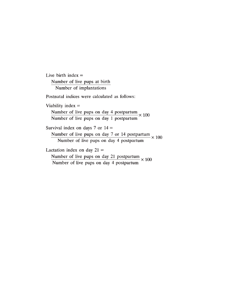
Presenting toxicology results
130
A1.7.2 Postnatal data
Post-implantation loss (Number of implantations—Total number of dead or live
pups at birth) was calculated for each female. Post-implantation loss covers only
the period between days 8–9 and the end of gestation and does not include the
first 2–3 days post-implantation, as any death occurring in this phase would leave
no remains visible on day 21 postpartum.
Group mean implantation loss was calculated as a mean of individual values.
Live birth index was calculated as follows:
The postnatal indices were calculated for each litter and then per group for these
values. The calculation of the number of living pups on day 4 postpartum was
established after culling, except for the viability index.
A1.8
Statistical Methods
Statistical analysis (Gad and Weil, 1989) was performed on the individual values
for dam parameters and by sex on the mean values for the litter and pup data.
Differences between the treated and control groups were tested using a decision
tree to select an appropriate hypothesis-testing procedure.
Parametric data, i.e. food consumption, body weight, body weight change, absolute
and relative organ weight, gestation length, litter size, fetal weight, number of
implantation sites, pre- and post-implantation loss, physical and functional
development and behavioral tests were analyzed in the following way. A Bartlett’s
test was carried out in order to verify the homogeneity of variance, and then:
•
when the variances were found to be homogeneous, an overall Analysis of
Variance was carried out by means of the F test followed, when significant,
by multiple and pairwise comparison of the group means using the Dunnett’s
test;

Example of a reproductive toxicology report
131
•
when the variances were found not to be homogeneous, the overall comparison
of the groups was carried out using the Kruskal-Wallis test followed, when
significant, by multiple and pairwise comparison of the group means using
the Wilcoxon Rank Sum test.
The final toxicologic interpretation of data considered other factors such as dose—
response relationships, biological plausibility and consistency.
Categorical data, i.e. mating and fertility indices, pregnancy rate, parturition
and live birth indices, percentages of stillborn or malformed fetuses, mortality of
pups, postnatal indices, pupillary reflex test and the water maze test were analyzed
by means of the Fisher’s Exact test.
A1.9
Archives
All raw data, specimens and other study documents pertaining to this study will
be stored in the respective research center archives.


133
12
Using References
The following are our recommendations for using references in toxicology reports.
12.1
Journals
12.1.1 References in the Text
Mention the authors and the year of publication: “(Dawson and Dupont, 1972)”.
Cite first author+et al.” for three or more authors: “(Dawson et al., 1985)”. If
there are 2 or more references, list them by alphabetic order of the authors and
then by increasing year, e.g. “(Dawson et al., 1926; Dawson et al., 1929; Lindsay,
1971)”.
12.1.2 References in the References Section
List the authors and year of publication, title, journal, volume number, first-page-
last-page, by alphabetic order of the authors and then by increasing year.
Example: Dawson A.B., Dupont C.D. and Durand E.F. (1985) Note on the staining
of the skeleton of cleared specimens with Alizarin red Stain. Tech., 1; 123–124.
For two or more references of the same author and same year, list them as follows:
“Dupont (1994a)” and “Dupont (1994b)”.
12.2
Books
12.2.1
References in the Text
State the author and the year of publication: “(Keenan, 1958)”. For two authors,
refer to both: “(Hannah and Blow, 1987)”. Cite the principal author+“et al.” for
three or more authors “(Blackwell et al., 1952)”.

Presenting toxicology results
134
12.2.2 References in the References Section
List the author and year of publication, title of the part of the book, “In:” Editor’s
surname, “ed.”,. title of the book, edition number (2nd or above), place of publication,
name of publisher, volume number, chapter number or page numbers if specific
pages are cited.
Example: Kavet J. (1976) Trends in the utilization of influenza vaccine: an
examination of the implementation of public policy in the United States. In: Selby
P, ed. Influenza: Virus, Vaccines, and Strategy, 2nd ed, Orlando, Fla.: Academic
Press Inc.; 297–308.
12.3 Unpublished/In-house Reports
12.3.1 References in the Text
Give the report number and the year of signature: “(Report PP 93–221, 1993)”.
12.3.2 References in the References Section
Cite the report number, the year of signature, the title and the name and location
of the testing facility.
Reports PP XX-XXX are listed by increasing report year and/or report
number.
Example: Report PP 93–016 (1993) PP 27567: Two-week oral toxicity study in
rats. Drug Safety Laboratories, Poisonous Prose Inc., Littlebrook, Surrey, UK.

135
13
References and Recommended
Reading
ALLEY, M. (1987) The Craft of Scientific Writing (Englewood Cliffs, N.J.: Prentice-Hall
International).
AMERICAN NATIONAL STANDARDS INSTITUTE, INC. (1979a) American national
standards for the preparation of scientific papers for written or oral presentation. ANSI
Z39.16–1979 (New York: American National Standards Institute, Inc.). (1979b) American
national standards for writing abstracts. ANSI Z39.14–1979 (New York: American National
Standards Institute, Inc.).
ANONYMOUS (1982) The Chicago Manual of Style, 13th edition (Chicago: The University
of Chicago Press). (1990) Mosby’s Dictionary, Medical, Nursing and Allied Health, 3rd
edition (St. Louis, Mo.: Mosby).
BLACK, H.E. (1994) Design and writing of the preclinical safety report, Toxicologic Pathology
22 (2), 202–5.
COMMITTEE OF GRADUATE TRAINING IN SCIENTIFIC WRITING (1989) Scientific
Writing for Graduate Students (Bethesda, Md.: Council of Biology Editors, Inc.).
COUNCIL OF BIOLOGY EDITORS STYLE MANUAL COMMITTEE (1983) CBE Style
Manual. A Guide for Authors, Editors and Publishers in the Biological Sciences, 5th
edition (Chicago, Ill.: Council of Biology Editors, Inc.).
DAY, R.A. (1991) How to Write and Publish a Scientific Paper, 3rd edition (Cambridge:
Cambridge University Press).
DEVLIN, J. (1987) A Dictionary of Synonyms and Antonyms (New York: Warner Books).
FARR, A.D. (1985) Science Writing for Beginners (Oxford: Blackwell Scientific Publications).
FOWLER, H.W. (1968) A Dictionary of Modern English Usage, 2nd edition (rev. by Sir
Ernest Gowers) (Oxford: Oxford University Press).
GOPEN, G.D. and SWAN, J.A. (1987) The science of scientific writing, American Scientist,
78, 550–8.
GOWERS, E. (1986) The Complete Plain Words (revised edition by S.Greenbaum and J.
Whitcut), 3rd edition (London: HMSO).
GREGORY, M.W. (1992) The infectiousness of pompous prose, Nature 360, 11–12.
HODGSON, E., MAILMAN, R.B. and CHAMBERS, J.E. (1988). Macmillan Dictionary
of Toxicology (London: Macmillan).

Presenting toxicology results
136
MATTHEWS, B.R., LEE, R.M., EISEN, S.M. and DEWS, I.M. (1994) How to Write an
Expert Report, B.R.Matthews (ed.) (MCRC Group, Romford, Essex RM7 7DA, UK:
Rostrum Publications).
SCIENTIFIC ILLUSTRATION COMMITTEE, COUNCIL OF BIOLOGY EDITORS (1988)
Illustrat-ing Science: Standards for Publication (Bethesda, Md.: Council of Biology
Editors, Inc.).
SHERMAN, T.A. and JOHNSON, S.S. (1992) Modern Technical Writing, 5th edition
(Englewood Cliffs, N.J.: Prentice Hall).
ZBINDEN, G. (1987). Predictive Values of Animal Studies in Toxicology Testing, CMR
Annual Lecture 1987 (Carshalton, Surrey, UK: Centre for Medicines Research).

137
abortion 54–5, 90
absorption 41, 42
abstract see summary
administration route 28
effects related to 37, 73
alterations 90, 92
anatomic pathology 75–88
blood pressure 59–60
body weight 55–6, 76
brevity 2
carcinogenicity study
conclusion 40
introduction 30
materials and methods section 33
reporting results 85–8
title 28
cardiovascular parameters 58–60
clarity 2
class effect 23
clinical pathology data
evaluation 63–5
in perspective 72–4
reporting 65–72
clinical signs 51–3, 68
glossary 52–3
cohabitation 89–90
compound designation 28
compound-related effects 23, 24, 34,
37, 66–8, 72–4
in histopathology 82–3, 84
at necropsy 79, 80
on organ weight 76, 77
concentrations 42
conceptus 90, 92
conclusion 37–40
control groups 64, 66
creatine phosphokinase (CPK) 73
developmental toxicity 90, 92–3
relationship with maternal
toxicity 97–8
diastolic blood pressure 59–60
direct developmental toxin 97
discussion 4–5, 36–7
dose levels, nominal/actual 31, 32
dose-related changes 68–9
in organ weight 76
electrocardiography (ECG) 58–9
embryofetal toxicity study 92, 93
embryogenesis 90
embryotoxicity 92
English
basic biomedical 1–15
myths concerning 2
evaluation 4–5, 36–7
expert review 36
fertilization 90
fetal development 90
Index

Presenting toxicology results
138
food consumption 56–7
general toxicology reports
example 99–112
results 34
title 28
generations, fertility studies 93
gestation, dating 90
grading
clinical signs 51
results 35, 67
graphs 41
histopathology 81–5
historical control data 64–5
hyperplasia 82, 85–8
implantation 90
IMRAD report structure 17–18
modified 18–19
in-life observations 35, 49–61
incidence of effects 69–70
variation over time 70–1
incoherence 2, 9–10, 12–13
independent review 2–3
indirect developmental toxin 97
introduction 3, 29–30
KISS 37
lactic dehydrogenase 73
language 1–15
LDH 73
litter data 97
malformations 90, 92
materials and methods section 3–4,
31–3
anatomic pathology 75
maternal toxicity, relationship with
developmental toxicity 97–8
mating 89–90
maximal tolerated dose 24, 38
misused words and expressions 11–12,
14–15
mortality 35, 49–51, 80
MTD 24, 38
multiple phases, fertility studies 93
necropsy 49, 79–81
NEL 24, 37
neoplasia 82, 85–8
no-effect level 24, 37
no observable adverse effect level
24, 37
no-observable effect level 24, 37, 98
NOAEL 24, 37
NOEL 24, 37, 98
ophthalmology 60–1
organ weights 76–9
organogenesis 90
p-values 35
percentages 34, 67–8
perinatal toxicity study, materials and
methods section 31–2
postnatal toxicity study 92–3
materials and methods section
31–2
prenatal toxicity study 92–3
pretest data 64–5
procedure-related effects 73
protocol 31
pup 90
qualitative data descriptions 34
RDRD report structure 19
reduction 34–5
references 72, 133–4
repeated-dose toxicology study report
mortality report 51
results 34
summary 23, 25
title 28
report
principles of writing 27–40
structure 3–5, 17–19
reproductive cycle phases 93, 94
reproductive toxicity study
definitions 93–4
embryofetal development
(Segment II) 54–5, 94
fertility and early embryonic
development (Segment I) 93–4
generations 93

Index
139
mortality report 51
multiple phases 93
pre- and postnatal development
(Segment III) 94
reports
conclusion 38, 39, 98
discussion 98
example 113–31
methods 95–6
outline 94–5
results 34, 96–8
summary 23, 25–6, 98
title 28
terminology and definitions 89–94
resorption 90
results 4, 33–6
anatomic pathology 75
tabular presentation 35
reversible effects 71–2
selective developmental toxin 97
severity, progressive increase in 70–1
sex 34, 70, 76
single-dose toxicity study
introduction 30
mortality report 50
summary 23, 24–5
title 28
spelling mistakes 7–9
statistical analysis of clinical
pathology data 63–4
study protocol 31
study types 28–9
summary 5, 21
conclusion of 24
discussion section 23–4
examples 24–6
introduction 21
materials and methods section
21–2
results section 22–3
synopsis 36
systolic blood pressure 59–60
tables 35, 67
tenses 5–7
teratology study 92
test species 29
thalidomide 92
titles of reports 27–9
toxicokinetic study
conclusion 37
data presentation 41–7
results 33
transient changes 71–2
typographic errors 7–9
units 42
variations 90, 92
vehicle-related effects 24, 37, 73
verbosity 9–10, 12–13
Wyszukiwarka
Podobne podstrony:
Bayesian Methods A General Introduction [jnl article] E Jaynes (1996) WW
Wavelet Transforms that Map Integers to Integers [jnl article] (1996) WW
Building GUIs with Matlab V 5 (1996) WW
Elementary Number Theory (Math 780 instructors notes) (1996) WW
Analysis J Hyland (1996) WW
Group Theory [jnl article] J Milne (1996) WW
Introduction to Linear Logic T Brauner (1996) WW
Connectionism Past, Present, and Future [jnl article] J Pollack WW
Presentationenzym
Product presentation XC100FC
PRESENTATION
Presentation1(1)
AC31 Presentation
Product presentation easyControl
Molecular Toxicology 8
Presentation1 2
159 Present Perfect
06 1996 55 58
więcej podobnych podstron