netter55

Slructure of the Heart
CARDIOYASCULAR PHYSIOLOGY
Mitral valve
Poslerior cusp-
Ascending aorta-v
L. cusp—v Posterior \ (noncoronary) cusp v Superior N.
Aortic
valve
Mem-
branous
septum
[ Anterior (aortic) cusp-R. pulmonary
Anterior (aortic) cusp of mitral valve-1
. Pulmonary trunk
L pulmonary
•Ascending aorta ■ Orifices of coronary, • R. auriclo
■ L. cusp •R.cusp
Aortic
• Outflow
Atrio \ ventricular
pan-N
Inter
ventricular part^—
■ Conus arteriosus
■ R. anterior papillary
Moderator 1 Jr Septal band J N L. ventride
Muscular
interventricular
R. atrium
Anterior
cusp
(retracted) Medial (septal) y
cusp —'
Posterior cusp-'
R. vcntridc-'
R. anterior papillary musde (cut)
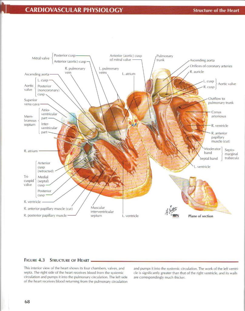
Figurę 4.3 Structure of Heart
This interior view of the heart shows its four chambers, valve$, and septa. The right side of the heart receives blood firom the systemie circulation and pumps it into the pulmonary circulation. The left side of the heart receives blood returning from the pulmonary circulation
and pumps it into the systemie circulation. The work of the left ventri-de is significantly greater than that of the right yentride, and its walls are correspondingly much thicker.
68
Wyszukiwarka
Podobne podstrony:
12434 netter57 Eleclrital Activity of the HeartCARDIOYASCULAR PHYSIOLOCY Aclion Potential of SA Node
77961 netter56 Conduction System of the HeartCARDIOYASCULAR PHYSIOLOGY Superior vena A. Right Side M
26834 netter121 Anatomy of the Kidney: The Nephron RENAL PHYSIOLOGY Cortical nephrons dilute the uri
netter161 Overview of Hormone ActionENDOCRINE PHYSIOLOGY Steroid Hnrmones Thyroid Hormones Vita
netter70 Monitoring of Blond PressureCARDIOVASCULAR PHYSIOLOGY Low-Pressure Baroreceptors ANP releas
netter70 Monitoring of Blond PressureCARDIOVASCULAR PHYSIOLOGY Low-Pressure Baroreceptors ANP releas
12897 netter104 Anatomy of Ihc Kidf»RENAL PHYSIOLOGY A. Antcrior surfacc of right kidney Superior ox
więcej podobnych podstron