netter59
Electrocardiogram: II
CARDIOVASCULAR PHYSIOLOGY
D. Late ventricular depolarization
As depolarization progresses over ventrides, vector shifts to hecome directed supenorly as well as to left, thus extending upward R wave in lead I and causing ncgatiye (downward) deflection (S wave) in lead aVF
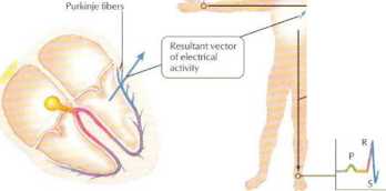
Rir< ording axis of lead I thorizontal, right to left)
- Recording axis of lead aVF (vertical, downward)

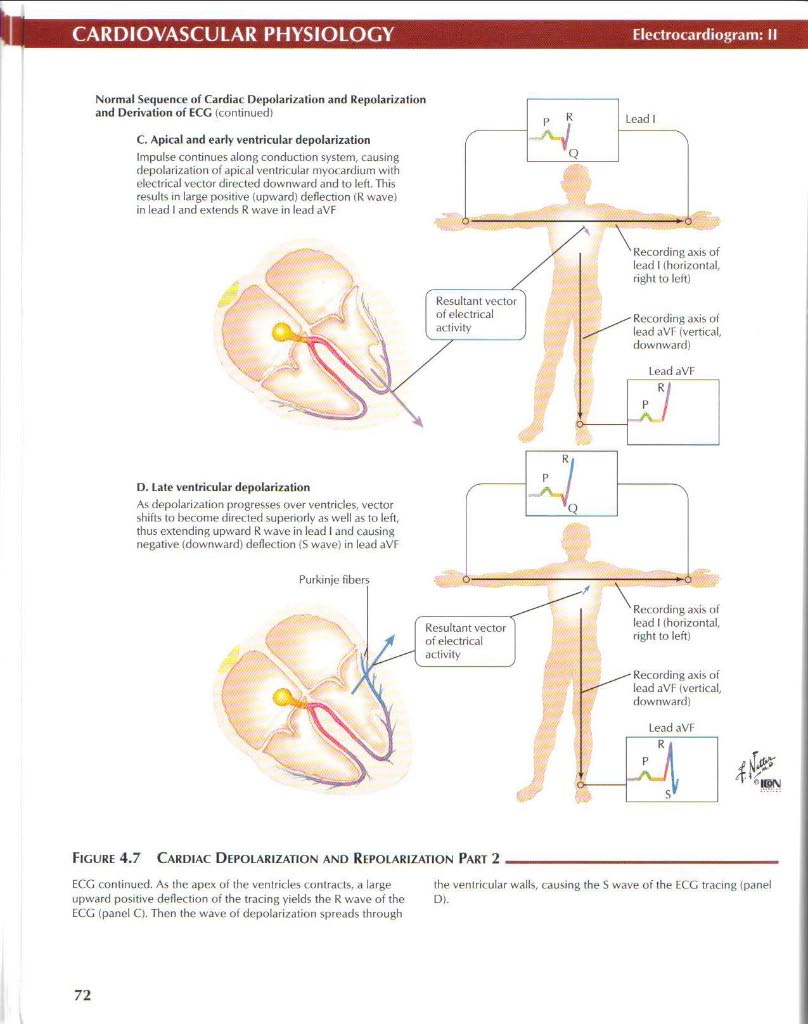
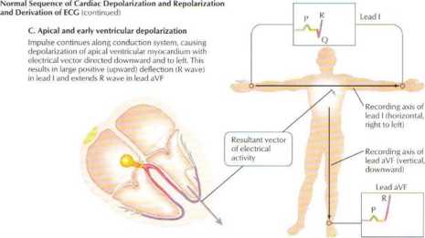
Figurę 4.7 Cardiac Depolarization and Repolarization Part 2
ECG continued. As the apex of the ventricles contracts, a large the venlricular walls, causing the S wave of the ECG tracing (panel
upward pos«tive deflection of the tracing yiełds the R wave of the D».
ECG (panel Q. Thcn the wavc of depolarization spreads through
72
Wyszukiwarka
Podobne podstrony:
netter60 Electrocardiogram: IIICARDIOVASCULAR PHYSIOLOGY Norm.il Sequence of Cardiac Depolarization
netter58 Bectrocardiogram: ICARDIOYASCULAR PHYSIOLOGY Normal $cqucncc ot Cardiac Depolarization and
netter144 Colonie MotilityGASTROINTESTINAL PHYSIOLOGY Adaptiw lelłOtation (intralumin.il pressure in
netter106 Glomerular FiltratinnRENAL PHYSIOLOGY Colloid osmotic pressure of plasma (Uln) U
netter11 NEUROPHYSIOLOGYCerebrospinal Fluid (CSF): Brain Ventricles and CSF Composition Frontal (ant
netter113 Urine DilutiorRENAL PHYSIOLOGY h2o Na Cl" h2o 375 —*t- Urea Notę: Figurcs given are e
netter126 Autonomie lnnervationGASTROINTESTINAL PHYSIOLOGY Figurę 7.5 Aijtonomic Innervation The inn
netter127 Autonomie lnnervationGASTROINTESTINAL PHYSIOLOGY m generał sympathetics decrease peristals
netter134 Castric DigestionGASTROINTESTINAL PHYSIOLOGY Figurę 7.12 Gastric Digestiye Function The st
netter61 Cardiac CycleCARDIOVASCULAR PHYSIOLOGY Figurę 4.9 Cardiac Cycle The cardiac cycle represent
więcej podobnych podstron