55 (174)
5: Deep mycoses


Figurę 5:9: Perinasal ulcer in a domestic short hair cal with cryptococcosis (courtesy of B. Hubert)
Figurę 5:10 :Nasal ulcer in a domestic short hair cat with cryptococcosis (courtesy ofB. Hubert)
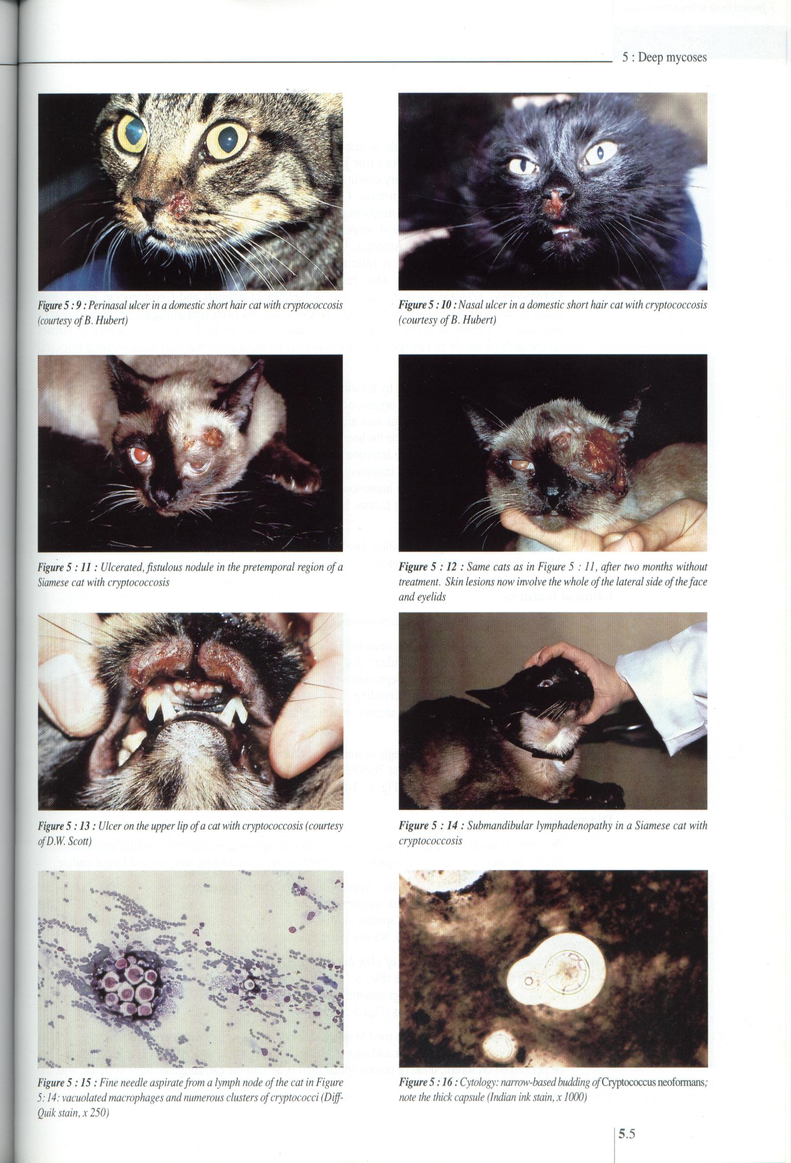
Figurę 5:11: Ulcerated,fistulous nodule in the pretemporal region ofa Siamese cat with cryptococcosis
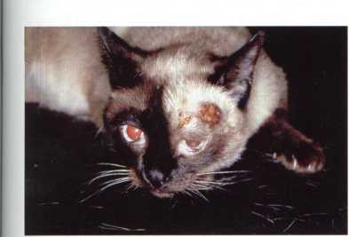
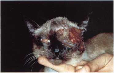
Figurę 5 :12 : Same cats as in Figurę 5 :11, after two months without treatment. Skin lesions now involve the whole ofthe lateral side of the face and eyelids
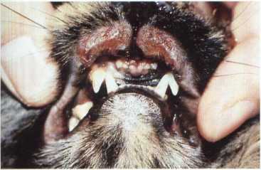

Figurę 5:13: Ulcer on the upper lip ofa cat with cryptococcosis (courtesy ofD.W. Scott)
Figurę 5 :14 : Submandibular lymphadenopathy in a Siamese cat with cryptococcosis
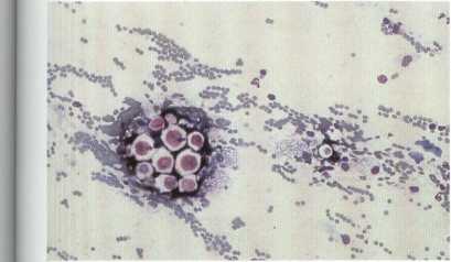
Figurę 5:15: Fine needle aspirate from a lymph node ofthe cat in Figurę 5:14: vacuolated macrophages and numerous clusters ofcryptococci (Diff-Quik stain, x 250)
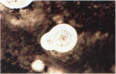
Figurę 5:16: Cytology: narrow-based budding o/Cryptococcus neoformans; notę the thick capsule (Indian ink stain, x 1000)
5.5
Wyszukiwarka
Podobne podstrony:
53 (169) 5 : Deep mycoses Figurę 5 :2 : Nasal nodule in a cat with phaeohyphomycosis Figurę 5:1: Ulc
Figurę 4: Implied Changes In Domestic Airline Demand Over Time years. Yet, the industry madę money i
27 (384) 2: Diagnostic approach Figurę 2 :11: Multiple dermal nodules in a cal with cryptococcosis F
29 (352) 2: Diagnostic approach Figurę 2:17: Sclerosis in a cat with morphea (courtesy of E. Bensign
68 (121) 6: Bacterial dermatoses Figurę6:9: Ulceratednodular lesion ina cat with leprosy (courtesy o
In my speech I will deal with the phenomenon of the development of the dance fonn of a solo performa
3 (1497) I 5 Deep mycoses Lluis Ferrer - Alessandra Fondati Deep mycoses are rare in the cat and may
fig13 Figurę 13 Silver Valkyrie figurę from Tuna in Sweden
fig15 Figurę 15 Silver Valkyrie figurę from Grodinge in Sweden
rulespage10 STRATEGYI Figurę 5. The player in this example will need to reorganize his Power Structu
rulespage10 STRATEGYI Figurę 5. The player in this example will need to reorganize his Power Structu
IMG@55 Osobiste zaufanie do polityków Persona! trust in potfócians 11,1
rulespage10 STRATEGYI Figurę 5. The player in this example will need to reorganize his Power Structu
Figurę 7.1. Economic growth In per cent 10 12---- - 12 ““ OECD members acceeded before 1990 “ “
Figurę 6: Domestic Market Share of Low-Cost Carriers from this figurę. First, LCCs in aggregate have
więcej podobnych podstron