
23
Nanoparticle Molecular Labels
James F. Hainfeld, Richard D. Powell, and Gerhard W. Hacker
23.1
Introduction
Nanotechnology means many things to many people, from Fantastic Voyage miniature
submarines traveling through a person’s arteries, to a ribosome that is a cellular “factory”
which synthesizes proteins, while material scientists believe that it means making better
materials from molecular building blocks.
The common theme seems to be “Nano” – that is, small – in the nanometer size range.
In a sense, nanotechnology has been around a long time, as both chemistry and biochem-
istry rely on molecules and complexes in this size range or smaller. For example, nylon
was first made in 1935 as a silk substitute, and was patterned after silk by making an
amide-bonded polymer, similar to silk, but with a slightly different repeating unit [1]. Na-
noparticles have also been around for a long time; presumably, the first nanoparticle was
recognized in 1570 with aurum potable (potable gold) and luna potable (potable silver)
which alchemists used as elixirs. Unfortunately, they did not make the consumer live for-
ever, as evidenced by the high incidence (100 %) of dead alchemists. As early as 1595, gold
colloids were incorporated into glass to make various colors, such as red, purple, violet,
brown, or black. Faraday was the first to recognize that the colors were related to particle
sizes [2].
This chapter deals with the biological application of metal nanoparticles to label
biomolecules. A number of uses benefit from this combination : the metal nanoparticles
make detection possible, easier or more sensitive. Colloidal gold particles, when adsorbed
to antibodies [3] or to other targeting agents such as proteins [4] or peptides [5–7], are
widely used as labels for the detection or microscopic localization of molecular and macro-
molecular targets. One popular pregnancy test kit develops a pink line which is in fact a
40-nm gold particle adsorbed to an antibody, anti-human chorionic gonadotropin (hCG).
This “pregnancy hormone” keeps the corpus luteum producing progesterone after con-
ception occurs. The urine sample flows over a capture stripe where anti-hCG has been
previously adsorbed to nitrocellulose; if hCG is present, it is bound, or “captured”.
The gold with anti-hCG then flows over, and if hCG is bound, the gold-antibody binds,
leaving a pink color on the stripe. The very high extinction coefficient of the 40-nm
353
Nanobiotechnology. Edited by Christof Niemeyer, Chad Mirkin
Copyright
c 2004 WILEY-VCH Verlag GmbH & Co. K aA, Weinheim
ISBN 3-527-30658-7
G

gold,
2
10
9
M
–1
cm
–1
compared with that of a fluorescent molecule (fluorescein :
7
10
4
), increases detection sensitivity by a factor of about 30 000. The gold nanopar-
ticle gives sufficient sensitivity to be perceived with the unaided eye, thereby producing an
inexpensive assay [8].
Nanobiotechnology implies both some degree of supramolecular organization or coop-
eration, and the incorporation of a desirable function, ability or property into a supramo-
lecular construct. The entry of metal particle bioconjugates as players in nanobiotechnol-
ogy has been facilitated by the development of molecular control over the site, nature and
formation of the link between the biological molecule and metal particle. This enables
both the conjugation of metal particles to molecules with potential applications in nano-
technology, and the selective attachment of metal nanoparticles to specific sites within bio-
logical structures where the properties of the metal particles impart potentially useful
functionality to the construct. This chapter will focus on the covalently linkable metal clus-
ter labels, principally Nanogold [9, 10] and undecagold [11–13], which have been used for
a number of such applications. Other types of particles, including unstabilized gold [14–
16], clusters of other metals, and larger gold particles with controlled chemical reactivity
[17], have also been utilized. In this chapter, both the use of metal particle labels to detect
and localize biological targets will be discussed, together with the preparation and poten-
tial applications of metal cluster bioconjugates with novel properties and functionality.
23.2
Immunogold-Silver Staining: A History
For more than 30 years, nanometer-sized gold particles (mostly colloidal gold, with dia-
meters ranging from 1 to 40 nm) have been the label of choice to demonstrate proteins
and peptides (and other substances against which specific antibodies can be made) by
transmission electron microscopy (TEM) [3]. Not only for TEM, but also for light micro-
scopy (LM) and for scanning electron microscopy (SEM), immunogold-staining (IGS)
methods show various advantages over other, nonparticulate immunostaining techniques,
including enhanced visibility and increased detection efficiency and sensitivity. Accumu-
lations of colloidal gold can be made visible with LM by the application of autometallogra-
phy (AMG) [18–28], a group of techniques whereby silver or gold ions are reduced in situ
to metal atoms and precipitated onto the surface of the gold particle nuclei. In the electron
microscope, it can be shown that gold particles thereby grow considerably in size, until
they conglomerate [29–31]. Combination of this reaction with colloidal gold-labeled en-
zyme histochemistry [21] and immunohistochemistry (IHC) [32, 33], both in 1983, led
to the introduction of immunogold-silver staining (IGSS), and this was a major break-
through in sensitivity and detection efficiency in the early 1980s. Initial attempts to
apply Holgate’s original IGSS method with a broader spectrum of antibodies used in gen-
eral immunohistopathology often resulted in very high levels of unwanted background
staining, and often the overall appearance of the stained section was “dirty”. Successful
attempts to modify IGSS to facilitate the highly sensitive demonstration of various
kinds of substances in routine paraffin sections were published by a number of research
teams. With the years, the methodology became increasingly sophisticated, and a variety
of protocols were used [34–46]. Modifications included the use of gold particle sizes smal-
354
ler than the ones originally used (1–5 nm in diameter), the use of fish gelatin, a number
of new AMG developers, the use of gold-labeled protein-A [47–50], utilization of the strep-
tavidin–biotin complex (S-ABC) principle [51, 52], and the introduction of antigen retrieval
techniques. For review and update see Ref. [53].
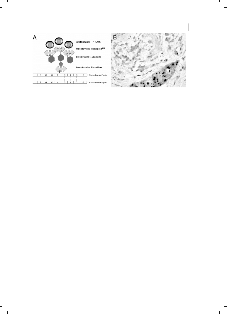
The introduction of covalently linked gold cluster labels rather than colloidal gold
marked another major advance in the methodology, giving researchers the ability to direct
the attachment of metal nanoparticles to biological molecules with submolecular preci-
sion. The undecagold [12] and Nanogold [9, 10] labels have been conjugated to many dif-
ferent molecules that cannot be labeled with colloidal gold, including proteins, peptides,
oligonucleotides, lipids, and small molecules, many of which have potential nanotechnol-
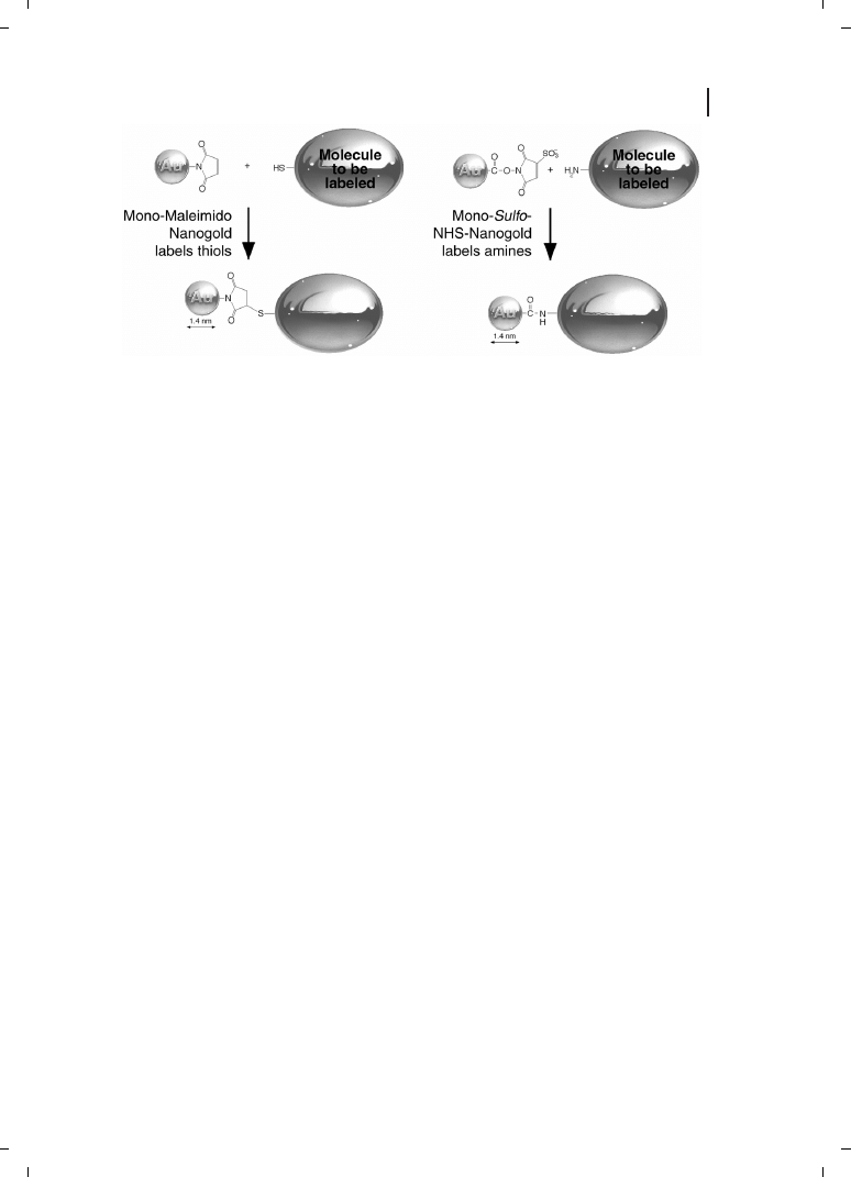
ogy applications, which are discussed in more detail below. Conjugation reactions are
shown in Figure 23.1. Antibody and protein conjugates give improved performance
over colloidal gold probes, including increased cellular penetration, labeling density,
and access to hindered antigens [54, 55].
For silver or gold amplification, numerous protocols had been described and are being
marketed by a number of companies. Danscher’s original protocols relied on the use of
silver lactate [18–20], and had to be applied in a darkened room or under red light to
give background-free preparations. Hacker et al. described a less light-sensitive modifica-
tion using silver acetate as the silver ion source [27], thereby introducing the possibility to
amplify gold-labeled LM preparations in normal laboratory daylight. It became possible
now to optimize staining under microscopic control. Comparisons of different silver
salts were reported and showed marked differences [46]. Most recently, AMG based on
gold ions has been introduced commercially (GoldEnhance, Nanoprobes, Inc.), a new tech-
nique that further improved the spatial staining resolution and signal-to-noise ratio [56–
59].
355
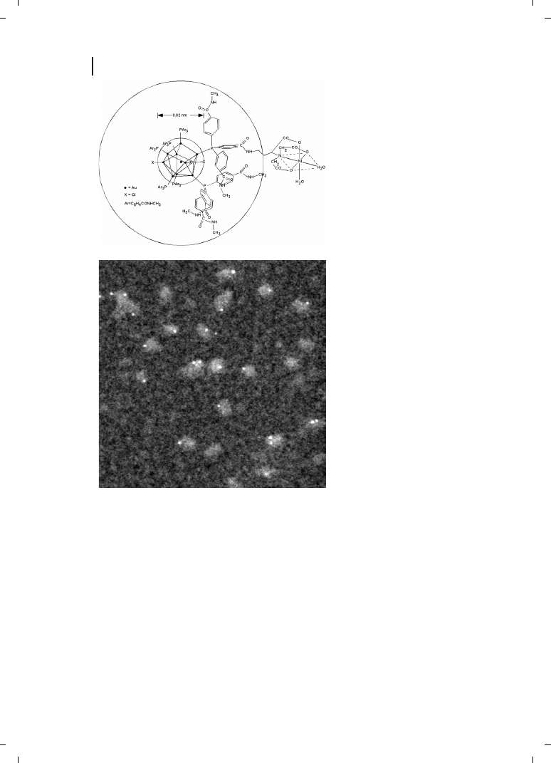
Figure 23.1
23.3
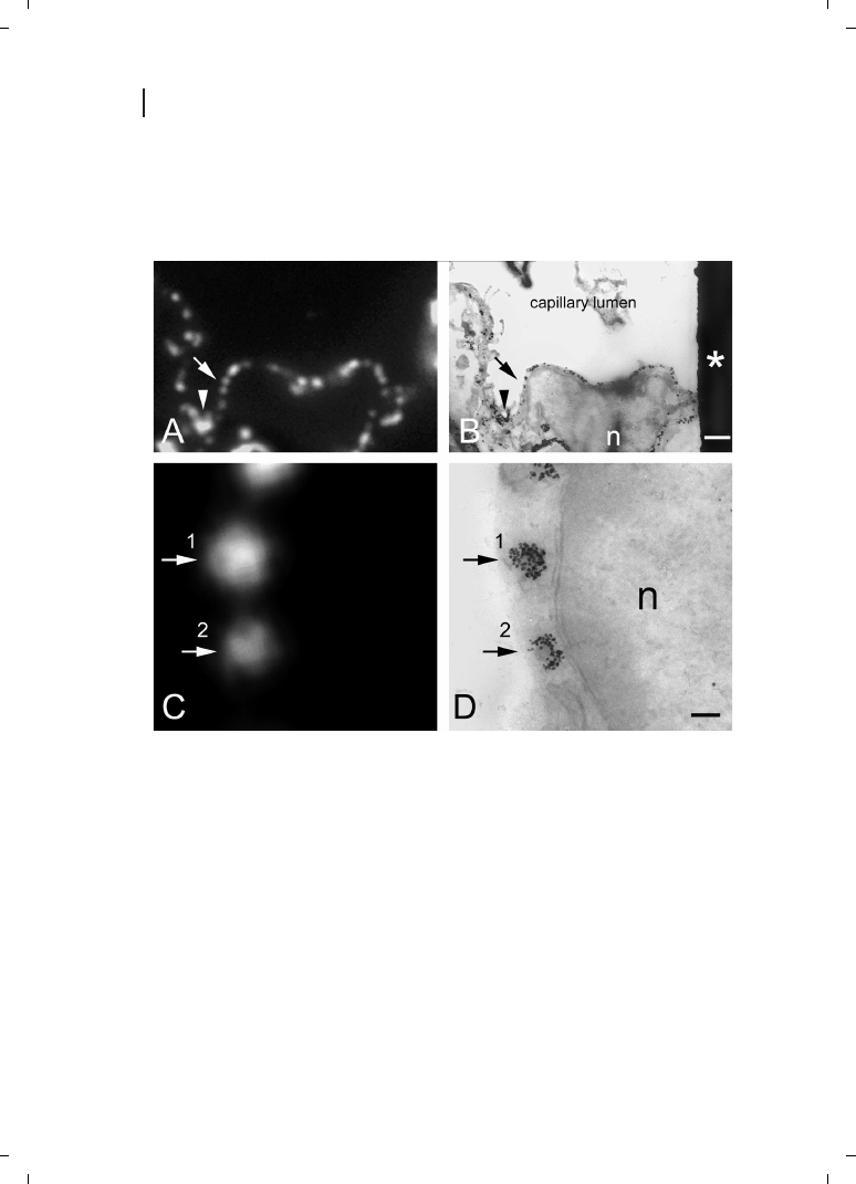
Combined Fluorescent and Gold Probes
Antibodies, or even antibody fragments, are sufficiently large that both gold clusters and
fluorescent labels may be attached, spaced sufficiently far apart that quenching by Förster
energy transfer [60] is minimal, fluorescence is largely maintained, and the probes are ef-
356
Figure 23.2

fective for both immunofluorescence and immunogold labeling [61]. Initially, secondary
Fab antibody probes labeled with both Nanogold and fluorescein were prepared by the
sequential conjugation of Monomaleimido Nanogold and fluorescein N-hydroxysuccini-
mide ester. These were used to label the SC35 pre-mRNA splicing factor in HeLa cells
[62, 63]. The same probe was also used to label human lymphocytes, and labeling was vi-
sualized using fluorescence microscopy, different modalities of LM, and TEM [64]. Proof
of principle was subsequently demonstrated using correlative light and electron micro-
scopy of specimens with features localized on indexed grids [65]. Combined Cy3 and Na-
nogold probes have also been developed, and demonstrate high specificity and sensitivity
both for immunocytochemistry and for in-situ hybridization [66, 67]; using Tyramide Sig-
nal Amplification (TSA) followed by combined Cy3/Nanogold-labeled streptavidin, the
fluorescence signal was sufficiently bright that staining was observed for HPV 16/18 in
SiHa cells, known to contain only one or two copies of the target. More recently, combined
Alexa Fluor 488 and 594 and Nanogold probes have been prepared and found to give
higher brightness and improved pH compatibility for fluorescent staining [68]. An exam-
ple of correlative fluorescence and electron microscopic localization of caveolin is shown
in Figure 23.2 [69]. It has also been reported that fluorescence is maintained even after a
brief period of silver enhancement, enabling observation by epifluorescence microscopy
[65]; preliminary results have also suggested that fluorescence may be sufficiently pre-
served to allow the preparation of dual-function antibody probes in which fluorescent la-
bels are combined with larger platinum clusters [70].
23.4
Methodology
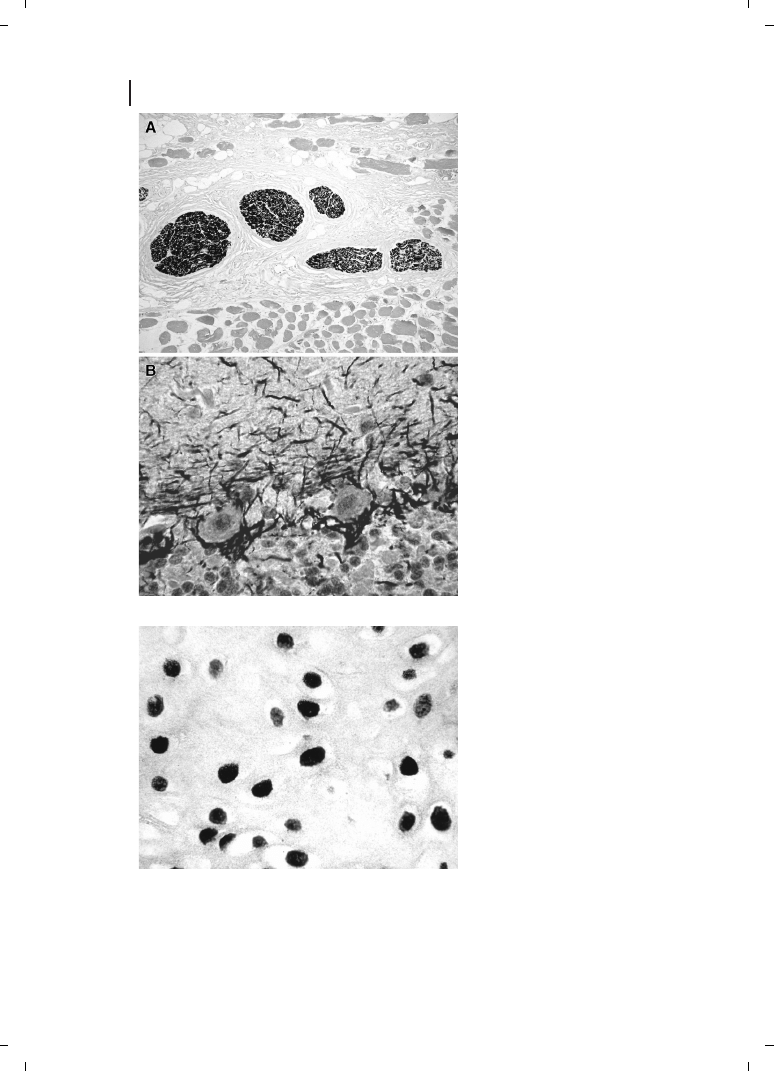
Choice of Gold and AMG Type
Originally, colloidal gold particles were mainly applied adsorbed to second layer antibodies
and used in an indirect, two-step IHC method [32–35]. Later on, it was found that the use
of covalently bound Nanogold–antibody and streptavidin conjugates, instead of colloidal
gold electrochemically adsorbed to antibodies, gave a further boost in signal-to-noise
ratio [9, 10]. A three-step S-ABC Nanogold technique was born that could be successfully
applied in IHC, and also for in-situ hybridization (ISH) [38, 57]. Conglomerations of clus-
tered gold particles amplified by silver- or gold-salt-based AMG now appeared as jet-black
precipitates with a distinctly sharper appearance than the reaction products of most en-
zyme-labeled preparations. Also, when compared to the “classical” indirect and silver-en-
hanced colloidal gold techniques, the staining results were often much clearer than those
achieved before (Figure 23.3). The new GoldEnhance technology (Nanoprobes, Inc.),
based on the catalytic deposition of gold rather than silver [58, 59], allows for a more
“metallic” appearance seen with LM – that is, the edges of staining appear clear-cut
and the staining itself is completely black, provided that the amount of the substance
to be detected is high enough and that the antigen–antibody reaction is adequate. A simi-
lar result is also most often obtained in ISH (Figure 23.4).
357
358
Figure 23.3
Figure 23.4

Iodinization
One important fact also should be re-addressed here : it had previously been reported by
the original authors of IGSS [32, 33] that pre-treating the sections with Lugol’s iodine is
essential for obtaining a high detection efficiency and sensitivity. Since then, most authors
have confirmed this finding, although the exact process yet still seems not clearly under-
stood. It had been suggested that the weak oxidizing activity of the halogen is responsible
for the fact that many or most antigens can only be demonstrated if this step is used. Al-
though some authors also suggested protocols without the Lugol’s iodine steps, we highly
recommend using it, for IHC as well as for ISH; in our experience, staining sensitivity
and signal-to-noise ratio is far better in most experiments. The effect sometimes, however,
appears to be less significant or even diverse in cryostat or in resin sections. Most proto-
cols referenced above do rely on the use of iodinization.
Sensitivity
In comparison to most other IHC techniques, IGSS methods are extremely sensitive and
detection efficient. Often, antigens can be detected with IGSS where other methods failed
or gave equivocal reactions [35]. Although in IGSS, colloidal gold most often produced sur-
prisingly good results, it was noted that often it could not clearly demonstrate intranuclear
structures such as steroid receptors, proliferation markers (e. g., Ki-67), or certain tumor
suppressor gene proteins (e. g., p53). For such applications, the use of streptavidin–Nano-
gold appears to be a very practical solution. Most likely due to its nonionizing character-
istics at near-neutral pH values, and in contrast to the isoelectric point of colloidal gold
( 8.4), Nanogold appears to produce much better labeling of proteins localized near
DNA [37, 71].
23.5
Applications for the Microscopical Detection of Antigens
Immunocytochemistry or histochemistry using gold particles as the label can be recom-
mended for many applications in routine and scientific detection experiments, especially
those where a high sensitivity is needed, or when there is a need for spectacular photomi-
crographs to be produced. One of the major advantages of the resulting black stain is that
conventional hematoxylin and eosin (HE) counterstaining is possible, thereby also enlar-
ging the diagnostic potential of routine pathology [35, 72]. Multiple immunostaining reac-
tions can be achieved which are of outstanding visual impact (e. g. [36]; Hacker in Refs.
[41, 73, 74]). The combination of Nanogold with a fluorescent label in one-and-the-
same preparation provides the fascinating new possibility of subsequently using fluores-
cence microscopy and transmitted light microscopy [61–64, 75]. Very recent experiments
(D. Schwertner and G. W. Hacker, unpublished results) have shown fascinating new pos-
sibilities when applying gold-silver techniques for three-dimensional full-color computer
light microscopy; for example, when using desmin-antibodies, the hexamer structure
359
can be readily experienced at the LM level. For uses at the EM level (not within the scope
of this chapter), the reader is referred to a few key articles in the literature (see Refs. [53,
54, 72, 75–81]).
Nanogold-silver staining has been used with conventional enzyme IHC for double-
staining experiments. In one study, m4R and ChAT or -opiate receptors were localized
for EM with silver-enhanced Nanogold-labeled secondary antibodies and peroxidase-
DAB respectively in rat brain sections [82]; in another example, cytokeratin-19 and -tubu-
lin were “simultaneously” localized at the EM level in CACO-2 cells. Both primary anti-
bodies (mAb anti-CK19 and rabbit anti- -tubulin) were added together. To avoid interac-
tions between the peroxidase and the silver enhancer of Nanogold, the Nanogold-silver en-
hancement procedure was completed first; the cells were then incubated with Fab anti-
rabbit IgG-peroxidase followed by diaminobenzidine development [83].
23.6
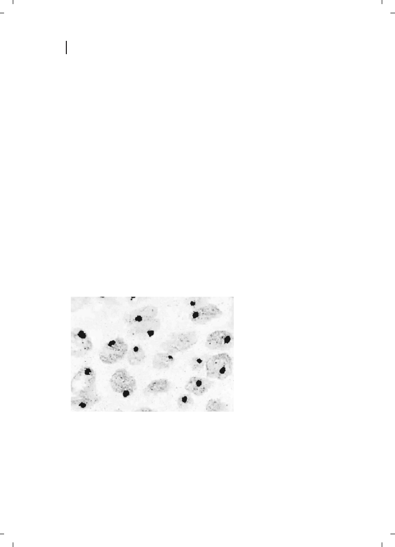
Detection of Nucleic Acid Sequences
A number of protocols using colloidal gold with silver enhancement for nucleic acid se-
quence detection were described during the 1980s [84-87] but have not been widely
adopted. ISH staining results at that time were often better when a peroxidase-based sys-
tem was used, but with the introduction of gold cluster labels this situation has slowly
changed. The nearly pH-neutral behavior of Nanogold also allowed a greatly improved ap-
plicability to the detection of gene sequences by ISH : Nucleic acid sequences present only
in minute amounts could be readily demonstrated using a simple, two-step indirect ISH
technique (Figure 23.5) [88].
360
Figure 23.5
Applications of gold and silver for in-situ polymerase chain reaction (IS-PCR) have also
been described [89–91], and in this still-emerging field numerous advantages of this
highly sensitive detection method are clear [88, 92–94]. Due not only to the occasionally
relatively low reproducibility and major specificity problems encountered with IS-PCR,
but also to the relatively higher costs, the authors have during more recent years placed
higher emphasis of label amplification techniques, rather than target (DNA/RNA) ampli-
fication. Another application of gold-silver for super-sensitive RNA-detection which is still
awaiting broader investigation is that of in-situ self-sustained sequence replication-based
amplification (3SR) [95].
The introduction of catalyzed reporter deposition (CARD), now commercially termed
“tyramide-signal-amplification” (TSA; PerkinElmer Life Sciences) or “catalyzed signal
amplification” (CSA; DakoCytomation, Glostrup, Denmark, and Carpinteria, CA, USA)
[96, 97], for the first time allowed the detection of single molecules of gene sequences
in the light microscope by using a relatively straightforward ISH and applying the Nano-
gold technique [56, 57, 98–104]. Today, the TSA protocol can be carried out fully automa-
tically [105, 106]. A bright-field (LM) method, GOLDFISH (gold-facilitated in-situ hybridi-
zation) has been developed for the detection of Her-2/neu gene amplification in paraffin-
embedded sections of invasive ductal carcinoma [107], and showed both high reproduci-
bility and excellent concordance with fluorescence in-situ hybridization (FISH) methods
[108] (Figure 23.6).
23.7
Applications for Microscopical Detection of Nucleic Acids
For ISH, a large spectrum of uses is applicable. The in-situ detection of specific DNA- or
RNA-sequences with molecular sensitivity gives rise to the bulk of routine uses for diag-
nostic pathology, for example, in the detection of tumor-associated viruses or of tumor
suppressor genes. Gold-silver-based ISH is also a very elegant way to demonstrate cancer
gene amplification by LM, with numerous advantages when compared to FISH, for exam-
361
Figure 23.6

ple, the direct applicability of automated computer-based image analysis in permanent
preparations without the need for special equipment. The GOLDFISH method yields a
dense, punctate staining pattern, which readily allows visualization of the underlying ul-
trastructure; this is important for a complete diagnosis. It also allows the use of other
stains. In transmission LM, conventional histochemical counterstains can be used (e. g.,
HE); although counterstains such as pontamine sky blue may also be used with FISH,
they do not provide as useful an interpretation as HE or Nuclear Fast Red counterstains.
In LM or transmission EM, Nanogold-silver/gold ISH or ICC may be combined with a
chromogenic second specific target stain, such as peroxidase-DAB-H
2
O
2
, to detect anti-
gens or other nucleic acid sequences on the same section; this is most effective if the en-
zymatic chromogen is sufficiently different in color. With LM, in addition, a light nuclear
counterstain such as hematoxylin may be applied, whilst with the TEM a light conven-
tional contrasting stain should be used. This is more convenient for the practicing pathol-
ogist as it uses the standard bright-field light microscope rather than requiring expensive
or less-accessible fluorescence optics. Furthermore, the interpretation is simpler because it
is based on the overall pattern rather than requiring spot counting [107].
23.8
Technical Guidelines and Laboratory Protocols
Most recently, a book was published which was dedicated solely to gold and silver staining
techniques [53] wherein a state-of-the-art review and exact technical guidelines on the
most promising molecular morphological technologies related to gold labels and autome-
tallography is provided. Constantly updated staining protocols are available on the inter-
net, under the web address http ://www.frontierquestions.com/labprotocols.htm
23.9
Gold Derivatives of Other Biomolecules
As gold nanoparticles can be covalently attached to antibodies, it is likely that they could
also be attached to other biomolecules such as proteins, peptides, drugs, viruses, carbohy-
drates, lipids, and nucleic acids. The introduction of chemically selective reactivity has en-
abled conjugation of metal particle labels to almost any biological molecule containing an
appropriate reactive group, in a similar manner to the conjugation of fluorescent labels. In
practice, the relatively larger size of the gold particle compared with many biomolecules of
interest means that conjugation should be approached with consideration of how the
properties and biological activity of the conjugate might be modified by the attached
gold particle. This is particularly appropriate with small molecules in which the gold
can perturb binding. For example, while undecagold-conjugated phalloidin can be used
to map the topography of actin filaments [109], a phalloidin conjugate prepared with
the larger Nanogold was reported not to show comparable activity [110].
362

Protein Labeling
Because gold cluster labeling proceeds through specific chemical reactions, it is selective
towards specific groups in the conjugated biomolecule. If the gold particle reacts with a
unique group in the biomolecule, the reaction is site-specific, and can be chosen so
that it does not interfere with biological function. As the reaction is no longer dependent
on the charge properties of the conjugate protein, many proteins that are not amenable to
conventional colloidal gold labeling may be conjugated with gold clusters.
This site-specificity implies higher resolution when the bound probe is microscopically
localized. This in turn enables a higher level of resolution in EM studies; while colloidal
gold labeling might typically localize targets within tissues, site-specific Nanogold labeling
can be used to localize specific functional elements at the macromolecular level, enabling
the localization or differentiation of different binding sites within multi-subunit protein
complexes, structures, or organelles. Examples include the determination of the quatern-
ary structure of the insulin-insulin receptor by cryoelectron microscopy using insulin la-
beled with Mono-Sulfo-NHS-Nanogold [111, 112], and the use of Nanogold to localize the
two dimers of L7/L12 within the structure of the 70S ribosome. Protein L7/L12 was re-
duced with 1 % mercaptoethanol and labeled with Monomaleimido Nanogold; two recon-
stitution approaches, together with cryoelectron microscopy and single particle recon-
struction, were used to determine the structure [113].
Subtle changes in binding can be differentiated by gold labeling and modifications in
the complex assembly and binding procedures. In a recent example, undecagold was
used to localize the site of microtubule-associated protein 2 (MAP2) and tau protein bind-
ing on the surface of pre-assembled microtubule protofilaments; Cryo-EM and helical
image analysis showed that both the IR and MAP2 elements lie along the exterior ridges
of microtubules [114]. In a subsequent study, tau protein, labeled with Nanogold at a re-
peat motif in the microtubule-binding domain, was used to study tau binding during mi-
crotubule assembly. Three-dimensional electron cryomicroscopy indicated that a repeat
motif occupies a similar site to taxol on the inner surface of the microtubules, supporting
the conclusion that one of the tau repeat loops is the natural substrate that occupies the
taxol-binding pocket in beta-tubulin [115]. The reactivity of the gold labeling reagent may
be used to localize a target chemical group. Monomaleimido Nanogold has been used to
localize and quantitate interprotamine disulfide bonds during spermiogenesis : the disap-
pearance of Nanogold labeling was an indicator of the formation of disulfide bonds by cy-
steine residues [116]. Nanogold may be prepared in a positively or negatively ionizing
form by incorporation of synthetically modified ligands, bearing aliphatic amines or car-
boxyls respectively, into its surface; the resulting charge can provide a method for labeling.
Prescianotto-Baschong and co-workers have used positively charged Nanogold to label
elements of the yeast endocytic pathway [117, 118]. Negatively charged Nanogold was
used to map the distribution of electrical charges over the surface of Plasmodium falci-
parum merozoites and erythrocytes. Atomic force microscopy with surface potential spec-
troscopy were used to map the surface charge directly; this was followed up by incubation
with negatively charged Nanogold, silver enhancement and gold toning, localized with
TEM [119].
363

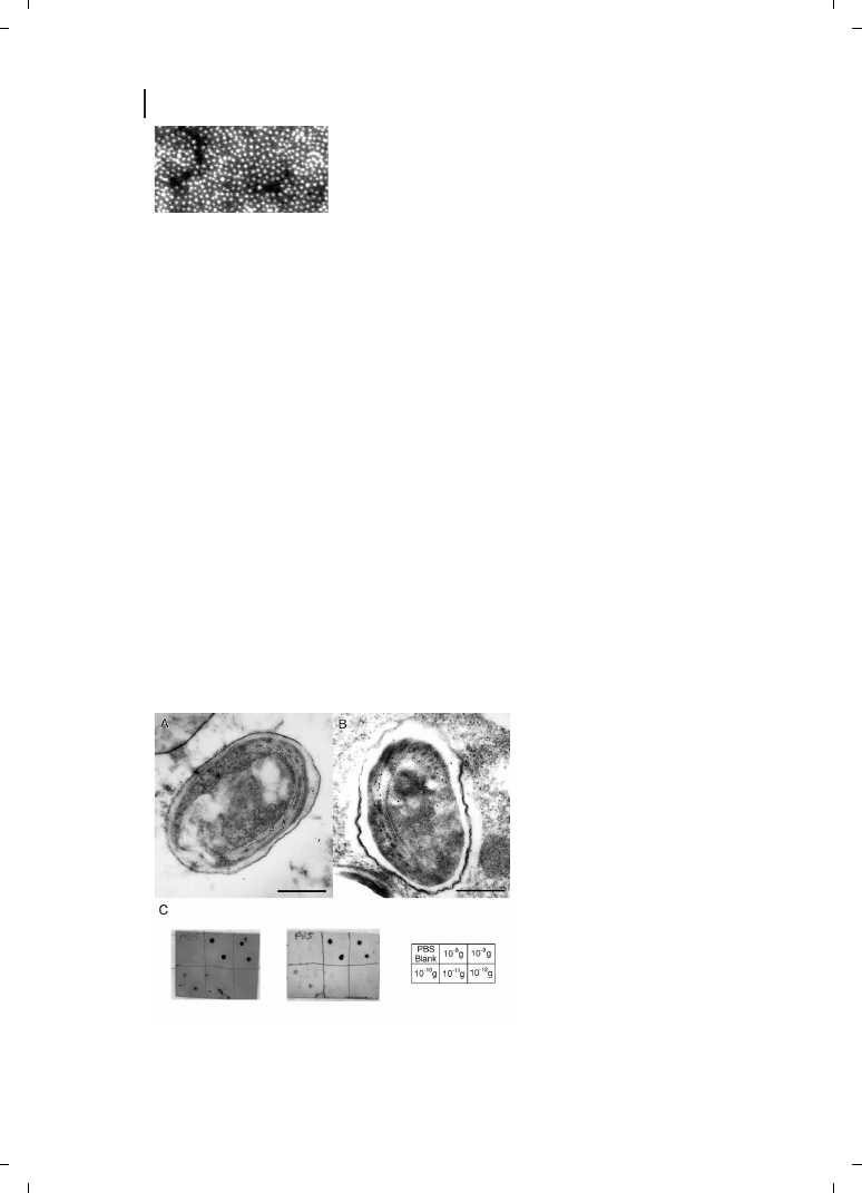
The extreme insolubility of the aberrantly folded isoform (PrPSc) of the prion protein
(PrP) responsible for Creutzfeldt–Jakob disease (CJD), bovine spongiform encephalopathy
(BSE) and other spongiform encephalopathies has prevented structural determination by
X-ray diffraction or nuclear magnetic resonance (NMR) imaging. However, 2-D electron
crystallography and Nanogold labeling of two truncated but still infectious variants, N-
terminally truncated PrPSc (PrP 27-30) and a miniprion (PrPSc106), yielded sufficient
structural information to construct models for the PrPSc structure. N-linked sugars
were oxidized with periodate, then selectively labeled using Monoamino Nanogold. Nega-
tive-stain EM and image processing allowed the extraction of limited structural informa-
tion to 7 Å resolution. The dimensions of the monomer and the locations of the deleted
segment and sugars, used as constraints in the construction of models for PrPSc, were
satisfied only by structures featuring parallel beta-helices as the key element – a signifi-
cant finding that will help derive an understanding of prion propagation and the process
of neurodegeneration associated with these prion diseases [120].
Gold Cluster-labeled Peptides
Conjugation of gold cluster labels to peptides has been described by a number of research-
ers, and these findings have been reviewed previously [10]. Two groups have used Nano-
gold to label antibody Fv fragments, thus generating a probe smaller than a Fab fragment
that, as the gold particle is linked at a site where it does not affect binding, retains the
immunoreactivity of the native antibody [121, 122]. Segond von Banchet has conjugated
Nanogold to the undecapeptide Substance P [123], and the tetradecapeptide somatostatin
[124] – the same peptide earlier conjugated with colloidal gold [5–7] – and used the labeled
peptides to localize substance P binding sites and somatostatin receptors in the rat spinal
cord. The larger peptide calmodulin, a 17 kDa protein that regulates the calcium release
channel (ryanodine receptor) in the sarcoplasmic reticulum of skeletal and cardiac mus-
cle, has been labeled with Nanogold at a cysteine residue (Cys27) and used as a high-re-
solution probe to directly visualize the binding and control site of calmodulin on isolated
single molecules of the calcium release channel cryoembedded in amorphous ice [125].
More recently, Nanogold-labeled calmodulins have been used, exchanged for delta, to
enable the localization of the delta subunit within the bridged, bilobal phosphorylase b
kinase holoenzyme complex by scanning TEM [126].
Gold Cluster Conjugates of Other Small Molecules
Even small molecules, appropriately functionalized, may be labeled with gold clusters.
The use of undecagold-conjugated phalloidin to map the topography of actin filaments
by STEM has been mentioned previously; the phalloidin was synthetically modified to in-
corporate an aliphatic amino-group, which was then reacted with bis-(4-nitrophenyl) adi-
pate followed by amino- undecagold [109]. In addition, a snake venom toxin, toxin- from
Naja nigricollis, has been derivatized with maleimido-undecagold to produce a small probe
with a high affinity for the cholinergic binding site of the Torpedo marmorata nicotinic re-
364
ceptor [127]; the use of small molecule probes such as these may enable further improve-
ments in the resolution achievable in EM labeling.
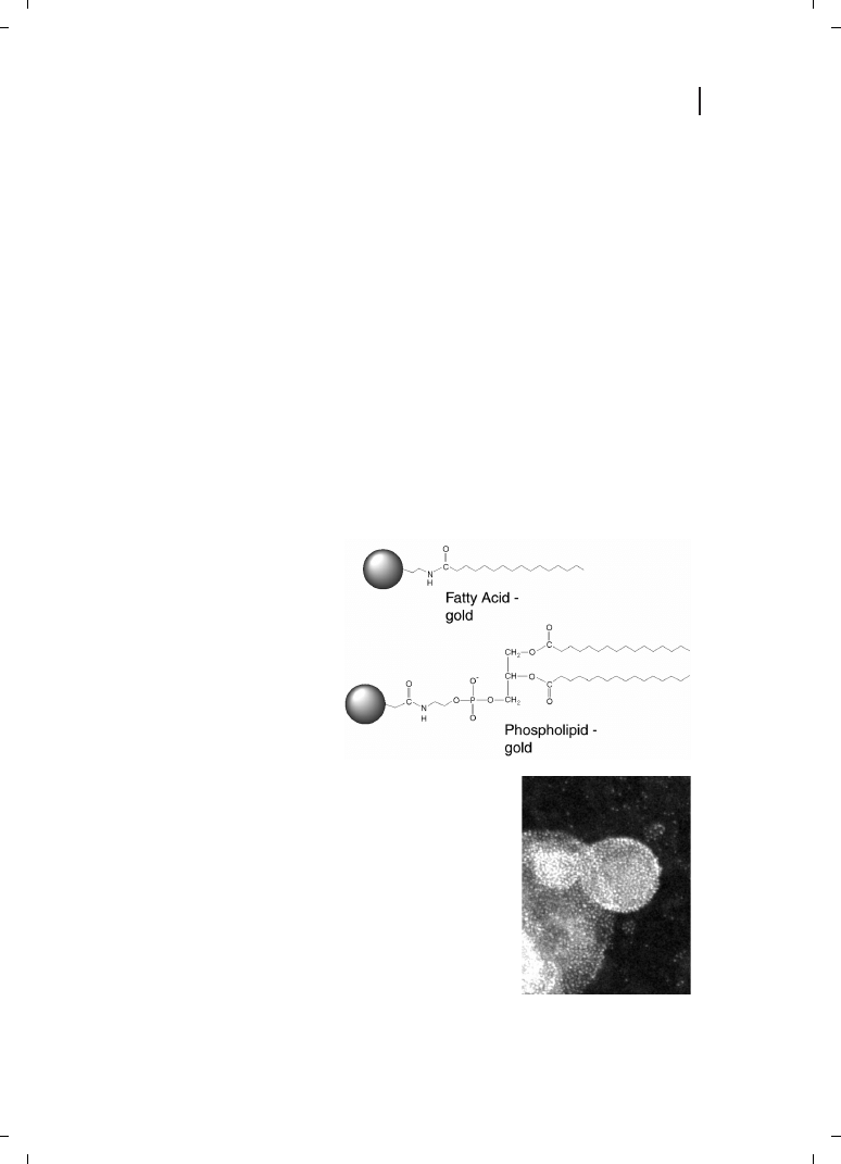
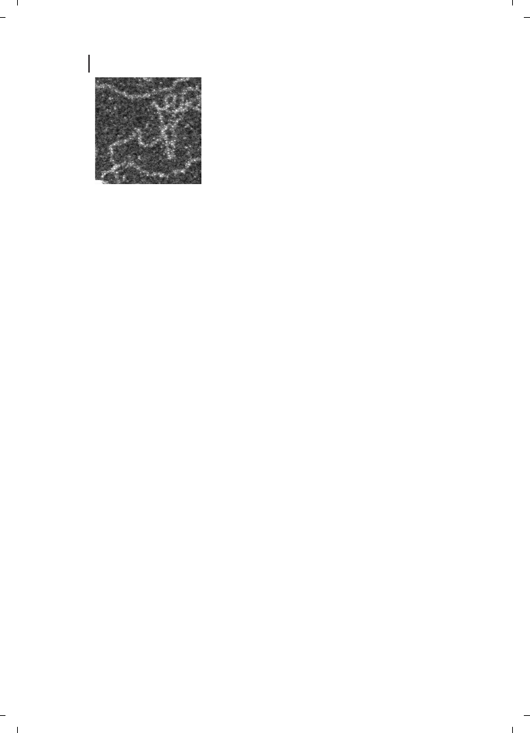
Gold–Lipids : Metallosomes
Fatty acids or phospholipids that bear an appropriate chemical group may also be conju-
gated with gold nanoparticles. Two such Nanogold conjugates, palmitoyl Nanogold and
diphosphatidyl ethanolamino- (DPPE) Nanogold, are shown in Figure 23.7. Lipids in
aqueous solutions form emulsions similar to mayonnaise. These possess the ability to
self-organize to give a wide variety of nanoscale structures and morphologies with poten-
tially useful applications : upon sonication, lipids form micelles, hollow liposome vesicles,
multilayer liposomes, sheets and tubes depending on the phase state.
Lipid–gold conjugates have applications both as probes for the microscopic localization
and tracking of liposomes, and as components for the templated assembly of supramole-
cular arrays of gold particles with a rich variety of morphologies. Liposomes are used to
encapsulate drugs : they both keep the drug separate from metabolic activity, and deliver
it to the target cells. These gold–lipid conjugates have been incorporated into antifungal
drug liposomes to aid in visualization of the delivery process [128], and also to demon-
strate the targeting of cationic liposomes to endothelial cells in tumors and chronic in-
365
Figure 23.7
Figure 23.8
flammation [129]. Sonication of gold–lipids in aqueous solution leads to formation of
gold–liposomes, which have been named “metallosomes” [130, 131] (Figure 23.8). Inter-
estingly, each lipid molecule has one 1.4-nm Nanogold cluster attached, such that a single
lipid molecule is then visualized. Lipids also form a monolayer when placed at an air–
water interface, and this can be picked up on an electron microscope grid and viewed
by EM (Figure 23.9).
23.10
Larger Covalent Particle Labels
Research is also being directed towards the bioconjugate chemistry of larger gold particles,
functionalized in a similar manner to Nanogold, by synthetic modification of small or-
ganic molecules coordinated to the gold surface. A preliminary report has described the
preparation and use of a covalent 10 nm gold–Fab conjugate for blotting and immunoe-
lectron microscopy (Figure 23.10) [17]. Fundamental changes are observed in the chemical
and electronic properties of gold particles from the small gold clusters such as undecagold
and Nanogold to even slightly larger gold nanoparticles 3 and 5 nm in diameter, and this
has important implications for the roles that these particles might serve in nanobiotech-
nology applications. Gold particles with controlled cross-linking functionality, in a range
of precisely defined sizes with a variety of properties, would be a valuable component
of this developing field.
366
Figure 23.9
Figure 23.10
23.11
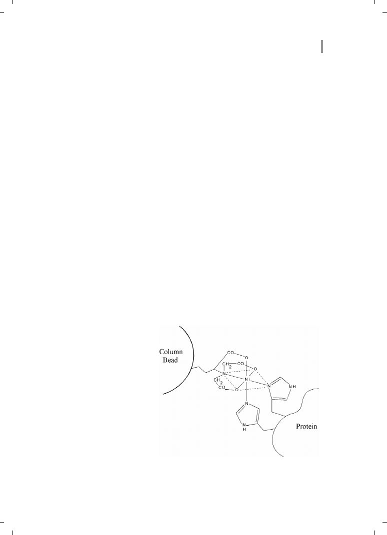
Gold Targeted to His Tags
A popular molecular biology technique is to transfect a cell so that an engineered DNA is
incorporated and the new protein expressed. Frequently, overexpression is an objective, so
that the new protein can be produced in quantity. Purification of the protein from the
other cell materials is then a challenge. It was discovered that histidine residues have
an affinity for metals, and by coding a sequence of six histidines (usually at the amino
or carboxyl end of the protein), the whole cell contents could be poured over a metal-func-
tionalized column, and only the his-tagged protein would bind. The bound protein can
then be eluted under more stringent conditions to yield highly purified bulk protein in
one step. Although other chelators and metals are sometimes used [132], the optimum
chromatography media was one derivatized with the nitrilotriacetic acid group (NTA)
[133] : this binds a hexacoordinate nickel(II) atom in a configuration that leaves two adja-
cent coordination sites unoccupied (Figure 23.11), creating a binding site with a strong,
selective affinity towards polyhistidine. Dissociation constants for NTA-Ni(II) binding to
polyhistidine are thought to be about 10
–7
M [134, 135]. This rivals that of antibodies,
and means that NTA-Ni(II) functionalized gold nanoparticles may be used as probes to
localize polyhistidine-tagged targets. Because they are much smaller than antibody conju-
gates, NTA-Ni(II) derivatives may be better able to penetrate and access sterically hindered
targets, and form more closely spaced supramolecular structures such as nanowires.
For biomolecular labeling of His-tagged proteins (one containing the 6
His tag), the
Ni-NTA group was incorporated synthetically into the organic shell of a gold particle
[136] (Figure 23.12). The nickel(II)-charged derivative has been used to quantitatively
label the three 6
His-tagged subunits of the 64-kDa adenovirus A12 knob protein for
STEM observation [136, 137] (Figure 23.13), and also to label N-terminal 6
His-tagged
PsbH protein which was then located within the Photosystem II multisubunit complex
by EM and image analysis [138].
367
Figure 23.11
23.12
Enzyme Metallography
Enzymes play a variety of roles in the sequestration, transport, and metabolic activity of
metals within organisms. Some bacterial cells accumulate magnetic particles that allow
them to navigate without a global positioning system (GPS), and some cells perform
metal pumping and sequestration, for growth in toxic environments. In similar manner,
our bodies store iron in ferritin, which consists of a protein shell with portals whereby
about 5000 iron atoms can enter and be stored in an insoluble Fe(III) oxidation state.
An interesting recent discovery was that enzymes can be controlled to deposit metal in
the zero oxidation state [139]. The beauty of enzyme reactions is that the enzymes are cat-
368
Figure 23.12
Figure 23.13
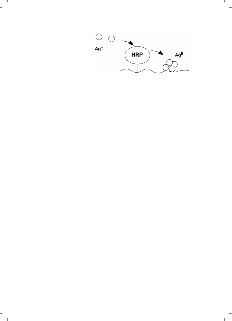
alysts, and can cycle a very large number of times. If such an enzyme is targeted to a gene
or antigen, it can then be used to locally deposit metal in the nanoscale range (Figure
23.14). If the enzyme is kept supplied with metal ion substrate, then macroscopic deposits
will result, making sensitive detection possible even when using all methods that might
be used to detect and localize metal deposits, including light scattering, reflectance, SERS,
absorption, density, and conductivity.
23.13
Gold Cluster Nanocrystals
Gold cluster compounds may be prepared with sufficient monodispersity that they can
deposit to form highly ordered 2-D arrays, or “nanocrystals”. The self-assembly of larger
colloidal gold particles has been documented [140], but we have observed similar behavior
in a gold cluster, similar to Nanogold but slightly larger, known as “Greengold” [141].
Greengold is a highly regular compound, thought to contain 73–75 gold atoms based
on MALDI mass spectroscopy data. Upon standing, it can form small microcrystals
(found by STEM to be usually 2-D planar sheets), but occasionally it may form thin 3-
D crystals. The center-to-center spacing between gold clusters is 2.6 nm, which is consis-
tent with a gold core about 1.4 nm in diameter and an organic ligand shell of 0.6 nm
thickness [142].
23.14
Gold Cluster–Oligonucleotide Conjugates : Nanotechnology Applications
The use of the supramolecular organizing properties of biomolecules to arrange metal na-
noparticles into extended arrays offers one of the most promising approaches to develop-
ing their nanotechnology applications. In this respect, nucleic acids occupy a unique place
in nanobiotechnology. The vast number of unique combinations available for their hybri-
dization allows the “programming” of many simultaneous, unique interactions, which
can be used to assemble complex structures, or even carry out complex processes such
as DNA computing. Combined with the variety of physical, optical and electronic proper-
ties imparted by metal nanoparticles, this promises to give rise to a highly diverse set of
nanotechnological applications for metal nanoparticle–nucleic acid conjugates.
369
Figure 23.14
DNA Nanowires
DNA has some interesting properties for use in wiring nanocircuitry. It is tiny in width
(2 nm), flexible, and may be easily synthesized in well-defined lengths. With other poly-
mers, the lengths are difficult to control, whereas with DNA each base is programmed so
that exact lengths can be mass-produced. The ends may be synthesized with unique nu-
cleotide sequences that will bind to (hybridize) with a complementary sequence target.
This means that the ends of such a DNA nanowire will self-assemble and connect to com-
plementary target pads, even in three dimensions (Figure 23.15). Current computers rely
on lithography and use wiring that is
0.3 m, so DNA would be a factor of 150 times
smaller. Packing density in two dimensions could then be increased by 1502 = 22 500,
or in three dimensions by 3 375 000. While this is optimistic, even several orders of mag-
nitude improvement would be significant.
A number of methods are available for attaching metal nanoparticles to DNA, including
covalent attachment to specific bases [143, 144], photoreaction, intercalation, and charge
binding. One method we have used is shown in Figure 23.15, which shows a positively
charged Nanogold cluster bound to the negatively charged DNA [145]. The average spac-
ing of gold quantum dots is
2 nm. Although conduction can occur through tunneling or
electron hopping, the dots may be processed by autometallography, using them as nuclei
to deposit additional metal, growing them to confluence if desired to make a continuous
wire.
3-D Nanostructured Mineralized Biomaterials
The assembly of complementary oligonucleotides with multiple hybridization sites into
complex, 3-D structures with multiple unique sites and potential functionalities was
first pursued by Mirkin, who used thiolated oligonucleotides coordinated directly to unsta-
bilized colloidal gold particles to construct large oligomers containing semi-regular arrays
of 13-nm gold particles [146]. The optical properties of the gold particles were quickly ap-
plied in a method for detecting DNA hybridization, using the color change that occurred
when labeled strands hybridized and brought the gold particles into proximity [16, 147].
370
Figure 23.15
Further developments and applications of this technology are described in other chapters
of this book; here, we will limit our discussion to the use of gold cluster labels.
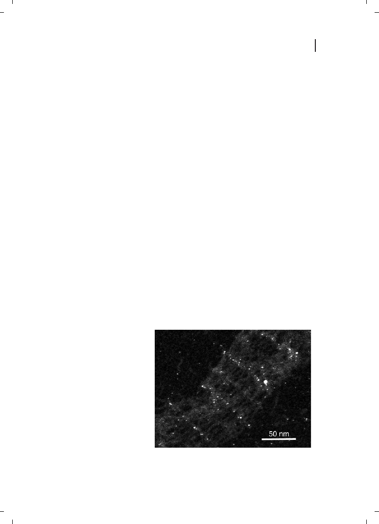
The chemical selectivity of covalent gold cluster labeling affords an additional dimen-
sion in the design of hybrid DNA–metal nanoparticle materials, by letting the researcher
choose a reaction to attach the gold particles directly at any suitably modified point within
the structure. Programmed self-assembly of Nanogold-labeled oligonucleotides has been
demonstrated by Liu and co-workers, who used Mono-Sulfo-NHS-Nanogold to label nano-
tube-forming oligonucleotides before assembly [148]. Kiehl and co-workers recently as-
sembled metallic nanoparticle arrays using DNA crystals, labeled site-specifically with Na-
nogold, as a programmable molecular scaffolding [149]. 2D DNA crystals as a scaffolding
potentially offers fundamental advantages over other self-assembly approaches for the pre-
cision, rigidity, and programmability of the assembled nanostructures, and this represents
a critical step toward the realization of DNA nanotechnology and its nanoelectronic appli-
cations. 2-D arrays were constructed by tiling together rigid DNA motifs composed of
double-crossover (DX) molecules containing DNA hairpins. The nanoparticles formed
precisely integrated components, covalently bonded to the DNA scaffolding. STEM
showed that the gold particles formed 2-D arrays with interparticle spacings of 4 and
64 nm (Figure 23.16). DNA–Nanogold conjugates were prepared from 5 -thiol-modified
C6 oligonucleotides, which were reacted with Monomaleimido Nanogold. DNA :Nanogold
labeling stoichiometry of the purified conjugate was estimated spectroscopically to be very
close to the desired 1 :1 product.
Another important development is the use of autometallography to further modify such
materials after assembly, and introduce new and useful functionality. This has been pos-
tulated as a method for forming metallic nanowires [145] and nanospheres or other struc-
tures [131] using gold-decorated DNA and liposomes respectively. Mirkin and co-workers
have reported the fabrication of a conductimetric biosensor based on these processes. The
device comprises lithographically prepared microelectrode pairs (separation 20 m), a
shorter “capture” oligonucleotide strand located in the gap between them, and a longer
“target” oligonucleotide in solution. The target oligonucleotide has contiguous recognition
elements complementary to the capture strand on one end and on the other to oligonu-
371
Figure 23.16

Wyszukiwarka
Podobne podstrony:
23 Nanoparticle Molecular Labels damaged version
PRK 23 10 2011 org
23 piątek
23 Metody montażu w mikroelektronice
23 Tydzień zwykły, 23 wtorek
Atrybucje 23 24
Cwiczenia 23 25 2007
Molecular Toxicology 8
23 sekcja
21 23
Doradztwo Podatkowe z 23 czerwca 08 (nr 121)
23 Pddzialywanie swiatla z materia
Podstawy rekreacji ćwiczenia 23 01 10x
brzuch i miednica 2003 2004 23 01
Dz U 2008 4 23
Molecular evolution of FOXP2, Nature
więcej podobnych podstron