
Expert Review
Organic
–Inorganic Surface Modifications for Titanium Implant Surfaces
Lise T. de Jonge,
1
Sander C. G. Leeuwenburgh,
1
Joop G. C. Wolke,
1
and John A. Jansen
1,2
Received February 11, 2008; accepted April 29, 2008; published online May 29, 2008
Abstract. This paper reviews current physicochemical and biochemical coating techniques that are
investigated to enhance bone regeneration at the interface of titanium implant materials. By applying
coatings onto titanium surfaces that mimic the organic and inorganic components of living bone tissue, a
physiological transition between the non-physiological titanium surface and surrounding bone tissue can
be established. In this way, the coated titanium implants stimulate bone formation from the implant
surface, thereby enhancing early and strong fixation of bone-substituting implants. As such, a continuous
transition from bone tissue to implant surface is induced. This review presents an overview of various
techniques that can be used to this end, and that are inspired by either inorganic (calcium phosphate) or
organic (extracellular matrix components, growth factors, enzymes, etc.) components of natural bone
tissue. The combination, however, of both organic and inorganic constituents is expected to result into
truly bone-resembling coatings, and as such to a new generation of surface-modified titanium implants
with improved functionality and biological efficacy.
KEY WORDS: calcium phosphate; ECM proteins; protein immobilization; surface modification; titanium
implants.
INTRODUCTION
The research field of bone tissue engineering applies the
principles of biology and engineering to develop functional
substitutes for damaged bone tissue (
). To restore, maintain
and improve bone tissue function, three key elements are
required: (1) a scaffold or carrier material combined with (2)
cells and/or (3) bone stimulating molecules (e.g. growth
factors). The scaffold provides mechanical support and serves
as a substrate upon which cells attach, proliferate and
undergo differentiation. In that respect, metallic implants
used in plastic and reconstructive surgery, orthopedic surgery,
craniofacial surgery, and oral implantology can be regarded as
scaffolds for load-bearing, bone-replacing/contacting applica-
tions such as joint and tooth replacement, fracture healing,
and reconstruction of congenital skeletal abnormalities. For
these implants, the ultimate goal is to obtain a life-long secure
anchoring of the implant in the native surrounding bone.
Commercially pure titanium (cpTi) and Ti
–6Al–4V alloys are
the most commonly used metallic implant materials, as they
are highly biocompatible materials with excellent mechanical
properties and corrosion resistance (
). The biocompatibil-
ity of titanium implants is attributed to the stable oxide layer
(with a thickness of 3
–10 nm) that spontaneously forms when
titanium is exposed to oxygen (
). This reaction prevents
the formation of fibrous tissue around the implant, and
creates direct contact to osseous tissue. Nevertheless, when
applying Ti(O
2
) as implant material, a non-physiological
surface is exposed to a physiological environment. However,
by generating a coating onto a titanium surface that mimics
the organic and inorganic components of living bone tissue, a
physiological transition between the non-physiological titani-
um surface and surrounding bone tissue can be established. In
this way, the coated titanium implant functions as scaffold for
improved bone cell attachment, proliferation and differenti-
ation. Such a coating is supposed to further enhance early and
strong fixation of a bone-substituting implant by stimulating
bone formation starting from the implant surface. As such, a
continuous transition from tissue to implant surface can be
induced. Consequently, research efforts have focused on
modifying the surface properties of titanium to control the
interaction between the implant and its biological surround-
ing. This paper reviews current physicochemical and bio-
chemical surface modification approaches to enhance bone
regeneration at the interface of titanium(-alloy) implants. The
first part of this review will present a brief description of the
biological processes that occur at the interface of the implant
surface upon implantation in bone tissue, followed by an
overview of both inorganic (calcium phosphate) and organic
(protein) coatings that stimulate bone formation to achieve
an improved and accelerated implant fixation.
THE BONE-IMPLANT INTERFACE
Bone
Bone tissue is a living organ, which can be described as a
natural composite composed of an organic matrix strength-
2357
0724-8741/08/1000-2357/0 # 2008 The Author(s)
Pharmaceutical Research, Vol. 25, No. 10, October 2008 (
#
2008)
DOI: 10.1007/s11095-008-9617-0
1
Department of Periodontology and Biomaterials, Radboud University
Nijmegen Medical Center, THK-309-PB, P.O. Box 9101, 6500 HB
Nijmegen, The Netherlands.
2
To whom correspondence should be addressed. (e-mail: j.jansen@
dent.umcn.nl)
ened by an inorganic calcium phosphate (CaP) phase. The
extracellular organic matrix (ECM) of bone consists of 90%
collagenous proteins (type I collagen 97% and type V
collagen 3%) and 10% non-collagenous proteins (osteocalcin
20%, osteonectin 20%, bone sialoproteins 12%, proteogly-
cans 10%, osteopontin, fibronectin, growth factors, etc.).
Regarding the inorganic component, the most abundant
mineral phase in human bone is carbonate rich hydroxyapa-
tite (with a carbonate content between 4% and 8%) (
). The
apatite in bone mineral is composed of small platelet-like
crystals of just 2
–4 nm in thickness, 25 nm in width, and 50 nm
in length (
). This calcified matrix embeds bone cells, which
participate in the maintenance and organization of bone.
Bone is subject to constant remodeling by osteoblasts and
osteoclasts, i.e., bone-forming and bone-resorbing cells.
Osteoblasts are responsible for the synthesis, deposition,
and mineralization of extracellular matrix. They are located
at bone surfaces and form a continuous layer. Upon
embedding in this matrix, osteoblasts finally transform into
quiescent osteocytes. Osteoclasts are large multinuclear cells
that are involved in bone resorption. A main feature of this
bone cell type is its ruffled border, which acts as a high
surface area interface for excretion of proteins and (hydro-
chloric) acid. The acid decreases the local pH and dissolves
CaP bone mineral. This dynamic process of bone formation
and destruction accounts for its remodeling, thereby enabling
bone regeneration.
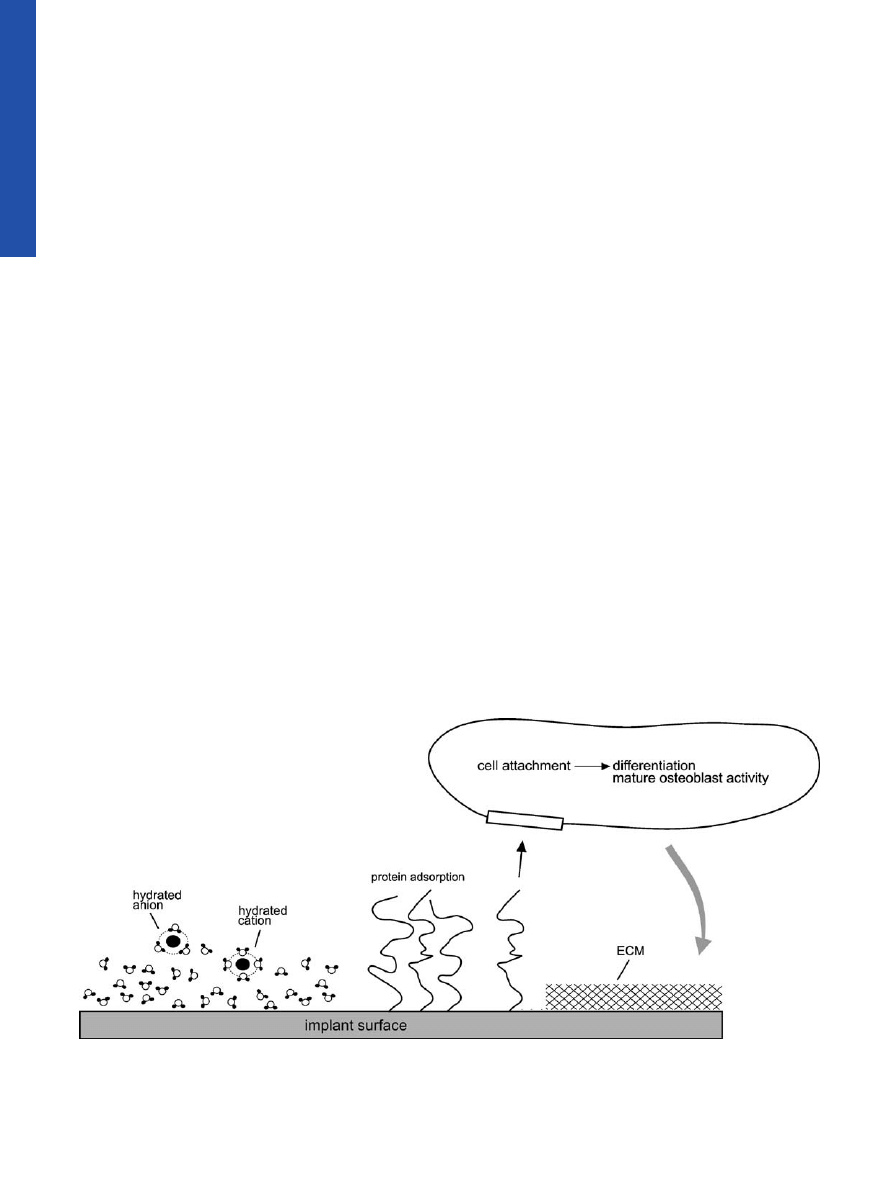
Cellular Interactions with Implant Surfaces
A sequence of complex and strongly interrelated events
takes place at the implant surface after implantation of the
material (Fig.
) (
). Immediately after implantation, water
molecules bind to the surface and form a water mono- or
bilayer. The arrangement of the water molecules depends on
the implant surface properties at the atomic scale. Hydrated
ions, such as Cl
−
, Na
+
, and Ca
2+
, are subsequently
incorporated into the surface water layer. Blood proteins
and tissue specific proteins adsorb and desorb to and from the
surface (
). This adsorption process is strongly dependent on
the implant surface features, such as its physicochemical,
biochemical and topographic characteristics. Inorganic,
physicochemical stimuli, such as release of Ca
2+
and PO
3
4
ions from calcium phosphates, can positively affect the
cellular response (
). Additionally, implants biochemically
modified with biomolecules immobilized on the surface, such
as growth factors or cell adhesion motifs, will induce certain
cell responses in the physiological surrounding by specific cell
signaling pathways. Next to that, implant surfaces that have
protrusions, cavities, gullies, etc., on a micro- and/or nano-
scale will induce biological interactions different from those
with a flat surface (
). As a result, both the exact mixture of
adsorbed proteins and their conformational state(s) are
largely controlled by the implant surface. This surface-
specific adsorbed biofilm subsequently determines cell
adhesion, since proteins act as contact for the attachment of
cells. This is accomplished by means of integrins, which are
specific transmembrane receptors that bind to adhesive
proteins on the biomaterials surface and to components of
the cytoskeleton through their extra- and intracellular
domains, respectively.
In general, the biocompatibility of bone-replacing im-
plant materials is closely related to osteoblast adhesion onto
their surface (
). Osteoblast attachment, adhesion and
spreading will influence the capacity of these cells to
proliferate and to differentiate itself upon contact with the
implant. These latter processes are quintessential for the
establishment of a mechanically solid interface with complete
fusion between the implant surface and bone tissue without
any intervening fibrous tissue layer.
Fig. 1. Schematic representation of events consecutively taking place at the titanium surface after implantation into living bone tissue. Water
binds to the surface, followed by incorporation of hydrated ions, adsorption and desorption of proteins, eventually leading to cell attachment.
After differentiation, mature osteoblasts produce the extracellular matrix (ECM).
2358
de Jonge, Leeuwenburgh, Wolke, and Jansen

SURFACE MODIFICATION OF TITANIUM IMPLANTS
Several reviews have summarized a wide variety of surface
modification approaches for titanium and titanium alloys in the
biomedical field (
). Traditionally, these approaches
focused on the modification of the implant surface topography
and morphology (
). These surface modifications mainly
included mechanical methods such as machining (
,
),
grinding, polishing (
), and chemical
methods such as acid etching (
), alkali etching (
,
and anodization (
) to alter the topography of the titanium
surface. Another approach towards the creation of a biologi-
cally active implant surface involves the application of an
additional coating onto the titanium surface by means of
physicochemical and biochemical deposition techniques
(
). In the following sections an overview will be given of
the physicochemical and biochemical methods to provide
titanium with components of the ECM as a surface coating
aimed at implant fixation within living bone tissue. First, calcium
phosphate coatings that are similar to the mineral phase in
natural bone will be reviewed on their use for biomedical
implant materials (
“
Inorganic Calcium Phosphate Coatings
”).
Thereafter, coating methods to immobilize various organic
biomolecules onto implant surfaces will be evaluated (
“
”), whereas organic–inorganic composite
coatings, which mimic the composition of natural bone even more,
will be discussed (
“
”).
Inorganic Calcium Phosphate Coatings
Calcium Phosphates
CaPs are often used in the biomedical field due to their
similarity with the mineral phase present in bone and teeth
(
). Hydroxyapatite, or more specifically carbonate apatite,
is by far the most abundant inorganic phase in the human
body. Apatites have the formula Ca
5
(PO
4
)X, where X may
represent several mono- and/or divalent anions such as F
−
,
OH
−
, or carbonate. The name apatite is derived from the
Greek
απαταω (Eng. “to deceive”), because the mineral was
frequently confused with other compounds such as
aquamarine, amethyst, etc. The apatite structure is very
tolerant for ionic substitutions. For example, Ca
2+
ions can
be partly or completely replaced by Ba
2+
, Sr
2+
or Pb
2+
. The
exact lattice parameters
—and many other properties of
apatites
—depends slightly on the mode of preparation
because of the frequent occurrence of nonstoichiometry.
Table
lists the chemical names, compositions and
frequently used abbreviations of the most important CaP
phases (
).
Carbonate apatite comprises a chemical composition
closer to bone and dental enamel than that of hydroxyapatite.
The relation between carbonate apatite and hydroxyapatite is
important, because carbonate increases the chemical reactiv-
ity of apatites. This occurs by an increase of the solubility of
the product and rate of dissolution in acids, and by reducing
the thermal stability (
). Since carbonate is known as an
effective crystal growth inhibitor, carbonate apatite consists of
smaller crystals than hydroxyapatite (
Bioactivity of Calcium Phosphates
Calcium phosphate (CaP) ceramics are known for their
bioactive properties (
). Generally, bioactive materials
interact with surrounding bone, resulting into the formation
of a chemical bond to this tissue (
“bone-bonding”). This
phenomenon of bioactivity is determined mainly by chemical
factors
—such as the crystal phase and molecular structure of
the material
—as well as physical factors, such as surface
roughness and porosity.
Bone-bonding occurs through a time-dependent kinetic
modification of the surface, triggered by their implantation
within the living bone (
,
). An ion-exchange reaction
between the bioactive implant and surrounding body fluids
results in the formation of a carbonate apatite layer on the
implant that is chemically and crystallographically equivalent
to the mineral phase in bone. The bone healing process is
therefore enhanced by this biological apatite layer (
The correlation between bioactivity and the formation of a
carbonate apatite layer is often inverted for preliminary in
vitro testing of the potential bioactivity of biomaterials. The
capacity to nucleate CaP formation under in vitro conditions
is then interpreted as a first indication of possible bioactivity
in vivo (
Calcium Phosphate Coatings
CaP ceramics are too brittle for use as bulk material
under loaded conditions, which makes that CaP ceramics are
frequently applied as coatings onto the surface of metallic
Table I. Ca/P Ratios, Composition, Names and Abbreviations for Various Calcium Phosphates
Ca/P ratio
Formula
Name
Abbreviation
0.5
Ca(H
2
PO
4
)
2
·H
2
O
Monocalcium phosphate monohydrate
MCPM
0.5
Ca(H
2
PO
4
)
2
Monocalcium phosphate anhydrous
MCPA
1.0
CaHPO
4
·2H
2
O
Dicalcium phosphate dihydrate
DCPD
1.0
CaHPO
4
Dicalcium phosphate anhydrous
DCPA
1.33
Ca
8
H
2
(PO
4
)
6
·5H
2
O
Octacalcium phosphate
OCP
1.5
Ca
3
(PO
4
)
2
Tricalcium phosphate
TCP
1.67
Ca
5
(PO
4
)
3
(OH)
Hydroxyapatite
HA/OHAp
1.67
Ca
5
(PO
4
)
3
F
Fluorapatite
FA/FAp
≥1.67
Ca
5
(PO
4
)
x
(CO
3
)
y
Carbonate apatite
CA/CO
3
Ap
2.0
CaO·Ca
3
(PO
4
)
2
Tetracalcium phosphate
TetCP
2359
Organic
–Inorganic Surface Modifications for Ti Implant Surfaces
implant materials in order to combine the mechanical
strength of metals with the excellent biological properties of
CaP ceramics.
CaP coatings for orthopaedic and dental implants were
introduced by de Groot and Geesink (
). Since then
numerous reports have been published about the osteocon-
ductive properties of CaP-coated implants (osteoconduction
refers to the ability of a biomaterial to support the growth of
bone over its surface). These CaP coatings are described to
induce an increased bone-to-implant contact (
,
), to
improve the implant fixation (
), and to facilitate the
bridging of small gaps between implant and surrounding
bone (
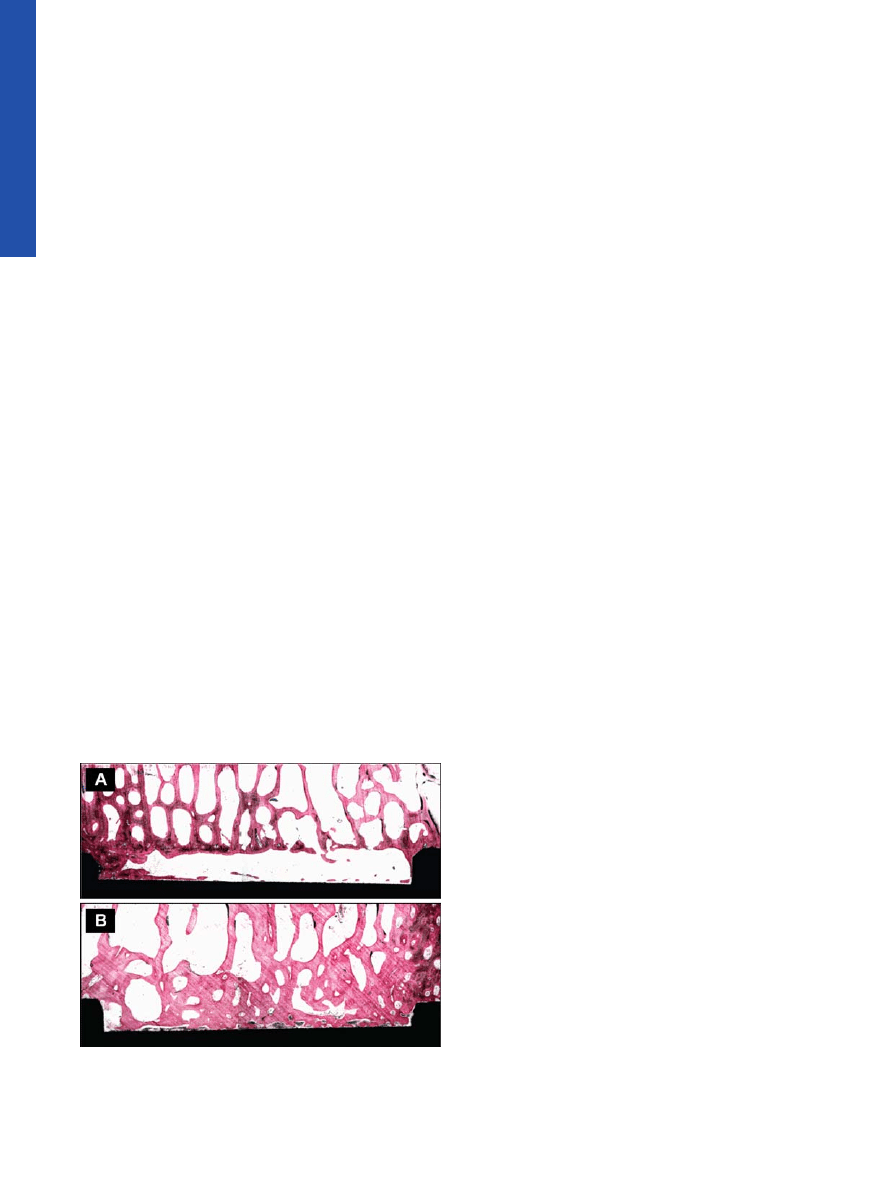
). As an example of the osteoconductive proper-
ties of CaP coatings, Fig.
shows the light micrographs of
histological sections of implant gaps either with or without
CaP coating. The CaP-layer guides bone growth along the
implant surface, and as a result bone formation now occurs
from both the surrounding tissue and the implant surface, in
which CaP functions as a physiological transition between the
non-physiological titanium surface and surrounding bone.
Calcium Phosphate Coating Techniques
From a commercial point of view, the most successful
method to apply CaP coatings to implants has been the
plasma-spraying technique, due to its high deposition rate and
the ability to coat large areas. Although the osteoconductive
and bone-bonding behavior of plasma-sprayed coatings is
confirmed by numerous studies (
–
), still some serious
concerns are related to the plasma-spraying technique (
):
&
Plasma-sprayed coatings must be at least 50
μm thick to
completely cover the implant. As a consequence, the
adhesion of the thick plasma-sprayed coatings tends to be
quite weak, which necessitates a pre-treatment of the
substrates such as grit blasting to roughen the substrate
and to increase the mechanical interlocking of the coating
–
substrate system.
&
Phase changes in the CaP powder particles during the
coating process are unpredictable due to the high temper-
ature differences in the plasma, leading to the formation of
undesired phases such as tetracalcium phosphate, calcium
oxide, and
α-tricalcium phosphate. Moreover, particularly
promising phases such as carbonate apatite (which is close
to bone composition) (
) and biological agents such as
growth factors cannot be deposited using plasma-spraying.
&
Particle release and delamination are specific drawbacks
for the plasma-spraying technique. The crystallinity of
plasma-sprayed coatings is not uniform, as the coatings
consist of crystalline and amorphous regions. When CaP
material is released from these heterogeneous coatings, the
resultant particles may initiate inflammation in surrounding
tissues.
&
Poor control over thickness and surface morphology.
Therefore, researchers have been continuously inspired
in the past two decades to explore alternative or complemen-
tary techniques for deposition of CaP coatings onto an
implant surface. To overcome the above mentioned draw-
backs of plasma-sprayed coatings, various deposition methods
have been proposed, including magnetron sputtering, elec-
trophoretic deposition, hot isostatic pressing, sol
–gel deposi-
tion, pulsed laser deposition, ion beam dynamic mixing
deposition, electrospray deposition, biomimetic deposition,
and electrolytic deposition. Table
presents the CaP coating
thickness and the most relevant advantages and disadvan-
tages of different CaP coating techniques. Clinically, each
application demands specific requirements, and in that
respect the wide range of available coating techniques offers
the possibility to select the most appropriate deposition
method for each specific implant application.
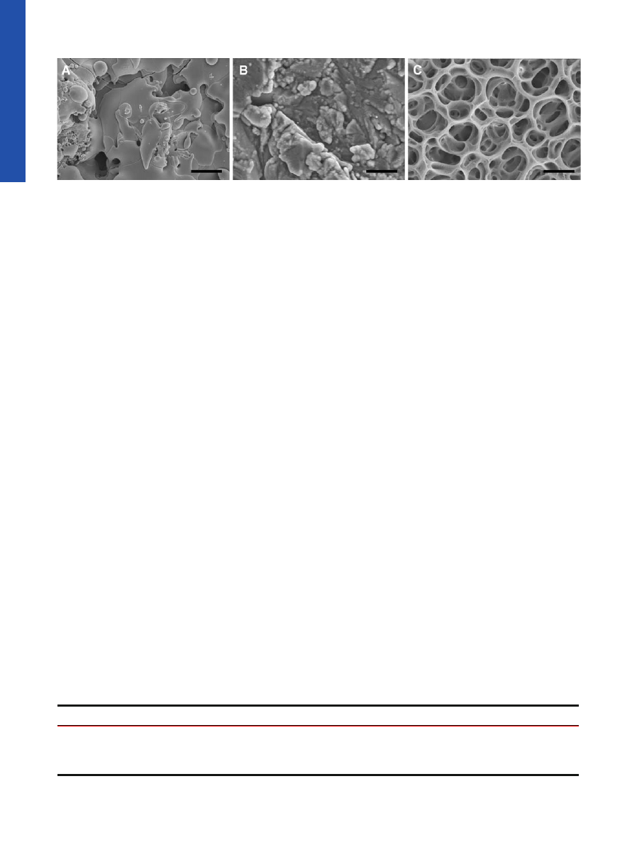
Summary and Outlook
Currently, a large variety of deposition methods is
available for application of CaP coatings onto titanium
implants. Generally, the properties of the produced coatings
differ considerably in terms of chemical structure, composi-
tion, thickness, mechanical properties, etc. (see Fig.
for an
illustration of the large variation in surface morphology of
three common CaP coating techniques). Therefore, caution
should always be taken when directly comparing the success
rates of these coating techniques without a proper under-
standing of the physicochemical nature of the specific CaP
coatings. Generally, it should be realized that conclusions
about the biological/clinical performance of CaP coatings
cannot be made without a complete set of characterizations
that enable correlation of material properties to biological
response.
Despite the proven efficacy of CaP-coatings for bone-
bonding purposes, universal acceptance of CaP-coated sys-
tems has not been achieved. Several factors are supposed to
be responsible for this phenomenon, such as commercially
based pricing strategies which determine that cemented
devices are currently cheaper. Still, the effect of marketing
efforts and national habit are suggested to be the main
determinants (
). Also, the large variability in quality of
hydroxyapatite coatings from different companies and even
between different batches has caused concerns about the
long-term reliability of CaP-coated systems. Therefore, qual-
Fig. 2. Light micrographs showing bone ingrowth at A uncoated
titanium implant and B an electrosprayed CaP coating, gap 1 mm
(original magnification ×2.5) (
).
2360
de Jonge, Leeuwenburgh, Wolke, and Jansen

ity reports should be available for each batch in order to
avoid the use of coatings of poor quality (
Organic Biomolecule Coatings
In addition to the physicochemical and morphological
surface modifications, biochemical methods to immobilize
proteins, enzymes and peptides on implant materials have
currently generated a great deal of interest (
). Many
different biologically functional molecules can be immobilized
onto titanium surfaces to enhance bone regeneration at the
interface of implant devices. In contrast to inorganic calcium
phosphate coatings, biomolecule surface modification utilizes
purely organic components of bone to affect tissue response.
Currently available organic coating approaches include (1)
immobilization of ECM proteins (such as collagen) or peptide
sequences as modulators for bone cell adhesion; (2) deposi-
tion of cell signaling agents (bone growth factors) to trigger
new bone formation; (3) immobilization of DNA for struc-
tural reinforcement; (4) enzyme-modified titanium surfaces
for enhanced bone mineralization.
Immobilization Approaches
Three major methods can be used to immobilize
biomolecules onto titanium surfaces: (1) physical adsorption
(via van der Waals or electrostatic interactions); (2) physical
entrapment (use of barrier systems); (3) covalent attachment.
Adsorption is a very simple immobilization method
performed under mild conditions, and therefore hardly
disruptive to the biomolecules. However, by dipping titanium
implants into a solution of proteins, biomolecule linkage is
highly dependent on experimental parameters such as pH,
temperature and solvent. Furthermore, surface loading is very
low compared to methods as covalent coupling. In addition,
biomolecules desorb from the surface in an uncontrolled
manner. Using the approach of physical entrapment of
biomolecules, the biomolecule is retained by a barrier but
not chemically bound to it. Therefore, this technique is
extremely mild and universal for any biomolecule. However,
barriers are often fragile, and tearing or eroding can cause
loss of biomolecules. Besides, this method is mostly used to
biosensor applications (
). For the delivering of biomole-
cules to the implant interface, biomolecules are incorporated
into coatings made of materials such as poly(
D,L
-lactide)
(PDLLA), ethylene vinyl acetate (EVAc) and collagen (
). In this way, biomolecule release from the implant
surface can be controlled, which makes it an attractive
approach for the immobilization of bone growth factors. For
the immobilization of peptides, enzymes and adhesive
proteins onto titanium surfaces, covalent attachment is widely
used, even though this approach is more complicated and
time consuming than other immobilization methods. Covalent
binding is advantageous over biomolecule adsorption and
entrapment due to very high surface loading and low protein
loss. Using covalent attachment, the titanium surface is
derivatized into reactive groups, such as amino groups or
aldehyde groups (
). Subsequently, the biomolecules are
conjugated to the surface by reacting with these groups. The
most commonly covalent immobilization methods use silane
chemistry.
T
able
II.
T
echniqu
es
for
Produc
ing
Calcium
Phosphat
e
Coatings
onto
T
itan
ium
Imp
lants
T
echn
ique
Coat
ing
thickne
ss
Adv
antage
Disa
dvantage
Referen
ces
Plasm
a
spra
ying
50
–250
μ
m
High
depositio
n
rates
Non
-uniform
coatin
g
crys
tallinity;
line
of
sight
tec
hnique
(
–
)
RF
magne
tron
sputt
ering
0.5
–5
μ
m
Uniform
and
dense
coatin
g;
strong
adh
esion
Lin
e
of
sigh
t
techn
ique;
tim
e
consu
ming;
low
depo
sition
rate
s
(
–
)
Elect
rospra
y
depo
sition
0.1
–5
μ
m
Co-depo
sition
of
biomolec
ules;
control
ove
r
coatin
g
co
mposition
and
morp
hology
Lo
w
mec
hanic
al
stre
ngth;
Line
of
sight
techniq
ue
(
–
)
Pulse
d
laser
dep
osition
0.05
–5
μ
m
Control
ove
r
coatin
g
ch
emistry
and
morp
holog
y
Lin
e
of
sigh
t
techn
ique
(
–
)
Hot
isostatic
pressin
g
0.2
–2
mm
Dense
co
atings
Th
ermal
expan
sion
mism
atch;
dif
ferences
in
elast
ic
prop
erties
(
)
Ion
beam
dyn
amic
mix
ing
depo
sition
0.05
–1
μ
m
High
adhesi
ve
stre
ngth
Lin
e
of
sigh
t
techn
ique;
req
uires
high
sintering
tempe
ratures
(
–
)
Sol
–gel
depositio
n
<1
μ
m
Coating
o
f
co
mplex
geo
metries;
low
processin
g
temperature
Req
uires
cont
rolled
atm
osphere
proce
ssing;
expen
sive
raw
materia
ls
(
–
)
Dip
coating
0.05
–0.5
mm
Coating
o
f
co
mplex
geo
metries;
quick
met
hod
Th
ermal
expan
sion
mism
atch;
high
sintering
tempe
ratures
(
)
Biomimetic
depo
sition
<30
μ
m
Coating
o
f
co
mplex
geo
metries;
co-d
eposit
ion
of
bio
molec
ules
T
ime
consum
ing;
require
s
co
ntrolle
d
pH
(
–
)
Elect
rophore
thic
dep
osition
0.1
–2
mm
Uniform
coating;
coating
of
complex
geo
metries;
high
depo
sition
rate
s
Dif
ficult
to
produc
e
crac
k-free
coatings;
low
adhesi
ve
strengt
h
(
–
)
2361
Organic
–Inorganic Surface Modifications for Ti Implant Surfaces
The preferred method of immobilization depends on the
working mechanism of the specific biomolecules, which
dictates for instance a short-term, transient immobilization
for growth factors and a long-term immobilization for
adhesion molecules and enzymes. Biomolecules immobilized
onto the implant surface have to interact with surrounding
cell populations for a period of time to initiate cellular events.
Moreover, the concentration of biomolecule must exceed the
threshold levels for cellular activity (
). However, exact
data regarding the required duration of exposure and
concentration of biomolecule for optimal cell and tissue
response are still lacking.
ECM Proteins and Peptide Sequence Immobilization
Because of the crucial role of extracellular matrix-
mediated adhesion in osteoblast functions, extensive studies
have been performed to functionalize titanium implant
surfaces with elements of ECM proteins. Contact of cells
with adjoining cells and the surrounding ECM are mediated
by cell adhesion receptors. The cell membrane receptor
family of integrins is involved in cell adhesion to ECM
proteins. These integrins bind to specific amino acid sequen-
ces within ECM molecules. In particular, the amino acid
sequence arginine
–glycine–aspartic (RGD) has been identi-
fied as a cell adhesion motif in many ECM proteins, including
fibronectin, vitronectin, type I collagen, osteopontin and bone
sialoprotein. Thus, by immobilizing ECM proteins or peptide
sequences onto titanium implant materials, bio-functional
surfaces are produced that bind adhesion receptors and
promote cell adhesion. Additionally, the ECM also takes an
active part in regulating the cellular processes and responses,
influencing not only adhesion, but also proliferation, migra-
tion, morphological change, gene expression and cell survival
by intracellular signaling. As such, the biological acceptance
of implants can be improved by modifying implant surfaces
with ECM components, thereby mimicking the natural
interface and influencing the response of osteoblastic cells.
Although surface immobilization of entire proteins, such
as fibronectin and vitronectin, is demonstrated to be effective
in enhancing cellular attachment (
,
), research has
focused on the design of materials representing only short
peptide fragments of ECM proteins. These peptide sequences
can possess similar functionalities, for example, receptor
specificity, binding affinity, and signaling of cell responses,
compared to their native proteins (
). A major opportunity
in using peptide sequences is to target specific cellular
interactions to a given sequence, while eliminating possible
undesired responses of an intact protein. Peptide sequences
can be produced synthetically, allowing precise control over
their chemical composition and avoiding issues related to
concerns on proteins from animal sources. As compared to
the long chain proteins, the short peptide sequences are
generally more resistant to denaturizing insults (
,
).
Furthermore, an entire ECM protein tends to be randomly
folded upon adsorption to the biomaterial surface, resulting
in a less effective availability of the receptor-binding domains
as compared to short peptides (
). By linking peptide
sequences to implant materials, an artificial ECM can be
generated onto the titanium surface providing suitable
biological cues to guide new tissue formation.
The most commonly used peptide sequence for surface
modification is the above mentioned cell adhesion motif
RGD (
). Additionally, various other peptide sequen-
ces have been immobilized onto implant materials (Table
–
). To provide a stable link, peptide sequences
are usually covalently attached to the titanium surface, e.g.
via functional groups like hydroxyl-, amino-, or carboxyl
groups. RGD-functionalized materials are reported to im-
prove early bone ingrowth and matrix mineralization in
implanted constructs (
,
) and to induce more bone
contact to the implant (
).
Table III. Peptide Sequences of Extracellular Matrix Proteins Used for Implant Surface Modifications
Peptide sequence
Origin
Function
References
RGD
Fibronectin, vitronectin, collagen type I, bone sialoprotein
Cell adhesion
,
)
YIGSR, IKVAV
Laminin
Cell adhesion
FHRRIKA
Heparin binding domain
Improve osteoblastic mineralization
KRSR
Heparin binding domain
Osteoblast adhesion
Fig. 3. Scanning electron micrographs of CaP coating morphologies of A plasma-spray coating, B RF magnetron sputter coating, and C
electrospray deposition coating (bar represents 10
μm).
2362
de Jonge, Leeuwenburgh, Wolke, and Jansen

Growth Factor Immobilization
Growth factors are proteins that serve as signalling agents
for cells, and are secreted by cells that act on the appropriate
target cell or cells to carry out a specific action. They promote
replication, differentiation, protein synthesis and/or migration
of proper cell types. Once a growth factor binds to a target cell
receptor, it induces an intracellular signal transduction system
that produces a biological response. Growth factors release
from an implant surface can increase the osteoblastic activity of
the bone tissue and therefore favour bone regeneration (
).
Critical to the success of growth factors is the ability to deliver
the molecules so that they will induce the desired biological
effect. The kinetics of release of growth factors from the
implant varies depending on the chemistry of both growth
factor and implant surface (influenced by factors such as
adsorption, roughness, electrostatic interactions, etc.). Opti-
mum growth factor dosage, release kinetics and duration are
highly dependent on the specific clinical situation and
therefore still subject to much debate (
).
Bone regeneration around implants can be strongly
enhanced by immobilizing growth factors such as bone
morphogenetic protein (BMP), transforming growth factor-
beta (TGF-
β), fibroblast growth factor (FGF), platelet-
derived growth factor (PDGF), and insulin-like growth factor
(IGF) to the titanium surface (Table
) (
). The most
common osteogenic growth factors used for biomedical
purposes are the members of the TGF-
β superfamily,
including the BMP family (
). In particular BMP-2, BMP-
7 and TGF-
β1 are promising growth factors for enhanced
bone formation around the implant (
–
). Growth
factors can be adsorbed or covalently bound to the titanium
surface (
,
), but are commonly added to CaP or
collagen-coated implants (
,
–
). Growth factors
immobilized on titanium implants pre-coated with collagen or
CaP were found to be more effective in inducing bone
formation than growth factors bound to untreated titanium
surfaces (
–
). This may be due to a sustained delivery
profile or a higher stability of the growth factor (
).
Overall, loading implants with growth factors has shown to
accelerate bone formation (
) and to facilitate
the bridging of small gaps between implant and surrounding
bone (
). In summary, coating implants with locally
acting growth factors can improve the remodelling process at
the tissue
–implant interface, and is therefore a promising
option for establishing an improved integration of implants
into healing bone.
Deoxyribonucleic Acid (DNA) Coatings
Another possibility for the surface modification of
implants using organic components of native bone tissue is
the generation of DNA-containing coatings. The structural
properties of DNA show high potential for this unique
biomolecule to be used as a biomaterial coating, regardless
of its genetic information. Vertebrate DNA, a natural
polymeric material, possesses non- or low immunogenic
properties unlike bacterial DNA, which is a potent stimulator
of immune reactions (
,
). Additionally, DNA can be
used as a drug delivery since its functional groups allow
incorporation of growth factors.
The structure of DNA enables its interaction with other
molecules via groove binding and intercalation (
). In
view of this, DNA loaded with molecules that elicit specific
cellular responses (cytokines, growth factors, antibiotics, etc.)
can deliver these signals at an implantation site. Further, the
high phosphate content in DNA may beneficially affect the
deposition of calcium phosphates due to the high affinity of
phosphate for calcium ions (
,
). Finally, DNA
–lipid
complexes, depending on composition, may exert antibacte-
rial activities (
). Since infections are common problems
associated with implantation procedures, a coating that
possesses antibacterial activity may diminish the incidence
of peri-implantitis.
The high solubility of DNA in water and susceptibility to
degradation by nuclease enzymes, hampers coating applica-
tions without modifications. Since the introduction by Decher,
the electrostatic self-assembly (ESA) technique, also known
as the layer-by-layer (LbL) assembly, has received a great
deal of attention as a versatile and simple coating technique
(
). Further, this technique has the advantage that it is
applicable on many different materials without limitations
regarding implant geometry. The LbL technique is based on
electrostatic interactions between positively (cationic) and
negatively charged (anionic) polyelectrolytes. The coatings
generated by this process are stable through electrostatic
interactions between anionic phosphate groups in the DNA
and cationic polyelectrolytes. Multilayered coatings with
DNA as the anionic component have been produced for
sensors or transfection purposes (
), but van den
Beucken et al. were the first to examine LbL applied DNA
coatings for biomaterial purposes (
). Their studies dem-
onstrated that DNA-based coatings improved the deposition
of CaP, favorable for direct apposition of bone tissue to the
implant surface (
). Furthermore, DNA-based coatings
proved to be eligible for functionalization with biologically
active growth factors, and hence can modulate cell response
(
). These beneficial effects on cell and tissue response
show potential for DNA-based surface modifications with
respect to immunology, drug-delivery, and apposition of bone
mineral.
Enzyme Coatings
A novel approach for surface modification utilizes
enzyme-modified titanium surfaces to enhance bone mineral-
ization along the implant surface. Biologists have been
extensively investigating enzymes with respect to the mech-
anism of bone mineralization, but their potential for biomed-
ical applications is rather unexplored. The enzyme alkaline
phosphatase (ALP) is known to play an important role in the
mineralization process of bone and cartilage. ALP appears to
act both to increase the local concentration of inorganic
phosphate (P
i
), required for physiological mineralization of
hard tissues, and to decrease the concentration of extracellu-
lar pyrophosphate (PP
i
), a potent inhibitor of mineralization
(
). Until now, ALP was mainly of interest for tissue
engineering purposes to predict neo-tissue mineralization by
means of the enzyme expression. De Jonge et al. described
the electrospray deposition of ALP on titanium surfaces to
enable enzyme-mediated mineralization onto the implants
(article submitted to Advanced Functional Materials). The
2363
Organic
–Inorganic Surface Modifications for Ti Implant Surfaces

Electrospray deposition technique has proven a very success-
ful method for the deposition of biomolecules (
–
). Due
to fast dehydration upon electrospraying, a thin biofilm can
be deposited onto implant surfaces without the occurrence of
detrimental effects on biomolecule bioactivity. Under physi-
ological conditions, ALP coatings accelerated mineralization
onto the titanium surface (
). These newly developed
enzyme coatings seem promising for an early and improved
implant fixation.
Organic
–Inorganic Composite Coatings
Since bone is composed of an organic matrix (of which
90% are collagenous proteins) strengthened by an inorganic
CaP phase (carbonated hydroxyapatite), research during the
last decade has focused on the development of bio-inspired
composite coatings that resemble the unique nano-composite
structure bone tissue, thereby offering an added value over
coatings consisting of merely organic or inorganic compo-
nents. Composite coatings made of both collagen and CaP
have therefore generated a great deal of interest for implant
surface modification. Moreover, CaP coatings have been
combined with biomolecules that elicit specific cellular
responses (cytokines, growth factors, antibiotics, etc.) to
enhance bone formation at the implant surface.
Most techniques used to prepare inorganic CaP coatings
are performed either at extremely high temperatures or
under extremely non-physiological conditions (Table
which preclude the incorporation of biomolecules (
,
,
). Investigations have attempted to circumvent this
difficulty by adsorbing biological agents onto the surfaces of
preformed inorganic layers (
). However, these super-
ficially adsorbed molecules will be rapidly released in an
uncontrollable single burst upon implantation (
,
Hence, coating procedures that incorporate biomolecules into
the CaP coating create a more sustained release profile and
are therefore of high interest. In this way, the molecules can
both sustain their biological activity for a considered period of
time and support the mechanical properties of the coating in
case of structural ECM components such as collagen. Both
the biomimetic and electrospray deposition process (Table
)
are among the most promising techniques for generating
organic
–inorganic composite coatings on implant materials
due to their physiological process conditions (
Collagen
–CaP Composite Coatings
A composite coating composed of collagen protein and
CaP minerals is considered to be bioactive and may enhance
bone growth and fixation of titanium implant materials.
Collagen, being the main organic component of the ECM,
induces positive effects concerning cellular adhesion, prolif-
eration, and differentiation of many cell types in culture
(
). Furthermore, collagen exhibits high in vivo bio-
degradability and excellent biocompatibility (
Uniform, homogeneous collagen
–CaP coatings were
generated by adding collagen to electrolytic (ELD) and
biomimetic coating deposition procedures (
). Biomi-
metic growth induced a denser and thicker coating with
higher crystallinity compared to ELD (
). These composite
coatings improve early bone ingrowth in implanted con-
structs, however, in the same amount as implants coated with
only calcium phosphate (
). Nevertheless, the composite of
collagen type I and hydroxyapatite behaved mechanically in a
superior way than the individual components (
). The
ductile properties of collagen increased the poor fracture
toughness of hydroxyapatites.
Growth Factor
–CaP Composite Coatings
Improvement of the osteoconductivity of CaP coatings
can be achieved by the addition of bone growth factors. Bone
regeneration around CaP-coated implants can be strongly
enhanced by immobilizing growth factors such as BMP-2 and
TGF-
β to the implant surface (Table
) (
).
Growth factors immobilized on CaP resulted in a delayed
delivery and a higher stability of the growth factor (
).
For obtaining sustained release of the biologically active
agents, the biomimetic coating process proved to be a
successful method (
). Compared to growth factor adsorp-
tion onto CaP-coated surface, this technique incorporates the
growth factors directly into the inorganic layer. In this way,
the molecules were shown to be conducive to a sustained
biological activity for a considered period of time.
Incorporation of growth factors into CaP coatings was
found to be very effective in enhancing bone formation at the
tissue
–implant interface (
). Additionally, the continu-
ous release of bone-stimulating agents is of great promise for
the integration of implants into healing bone.
Table IV. Growth Factors for Bone Repair
Growth factor
Origin
Function
Transforming growth factor-beta (TGF-
β)
Platelets, bone extracellular matrix (ECM)
Stimulates undifferentiated mesenchymal cell
proliferation and osteoblast proliferation
Bone morphogenetic protein (BMP)
Osteoprogenitor cells, bone ECM
Promotes differentiation of mesenchymal stem
cells and osteoprogenitor cells to osteoblasts
Fibroblast growth factor (FGF)
Macrophages, mesenchymal cells,
chondrocytes, osteoblasts
Promotes replication of mesenchymal stem cells
and osteoblasts
Insulin-like growth factor (IGF)
Bone ECM, osteoblasts, chondrocytes
Promotes proliferation and differentiation of
osteoprogenitor cells
Platelet-derived growth factor (PDGF)
Platelets, osteoblasts
Promotes replication of osteoblasts
2364
de Jonge, Leeuwenburgh, Wolke, and Jansen

CONCLUSIONS
The biological performance of titanium implants can be
significantly improved by modifying the non-physiological
surface of these metallic implants through the application of
biologically active coatings. Therefore, various approaches
have been extensively investigated that use inorganic (CaP)
and organic (ECM components, growth factors, enzymes,
etc.) components of natural bone tissue, in that way directly
influencing the local response of surrounding tissues and
improving the apposition of newly formed bone. In that
respect, the combination of both organic and inorganic
constituents into composite coatings is believed to result into
truly bone-resembling coatings, and as such to a new
generation of surface-modified titanium implants with im-
proved functionality and biological efficacy.
Open Access
This article is distributed under the terms of the
Creative Commons Attribution Noncommercial License which
permits any noncommercial use, distribution, and reproduction in
any medium, provided the original author(s) and source are
credited.
REFERENCES
1. M. Balazic, J. Kopac, J. M. Jackson, and W. Ahmed. Review:
titanium and titanium alloys in medicine. Int. J. Nano Biomater.
1:3
–34 (2007).
2. J. Breme, E. Steinhauser, and G. Paulus. Commercially pure
titanium Steinhauser plate
–screw system for maxillofacial
surgery. Biomaterials. 9:310
–313 (1988).
3. X. Liu, P. K. Chu, and C. Ding. Surface modification of
titanium, titanium alloys, and related materials for biomedical
applications. Mater. Sci. Eng. R47:49
–121 (2004).
4. M. Niinomi. Mechanical biocompatibilities of titanium alloys
for biomedical applications. J. Mech. Behav. Biomed. Mater.
1:30
–42 (2008).
5. G. N. Raikar, J. C. Gregory, J. L. Ong, L. C. Lucas, J. E. Lemons,
D. Kawahara, and M. Nakamura. Surface characterization of
titanium implants. J. Vac. Sci. Technol. 13:2633
–2637 (1995).
6. Y. Sul, C. B. Johansson, S. Petronis, A. Krozer, Y. Jeong, A.
Wennerberg, and T. Albrektsson. Characteristics of the surface
oxides on turned and electrochemically oxidized pure titanium
implants up to dielectric breakdown: the oxide thickness,
micropore configurations, surface roughness, crystal structure
and chemical composition. Biomaterials. 23:491
–501 (2002).
7. R. Z. LeGeros. Calcium Phosphates in Oral Biology and
Medicine. Karger, Basel, 1991.
8. L. L. Hench. Bioceramics. J. Am. Ceram. Soc. 81:1705
–1728
(1998).
9. L. Vroman, A. L. Adams, and M. Klings. Interactions among
human blood proteins at interfaces. Fed. Proc. 30:1494
–1502 (1971).
10. R. Z. LeGeros. Biodegradation and bioresorption of calcium
phosphate ceramics. Clin. Mater. 14:65
–88 (1993).
11. D. A. Puleo, and M. V. Thomas. Implant surfaces. Dent. Clin.
North Am. 50:323
–338 (2006).
12. K. Anselme, B. Noel, and P. Hardouin. Human osteoblast
adhesion on titanium alloy, stainless steel, glass and plastic
substrates with same surface topography. J. Mater. Sci Mater.
Med. 10:815
–819 (1999).
13. J. C. Keller, J. G. Collins, G. G. Niederauer, and T. D. McGee.
In vitro attachment of osteoblast-like cells to osteoceramic
materials. Dent. Mater. 13:62
–68 (1997).
14. O. Zinger, K. Anselme, A. Denzer, P. Habersetzer, M. Wieland,
J. Jeanfils, P. Hardouin, and D. Landolt. Time-dependent
morphology and adhesion of osteoblastic cells on titanium
model surfaces featuring scale-resolved topography. Biomate-
rials. 25:2695
–2711 (2004).
15. M. Morra. Biomolecular modification of implant surfaces.
Expert. Rev. Med. Devices. 4:361
–372 (2007).
16. M. Schuler, D. Trentin, M. Textor, and S. G. Tosatti. Biomed-
ical interfaces: titanium surface technology for implants and cell
carriers. Nanomed. 1:449
–463 (2006).
17. D. M. Brunette. The effects of implant surface topography on
the behavior of cells. Int. J. Oral Maxillofac. Implants. 3:231
–
246 (1988).
18. L. F. Cooper. A role for surface topography in creating and
maintaining bone at titanium endosseous implants. J. Prosthet.
Dent. 84:522
–534 (2000).
19. J. P. Lucchini, J. L. Aurelle, M. Therin, K. Donath, and W.
Becker. A pilot study comparing screw-shaped implants.
Surface analysis and histologic evaluation of bone healing. Clin.
Oral Implants Res. 7:397
–404 (1996).
20. I. Watanabe, S. Kiyosue, C. Ohkubo, T. Aoki, and T. Okabe.
Machinability of cast commercial titanium alloys. J. Biomed.
Mater. Res. 63:760
–764 (2002).
21. M. E. Barbour, D. J. O
’Sullivan, H. F. Jenkinson, and D. C.
Jagger. The effects of polishing methods on surface morphol-
ogy, roughness and bacterial colonisation of titanium abut-
ments. J. Mater. Sci Mater. Med. 18:1439
–1447 (2007).
22. Y. H. Kim, J. Y. Koak, I. T. Chang, A. Wennerberg, and S. J.
Heo. A histomorphometric analysis of the effects of various
surface treatment methods on osseointegration. Int. J. Oral
Maxillofac. Implants. 18:349
–356 (2003).
23. A. Wennerberg, T. Albrektsson, B. Andersson, and J. J. Krol. A
histomorphometric and removal torque study of screw-shaped
titanium implants with three different surface topographies.
Clin. Oral Implants Res. 6:24
–30 (1995).
24. S. A. Cho, and K. T. Park. The removal torque of titanium
screw inserted in rabbit tibia treated by dual acid etching.
Biomaterials. 24:3611
–3617 (2003).
25. S. Szmukler-Moncler, D. Perrin, V. Ahossi, G. Magnin, and J. P.
Bernard. Biological properties of acid etched titanium implants:
effect of sandblasting on bone anchorage. J. Biomed. Mater.
Res. B Appl. Biomater. 68:149
–159 (2004).
26. S. Nishiguchi, H. Kato, H. Fujita, H. M. Kim, F. Miyaji, T.
Kokubo, and T. Nakamura. Enhancement of bone-bonding
strengths of titanium alloy implants by alkali and heat treat-
ments. J. Biomed. Mater. Res. 48:689
–696 (1999).
27. S. Nishiguchi, S. Fujibayashi, H. M. Kim, T. Kokubo, and T.
Nakamura. Biology of alkali- and heat-treated titanium
implants. J. Biomed. Mater. Res. A. 67:26
–35 (2003).
28. K. H. Park, S. J. Heo, J. Y. Koak, S. K. Kim, J. B. Lee, S. H.
Kim, and Y. J. Lim. Osseointegration of anodized titanium
implants under different current voltages: a rabbit study. J. Oral
Rehabil. 34:517
–527 (2007).
29. W. W. Son, X. Zhu, H. I. Shin, J. L. Ong, and K. H. Kim. In vivo
histological response to anodized and anodized/hydrothermally
treated titanium implants. J. Biomed. Mater. Res. B Appl.
Biomater. 66:520
–525 (2003).
30. M. Morra. Biochemical modification of titanium surfaces:
peptides and ECM proteins. Eur. Cell Mater. 12:1
–15 (2006).
31. R. Narayanan, S. K. Seshadri, T. Y. Kwon, and K. H. Kim. Calcium
phosphate-based coatings on titanium and its alloys: a review. J.
Biomed. Mater. Res. B Appl. Biomater. 85:279
–299 (2008).
32. C. Rey. Calcium phosphate biomaterials and bone mineral.
Differences in composition, structures and properties. Bioma-
terials. 11:13
–15 (1990).
33. J. C. Elliott. Structure and Chemistry of the Apatites and other
Calcium Phosphates. Elsevier, Amsterdam, 1994.
34. R. Z. LeGeros, and M. S. Tung. Chemical stability of
carbonate- and fluoride-containing apatites. Caries Res.
17:419
–429 (1983).
35. K. de Groot, J. G. Wolke, and J. A. Jansen. Calcium phosphate
coatings for medical implants. Proc. Inst. Mech. Eng., H.
212:137
–147 (1998).
36. R. Z. LeGeros. Calcium phosphate materials in restorative
dentistry: a review. Adv. Dent. Res. 2:164
–180 (1988).
2365
Organic
–Inorganic Surface Modifications for Ti Implant Surfaces

37. P. Saravanapavan, J. R. Jones, R. S. Pryce, and L. L. Hench.
Bioactivity of gel
–glass powders in the CaO–SiO
2
system: a
comparison with ternary (CaO
–P
2
O
5
–SiO
2
) and quaternary
glasses (SiO
2
–CaO–P
2
O
5
–Na
2
O). J. Biomed. Mater. Res. A.
66:110
–119 (2003).
38. F. Barrere, C. M. van der Valk, G. Meijer, R. A. Dalmeijer, K. de
Groot, and P. Layrolle. Osteointegration of biomimetic apatite
coating applied onto dense and porous metal implants in femurs
of goats. J. Biomed. Mater. Res. B Appl. Biomater. 67:655
–665
(2003).
39. H. F. Morris, S. Ochi, J. R. Spray, and J. W. Olson. Periodontal-
type measurements associated with hydroxyapatite-coated and
non- HA-coated implants: uncovering to 36 months. Ann.
Periodontol. 5:56
–67 (2000).
40. T. Kokubo, and H. Takadama. How useful is SBF in predicting
in vivo bone bioactivity? Biomaterials. 27:2907
–2915 (2006).
41. K. de Groot, R. Geesink, C. P. Klein, and P. Serekian. Plasma
sprayed coatings of hydroxylapatite. J. Biomed. Mater. Res.
21:1375
–1381 (1987).
42. R. G. T. Geesink, C. P. A. T. Klein, and K. de Groot. Chemical
implant fixation using hydroxylapatite coatings. Clin. Orthop.
225:147
–169 (1987).
43. W. J. Dhert, C. P. Klein, J. A. Jansen, E. A. van der Velde, R. C.
Vriesde, P. M. Rozing, and K. de Groot. A histological and
histomorphometrical investigation of fluorapatite, magnesium-
whitlockite, and hydroxylapatite plasma-sprayed coatings in
goats. J. Biomed. Mater. Res. 27:127
–138 (1993).
44. S. C. Leeuwenburgh, J. G. Wolke, M. C. Siebers, J. Schoonman,
and J. A. Jansen. In vitro and in vivo reactivity of porous,
electrosprayed calcium phosphate coatings. Biomaterials.
27:3368
–3378 (2006).
45. K. A. Thomas, C. D. Cook, R. J. Ray, and M. Jarcho. Biologic
response to hydroxylapatite coated titanium hips. J. Arthroplast.
4:43
–53 (1989).
46. K. Soballe, E. S. Hansen, H. Brockstedt-Rasmussen, and C.
Bunger. Hydroxyapatite coating converts fibrous tissue to bone
around loaded implants. J. Bone Jt. Surg. Br. 75:270
–278 (1993).
47. K. Soballe, E. S. Hansen, H. Brockstedt-Rasmussen, V. E.
Hjortdal, G. I. Juhl, C. M. Pedersen, I. Hvid, and C. Bunger.
Gap healing enhanced by hydroxyapatite coating in dogs. Clin.
Orthop. Relat Res. 272:300
–307 (1991).
48. P. K. Stephenson, M. A. Freeman, P. A. Revell, J. Germain, M.
Tuke, and C. J. Pirie. The effect of hydroxyapatite coating on
ingrowth of bone into cavities in an implant. J. Arthroplast.
6:51
–58 (1991).
49. W. J. A. Dhert. Retrieval studies on CaP-coated implants. Med.
Prog. Technol. 20:143
–154 (1994).
50. R. G. Geesink. Osteoconductive coatings for total joint
arthroplasty. Clin. Orthop. Relat Res. 395:53
–65 (2002).
51. W. R. Lacefield. Current status of ceramic coatings for dental
implants. Implant. Dent. 7:315
–322 (1998).
52. F. Lusquinos, A. De Carlos, J. Pou, J. L. Arias, M. Boutinguiza,
B. Leon, M. Perez-Amor, F. C. Driessens, K. Hing, I. Gibson, S.
Best, and W. Bonfield. Calcium phosphate coatings obtained by
Nd:YAG laser cladding: physicochemical and biologic proper-
ties. J. Biomed. Mater. Res. A. 64:630
–637 (2003).
53. Y. L. Chang, D. Lew, J. B. Park, and J. C. Keller. Biomechanical
and morphometric analysis of hydroxyapatite-coated implants
with varying crystallinity. J. Oral Maxillofac. Surg. 57:1096
–1108
(1999).
54. K. de Groot, J. G. C. Wolke, and J. A. Jansen. State of the art:
hydroxylapatite coating for dental implants. J. Oral Implantol.
20:232
–234 (1994).
55. Y. Kim, J. LeGeros, and R. Z. LeGeros. Characterization of
commercial HA-coated implants. J. Dent. Res. 73:173 (1994).
56. J. J. Lee, L. Rouhfar, and O. R. Beirne. Survival of hydroxy-
apatite-coated implants: a meta-analytic review. J. Oral Max-
illofac. Surg. 58:1372
–1379 (2000).
57. R. Z. LeGeros, J. LeGeros, Y. Kim, R. Kijkowska, R. Zheng,
and C. Bautista. Calcium phosphates in plasma-sprayed HA
coatings. Ceram. Trans. 48:173
–189 (1995).
58. D. E. MacDonald, F. Betts, M. Stranick, S. Doty, and A. L.
Boskey. Physicochemical study of plasma-sprayed hydroxyapa-
tite-coated implants in humans. J. Biomed. Mater. Res. 54:480
–
490 (2001).
59. E. Park, R. A. Condrate Sr., D. T. Hoelzer, and G. S. Fischman.
Interfacial characterization of plasma-spray coated calcium
phosphate on Ti
–6Al–4V. J. Mater. Sci Mater. Med. 9:643–649
(1998).
60. D. Tinsley, C. J. Watson, and J. L. Russell. A comparison of
hydroxylapatite coated implant retained fixed and removable
mandibular prostheses over 4 to 6 years. Clin. Oral Implants
Res. 12:159
–166 (2001).
61. Y. C. Tsui, C. Doyle, and T. W. Clyne. Plasma sprayed
hydroxyapatite coatings on titanium substrates. Part 1: mechan-
ical properties and residual stress levels. Biomaterials. 19:2015
–
2029 (1998).
62. Y. C. Tsui, C. Doyle, and T. W. Clyne. Plasma sprayed
hydroxyapatite coatings on titanium substrates. Part 2: optimi-
sation of coating properties. Biomaterials. 19:2031
–2043 (1998).
63. J. A. Jansen, J. G. Wolke, S. Swann, J. P. Van der Waerden, and
K. de Groot. Application of magnetron sputtering for produc-
ing ceramic coatings on implant materials. Clin. Oral Implants
Res. 4:28
–34 (1993).
64. J. G. Wolke, K. van Dijk, H. G. Schaeken, K. de Groot, and J.
A. Jansen. Study of the surface characteristics of magnetron-
sputter calcium phosphate coatings. J. Biomed. Mater. Res.
28:1477
–1484 (1994).
65. J. G. Wolke, J. P. Van der Waerden, H. G. Schaeken, and J. A.
Jansen. In vivo dissolution behavior of various RF magnetron-
sputtered Ca
–P coatings on roughened titanium implants.
Biomaterials. 24:2623
–2629 (2003).
66. Y. Yang, K. H. Kim, and J. L. Ong. A review on calcium
phosphate coatings produced using a sputter process
—an
alternative to plasma spraying. Biomaterials. 26:327
–337 (2005).
67. M. Yoshinari, T. Hayakawa, J. G. Wolke, K. Nemoto, and J. A.
Jansen. Influence of rapid heating with infrared radiation on
RF magnetron-sputtered calcium phosphate coatings. J.
Biomed. Mater. Res. 37:60
–67 (1997).
68. J. Huang, S. N. Jayasinghe, S. M. Best, M. J. Edirisinghe, R. A.
Brooks, and W. Bonfield. Electrospraying of a nano-hydroxy-
apatite suspension. J. Mater. Sci. 39:1029
–1032 (2004).
69. A. Jaworek. Micro- and nanoparticle production by electro-
spraying. Powder Technol. 176:18
–35 (2007).
70. S. Leeuwenburgh, J. Wolke, J. Schoonman, and J. Jansen.
Electrostatic spray deposition (ESD) of calcium phosphate
coatings. J. Biomed. Mater. Res. A. 66:330
–334 (2003).
71. S. Leeuwenburgh, J. Wolke, J. Schoonman, and J. A. Jansen.
Influence of deposition parameters on chemical properties of
calcium phosphate coatings prepared by using electrostatic
spray deposition. J. Biomed. Mater. Res. A. 74:275
–284 (2005).
72. S. C. G. Leeuwenburgh, M. Heine, J. G. C. Wolke, S. Pratsinis, J.
Schoonman, and J. A. Jansen. Morphology of calcium phosphate
coatings for biomedical applications deposited using electrostatic
spray deposition. Thin Solid Films. 503:69
–78 (2006).
73. X. Li, J. Huang, Z. Ahmad, and M. Edirisinghe. Electro-
hydrodynamic coating of metal with nano-sized hydroxyapatite.
Biomed. Mater. Eng. 17:335
–346 (2007).
74. E. S. Thian, J. Huang, Z. Ahmad, M. J. Edirisinghe, S. N.
Jayasinghe, D. C. Ireland, R. A. Brooks, N. Rushton, S. M.
Best, and W. Bonfield. Influence of nanohydroxyapatite
patterns deposited by electrohydrodynamic spraying on osteo-
blast response. J. Biomed. Mater. Res. A. 85:188
–194 (2008).
75. J. L. Arias, M. B. Mayor, J. Pou, Y. Leng, B. Leon, and M.
Perez-Amor. Micro- and nano-testing of calcium phosphate
coatings produced by pulsed laser deposition. Biomaterials.
24:3403
–3408 (2003).
76. L. Cleries, E. Martinez, J. M. Fernandez-Pradas, G. Sardin, J.
Esteve, and J. L. Morenza. Mechanical properties of calcium
phosphate coatings deposited by laser ablation. Biomaterials.
21:967
–971 (2000).
77. L. Cleries, J. M. Fernandez-Pradas, and J. L. Morenza. Bone
growth on and resorption of calcium phosphate coatings obtained
by pulsed laser deposition. J. Biomed. Mater. Res. 49:43
–52 (2000).
78. Y. Hashimoto, M. Kawashima, R. Hatanaka, M. Kusunoki, H.
Nishikawa, S. Hontsu, and M. Nakamura. Cytocompatibility of
calcium phosphate coatings deposited by an ArF pulsed laser. J.
Mater. Sci. Mater. Med. 19:327
–333 (2008).
79. H. Wie, H. Hero, and T. Solheim. Hot isostatic pressing-
processed hydroxyapatite-coated titanium implants: light mi-
2366
de Jonge, Leeuwenburgh, Wolke, and Jansen

croscopic and scanning electron microscopy investigations. Int.
J. Oral Maxillofac. Implants. 13:837
–844 (1998).
80. J. M. Choi, H. E. Kim, and I. S. Lee. Ion-beam-assisted
deposition (IBAD) of hydroxyapatite coating layer on Ti-based
metal substrate. Biomaterials. 21:469
–473 (2000).
81. T. Hayakawa, M. Yoshinari, H. Kiba, H. Yamamoto, K.
Nemoto, and J. A. Jansen. Trabecular bone response to surface
roughened and calcium phosphate (Ca
–P) coated titanium
implants. Biomaterials. 23:1025
–1031 (2002).
82. M. Yoshinari, Y. Ohtsuka, and T. Derand. Thin hydroxyapatite
coating produced by the ion beam dynamic mixing method.
Biomaterials. 15:529
–535 (1994).
83. H. W. Kim, H. E. Kim, V. Salih, and J. C. Knowles. Sol
–gel-
modified titanium with hydroxyapatite thin films and effect on
osteoblast-like cell responses. J. Biomed. Mater. Res. A. 74:294
–
305 (2005).
84. D. M. Liu, Q. Yang, and T. Troczynski. Sol
–gel hydroxyapatite
coatings on stainless steel substrates. Biomaterials. 23:691
–698
(2002).
85. D. Wang, C. Chen, T. He, and T. Lei. Hydroxyapatite coating
on Ti6Al4V alloy by a sol
–gel method. J. Mater. Sci Mater. Med.
19:2281
–2286 (2008).
86. A. A. Campbell, L. Song, X. S. Li, B. J. Nelson, C. Bottoni, D.
E. Brooks, and E. S. DeJong. Development, characterization,
and anti-microbial efficacy of hydroxyapatite
–chlorhexidine
coatings produced by surface-induced mineralization. J.
Biomed. Mater. Res. 53:400
–407 (2000).
87. W. Weng, and J. L. Baptista. Alkoxide route for preparing
hydroxyapatite and its coatings. Biomaterials. 19:125
–131
(1998).
88. L. Muller, E. Conforto, D. Caillard, and F. A. Muller.
Biomimetic apatite coatings
—carbonate substitution and
preferred growth orientation. Biomol. Eng. 24:462
–466
(2007).
89. M. Nagano, T. Kitsugi, T. Nakamura, T. Kokubo, and M. Tanahashi.
Bone bonding ability of an apatite-coated polymer produced using a
biomimetic method: a mechanical and histological study in vivo. J.
Biomed. Mater. Res. 31:487
–494 (1996).
90. W. Q. Yan, T. Nakamura, K. Kawanabe, S. Nishigochi, M. Oka,
and T. Kokubo. Apatite layer-coated titanium for use as bone
bonding implants. Biomaterials. 18:1185
–1190 (1997).
91. P. Ducheyne, S. Radin, M. Heughebaert, and J. C. Heughebaert.
Calcium phosphate ceramic coatings on porous titanium: effect of
structure and composition on electrophoretic deposition, vacuum
sintering and in vitro dissolution. Biomaterials. 11:244
–254
(1990).
92. X. Meng, T. Y. Kwon, Y. Yang, J. L. Ong, and K. H. Kim.
Effects of applied voltages on hydroxyapatite coating of
titanium by electrophoretic deposition. J. Biomed. Mater. Res.
B Appl. Biomater. 78:373
–377 (2006).
93. M. Wei, A. J. Ruys, M. V. Swain, S. H. Kim, B. K. Milthorpe,
and C. C. Sorrell. Interfacial bond strength of electrophoreti-
cally deposited hydroxyapatite coatings on metals. J. Mater. Sci.
Mater. Med. 10:401
–409 (1999).
94. I. Zhitomirsky, and L. Gal-Or. Electrophoretic deposition of
hydroxyapatite. J. Mater. Sci. Mater. Med. 8:213
–219 (1997).
95. J. A. Shepperd, and H. Apthorp. A contemporary snapshot of
the use of hydroxyapatite coating in orthopaedic surgery. J.
Bone Jt. Surg. Br. 87:1046
–1049 (2005).
96. K. Soballe, and S. Overgaard. The current status of hydroxyapatite
coating of prostheses. J. Bone Jt. Surg. Br. 78:689
–691 (1996).
97. S. Bierbaum, U. Hempel, U. Geissler, T. Hanke, D. Scharnweber,
K. W. Wenzel, and H. Worch. Modification of Ti6AL4V surfaces
using collagen I, III, and fibronectin. II. Influence on osteoblast
responses. J. Biomed. Mater. Res. A. 67:431
–438 (2003).
98. D. E. MacDonald, B. Markovic, M. Allen, P. Somasundaran,
and A. L. Boskey. Surface analysis of human plasma fibronectin
adsorbed to commercially pure titanium materials. J. Biomed.
Mater. Res. 41:120
–130 (1998).
99. S. Roessler, R. Born, D. Scharnweber, H. Worch, A. Sewing,
and M. Dard. Biomimetic coatings functionalized with adhesion
peptides for dental implants. J. Mater. Sci. Mater. Med. 12:871
–
877 (2001).
100. D. R. Sumner, T. M. Turner, A. F. Purchio, W. R. Gombotz, R.
M. Urban, and J. O. Galante. Enhancement of bone ingrowth
by transforming growth factor-beta. J. Bone Jt. Surg. Am.
77:1135
–1147 (1995).
101. J. J. van den Beucken, M. R. Vos, P. C. Thune, T. Hayakawa, T.
Fukushima, Y. Okahata, X. F. Walboomers, N. A. Sommerdijk,
R. J. Nolte, and J. A. Jansen. Fabrication, characterization, and
biological assessment of multilayered DNA-coatings for bioma-
terial purposes. Biomaterials. 27:691
–701 (2006).
102. W. H. Scouten, J. H. T. Luong, and R. S. Brown. Enzyme or
protein immobilization techniques for applications in biosensor
design. Trends Biotechnol. 13:178
–187 (1995).
103. U. Fischer, U. Hempel, D. Becker, S. Bierbaum, D. Scharnweber,
H. Worch, and K. W. Wenzel. Transforming growth factor beta1
immobilized adsorptively on Ti6Al4V and collagen type I coated
Ti6Al4V maintains its biological activity. Biomaterials. 24:2631
–
2641 (2003).
104. G. Schmidmaier, M. Lucke, P. Schwabe, M. Raschke, N. P.
Haas, and B. Wildemann. Collective review: bioactive implants
coated with poly(D,L-lactide) and growth factors IGF-I, TGF-
beta1, or BMP-2 for stimulation of fracture healing. J. Long-
Term Eff. Med. Implants. 16:61
–69 (2006).
105. W. R. Welsh, H. D. Kim, Y. S. Jong, and R. F. Valentini.
Controlled release of platelet-derived growth factor using
ethylene vinyl acetate copolymer (EVAc) coated on stainless-
steel wires. Biomaterials. 16:1319
–1325 (1995).
106. K. Duan, and R. Wang. Surface modifications of bone implants
through wet chemistry. J. Mater. Chem. 16:2309
–2321 (2006).
107. D. A. Puleo, and A. Nanci. Understanding and controlling the
bone
–implant interface. Biomaterials. 20:2311–2321 (1999).
108. Y. Ku, C. Chung, and J. Jung. The effect of the surface
modification of titanium using a recombinant fragment of
fibronectin and vitronectin on cell behavior. Biomaterials.
26:5153
–5157 (2005).
109. J. G. Steele, G. Johnson, C. McFarland, B. A. Dalton, T. R.
Gengenbach, R. C. Chatelier, P. A. Underwood, and H. J.
Griesser. Roles of serum vitronectin and fibronectin in initial
attachment of human vein endothelial cells and dermal fibroblasts
on oxygen- and nitrogen-containing surfaces made by radio-
frequency plasmas. J. Biomater. Sci Polym. Ed. 6:511
–532 (1994).
110. J. A. Hubbell. Bioactive biomaterials. Curr. Opin. Biotechnol.
10:123
–129 (1999).
111. H. Shin, S. Jo, and A. G. Mikos. Biomimetic materials for tissue
engineering. Biomaterials. 24:4353
–4364 (2003).
112. S. P. Massia, and J. A. Hubbell. An RGD spacing of 440 nm is
sufficient for integrin alpha V beta 3-mediated fibroblast
spreading and 140 nm for focal contact and stress fiber
formation. J. Cell Biol. 114:1089
–1100 (1991).
113. B. Elmengaard, J. E. Bechtold, and K. Soballe. In vivo effects of
RGD-coated titanium implants inserted in two bone-gap
models. J. Biomed. Mater. Res. A. 75:249
–255 (2005).
114. D. M. Ferris, G. D. Moodie, P. M. Dimond, C. W. Gioranni, M.
G. Ehrlich, and R. F. Valentini. RGD-coated titanium implants
stimulate increased bone formation in vivo. Biomaterials.
20:2323
–2331 (1999).
115. H. Schliephake, D. Scharnweber, M. Dard, A. Sewing, A. Aref,
and S. Roessler. Functionalization of dental implant surfaces
using adhesion molecules. J. Biomed. Mater. Res. B Appl.
Biomater. 73:88
–96 (2005).
116. K. C. Dee, T. T. Andersen, and R. Bizios. Design and function
of novel osteoblast-adhesive peptides for chemical modification
of biomaterials. J. Biomed. Mater. Res. 40:371
–377 (1998).
117. W. J. Grzesik, and P. G. Robey. Bone matrix RGD glycoproteins:
immunolocalization and interaction with human primary osteo-
blastic bone cells in vitro. J. Bone Miner. Res. 9:487
–496 (1994).
118. R. G. Lebaron, A. Kyriacos, and P. E. Athanasiou. Extracellu-
lar matrix cell adhesion peptides: functional applications in
orthopedic materials. Tissue Eng. 6:85
–103 (2000).
119. J. P. Ranieri, R. Bellamkonda, E. J. Bekos, T. G. Vargo, J. A.
Gardella Jr., and P. Aebischer. Neuronal cell attachment to
fluorinated ethylene propylene films with covalently immobi-
lized laminin oligopeptides YIGSR and IKVAV. II. J. Biomed.
Mater. Res. 29:779
–785 (1995).
120. A. Rezania, and K. E. Healy. Biomimetic peptide surfaces that
regulate adhesion, spreading, cytoskeletal organization, and
mineralization of the matrix deposited by osteoblast-like cells.
Biotechnol. Prog. 15:19
–32 (1999).
2367
Organic
–Inorganic Surface Modifications for Ti Implant Surfaces

121. H. Schliephake, D. Scharnweber, M. Dard, S. Rossler, A.
Sewing, J. Meyer, and D. Hoogestraat. Effect of RGD peptide
coating of titanium implants on periimplant bone formation in
the alveolar crest. An experimental pilot study in dogs. Clin.
Oral Implants Res. 13:312
–319 (2002).
122. B. Elmengaard, J. E. Bechtold, and K. Soballe. In vivo study of
the effect of RGD treatment on bone ongrowth on press-fit
titanium alloy implants. Biomaterials. 26:3521
–3526 (2005).
123. A. Rezania, C. H. Thomas, A. B. Branger, C. M. Waters, and K.
E. Healy. The detachment strength and morphology of bone cells
contacting materials modified with a peptide sequence found
within bone sialoprotein. J. Biomed. Mater. Res. 37:9
–19 (1997).
124. K. TAshiro, G. C. Sephel, B. Weeks, M. Sasaki, G. R. Martin,
H. K. Kleinman, and Y. Yamada. A synthetic peptide containing
the IKVAV sequence from the A chain of laminin mediates cell
attachment, migration, and neurite outgrowth. J. Biol. Chem.
264:16174
–16182 (1989).
125. J. R. Lieberman, A. Daluiski, and T. A. Einhorn. The role of
growth factors in the repair of bone. Biology and clinical
applications. J. Bone Jt. Surg. Am. 84:-A:1032
–1044 (2002).
126. J. E. Babensee, L. V. McIntire, and A. G. Mikos. Growth factor
delivery for tissue engineering. Pharm. Res. 17:497
–504 (2000).
127. E. Solheim. Growth factors in bone. Int. Orthop. 22:410
–416
(1998).
128. S. D. Cook, G. C. Baffes, M. W. Wolfe, T. K. Sampath, and D.
C. Rueger. Recombinant human bone morphogenetic protein-7
induces healing in a canine long-bone segmental defect model.
Clin. Orthop. Relat Res. 301:302
–312 (1994).
129. J. Hall, R. G. Sorensen, J. M. Wozney, and U. M. Wikesjo.
Bone formation at rhBMP-2-coated titanium implants in the rat
ectopic model. J. Clin. Periodontol. 34:444
–451 (2007).
130. M. Lind, S. Overgaard, K. Soballe, T. Nguyen, B. Ongpipattanakul,
and C. Bunger. Transforming growth factor-beta 1 enhances bone
healing to unloaded tricalcium phosphate coated implants: an
experimental study in dogs. J. Orthop. Res. 14:343
–350 (1996).
131. G. Schmidmaier, B. Wildemann, F. Cromme, F. Kandziora, N. P.
Haas, and M. Raschke. Bone morphogenetic protein-2 coating
of titanium implants increases biomechanical strength and
accelerates bone remodeling in fracture treatment: a biome-
chanical and histological study in rats. Bone. 30:816
–822
(2002).
132. M. C. Siebers, X. F. Walboomers, S. C. G. Leeuwenburgh, J. G. C.
Wolke, O. C. Boerman, and J. A. Jansen. Transforming growth
factor-b1 release from a porous electrostatic spray deposition-
derived calcium phosphate coatings. Tissue Eng. 12:2249
–2256
(2006).
133. J. M. Wozney, and V. Rosen. Bone morphogenetic protein and
bone morphogenetic protein gene family in bone formation and
repair. Clin. Orthop. Relat Res. 346:26
–37 (1998).
134. H. P. Jennissen. Accelerated and improved osteointegration of
implants biocoated with bone morphogenetic protein 2 (BMP-
2). Ann. NY Acad. Sci. 961:139
–142 (2002).
135. B. J. Cole, M. P. Bostrom, T. L. Pritchard, D. R. Sumner, E.
Tomin, J. M. Lane, and A. J. Weiland. Use of bone morpho-
genetic protein 2 on ectopic porous coated implants in the rat.
Clin. Orthop. Relat Res. 345:219
–228 (1997).
136. G. Herr, C. H. Hartwig, C. Boll, and W. Kusswetter. Ectopic
bone formation by composites of BMP and metal implants in
rats. Acta Orthop. Scand. 67:606
–610 (1996).
137. Y. Liu, E. B. Hunziker, P. Layrolle, J. D. De Bruijn, and K. de
Groot. Bone morphogenetic protein 2 incorporated into
biomimetic coatings retains its biological activity. Tissue Eng.
10:101
–108 (2004).
138. H. Bentz, J. A. Schroeder, and T. D. Estridge. Improved local
delivery of TGF-beta2 by binding to injectable fibrillar collagen
via difunctional polyethylene glycol. J. Biomed. Mater. Res.
39:539
–548 (1998).
139. J. De Groot. Carriers that concentrate native bone morphoge-
netic protein in vivo. Tissue Eng. 4:337
–341 (1998).
140. H. Uludag, D. D
’Augusta, R. Palmer, G. Timony, and J.
Wozney. Characterization of rhBMP-2 pharmacokinetics
implanted with biomaterial carriers in the rat ectopic model. J.
Biomed. Mater. Res. 46:193
–202 (1999).
141. H. Uludag, T. Gao, T. J. Porter, W. Friess, and J. M. Wozney.
Delivery systems for BMPs: factors contributing to protein
retention at an application site. J. Bone Jt. Surg. Am. 83-A
(Suppl 1):S128
–S135 (2001).
142. O. Hanisch, D. N. Tatakis, M. M. Boskovic, M. D. Rohrer, and
U. M. Wikesjo. Bone formation and reosseointegration in peri-
implantitis defects following surgical implantation of rhBMP-2.
Int. J. Oral Maxillofac. Implants. 12:604
–610 (1997).
143. U. M. Wikesjo, M. Qahash, R. C. Thomson, A. D. Cook, M. D.
Rohrer, J. M. Wozney, and W. R. Hardwick. rhBMP-2
significantly enhances guided bone regeneration. Clin. Oral
Implants. Res. 15:194
–204 (2004).
144. D. R. Sumner, T. M. Turner, R. M. Urban, T. Turek, H.
Seeherman, and J. M. Wozney. Locally delivered rhBMP-2
enhances bone ingrowth and gap healing in a canine model. J.
Orthop. Res. 22:58
–65 (2004).
145. X. Wang, Y. Jin, B. Liu, S. Zhou, L. Yang, X. Yang, and F. H.
White. Tissue reactions to titanium implants containing bovine
bone morphogenetic protein: a scanning electron microscopic
investigation. Int. J. Oral Maxillofac. Surg. 23:115
–119 (1994).
146. A. M. Krieg, A. K. Yi, S. Matson, T. J. Waldschmidt, G. A.
Bishop, R. Teasdale, G. A. Koretzky, and D. M. Klinman. CpG
motifs in bacterial DNA trigger direct B-cell activation. Nature.
374:546
–549 (1995).
147. A. J. McMichael. Antigens and MHC systems. In J. O. D.
McGee, P. G. Isaacson, and N. A. Wright (eds.), Oxford
textbook of pathology, Oxford University Press, Oxford, 1992.
148. A. Goldman, and T. Glumoff. Interaction of proteins with
nucleid acids. In G. M. Blackburn, and M. J. Gait (eds.),
Nucleid acids in chemistry and biology, Oxford University Press,
Oxford, 1996.
149. M. H. Werner, A. M. Gronenborn, and G. M. Clore. Interca-
lation, DNA kinking, and the control of transcription. Science.
271:778
–784 (1996).
150. W. D. Wilson. Reversible interactions of nucleid acids with small
molecules. In G. M. Blackburn, and M. J. Gait (eds.), Nucleid acids
in chemistry and biology, Oxford University Press, Oxford, 1996.
151. S. Kamei, N. Tomita, S. Tamai, K. Kato, and Y. Ikada.
Histologic and mechanical evaluation for bone bonding of
polymer surfaces grafted with a phosphate-containing polymer.
J. Biomed. Mater. Res. 37:384
–393 (1997).
152. O. N. Tretinnikov, K. Kato, and Y. Ikada. In vitro hydroxyap-
atite deposition onto a film surface-grated with organophos-
phate polymer. J. Biomed. Mater. Res. 28:1365
–1373 (1994).
153. Y. Inoue, T. Fukushima, T. Hayakawa, H. Takeuchi, H. Kaminishi,
K. Miyazaki, and Y. Okahata. Antibacterial characteristics of
newly developed amphiphilic lipids and DNA
–lipid complexes
against bacteria. J. Biomed. Mater. Res. A. 65:203
–208 (2003).
154. G. Decher, J. D. Hong, and J. Schmitt. Build up of ultra thin
multilayer films by self-assembly process: III. Consecutively
alternating adsorption of anionic and cationic polyelectrolytes
on charged surfaces. Thin Solid Films. 210:831
–835 (1992).
155. G. Decher. Fuzzy nanoassemblies: toward layered polymeric
multicomposites. Science. 277:1232
–1237 (1997).
156. X. CHen, J. Lang, and M. Liu. Layer-by layer assembly of
DNA
–dye complex films. Thin Solid Films. 409:227–232 (2002).
157. L. Luo, J. Liu, Z. Y. Wang, X. S. Dong, and E. Wang.
Fabrication of layer-by-layer deposited multilayer films con-
taining DNA and its interaction with methyl green. Biophys.
Chem. 94:11
–22 (2001).
158. M. Sastry, M. Rao, and K. M. Ganesh. Electrostatic assembly of
nanoparticles and biomacromolecules. Acc. Chem. Res. 35:847
–
855 (2002).
159. J. J. van den Beucken, X. F. Walboomers, S. C. Leeuwenburgh,
M. R. Vos, N. A. Sommerdijk, R. J. Nolte, and J. A. Jansen.
Multilayered DNA coatings: in vitro bioactivity studies and effects
on osteoblast-like cell behavior. Acta Biomater. 3:587
–596 (2007).
160. J. J. van den Beucken, X. F. Walboomers, O. C. Boerman, M.
R. Vos, N. A. Sommerdijk, T. Hayakawa, T. Fukushima, Y.
Okahata, R. J. Nolte, and J. A. Jansen. Functionalization of
multilayered DNA-coatings with bone morphogenetic protein
2. J. Control Release. 113:63
–72 (2006).
161. J. J. van den Beucken, X. F. Walboomers, S. T. Nillesen, M. R.
Vos, N. A. Sommerdijk, T. H. van Kuppevelt, R. J. Nolte, and J.
A. Jansen. In vitro and in vivo effects of deoxyribonucleic acid-
based coatings funtionalized with vascular endothelial growth
factor. Tissue Eng. 13:711
–720 (2007).
2368
de Jonge, Leeuwenburgh, Wolke, and Jansen

162. E. E. Golub, and K. Boesze-Battaglia. The role of alkaline
phosphatase in mineralization. Curr. Opin. Orthop. 18:444
–448
(2007).
163. N. V. Avseenko, T. Y. Morozova, F. I. Ataullakhanov, and V. N.
Morozov. Immobilization of proteins in immunochemical micro-
arrays fabricated by electrospray deposition. Anal. Chem.
73:6047
–6052 (2001).
164. B. Lee, J. Kim, K. Ishimoto, Y. Yamagata, A. Anioka, and T.
Namagune. Fabrication of protein microarrays for immunoas-
say using the electrospray deposition (ESD) method. J. Chem.
Eng. Japan. 36:1370
–1375 (2003).
165. V. N. Morozov, and T. Y. Morozova. Electrospray deposition as
a method to fabricate functionally active protein films. Anal.
Chem. 71:1415
–1420 (1999).
166. I. Uematsu, H. Matsumoto, K. Morota, M. Minagawa, A.
Tanioka, Y. Yamagata, and K. Inoue. Surface morphology and
biological activity of protein thin films produced by electrospray
deposition. J. Coll. Interf. Sci. 269:336
–340 (2004).
167. L. T. de Jonge, S. C. G. Leeuwenburgh, J. J. J. P. van de
Beucken, J. G. C. Wolke, and J. A. Jansen. Electrosprayed
enzyme coatings as bio-inspired alternatives to conventional
bioceramic coatings for orthopedic and oral implants. Adv.
Funct. Mater. (2008) (in press).
168. M. I. Alam, I. Asahina, K. Ohmamiuda, K. Takahashi, S.
Yokota, and S. Enomoto. Evaluation of ceramics composed of
different hydroxyapatite to tricalcium phosphate ratios as
carriers for rhBMP-2. Biomaterials. 22:1643
–1651 (2001).
169. M. Lind, S. Overgaard, H. Glerup, K. Soballe, and C. Bunger.
Transforming growth factor-beta1 adsorbed to tricalciumphos-
phate coated implants increases peri-implant bone-remodeling.
Biomaterials. 22:189
–193 (2001).
170. I. Ono, H. Gunji, F. Kaneko, T. Saito, and Y. Kuboki. Efficacy
of hydroxyapatite ceramic as a carrier for recombinant human
bone morphogenetic protein. J. Craniofac. Surg. 6:238
–244
(1995).
171. H. Yuan, P. Zou, Z. Yang, X. Zang, J. D. De Bruijn, and K. de
Groot. Bone morphogenetic protein and ceramic-induced
osteogenesis. J. Mater. Sci Mater. Med. 9:717
–721 (1998).
172. C. Du, G. B. Schneider, R. Zaharias, C. Abbott, D. Seabold, C.
Stanford, and J. Moradian-Oldak. Apatite/amelogenin coating
on titanium promotes osteogenic gene expression. J. Dent. Res.
84:1070
–1074 (2005).
173. Y. Liu, E. B. Hunziker, N. X. Randall, K. de Groot, and P.
Layrolle. Proteins incorporated into biomimetically prepared
calcium phosphate coatings modulate their mechanical strength
and dissolution rate. Biomaterials. 24:65
–70 (2003).
174. Y. Liu, E. B. Hunziker, P. Layrolle, J. D. De Bruijn, and K. de Groot.
Bone morphogenetic protein 2 incorporated into biomimetic coat-
ings retains its biological activity. Tissue Eng. 10:101
–108 (2004).
175. K. Anselme. Osteoblast adhesion on biomaterials. Biomaterials.
21:667
–681 (2000).
176. H. K. Kleinman, R. J. Klebe, and G. R. Martin. Role of
collagenous matrices in the adhesion and growth of cells. J. Cell
Biol. 88:473
–485 (1981).
177. M. Morra, C. Cassinelli, G. Cascardo, P. Cahalan, L. Cahalan,
M. Fini, and R. Giardino. Surface engineering of titanium by
collagen immobilization. Surface characterization and in vitro
and in vivo studies. Biomaterials. 24:4639
–4654 (2003).
178. C. H. Lee, A. Singla, and Y. Lee. Biomedical applications of
collagen. Int. J. Pharm. 221:1
–22 (2001).
179. Y. Fan, K. Duan, and R. Wang. A composite coating by
electrolysis-induced collagen self-assembly and calcium phos-
phate mineralization. Biomaterials. 26:1623
–1632 (2005).
180. K. Hu, X. Yang, Y. Cai, Z. Cui, and Q. Wei. Comparison of
physical characteristics and cell culture test of hydroxyapatite/
collagen composite coating on NiTi SMA: electrochemical
deposition and chemically biomimetic growth. Front. Mater.
Sci. China. 1:229
–236 (2007).
181. H. Schliephake, D. Scharnweber, M. Dard, S. Robetaler, A.
Sewing, and C. Huttman. Biological performance of biomimetic
calcium phosphate coating of titanium implants in the dog
mandible. J. Biomed. Mater. Res. A. 64:225
–234 (2003).
182. D. A. Wahl, and J. T. Czernuszka. Collagen
–hydroxyapatite
composites for hard tissue repair. Eur. Cell Mater. 11:43
–56 (2006).
183. Y. Liu, K. de Groot, and E. B. Hunziker. BMP-2 liberated from
biomimetic implant coatings induces and sustains direct ossifi-
cation in an ectopic rat model. Bone. 36:745
–757 (2005).
184. Y. Liu, R. O. Huse, K. de Groot, D. Buser, and E. B. Hunziker.
Delivery mode and efficacy of BMP-2 in association with
implants. J. Dent. Res. 86:84
–89 (2007).
185. P. J. Manders, J. G. Wolke, and J. A. Jansen. Bone response
adjacent to calcium phosphate electrostatic spray deposition
coated implants: an experimental study in goats. Clin. Oral
Implants Res. 17:548
–553 (2006).
2369
Organic
–Inorganic Surface Modifications for Ti Implant Surfaces
Document Outline
- Organic–Inorganic Surface Modifications for Titanium Implant Surfaces
Wyszukiwarka
Podobne podstrony:
Modyfikacja wysokosci id 306016 Nieznany
Abolicja podatkowa id 50334 Nieznany (2)
4 LIDER MENEDZER id 37733 Nieznany (2)
katechezy MB id 233498 Nieznany
metro sciaga id 296943 Nieznany
perf id 354744 Nieznany
interbase id 92028 Nieznany
Mbaku id 289860 Nieznany
Probiotyki antybiotyki id 66316 Nieznany
miedziowanie cz 2 id 113259 Nieznany
LTC1729 id 273494 Nieznany
D11B7AOver0400 id 130434 Nieznany
analiza ryzyka bio id 61320 Nieznany
pedagogika ogolna id 353595 Nieznany
Misc3 id 302777 Nieznany
cw med 5 id 122239 Nieznany
więcej podobnych podstron