
1
Growth-Related Metabolism of the Carbon Storage Poly-3-Hydroxybutyrate in
Legionella pneumophila
Nadine Gillmaier
1#
, Eva Schunder
2#
, Erika Kutzner
1
, Hana Tlapák
2
, Kerstin Rydzewski
2
, Vroni
Herrmann
2
, Maren Stämmler
3
, Peter Lasch
3
, Wolfgang Eisenreich
1§
and Klaus Heuner
2§
1
Lehrstuhl für Biochemie, Technische Universität München, Lichtenbergstr. 4, 85747 Garching,
Germany
2
Working group "Cellular Interactions of Bacterial Pathogens", ZBS 2, Robert Koch-Institute, Seestr.
10, 13353 Berlin, Germany
3
ZBS 6 "Proteomics and Spectroscopy", Robert Koch-Institute, Nordufer 20, 13353 Berlin, Germany
# both authors contributed equally to this work
§
To whom correspondence should be addressed: Klaus Heuner, Working group "Cellular Interactions
of Bacterial Pathogens", ZBS 2, Robert Koch Institute, Seestraße 10, 13353 Berlin, Germany, Tel.: 49-
30-18754-2226; Fax: 49-30-18754-2328; E-mail:
or Wolfgang Eisenreich, Lehrstuhl
für Biochemie, Technische Universität München, Lichtenbergstr. 4, 85747 Garching, Germany, Tel.:
49-89-289-13336; Fax: 49-89-289-13363; E-mail: wolfgang.eisenreich@ch.tum.de.
Key words: metabolism, biosynthesis, Legionella,
13
C-glucose,
13
C-serine, polyhydroxybutyrate,
isotopologue profiling
Running title: Metabolism of PHB in L. pneumophila
The abbreviations used are: Ac-CoA, acetyl-CoA; AYE, N-(2-Acetoamido)-2-aminoethanesulphonic
acid-buffered yeast extract; BCYE, buffered charcoal-yeast extract; CIT, citrate; E, exponential; ED,
Entner-Doudoroff pathway; EE, early exponential; FT-IR, Fourier transform infrared; ISO, isocitrate
dehydrogenase; GAP, glyceraldehyde-3-phosphate; HB, 3-hydroxybutyrate; KG,
-ketoglutarate;
MOI, multiplicity of infection; LCV, Legionella-containing vacuole; LE, late exponential; Lp,
Legionella pneumophila; MAL, malate; MIF, mature intracellular form; LE, late exponential; OAA,
oxaloacetate; PE, post exponential; PHB, poly-3-hydroxybutyrate; PYG, peptone yeast glucose 712
medium; S, stationary; TBDMS, N-(tert-butyldimethylsilyl); TCA, citrate cycle; mTCA, methylcitrate
cycle; TMS, trimethylsilyl; VBNC, viable but non-culturable; WT, wild-type.
http://www.jbc.org/cgi/doi/10.1074/jbc.M115.693481
JBC Papers in Press. Published on January 20, 2016 as Manuscript M115.693481
Copyright 2016 by The American Society for Biochemistry and Molecular Biology, Inc.
http://www.jbc.org/
Downloaded from

ABSTRACT
Legionella
pneumophila
(Lp),
the
causative agent of Legionnaires disease, has a
biphasic life cycle with a switch from a
replicative to a transmissive phenotype.
During the replicative phase, the bacteria
grow within host cells in Legionella-containing
vacuoles (LCVs). During the transmissive
phenotype and the post-exponential (PE)
growth phase, the pathogens express virulence
factors, become flagellated, and leave the
LCVs. Using
13
C-labeling experiments, we now
show that, under in vitro conditions, serine is
mainly metabolized during the replicative
phase for the biosynthesis of some amino acids
and for energy generation. During the PE
phase, these carbon fluxes are reduced and
glucose serves as an additional carbon
substrate also to feed the biosynthesis of poly-
3-hydroxybuyrate (PHB), an essential carbon
source for transmissive Lp. Whole-cell FT-IR
analysis
and
comparative
isotopologue
profiling further reveal that a putative 3-
ketothiolase (Lpp1788) and a PHB polymerase
(Lpp0650), but not enzymes of the crotonyl-
CoA pathway (Lpp0931-0933) are involved in
PHB metabolism during the PE phase.
However, the data also reflect that additional
bypassing reactions for PHB synthesis exist, in
agreement with in vivo competition assays
using Acanthamoeba castellannii or human
macrophage-like U937 cells as host cells. The
data suggest that substrate usage and PHB
metabolism are coordinated during the life
cycle of the pathogen.
INTRODUCTION
In fresh water habitats, Legionella
pneumophila (Lp) replicates in protozoa, mainly
amoebae, but the Gram-negative bacteria can also
be found within biofilms. Accidentally, Lp can be
transmitted by contaminated aerosols to humans
where it replicates within alveolar macrophages
leading to an atypical pneumonia (Legionnaires’
disease). Intracellularly, Lp replicates in vacuoles
(Legionella-containing vacuoles, LCV). When
nutrients become limiting, Lp differentiates into
the mature intracellular form (MIF). This phase
corresponds to the transmissive phase, in which
Lp becomes flagellated, expresses its virulence
factors and seems to be metabolically dormant
(1-4). This biphasic life cycle is also observed
during growth in liquid media and, therefore, in
vitro experiments are considered as valid models
to analyse the specific features encountered
during both phases (3). In the transmissive phase
of Lp, high amounts of cytoplasmic granules of
poly-3-hydroxybutyrate (PHB) are observed in
Lp. Generally, this polymer is known as an
important energy and carbon storage for some
bacteria (1,5-8). Indeed, PHB is also essential for
the survival of Lp in the environment where it is
catabolized during the viable but non-culturable
state (VBNC) of Lp (7-10). However, less is
known about the temporary amounts of PHB and
the dynamics of PHB metabolism during the life
cycle of Lp (1,7,10-12). PHB seems to be
synthesized from acetyl-CoA (Ac-CoA), when
the NAD(P)H concentration in the bacterium
increases, the activity of the TCA cycle is
reduced, and the genes encoding enzymes of
PHB formation are induced (5,7,13). In the first
step of PHB biosynthesis, the enzyme 3-
ketothiolase catalyses the reaction of Ac-CoA to
acetoacetyl-CoA.
Acetoacetyl-CoA
is
then
reduced to (R)-3-hydroxybutanoyl-CoA by a
reductase.
In
the
last
step,
(R)-3-
hydroxybutanoyl-CoA is polymerized into PHB
(Fig. 1). In Lp Paris, three putative 3-
ketothiolases (Lpp1788, Lpp1555 and Lpp1307),
three
putative
acetoacetyl-CoA
reductases
(Lpp0620, Lpp0621 and Lpp2322) and four
putative PHB synthases (Lpp0650, Lpp2038,
Lpp2214 and Lpp2323) can be assigned on the
basis of sequence homologies (13,14). However,
a functional assignment of these proteins is
missing. Even the carbon substrates providing the
Ac-CoA precursors are still obscure. Using
radiotracers, it was shown earlier that carbon
from Leu and acetone enters the lipid fraction of
Lp also containing PHB (15) (see also Fig. 1).
Alternatively, carbon flux into PHB was
suggested to start from fatty acid degradation
(involving Lpp0932) (16-18). However, in earlier
13
C-experiments using steady-state labeling
experiments until the post-exponential growth
phase of Lp, PHB acquired label from [U-
13
C
3
]serine and to a minor extent from [U-
13
C
6
]glucose via [
13
C
2
]-Ac-CoA (19).
Mainly on the basis of genome
sequencing and studies under in vitro conditions,
the core metabolic capabilities of Lp appear to be
known (20). It is now established knowledge that
amino acids, e.g. serine, are main carbon and
energy sources for Lp during growth in medium
(15,21-26). Using
13
C-isotopologue profiling with
Lp growing under in vitro conditions until the
late exponential phase, serine was efficiently
converted into pyruvate and further into Ac-CoA
which can be shuffled into the TCA (19) (Fig. 1).
Amino acids also play an important role as
http://www.jbc.org/
Downloaded from

3
nutrients during growth within host cells
(14,20,27-29).
It was repeatedly reported that glucose is
not a major carbon substrate of Lp (16,30,31),
although genome analyses revealed the presence
of the Embden-Meyerhof-Parnass pathway and
the Entner-Doudoroff (ED) pathway (16,20,32).
More recently,
13
C-labeling experiments under in
vitro conditions demonstrated that exogenous
glucose can indeed be utilized through the ED
pathway finally providing pyruvate, oxaloacetate,
and a-ketoglutarate as precursors for some amino
acids and acetyl-CoA for PHB biosynthesis (19)
(Fig. 1). It was also reported that the ED pathway
is necessary during the intracellular life cycle of
Lp (33). Indeed, host cell’s glycogen could be
degraded to glucose by action of the bacterial
glucoamylase GamA (19,34). Further supporting
the role of glucose as a nutrient for intracellular
Lp, glucose uptake was found to be increased
during the late phases of growth (33) and
Legionella species-specific differences in their
usages of glucose and serine as carbon substrates
were suggested recently (35, 36). However, the
differential transfer of substrates during the
different growth phases of Lp has not yet been
directly shown.
We have now analysed by growth-phase
dependent whole-cell FT-IR spectroscopy and
isotopologue profiling the relative amounts of
PHB, the pathways in PHB formation and
degradation, and the underlying metabolic fluxes
starting from different substrates during the
various growth phases of Lp strain Paris.
EXPERIMENTAL PROCEDURES
Strains, growth conditions, media and
buffers - L. pneumophila Paris wild-type was
used in this study (32). The following isogenic
mutant strains were used: Δketo (lpp1788, acetyl-
CoA acetyltransferase, β-ketothiolase (14), Δzwf
[lpp0483,
glucose-6-phosphate-dehydrogenase
(19)], ΔgamA (lpp0489, glucoamylase) (34),
Δlpp0931-33, Δketo/lpp0931-33 and Δlpp0650
mutant
strains
(this
work,
see
below).
Escherichia coli DH5α, serving as host for
amplification of recombinant plasmid DNA, was
grown in lysogeny broth (LB) or on LB agar
(37,38).
Acanthamoeba castellanii ATCC 30010
was cultured in PYG 712 medium (2% proteose
peptone, 0.1% yeast extract, 0.1 M glucose, 4
mM MgSO
4
x 7 H
2
O, 0.4 M CaCl
2
x 2 H
2
O, 0.1%
sodium
citrate
dihydrate,
0.05
mM
Fe(NH
4
)
2
(SO
4
)
2
x
6 H
2
O, 2.5 mM NaH
2
PO
4
, and
2.5 mM K
2
HPO
4
) at 20 °C. The Acanthamoeba
(Ac) buffer was PYG 712 medium without
peptone, yeast extract and glucose. The U937
human macrophage-like cell line ATCC CRL-
1593.2 was cultivated in RPMI 1640 +10% FCS
medium (PAA/GE Healthcare Europe GmbH,
Freiburg, Germany) at 37 °C and 5% CO
2
.
Lp was grown in ACES-buffered yeast
extract (AYE) broth consisting of 10 g of ACES
[N-(2-acetoamido)-2-aminoethanesulphonic
acid], 10 g of yeast extract, 0.4 g of L-Cys, and
0.25 g of ferric pyrophosphate per liter (adjusted
to pH 6.8 with 3 M KOH and sterile filtrated) at
37 °C with agitation at 250 rpm or on buffered
charcoal-yeast extract (BCYE) agar for 3 days at
37 °C. For cultivation of Lp on agar plates,
kanamycin was used in a final concentration of
12.5 μg/ml. Bacterial growth in AYE medium
was monitored by determining the optical density
at 600 nm (OD
600
) with a Thermo Scientific
GENESYS 10 Bio spectrophotometer (VWR,
Darmstadt, Germany). When appropriate, media
were supplemented with antibiotics to final
concentrations of kanamycin at 8 or 40 μg/ml for
Lp or E. coli, respectively, and ampicillin at 100
μg/ml for E. coli.
Intracellular replication (infection) assay
in A. castellanii and U937 cells - The
intracellular multiplication assays were carried
out at a growth temperature of 37 °C as described
earlier (19, 39).
DNA techniques and sequence analysis -
Genomic and plasmid DNAs were prepared
according to standard protocols and the
manufacturer’s instructions (40). PCR was
carried
out
using
a
TRIO-Thermoblock
(Biometra, Göttingen, Germany) and Taq DNA
polymerase (Qiagen, Hilden, Germany). Foreign
DNA
was
introduced
into
E.
coli
by
electroporation with a gene pulser (Bio-Rad,
Munich, Germany) according to manufacturer’s
specifications at 1.7 kV, 100 Ω and 25 µF.
Plasmid DNA was sequenced with infrared, dye-
labeled primers by using an automated DNA
sequencer (LI-COR-DNA 4000; MWG-Biotech,
Ebersberg, Germany). Primers were obtained
from Eurofins MWG Operon (Ebersberg,
Germany). Restriction enzymes were from New
England Biolabs (Frankfurt a.M., Germany).
Gene cloning and construction of L.
pneumophila Paris mutants - The knock-out
mutants
of
genes
Δlpp0931-33
and
Δketo/lpp0931-33 were constructed as described
before (13). In brief, lpp0931-33 was inactivated
by insertion of a gentamicin (GmR_U, GmR_R)
resistance cassette into the chromosomal gene.
The
chromosomal
region
containing
the
http://www.jbc.org/
Downloaded from

4
respective flanking regions were PCR-amplified
(primers 0931-1F, -2R) and the product was
cloned into the pGEM-T Easy vector (Promega)
resulting in pVH11. On these templates, an
inverse PCR was performed introducing an XbaI
restriction site. They were religated and XbaI-
digested (0931-3R2, -4F, pVH12). A gentamicin
cassette with XbaI restriction sites was cloned
into
pVH12
resulting
in
pVH13.
For
chromosomal recombination, the construct was
amplified per PCR. Natural transformation of Lp
Paris was done as described before with
modification (14,41). The Δketo/lpp0931-33
double mutant was constructed by using the
Δketo strain as the acceptor strain for natural
transformation using the PCR product of the
lpp0931-33-Gm
R
cassette construct (see above).
Selection for double mutants was done by
screening on agar plates containing kanamycin
and gentamicin. Three independent Δ mutant
strains were generated for each gene and
confirmed by PCR analysis (data not shown).
The knock-out mutant of gene Δlpp0650
was constructed using the In-Fusion-Cloning Kit
(Takara clontech, www.clontech.com) according
to the manufacturers’ instructions. To generate
the construct for natural transformation, regions
of 900 bp flanking the gene lpp0650 and a
kanamycin cassette were amplified by PCR. The
amplification of the flanking regions (primers
iLpp_0650_1U/2R and iLpp_0650_5U/6R) was
done with chromosomal DNA from Lp Paris WT
and for the kanamycin cassette, pChA12 was the
target (primers iLpp_0650_3U/4R). The primers
were constructed with an overlap according to the
instructions of the In-Fusion manual. The cloning
enhancer treated fragments were fused with the
open
vector
pGEMTeasy
(Promega)
and
transformed into the stellar competent cells
(Takara Clontech). Afterwards, the cells were
plated on LB kanamycin plates for selection. A
PCR amplification confirmed colonies carrying
plasmids with the flanking regions surrounding
the kanamycin cassette in the vector pGEMTeasy
(control primers iLpp_650T1U/6R). The plasmid
pES0650_18 was confirmed by sequencing and
used for the amplification of the kanamycin
cassette with the flanking regions (primers
M13U/R). The amplified and purified PCR
product was used for two independent natural
transformations of Lp Paris WT as described
above. The successful generation of the Lp Paris
Δ0650 mutants was confirmed via PCR (primers
Lpp_0650_Mut1U/2R,
Lpp_0650_Wt_1U/2R).
Two independent mutants were generated. For
more details, see also Table 1.
SDS-PAGE
and
immunoblotting
-
Flagellin detection was carried out by sodium
dodecyl
sulfate-polyacrylamide
gel
electrophoresis
(SDS-PAGE)
and
Western
blotting. SDS-PAGE was performed as described
previously (42). Equal amounts of Legionella,
grown in AYE broth to early exponential (EE),
late exponential (LE), post-exponential (PE), and
stationary (S) phase were boiled for 10 min in
Laemmli buffer and loaded onto a 12% SDS
polyacrylamide gel. Western blotting was carried
out by using polyclonal anti-FlaA antisera diluted
in 1% milk/TBS (1:1,000) (43). A horseradish
peroxidase-conjugated goat anti-rabbit antibody
was used as secondary antibody (1:1,000). FlaA
was visualized by incubation of the blot with 50
ml colour reaction solution (47 ml TBS, 3 ml 4-
chloro-1-naphthol and 80 µl H
2
O
2
), and the
reaction was stopped with distilled water. Data
were obtained from at least two independent
experiments.
Isotopologue profiling of L. pneumophila
wild-type and Δketo in medium containing [U-
13
C
3
]serine or [U-
13
C
6
]glucose - The cultivation
of all strains and the
13
C labeling experiments
were performed according to Eylert et al. (19),
with the exception of using different time points
for tracer addition and harvest of bacterial cells
(Fig. 2A). Briefly, 1 ml of an overnight culture of
the strains was added to 250 ml AYE medium
supplemented with 2 g/l of [U-
13
C
6
]glucose or
0.25
g/l
of
[U-
13
C
3
]serine,
respectively.
Incubation was conducted at 37 °C and 220 rpm.
The labeling experiments were performed from
OD
600
=0.1 (addition of the tracer) to OD
600
=1.0
(EE phase; harvest), from OD
600
=1.0 (addition of
the tracer) to 1.5 (LE phase; harvest), from
OD
600
=1.5 (addition of the tracer) to 1.9 (PE
phase; harvest), or from OD
600
=1.9 (addition of
the tracer) plus additional 17 h of growth (S
phase; harvest), respectively. Growth was
stopped by addition of 10 mM sodium azide.
Bacteria were pelleted at 5,500 x g at 4 °C for 15
min. The pellets were washed twice with 200 ml
of water and once again with 2 ml of water. The
supernatants
were
discarded.
Finally,
the
bacterial pellets were autoclaved at 120 °C for 20
min.
Workup of L. pneumophila cells -
Extraction with dichloromethane and the acidic
hydrolysis of the residual bacterial pellets were
done as described earlier (19). The acidic
treatment converted Asn and Gln into Asp and
Glu. The labeling data given for Asp and Glu
therefore represent Asn/Asp and Gln/Glu
averages, respectively. Cys, Trp and Met were
http://www.jbc.org/
Downloaded from

5
destroyed during the harsh conditions of acidic
hydrolysis. The resulting amino acids (from
proteins) and 3-hydroxybutyrate (from PHB)
were converted into N-(tert-butyldimethylsilyl)
(TBDMS)-derivatives or trimethylsilyl (TMS)-
derivatives, respectively, as described (19).
Mass Spectrometry and isotopologue
analysis - N-(Tert-butyldimethylsilyl) (TBDMS)-
amino
acids
and
trimethylsilyl
(TMS)-3-
hydroxybutyrate (HB) were analysed by GC-MS
using a GCMS-QP 2010 Plus spectrometer
(Shimadzu, Duisburg, Germany) as described
earlier (19). The yields of (TBDMS)-Arg were
too low for isotopologue analysis. Data were
collected using the GC-MS solution Ver.2
software (Shimadzu). Samples were analyzed at
least three times. The overall
13
C excess (mol-%)
and the relative contributions of isotopomers (%)
were computed by an Excel-based in-house
software package according to Lee et al. (19,44).
NMR spectroscopy -
13
C-NMR spectra
were recorded at 25 °C using an Avance III 500
MHz
spectrometer
(Bruker
Instruments,
Karlsruhe,
Germany).
Extracts
with
dichloromethane were measured in CDCl
3
.
Fourier transform infrared (FT-IR)
spectroscopy of whole Lp cells to quantify PHB -
Bacteria were grown in AYE broth to EE, LE, PE
and S phase. After centrifugation of the bacterial
suspensions (7 ml, OD
600nm
= 1) at 4,600 g for 15
min, the bacterial pellets were washed three times
with distilled water and then resuspended, while
the amount of distilled water was specifically
adjusted to the pellet size. A suspension volume
of 35 µl was then transferred onto a ZnSe sample
holder and dried to a film in a desiccator under
moderate vacuum (0.9 bar) over P
2
O
10
(Sicapent,
Merck) for approximately 30 min. Prior to FT-IR
measurements, the sample holder was sealed with
a KBr cover plate. FT-IR test measurements with
eight individual sample scans were subsequently
conducted in order to assure that the absorption
values of the most intensive IR band, the amide I
band (1,620 – 1,690 cm
-1
), varied between
predefined quality-test threshold values of 0.345
and 1.245 absorbance units (45). New samples
were prepared in cases where the quality tests
failed and checked again by the quality test.
FT-IR spectra were acquired from
bacterial samples (three independent cultivations
for each strain and growth phase) by means of an
IFS 28/B FT-IR spectrometer from Bruker Optics
(Ettlingen, Germany). The instrument was
equipped with a deuterated triglycine sulfate
(DTGS) detector, a mid-IR globar source, a KBr
beam splitter and a 15 position multisampling
sample wheel that allowed for automated
measurements of dried film samples. Background
spectra were collected from an empty position of
the ZnSe sample wheel. The software to record
and analyze the FT-IR spectra was OPUS 5.0
(Bruker Optics). Sample and background spectra
were measured by co-adding 64 individual
sample scans. Spectra were acquired in
absorbance/transmission mode in the spectral
range between 500 cm
-1
and 4,000 cm
-1
. Nominal
resolution was 6 cm
-1
and a zero-filling factor of
4 was applied giving a point spacing of
approximately 1 cm
-1
.
RESULTS AND DISCUSSION
Construction
and
growth
characterization of Lp mutant strains defective in
PHB formation - Lpp0650 encodes one of the
four putative PHB polymerases in Lp, whereas
Lpp1788 putatively encodes the 3-ketothiolase
reaction (14,32) (Fig. 1). The gene cluster
lpp0931-33 encodes an acyl-CoA dehydrogenase
(lpp0931), an enoyl-CoA hydratase (lpp0932)
and a crotonyl-CoA hydratase involved in fatty
acid metabolism. However, these enzymes might
also be involved in PHB formation from
butanoyl-CoA (generated by degradation of fatty
acids) via crotonyl-CoA to (R)-OH-butanoyl-
CoA, thereby bypassing the 3-ketothiolase
reaction (Fig. 1). To substantiate the roles of
these gene products in PHB metabolism, we
constructed deletion mutants of Lp devoid of
lpp0650 (ΔPHB-polymerase), lpp1788 (Δketo),
or lpp0931-33. Moreover, Δlpp0931-33/Δketo
double mutant strains were constructed. All genes
mentioned above are generally present in the yet
available
genomes
of
Legionella
strains,
underlining the general character of this study.
In AYE medium at 37 °C, all of these
mutants grew nearly similar as the wild type
strain (Fig. 3A). However, we recognized that the
Δketo strain exhibited a prolonged lag-phase, but
then it replicated as fast as the wild type strain. In
addition, no defect of the Δketo mutant strain
could be detected in a replication/survival assay
using A. castellanii as host cells (14). The
Δlpp0931-33, the Δketo/Δlpp0931-33 and the
Δlpp0650 mutant strains also showed no defect in
the intracellular replication assay over 72 h using
A. castellanii as the host (Fig. 4B). Obviously,
PHB metabolism was not affected in the mutants
under study or it was not relevant for the
intracellular conditions in the replication/survival
assays until the late exponential phase of L.
pneumophila in A. castellanii. However, it cannot
http://www.jbc.org/
Downloaded from

6
be ruled out that survival and infectivity of the Lp
mutants are impaired in infected amoebae under
environmental VBNC conditions.
The growth behaviour of Lp mutants in
A. castellanii could also be observed in infection
assays using human macrophage-like U937 cells,
with the exception of strain Δlpp0931-33 which
displayed a slightly reduced capacity of
intracellular replication (Fig. 4A). Notably, this
phenotype was observed for two independently
generated Δlpp0931-33 mutant strains and it
therefore appears less probable that second site
mutations caused this effect. Indeed, the reduced
growth might be explained by a ΔLpp0931-33-
dependent decrease in the concentrations of
acylated acyl carrier proteins, which are
measured by the stringent response enzyme SpoT
in Lp and could lead to a change in the
expression of the transmissive phenotpye (cell
cycle), as reported earlier (46). On the other
hand, this phenotype could be suppressed by the
additional inactivation of the ketothiolase in the
Δketo/Δlpp0931-33 double mutant by blocking
the conversion of Ac-CoA into acetoacetyl-CoA,
thereby also influencing (i.e. increasing) the
amounts of acetylated acyl carrier proteins, and
finally resulting in the unaffected growth
behavior of the Δketo/Δlpp0931-33 double
mutant.
Determination
of
PHB
by
FT-IR
measurements of whole cells - To directly address
the question of PHB metabolism, we now
quantified the relative PHB amounts in the strains
under study by means of Nile red staining (data
not shown) and Fourier transform infrared (FT-
IR) spectroscopy of whole intact cells from
different growth phases. For this purpose,
absorbance spectra from three independent
cultivations per Lp strain and growth phase were
measured and pre-processed. Pre-processing
involved vector-normalization in the spectral
region of the amid II band between 1,480-1,590
cm
-1
and baseline-correction (Fig. 5), which
assures equal scaling of the spectra in the amide
II region. The amide II band can be considered as
a measure of the total protein mass of microbial
cells, while the amount of PHB is represented by
the intensity of the ester carbonyl band at 1,739
cm
-1
. On this basis, relative amounts of PHB can
be determined from the pre-processed FT-IR
spectra by calculating the integral absorbance of
the carbonyl ester band between 1,727 and 1,750
cm
-1
(Fig. 5, lower panel). Furthermore,
percentage values with regard to the PHB content
of Lpp WT in the PE phase were obtained by
setting this specific value to 100%.
Using this procedure, we analyzed the Lp
Paris wild-type, Δketo, the Δlpp0931-33, the
Δlpp0931-33/Δketo double and the lpp0650
mutant strains. For this purpose, the mentioned
strains were grown at 37 °C in YAE medium
(inoculation, OD
600
=0.3) and harvested at
OD
600
=1.0 (EE phase), OD
600
=1.5 (LE phase),
OD
600
=1.9 (PE phase) and OD
600
=1.9 plus
additional 17 h of growth (S phase), respectively
(Fig. 3A). As a control for the growth phases, we
analyzed the expression of flagellin (FlaA) by Lp
harvested at the indicated growth phase, since it
is known that the expression of flagellin is highly
induced in PE phase of Lp (2,47). As expected,
the bacteria did not express flagellin in the
replicative phase (EE + LE), whereas FlaA was
detected in PE and S phases (Fig. 3B).
In Figure 3C, the absorbance spectra used
to determine the relative PHB amounts of the
different
strains
under
study
are
given
exemplarily for Lp WT and the isogenic Δketo
mutant strain. Table 2 shows the relative amounts
of PHB normalized to the PHB content of Lp WT
cells in the PE phase (see also Figure 3D). The
spectra in Figure 3C and the relative PHB values
in Figure 3D demonstrate for the wild type strain
a reduced PHB content during the replicative
phase varying between 37-45% with respect to
the PHB content in the PE phase (Table 2). The
PHB content increased from the late exponential
(LE, 37%) to the post-exponential growth phase
(PE, 100% PHB), then the amount of PHB again
decreased (46%, see Fig. 3D and Table 2),
corroborating that PHB was catabolized during
the stationary phase of growth. It can be
concluded that Lp WT assembles PHB until the
PE phase when entering the transmissive phase
where the bacteria then use their PHB storage as
an energy source and probably also to provide
NADPH by PHB degradation, and as a carbon
source to provide Ac-CoA for the reduced carbon
metabolism during the transmissive phase.
In comparison to the WT, the amount of
PHB in the Δketo mutant strain was found to be
increased during the late PE and the S phase
(~200%) (Fig. 3D and Table 2). In sharp contrast
to the WT, the increased amount of PHB did not
significantly decrease during the S phase (Fig. 3C
and 3D, Δketo). Surprisingly, the relative amount
of PHB of the lpp0650 mutant devoid of one of
the putative PHB polymerases was only about
15% of that of the wild type strain at PE phase,
although only one out of the four PHB
polymerases was inactivated (Fig. 3D and Table
2). This indicates that lpp0650 is the major PHB
polymerase during in vitro growth of Lp Paris at
http://www.jbc.org/
Downloaded from

7
37 °C. However, the deletion of the FAD-
dependent crotonyl-CoA pathway (lpp0931-33)
had only a small influence on the synthesis of
PHB (79% in comparison to WT level). The
double mutant strain behaved like the Δketo
mutant strain, the synthesis of PHB during the
replicative phase of both mutant strains was
increased (about 200% of WT level, see Fig. 3D
and Table 2). These results reflected that the
FAD-dependent
crotonyl-CoA
pathway
(lpp0931-33) has only a limited influence on the
metabolism of PHB. Furthermore, in the Δketo
mutant (lpp1788), PHB was not significantly
degraded during the S phase (Table 2),
demonstrating that lpp1788 is important for the
degradation of PHB, as well as for the synthesis
of PHB (Fig. 1). An earlier study reported that a
bdhA-patD mutant strain of Lp Philadelphia-1
exhibits a two-fold increased amount of PHB,
when compared to the WT strain (48). BdhA is a
3-hydroxybutyrate
dehydrogenase,
and
the
authors hypothesized that this enzyme is involved
in the degradation of PHB. The homolog of bdhA
in Lp Paris is lpp2264 (see Fig. 1). Interestingly,
the inactivation of PHB-degradation by deletion
of lpp1788 in Lp Paris or bdhA in Lp
Philadelphia-1 (lpp2264-homolog) led to a
similar double-fold increased amount of PHB in
the respective bacteria (48).
In an additional experiment, we found
that a Δzwf mutant of Lp (zwf gene encodes the
first
enzyme
[glucose
6-phosphate
dehydrogenase] of the ED pathway) synthesized
less amounts of PHB compared with the wild-
type (68%, Fig. 6A), which is an indication that
the ED pathway of glucose catabolism is
connected with PHB biosynthesis (19). In
addition, it also supports the published role of the
ED pathway for the life cycle of Lp (19,33).
The
gamA gene encodes a glucoamylase, responsible
for the glycogen-degrading activity of Lp Paris,
but the inactivation of gamA had no effect on
intracellular replication in A. castellanii (34). As
expected, the amount of PHB of the Δgam
mutant strain was similar to that of the WT strain
(Fig. 6A). Furthermore, this experiment also
revealed that the amount of PHB in the wild-type
strain was rapidly degraded during prolonged
incubation in medium or on agar plates (Fig. 6B).
However, the amount of PHB in the Δketo
mutant strain remained nearly constant during
stationary growth (measured up to 108 h) in
medium, whereas on agar plates the amount of
PHB was decreased during prolonged stationary
growth. Consequently, the metabolism of PHB in
the Δketo mutant strain depends on the growth
conditions (i.e. grown in liquid medium or on a
surface). This could point at a distinct role of
PHB degradation in VBNC Lp WT and its Δketo
mutant strain.
Growth phase-dependent utilization of
serine and glucose by Lp Paris WT -To now
investigate in more detail the role of potential
carbon sources in PHB formation during the
different growth phases of Lp, we analyzed the
utilization of exogenous serine and glucose
throughout the life cycle of the bacterium. For
this purpose, we performed labeling experiments
of Lp Paris growing in AYE medium containing
[U-
13
C
3
]-Ser or [U-
13
C
6
]glucose, respectively.
The bacteria were grown at 37 °C with one of the
labeled substrates from the inoculation time
(OD
600
= 0.1) to OD
600
=1.0 (EE phase), from
OD
600
=1.0 to 1.5 (LE phase), from 1.5 to 1.9 (PE
phase), and from 1.9 plus additional 17 h of
growth (S phase), respectively (see Fig. 2A). The
cells were extracted with dichloromethane and
the extract was analyzed by NMR spectroscopy.
The
13
C-NMR spectra displayed four intense
signals with the known chemical shifts of PHB
(19) (data not shown). Because of multiple
13
C-
labeling, each of these signals (corresponding to
C-1 – C-4 of the 3-hydroxybutyrate units in
PHB) was characterized by a central singlet
(corresponding to
13
C
1
-isotopologues) and a
doublet (corresponding to
13
C
2
-isotopologues as
described earlier (19). On the basis of the
coupling constants gleaned from the doublets, it
was clearly evident that labeled PHB consisted of
a mixture of [1,2-
13
C
2
]- and [3,4-
13
C
2
]-
isotopologues that can be explained by the
biosynthetic pathway starting from [1,2-
13
C
2
]-Ac-
CoA (see also Fig. 1). Notably, alternative
coupling patterns reflecting different routes (i.e.
not
via
[1,2-
13
C
2
]-
or
[3,4-
13
C
2
]-3-
hydroxybutyryl-CoA made from [1,2-
13
C
2
]-Ac-
CoA) were not detected in any of the PHB
samples. On the other hand, the relative rates of
13
C-incorporation of [1,2-
13
C
2
]-Ac-CoA into PHB
were clearly different in the various mutants, as
seen from the relative signal intensities of the
13
C-coupled signal pairs in comparison to the
central signals. For example, the relative sizes of
the
13
C-coupled doublets appeared smaller in
PHB from the
keto mutant than from the wild
type strain. This was surprising since the amount
of PHB in the
keto mutant was much higher
than in the wild type strain (see above).
These differences in
13
C-enrichments
were therefore analyzed in closer detail by GC-
MS analysis of the PHB hydrolysates. For this
purpose, PHB (used for NMR analyses) and the
http://www.jbc.org/
Downloaded from

8
residual cell mass as well (i.e. after hexane
extraction) were hydrolysed under acidic
conditions. The resulting 3-hydroxybutyrate and
amino acids were silylated and analysed by mass
spectrometry. MS-based isotopologue profiling
of amino acids (from the proteins) and 3-
hydroxybutyrate (from PHB) provided accurate
quantitative information about the relative
incorporation of
13
C-labeled Ser or glucose
during the various growth phases of Lp (Fig. 2B).
Using [U-
13
C
6
]glucose as a supplement,
we could not detect significant
13
C incorporation
(> 1%
13
C-excess) into Ser, His, Ile, Leu, Val and
Thr during any growth phase. Apparently, these
amino acids were taken up (in unlabeled form)
from the complex YAE medium and directly
incorporated into bacterial protein. On the other
hand, Ala, Asp, Glu, Gly, and 3-hydroxybutyrate
acquired significant label from [U-
13
C
6
]glucose,
in agreement to our earlier studies (19). Notably
and in extension to the results from the earlier
study, the incorporation rates of [U-
13
C
6
]glucose
into these metabolites significantly varied during
the growth phases under study. Specifically, the
relative incorporation of glucose into amino acids
was very low during the EE phase (Ala, 1.5%;
Glu, 0.5%; Asp, 0.2%). 3-Hydroxybutyrate from
PHB was not detectable from these early
bacteria. However, the incorporation of glucose
strongly increased from the LE (Ala, 2.2%; Glu,
0.9%; Asp, 0.3%; PHB, 2.6%) to the PE phase
(Ala, 5.3%; Glu, 3%; Asp, 1.2%; PHB, 6.2%)
(Fig. 7A). The corresponding incorporation
values detected in the S phase were nearly the
same as in PE phase, with the exception of Gly
that only acquired label from glucose during the
S
phase
(Fig.
2B).
The
isotopologue
compositions in the labeled amino acids (data not
shown) reflected the well-known glucose
degradation via the Entner-Doudoroff pathway
and the citrate cycle, as already described earlier
in detail (19). The mass pattern in 3-
hydroxybutyrate again confirmed PHB formation
using [1,2-
13
C
2
]-Ac-CoA units as precursors.
Notably, in all metabolites under study the
relative fractions of the key isotopologues did not
significantly change during the different phases
of growth. This indicated that the pathways of
glucose
utilization
were
growth-phase
independent. However, it should be emphasized
again that the efficiencies to use these pathways
were growth-rate dependent, as seen from the
different overall
13
C-enrichments (Fig. 2B).
In sharp contrast to the
13
C-glucose
experiment,
[U-
13
C
3
]-Ser
was
efficiently
incorporated into amino acids already during the
EE phase (Ala, 18%; Glu, 8%; Asp, 4%; Ser,
54%) and into amino acids and PHB during the
LE phase (Ala, 13%; Glu, 5.5%; Asp, 2.3%; Ser,
28%; 3-hydroxybutyrate, 11.8%). When reaching
the PE phase, these values further decreased (Ala,
4.6%; Glu, 1.8%; Asp, 1%; Ser, 6.5%; 3-
hydroxybutyrate, 1.9%) (Fig. 2B).
Notably, in the PE phase the
13
C-excess
value for Ser was only 6.5%. In the S phase, the
incorporation rate again slightly increased (Ala,
6%; Glu, 2.7%; Asp, 1.4%; Ser, 7% and 3-
hydroxybutyrate, 7.2%), probably due to a
specific serine transport protein (Lpp2269) whose
expression is induced during the transmissive
phase (13). Interestingly, from the PE to the S
phase the amounts of M+1 and M+2 of
13
C-Ser
increased, although ”fresh” [U-
13
C
3
]-Ser was
added and was therefore still present in the
medium, but was probably not taken up by Lp.
Rather, the increasing amounts of M+1 and M+2
of
13
C-Ser may be due to anaplerotic reactions
generating [
13
C
2
]pyruvate from [
13
C
2
]-OAA by
PEPC activity or from malate by malic enzyme
(MEZ) activity. The transcription of this gene
(lpp3043) is upregulated in the PE phase (13).
A summarizing model for the growth
phase-dependent carbon flux from Ser and
glucose is shown in Figure 7A. In the replicative
phase (EE + LE), Ser is directly used for protein
biosynthesis (54 mol%
13
C
3
-Ser), but also
converted into
13
C
3
-pyruvate (as shown by the
detection of M+3 Ala). Moreover,
13
C
3
-pyruvate
affords
13
C
2
-Ac-CoA, and, via the TCA,
13
C
2
-
-
ketoglutarate (KG) and
13
C
2
-oxaloacetate (OAA)
(as shown by the detection of M+2 Glu and Asp,
respectively). High activity of the TCA could
also indicate the large demand for energy during
the replicative phase. During the LE phase, there
is also considerable flux from Ser into PHB via
Ac-CoA. In sharp contrast, label from glucose is
not efficiently transferred into pyruvate and Ac-
CoA used for amino acid and PHB formation
during the replicative phase of growth, which is
in good agreement with the observation that
glucose is not efficiently taken up by Lp during
the exponential phase of growth (33). Thus, the
results indicate that until the LE phase, the citrate
cycle is highly active where the majority of Ac-
CoA enters the citrate cycle enabling NADH and
NADPH formation that are important for ATP
synthesis and for other biosynthesis reactions,
respectively. This is supported by the results of
transcriptome studies demonstrating that, for
example,
genes
encoding
pyruvate
dehydrogenase, NADH dehydrogenase, H
+
-ATP
synthase and genes involved in fatty acid
http://www.jbc.org/
Downloaded from

9
synthesis are upregulated in the exponential
phase (13).
The utilization of Ser and glucose by Lp
Paris changed when entering the PE phase of
growth. Now, carbon flux from glucose to
pyruvate/Ala and PHB via Ac-CoA increased.
Glucose-derived Ac-CoA was also shuffled to
some extent into the TCA as shown by
incorporation
of
13
C-glucose
into
-
ketoglutarate/Glu and oxaloacetate/Asp during
this period. On the other hand, incorporation
from Ser decreased during the PE phase as
compared to the replicative phase. The slight
increase of flux from Ser to some amino acids
during the S phase (Fig. 2B) may be explained by
the depletion of amino acids and nutrients from
the medium, which then could again lead to an
increased uptake of the supplemented
13
C
3
-Ser
from the medium. This suggestion is supported
by the above mentioned expression of a specific
Ser uptake protein (Lpp2269) in the PE phase of
growth (13).
In summary, the results
provide strong
evidence that Ser (or amino acids in general) is
(are) the dominant carbon source(s) during
replication, whereas glucose is additionally used
during the PE phase mainly to generate PHB, the
carbon and energy resource of Lp. However, it
cannot be excluded that glucose (or sugars in
general)
is
(are)
also
incorporated
into
carbohydrates and cell wall components of Lp,
since these products were not analyzed in our
study. Remarkably, the role of gluconeogenesis
for the metabolism of Lp is still unclear (16,20).
Probably, NADPH generated by the
degradation of glucose via the ED pathway and
the citrate cycle involving an NADP-dependent
isocitrate dehydrogenase (Fig. 7A), is directly
connected to PHB biosynthesis. Indeed, it was
suggested that PHB in bacteria plays a role as a
redox regulator (5). Further substantiating this
hypothesis, in the PE phase of Lp, genes
responsible for anaplerotic reactions (malic
enzyme [lpp3043], pyruvate decarboxylase
[lpp1157],
or
the
3-hydroxybutyrate
dehydrogenase [lpp2264]), as well as genes
encoding proteins for PHB synthesis (see Fig. 1),
the citrate synthase, aconitase and Glu/Asp
transaminase were reported to be upregulated
(13). In addition, high enzymatic activities (see
Fig. 7A, PE, + to +++) were demonstrated for
some of these gene products (30).
Growth-phase dependent usage of Ser
and glucose by the Δketo strain of Lp Paris - In
order to understand why the amount of PHB was
found increased in the Δketo mutant devoid of
Lpp1788, a putative key enzyme in providing the
3-hydroxybutyryl-CoA
precursor
for
PHB
biosynthesis (see Fig. 2A), we performed the
growth-phase dependent labeling experiments
with the Δketo mutant strain, as described above
for the wild type strain. Surprisingly, during all
growth phases of the mutant, the incorporation of
[U-
13
C
6
]glucose or [U-
13
C
3
]-Ser into PHB was
very low, despite the increased amounts of PHB,
as compared to the wild type strain (Fig. 3D). On
the other hand, the
13
C-enrichments (Fig. 2C) and
isotopologue profiles of amino acids were quite
similar to the corresponding data in the wild type
strain. More specifically,
13
C-excess values using
[U-
13
C
3
]-Ser as a substrate were only slightly
decreased in the mutant during the EE and LE
phases (EE: [mutant//wt]: Ala, 15%//18%; Glu,
6%//8%; Asp, 3%//4%; Ser, 47%//54%), but
increased during the PE and S phases (PE:
[mutant//wt]: Ala, 9%//4.6%; Glu, 3.8%//1.8%;
Asp, 1.0%//4%; Ser, 12.8%//6.5%) (Fig. 2C).
Thus, whereas the incorporation of Ser into PHB
of the mutant was highly reduced (by about 72%
in comparison to the wild-type), incorporation
into amino acids was only moderately reduced in
the Δketo mutant (by 1-10%). This indicates that
Lpp1788 is the key enzyme in the formation of 3-
hydroxybutyryl-CoA during the replicative phase
of Lp.
Carbon flux from glucose into PHB from
the Δketo mutant was also decreased, in
agreement with the conclusion made above. The
fact that some amino acids from the Δketo mutant
acquired more
13
C-label from [U-
13
C
6
]glucose
could reflect that the carbon flux from Ac-CoA
(that is not consumed because of the loss of
Lpp1788) is now shuffled into the citrate cycle
(Glu, Asp) and to pyruvate (Ala). However, the
still existing formation of labeled PHB in the
mutant may be explained by the activity of two
further enzymes (Lpp1307 and Lpp1555) with
putative 3-ketothiolase activity and/or by
additional
pathways
feeding
the
PHB
biosynthesis pathway (e.g. via degradation of
fatty acids or ketogenic amino acids such as Leu
or Lys, see Fig. 1). However, the origin of the
high amount of unlabeled PHB in the Δketo
mutant strain is not known. Probably, there is
another link from fatty acids (another than
lpp0931-33), like the 3-ketoacyl-CoA reductase
activity in pseudomonads (49,50) or from
ketogenic amino acids. Further research is
necessary to complete the metabolic network
involved in PHB biosynthesis and degradation.
However, the present study provides for the first
time functional information about the key players
http://www.jbc.org/
Downloaded from

10
in this network during the various growth phases
of Lp.
CONCLUSION
13
C-Labeling experiments and whole cell
FT-IR
analyses
show
that
poly-3-
hydroxybutyrate (PHB) is generated by Lp
mainly during the post exponential growth phase
using Ac-CoA units. During this late phase of
growth,
exogenous
glucose
significantly
contributes to the formation of the Ac-CoA
precursor units, whereas during the earlier growth
phase, serine is among the major carbon
substrates of Lp. Comparative analyses using a
set of mutants of Lp defective in potential key
elements of PHB metabolism demonstrate that
Lpp0650, one of the four potential PHB
polymerases in Lp, is involved in the biosynthesis
of most PHB (> 80 %). Although the
lpp0650
mutant was not significantly hampered in
intracellular growth inside the natural host,
Acanthamoeba castellannii, as well as in the
human macrophage like U937 cells, the enzyme
could play an essential role during the life cycle
of environmental Lp e.g. under extracellular
conditions when forming biofilms. Our data also
show that the putative 3-ketothiolase, Lpp1788,
but not enzymes of the crotonyl-CoA pathway,
Lpp0931-33, are relevant for PHB degradation
under the experimental in vitro conditions of our
study. While during the stationary growth phase
the amount of PHB was decreased in the wild-
type strain, this degradation was not observed in
the
lpp1788 mutant. Interestingly, however, the
biosynthesis of PHB was not decreased by loss of
the same ketothiolase Lpp1788, since the
lpp1788 mutant assembled even more PHB than
the wild-type. Since the incorporation rates of
exogenous
13
C-serine or glucose into mutant PHB
were lower, it must be assumed that another
unlabeled substrate present in the medium
efficiently serves as an alternative substrate to
provide precursors for PHB. The example shows
the adaptive capabilities of Lp under changing
environmental conditions.
Another example for this adaptive
response upon changing metabolic conditions is
given by the observed differential usages of
serine or glucose as carbon substrates during the
growth phases of Lp. Specifically, serine is a
preferred substrate during the exponential growth
phase. Serine (and other amino acids) is then
directly used for protein biosynthesis, but also
catabolized mainly via the TCA cycle to generate
precursors for other biosynthetic reactions
including amino acids, and reduction equivalents
for ATP synthesis. Recently, we demonstrated
that this substrate usage is also true for
intracellular multiplying Lp in A. castellanii (14).
In contrast, glucose is metabolized during
the post-exponential phase, where it contributes
to PHB formation by providing its Ac-CoA
precursors via degradation to pyruvate by the ED
pathway and further to Ac-CoA. Following this
feature, the synthesis of PHB is also induced
during the PE phase. In line with earlier
observations (15), glucose may therefore be an
important additional substrate under intracellular
conditions to feed the biosynthesis of PHB when
the bacteria become virulent, leave the vacuoles,
and meet new substrates such as glucose in the
cytosolic compartment of the host cell (20, 52).
Notably, glucose could also be generated from
cytosolic glycogen of the host cells by the action
of the bacterial glycogen-degrading enzyme
GamA (34). In any case, the observed shifts in
substrate usages can be taken as another piece of
evidence that metabolic adaptation is a key
element in the life style of Legionella.
ACKNOWLEDGMENTS
This work was supported by Grants EI 384/4-2 and HE 2845/6-1 from the Deutsche
Forschungsgemeinschaft DFG SPP1316, Bonn, Germany (to W.E. and K.H., respectively) and the
Robert Koch Institute, Berlin, Germany.
CONFLICTS OF INTEREST
The authors declare that they have no conflicts of interest with the contents of this article.
AUTHORS CONTRIBUTIONS
KH and WE designed the study and wrote the paper. NG and EK performed isotopologue profiling.
ES, HT, KR and VH performed the biological experiments. MS and PL performed whole-cell FT-IR
experiments. All authors reviewed the results and approved the final version of the manuscript.
http://www.jbc.org/
Downloaded from

11
REFERENCES
1.
Garduno, R. A., Garduno, E., Hiltz, M., and Hoffman, P. S. (2002) Intracellular growth of Legionella
pneumophila gives rise to a differentiated form dissimilar to stationary-phase forms. Infect. Immun. 70,
6273-6283
2.
Heuner, K., Brand, B. C., and Hacker, J. (1999) The expression of the flagellum of Legionella
pneumophila is modulated by different environmental factors. FEMS Microbiol. Lett. 175, 69-77
3.
Molofsky, A. B., and Swanson, M. S. (2004) Differentiate to thrive: lessons from the Legionella
pneumophila life cycle. Mol. Microbiol. 53, 29-40
4.
Robertson, P., Abdelhady, H., and Garduno, R. A. (2014) The many forms of a pleomorphic bacterial
pathogen-the developmental network of Legionella pneumophila. Front. Microbiol. 5, 670
5.
Anderson, A. J., and Dawes, E. A. (1990) Occurrence, metabolism, metabolic role, and industrial uses
of bacterial polyhydroxyalkanoates. Microbiol. Rev. 54, 450-472
6.
Anderson, A. J., Haywood, G. W., and Dawes, E. A. (1990) Biosynthesis and composition of bacterial
poly(hydroxyalkanoates). Int. J. Biol. Macromol. 12, 102-105
7.
James, B. W., Mauchline, W. S., Dennis, P. J., Keevil, C. W., and Wait, R. (1999) Poly-3-
hydroxybutyrate in Legionella pneumophila, an energy source for survival in low-nutrient
environments. Appl. Environ. Microbiol. 65, 822-827
8.
Kadouri, D., Jurkevitch, E., Okon, Y., and Castro-Sowinski, S. (2005) Ecological and agricultural
significance of bacterial polyhydroxyalkanoates. Crit. Rev. Microbiol. 31, 55-67
9.
Mauchline, W. S., Araujo, R., Wait, R., Dowsett, A. B., Dennis, P. J., and Keevil, C. W. (1992)
Physiology and morphology of Legionella pneumophila in continuous culture at low oxygen
concentration. J. Gen. Microbiol. 138, 2371-2380
10.
Rowbotham, T. J. (1986) Current views on the relationships between amoebae, legionellae and man.
Isr. J. Med. Sci. 22, 678-689
11.
Ngo Thi, N. A., and Naumann, D. (2007) Investigating the heterogeneity of cell growth in microbial
colonies by FTIR microspectroscopy. Anal. Bioanal. Chem. 387, 1769-1777
12.
Oldham, L. J., and Rodgers, F. G. (1985) Adhesion, penetration and intracellular replication of
Legionella pneumophila: an in vitro model of pathogenesis. J. Gen. Microbiol. 131, 697-706
13.
Brüggemann, H., Hagman, A., Jules, M., Sismeiro, O., Dillies, M. A., Gouyette, C., Kunst, F., Steinert,
M., Heuner, K., Coppee, J. Y., and Buchrieser, C. (2006) Virulence strategies for infecting phagocytes
deduced from the in vivo transcriptional program of Legionella pneumophila. Cell. Microbiol. 8, 1228-
1240
14.
Schunder, E., Gillmaier, N., Kutzner, E., Herrmann, V., Lautner, M., Heuner, K., and Eisenreich, W.
(2014) Amino acid uptake and metabolism of Legionella pneumophila hosted by Acanthamoeba
castellanii. J. Biol. Chem. 289, 21040-21054
15.
Tesh, M. J., Morse, S. A., and Miller, R. D. (1983) Intermediary metabolism in Legionella
pneumophila: Utilization of amino acids and other compounds as energy sources. J. Bacteriol. 154,
1104-1109
16.
Hoffman, P. S. (2008) Microbial Physiology. in Legionella pneumophila: Pathogenesis and Immunity
(Hoffman, P. S., Klein T, Friedman H. ed.), Springer Publishing Corp. pp 113-131
17.
Hayashi, T., Nakamichi, M., Naitou, H., Ohashi, N., Imai, Y., and Miyake, M. (2010) Proteomic
analysis of growth phase-dependent expression of Legionella pneumophila proteins which involves
regulation of bacterial virulence traits. PLoS One 5, e11718
18.
Reich-Slotky, R., Kabbash, C. A., Della-Latta, P., Blanchard, J. S., Feinmark, S. J., Freeman, S.,
Kaplan, G., Shuman, H. A., and Silverstein, S. C. (2009) Gemfibrozil inhibits Legionella pneumophila
and Mycobacterium tuberculosis enoyl coenzyme A reductases and blocks intracellular growth of these
bacteria in macrophages. J. Bacteriol. 191, 5262-5271
19.
Eylert, E., Herrmann, V., Jules, M., Gillmaier, N., Lautner, M., Buchrieser, C., Eisenreich, W., and
Heuner, K. (2010) Isotopologue profiling of Legionella pneumophila: role of serine and glucose as
carbon substrates. J. Biol. Chem. 285, 22232-22243
20.
Fonseca, M. V., and Swanson, M. S. (2014) Nutrient salvaging and metabolism by the intracellular
pathogen Legionella pneumophila. Front. Cell. Infect. Microbiol. 4, 12
21.
George, J. R., Pine, L., Reeves, M. W., and Harrell, W. K. (1980) Amino acid requirements of
Legionella pneumophila. J. Clin. Microbiol. 11, 286-291
22.
Pine, L., George, J. R., Reeves, M. W., and Harrell, W. K. (1979) Development of a chemically defined
liquid medium for growth of Legionella pneumophila. J. Clin. Microbiol. 9, 615-626
http://www.jbc.org/
Downloaded from

12
23.
Tesh, M. J., and Miller, R. D. (1981) Amino acid requirements for Legionella pneumophila growth. J.
Clin. Microbiol. 13, 865-869
24.
Reeves, M. W., Pine, L., Hutner, S. H., George, J. R., and Harrell, W. K. (1981) Metal requirements of
Legionella pneumophila. J. Clin. Microbiol. 13, 688-695
25.
Ristroph, J. D., Hedlund, K. W., and Allen, R. G. (1980) Liquid medium for growth of Legionella
pneumophila. J. Clin. Microbiol. 11, 19-21
26.
Ristroph, J. D., Hedlund, K. W., and Gowda, S. (1981) Chemically defined medium for Legionella
pneumophila growth. J. Clin. Microbiol. 13, 115-119
27.
Wieland, H., Ullrich, S., Lang, F., and Neumeister, B. (2005) Intracellular multiplication of Legionella
pneumophila depends on host cell amino acid transporter SLC1A5. Mol. Microbiol. 55, 1528-1537
28.
Sauer, J. D., Bachman, M. A., and Swanson, M. S. (2005) The phagosomal transporter A couples
threonine acquisition to differentiation and replication of Legionella pneumophila in macrophages.
Proc. Natl. Acad. Sci. U. S. A. 102, 9924-9929
29.
Price, C. T., Al-Quadan, T., Santic, M., Rosenshine, I., and Abu Kwaik, Y. (2011) Host proteasomal
degradation generates amino acids essential for intracellular bacterial growth. Science 334, 1553-1557
30.
Hoffman, P. S., Keen, M. G. (1984) Metabolic pathways and nitrogen metabolism in Legionella
pneumophila. Curr. Microbiol. 11, 81-88
31.
Fonseca, M. V., Sauer, J-D, Swanson, MS. (2008) Nutrient acquisition and assimilation strategies of
Legionella pneumophila. in Legionella - Molecular Microbiology (Heuner, K., Swanson MS ed.),
Horizon Scientific Press, U. K. pp 213-226
32.
Cazalet, C., Rusniok, C., Brüggemann, H., Zidane, N., Magnier, A., Ma, L., Tichit, M., Jarraud, S.,
Bouchier, C., Vandenesch, F., Kunst, F., Etienne, J., Glaser, P., and Buchrieser, C. (2004) Evidence in
the Legionella pneumophila genome for exploitation of host cell functions and high genome plasticity.
Nat. Genet. 36, 1165-1173
33.
Harada, E., Iida, K., Shiota, S., Nakayama, H., and Yoshida, S. (2010) Glucose metabolism in
Legionella pneumophila: dependence on the Entner-Doudoroff pathway and connection with
intracellular bacterial growth. J. Bacteriol. 192, 2892-2899
34.
Herrmann, V., Eidner, A., Rydzewski, K., Bladel, I., Jules, M., Buchrieser, C., Eisenreich, W., and
Heuner, K. (2011) GamA is a eukaryotic-like glucoamylase responsible for glycogen- and starch-
degrading activity of Legionella pneumophila. Int. J. Med. Microbiol. 301, 133-139
35.
Brzuszkiewicz, E., Schulz, T., Rydzewski, K., Daniel, R., Gillmaier, N., Dittmann, C., Holland, G.,
Schunder, E., Lautner, M., Eisenreich, W., Luck, C., and Heuner, K. (2013) Legionella oakridgensis
ATCC 33761 genome sequence and phenotypic characterization reveals its replication capacity in
amoebae. Int. J. Med. Microbiol. 303, 514-528
36.
Heuner, K., and Eisenreich W. (2016) Crosstalk between metabolism and virulence of Legionella
pneumophila. In: Host - Pathogen lnteraction: Microbial Metabolism, Pathogenicity and Antiinfectives"
Part A: Adaptation of microbial metabolism in host-pathogen interaction. (Eds G. Unden and E.
Thines), in press.
37.
Bertani, G. (2004) Lysogeny at mid-twentieth century: P1, P2, and other experimental systems. J.
Bacteriol. 186, 595-600
38.
Bertani, G. (1951) Studies on lysogenesis. I. The mode of phage liberation by lysogenic Escherichia
coli. J. Bacteriol. 62, 293-300
39.
Lautner, M., Schunder, E., Herrmann, V., and Heuner, K. (2013) Regulation, integrase-dependent
excision, and horizontal transfer of genomic islands in Legionella pneumophila. J. Bacteriol. 195, 1583-
1597
40.
Sambrook, J., Fritsch, E.F., Maniatis, T. (1989) Molecular Cloning: A Laboratory Manual (third ed.).
Cold Spring Harbor Laboratory Press, Cold Spring Harbor (1989)
41.
Stone, B. J., and Kwaik, Y. A. (1999) Natural competence for DNA transformation by Legionella
pneumophila and its association with expression of type IV pili. J. Bacteriol. 181, 1395-1402
42.
Laemmli, U. K. (1970) Cleavage of structural proteins during the assembly of the head of bacteriophage
T4. Nature 227, 680-685
43.
Schulz, T., Rydzewski, K., Schunder, E., Holland, G., Bannert, N., and Heuner, K. (2012) FliA
expression analysis and influence of the regulatory proteins RpoN, FleQ and FliA on virulence and in
vivo fitness in Legionella pneumophila. Arch. Microbiol. 194, 977-989
44.
Lee, W. N., Byerley, L. O., Bergner, E. A., and Edmond, J. (1991) Mass isotopomer analysis:
theoretical and practical considerations. Biol. Mass Spectrom. 20, 451-458
45.
Naumann, D. (2008) FT-IR spectroscopy of microorganisms at the Robert Koch-Institute: Experiences
gained during a successful project. Proc. SPIE 6853
46.
Edwards, R. L., Dalebroux, Z. D. and Swanson, M. S. (2009) Legionella pneumophila couples fatty
acid flux to microbial differentiation and virulence. Mol. Microbiol. 71, 1190-1204.
http://www.jbc.org/
Downloaded from

13
47.
Heuner, K., Bender-Beck, L., Brand, B. C., Luck, P. C., Mann, K. H., Marre, R., Ott, M., and Hacker, J.
(1995) Cloning and genetic characterization of the flagellum subunit gene (flaA) of Legionella
pneumophila serogroup 1. Infect. Immun. 63, 2499-2507
48.
Aurass, P., Pless, B., Rydzewski, K., Holland, G., Bannert, N., and Flieger, A. (2009) bdhA-patD
operon as a virulence determinant, revealed by a novel large-scale approach for identification of
Legionella pneumophila mutants defective for amoeba infection. Appl. Environ. Microbiol. 75, 4506-
4515
49.
Poirier, Y. (2002) Polyhydroxyalknoate synthesis in plants as a tool for biotechnology and basic studies
of lipid metabolism. Prog. Lipid Res. 41, 131-155
50.
Ayub, N. D., Julia Pettinari, M., Mendez, B. S., and Lopez, N. I. (2006) Impaired polyhydroxybutyrate
biosynthesis from glucose in Pseudomonas sp. 14-3 is due to a defective beta-ketothiolase gene. FEMS
Microbiol. Lett. 264, 125-131
51.
O'Shaughnessy, J. B., Chan, M., Clark, K., and Ivanetich, K. M. (2003) Primer design for automated
DNA sequencing in a core facility. Biotechniques 35, 112-121
52.
Molmeret, M., Jones, S., Santic, M., Habyarimana, F., Esteban, M.T. and Kwaik, Y.A. (2010) Temporal
and spatial trigger of post-exponential virulence-associated regulatory cascades by Legionella
pneumophila after bacterial escape into the host cell cytosol. Environ. Microbiol. 12, 704-715
FIGURE LEGENDS
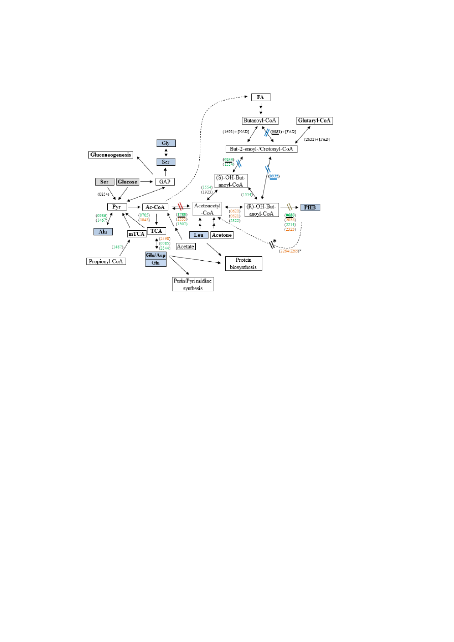
FIGURE 1. Overview of the metabolic pathways in L. pneumophila Paris relevant for PHB formation
and degradation. Key reactions investigated in this study are highlighted by underlined gene numbers.
13
C-Labeled substrates used in this study are indicated by grey boxes. Analyzed metabolites are
indicated by blue boxes. Gene numbers (lpp) are indicated in parentheses, genes given in green are
higher expressed in the exponential phase, whereas genes given in red are induced in the transmissive
(PE) phase (13). Reactions affected in the mutant strains are indicated. FA, fatty acids; *, refers to
(48). The genes indicated here are generally present in all known genomes of Legionella strains.
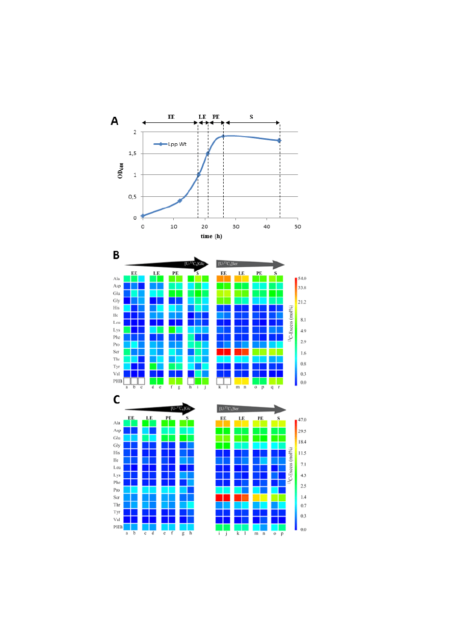
FIGURE 2. Growth-dependent incorporation of glucose or serine into L. pneumophila grown in liquid
culture. A. Schematic growth curve of L. pneumophila in AYE medium at 37 °C with indicated
periods of
13
C-labeling (EE, LE, PE and S). B.
13
C-Enrichments of amino acids and PHB of L.
pneumophila Paris wild-type (WT) grown in AYE medium at 37 °C during various growth phases
(EE, LE, PE, and S) using [U-
13
C
6
]glucose or [U-
13
C
3
]serine as precursors, respectively. Overall
13
C
excess (mol%) of labeled amino acids and PHB is given by a color map in a quasi-logarithmic form to
show even relatively small
13
C excess values. PHB indicated by white boxes could not be measured.
Each sample (from
individual labeling experiments indicated by a - r) was measured three times; the
color for each amino acid correlates with the mean value of the three measurements. Arrows on top of
the color code indicates the change in the relative incorporation rates during the growth phases. C.
Corresponding labeling data for the Δketo mutant devoid of Lpp1788, a putative key enzyme in
providing the 3-hydroxybutyryl-CoA precursor for PHB biosynthesis.
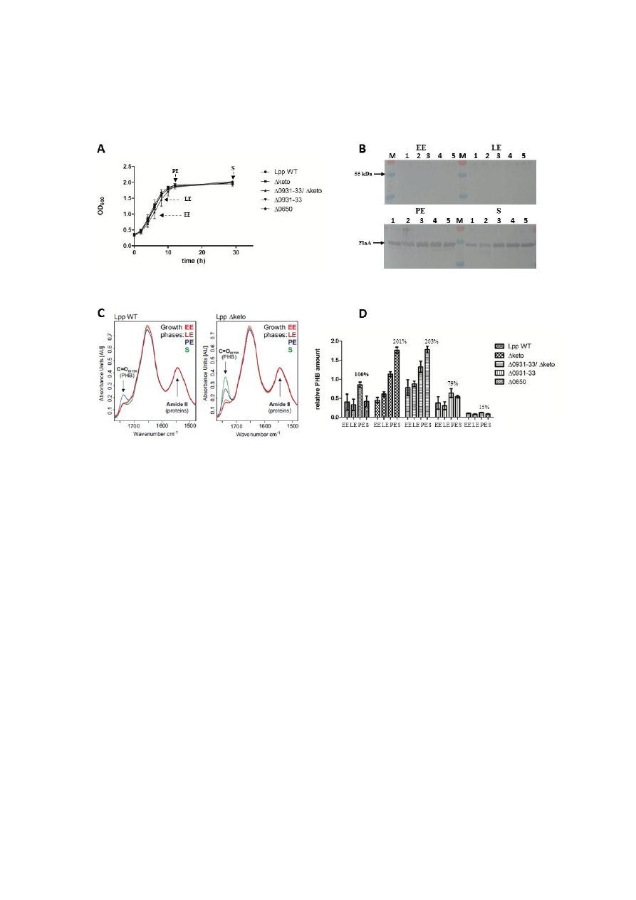
FIGURE 3. Growth phase-dependent amounts of PHB in L. pneumophila Paris wild-type (WT) and
the isogenic mutant strains Δketo, Δlpp0931-33, Δlpp0931-33/Δketo and Δlpp0650. A. Growth curve
of Lp strains grown in AYE medium at 37 °C. Time points (EE, LE, PE, S) of PHB measurement are
indicated by arrows. B. Western blot analysis of Lp Paris WT (1), Lpp Δketo (2), Lpp Δ0931-33/Δketo
(3), Lpp Δ0931-33 (4) and Lpp Δ0650 (5) using an anti-FlaA antiserum. M, protein marker. C. Pre-
processed FT-IR spectra demonstrating the relative amount of PHB of Lp Paris (Lpp WT) and
isogenic Δketo mutant strain (Lpp Δketo) in AYE medium at 37 °C at EE, LE, PE and S phases of
growth. The C=O ester (PHB) and Amide II (proteins) bands are indicated. The amount of PHB in the
Lpp Δketo mutant strain increased in the PE and S phase when compared to the Lpp WT strain D.
Relative PHB amounts in Lp Paris strains investigated by FT-IR spectroscopy. All values are mean
values of triplicate determinations (± the standard deviation) and are given in relative intensity units
(see Fig. 5). Given additional values are in percent with respect to the relative PHB content of Lpp WT
in the PE phase.
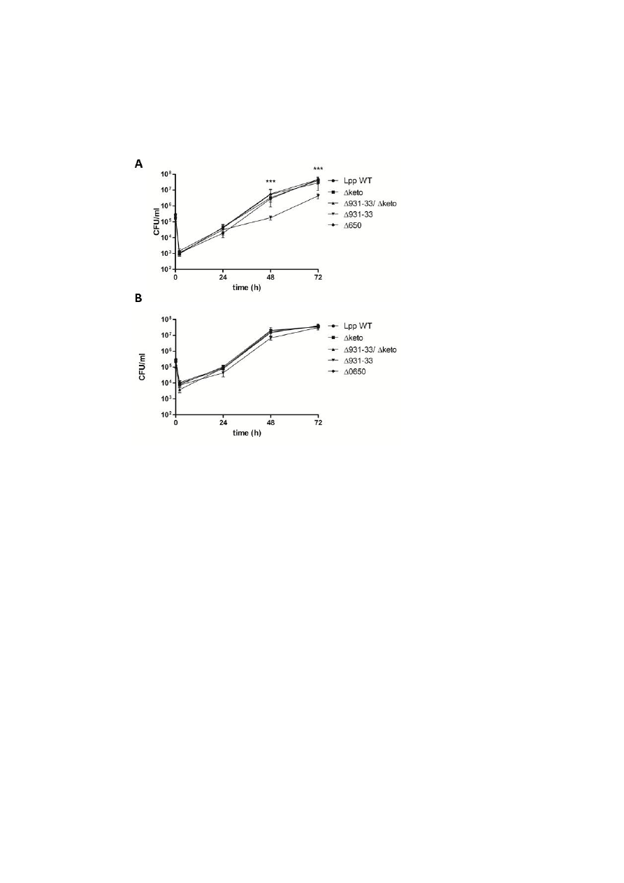
FIGURE 4. Co-culture of various L. pneumophila Paris strains with U937 cells (A) and A. castellanii
http://www.jbc.org/
Downloaded from

14
(B). Bacteria were used to infect host cells at a multiplicity of infection of 1 for 72 h. At various time
points post inoculation, bacteria were quantified by plating aliquots on BCYE agar plates to determine
the CFU/ml. Results are mean standard deviations of duplicate samples and are representative of at
least three independent experiments. Statistically significant differences in growth of Δlpp0931-33
strain to wild-type strain (determined by a student's t-test, p < 0.001) are indicated (***). Lpp, L.
pneumophila Paris; Δ, isogenic mutant strains of Lpp; 0931-33, lpp0931-33; 0650, lpp0650; keto,
lpp1788.
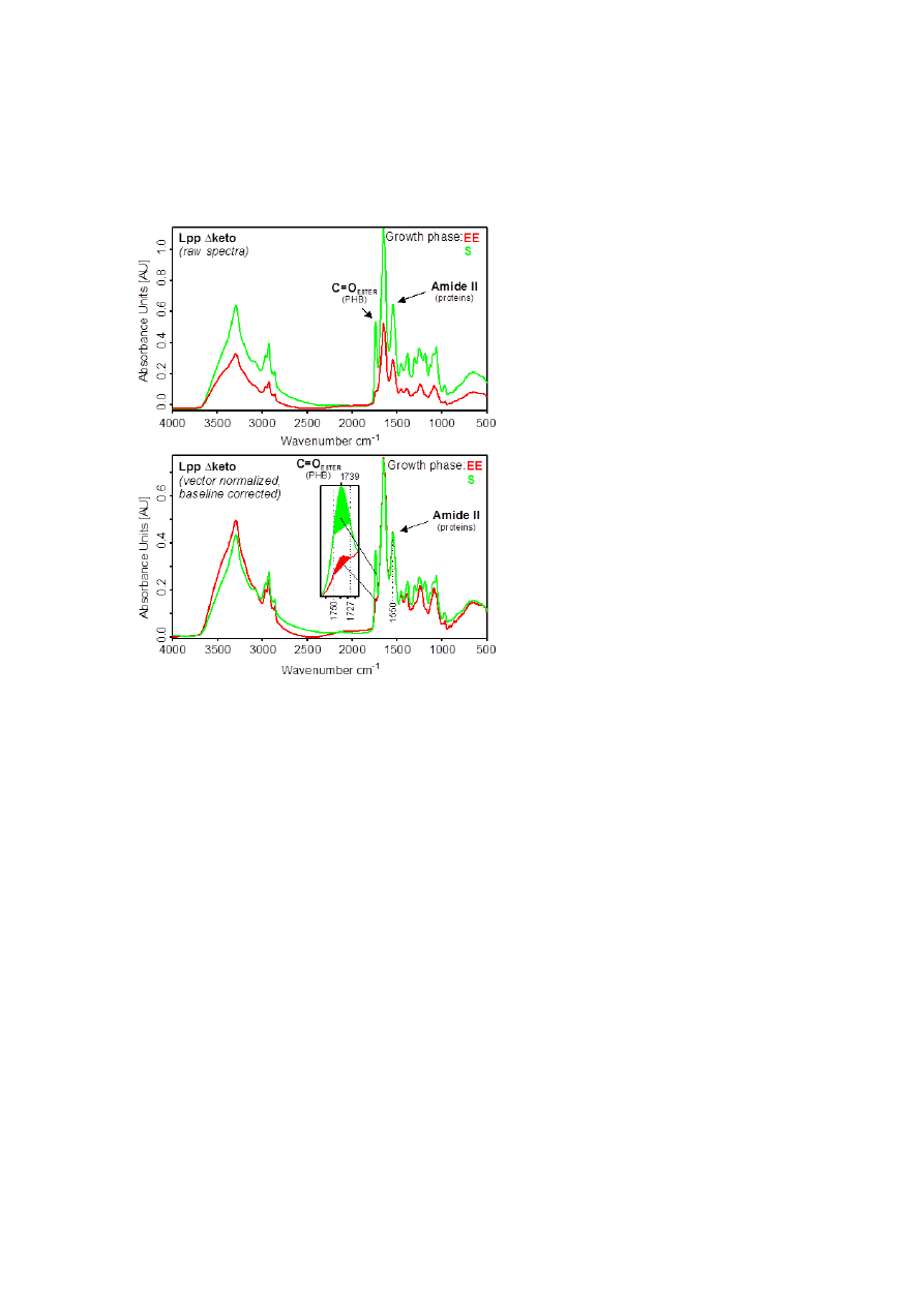
FIGURE 5. Determination of the relative PHB amounts from whole intact cells of L. pneumophila.
Upper panel, original (raw) absorbance spectra of L. pneumophila Paris Δketo (Lpp Δketo). EE: early
exponential phase; S: late stationary phase. Lower panel, pre-processed absorbance spectra of L.
pneumophila Paris Δketo. Pre-processing: vector-normalization in the amide II region (1520-1570 cm
-
1
) and offset-correction. The intensity of the ester carbonyl band around 1739 cm
-1
of spectra
normalized to the amide II band can be used to determine the relative amount of PHB present in the
cells. For this purpose, the areas under the curves are calculated between 1727 and 1750 cm
-1
(see
inset of the lower panel).
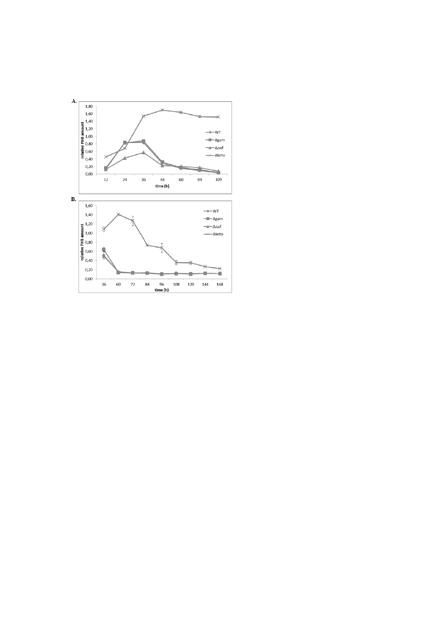
FIGURE 6. Growth-dependent relative amounts of PHB in L. pneumophila Paris wild-type (WT) and
the isogenic mutant strains Δgam, Δzwf and Δketo at 37 °C in AYE medium (A) and on BCYE agar
plates (B), investigated by FT-IR spectroscopy. All values are mean values of duplicate determinations
and are given in relative integrated intensity units (see Fig. 5).
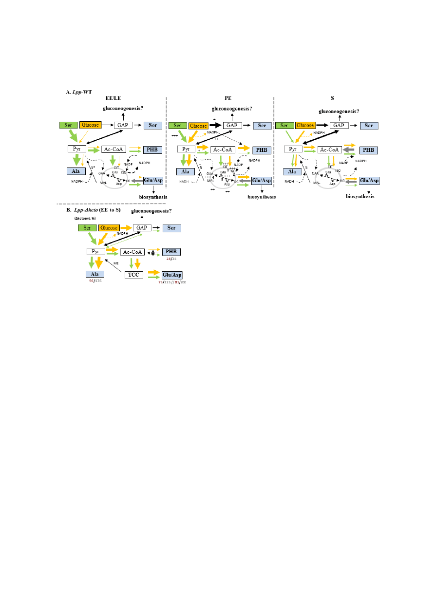
FIGURE 7. Flux model for PHB and amino acid metabolism in L. pneumophila Paris growing in
YAE medium during different growth phases. A. Carbon flux in Lp Paris during the exponential (EE,
LE), post-exponential (PE) and late stationary phase (S) using serine (green arrows) or glucose
(yellow arrows) as a substrate. Relative carbon fluxes are indicated by the thickness of the arrows. The
citrate cycle is indicated in grey. + to +++, enzymatic activity measured by Hoffman and Keen, (30);
1, aconitase; 2, isocitrate dehydrogenase; 3, Glu/Asp transaminase; 4, malate dehydrogenase; 5, malic
enzyme (MEZ, lpp3043 and 5* lpp0705). B. Carbon flux in Lp Paris Δketo mutant strain in
comparison to the wild type strain measured at the end of the growth phase. The ratios (Δketo mutant
/wild-type) of incorporation (
13
C mol%) of serine and glucose into selected amino acids or PHB are
indicated by red or grey numbers, respectively. *, inactivated lpp1788 (beta-ketothiolase). GAP,
glyceraldehyde-3-phosphate; ISO, isocitrate; CIT, citrate; KG, ketoglutarate; MAL, malate; OAA,
oxaloacetate.
http://www.jbc.org/
Downloaded from

15
TABLE 1. Primers used in this study.
Name
Tm
[°C]
Sequence 5‘ → 3‘
Reference
0931-1F
GCGAACATTAGGCTTGTCAATA
(this work)
0931-2R
GAGATTCAATCATTTTATTGCTCCACT
(this work)
0931-3R2
CATTTCTAGAAATGCCAAATGTTCATC
(this work)
0931-4F
GCTTGCTGTCATAAGGAAGTATC
(this work)
iLpp_0650_1U
CCGCGGGAATTCGATATCCTTTTAGCCACGATTT
ACTCCACTT
(this work)
iLpp_0650_2R
TAGAAGCTGACATTCTAGCTCCTGAAAGCAAAT
AATCGAA
(this work)
iLpp_0650_5U
TAGACACGATGGCCGTGGATGCCCCAGGGAGTT
ATGTACT
(this work)
iLpp_0650_6R
GAATTCACTAGTGATATCAGCCCTTATTTTAGCC
TTTGTTGTC
(this work)
iLpp_0650_3U
TGCTTTCAGGAGCTAGAATGTCAGCTTCTAGAC
TATCTGG
(this work)
iLpp_0650_4R
TCCCTGGGGCATCCACGGCCATCGTGTCTAGAC
ACTCCTG
(this work)
iLpp_0650T1U
CTTTTAGCCACGATTTACTCCACTT
(this work)
iLpp_0650T6R
AGCCCTTATTTTAGCCTTTGTTGTC
(this work)
Lpp_0650_Mut
_1U
TCAGGTTCGCCTTTTATTGC
(this work)
Lpp_0650_Mut
2R
AATTCCTGTCCTGCCTTCAG
(this work)
Lpp_0650_Wt_
1U
CTTTCATCGCTGGTCAGTCA
(this work)
Lpp_0650_Wt_
2R
ATGAACCGGAGTGTTCCTTG
(this work)
M13R
54.5
GGAAACAGCTATGACCATG
51
M13U
52.8
GTAAAACGACGGCCAGT
51
TABLE 2. PHB content [in %] of L. pneumophila strains as seen by FT-IR spectroscopy.
Percentage values (mean values from three independent cultivations) were obtained from experimental
FT-IR spectra with respect to the PHB content of Lpp WT in the PE phase.
Lpp strain
EE
LE
PE
S
WT
45
37
100
46
Δketo
51
69
131
201
Δ0931-33
55
37
79
62
Δ0931-33/Δketo
84
100
152
203
Δ0650
12
10
15
10
http://www.jbc.org/
Downloaded from

Heuner
Vroni Herrmann, Maren Stämmler, Peter Lasch, Wolfgang Eisenreich and Klaus
Nadine Gillmaier, Eva Schunder, Erika Kutzner, Hana Tlapák, Kerstin Rydzewski,
Legionella pneumophila
Growth-Related Metabolism of the Carbon Storage Poly-3-Hydroxybutyrate in
published online January 20, 2016
J. Biol. Chem.
Access the most updated version of this article at doi:
Alerts:
When a correction for this article is posted
to choose from all of JBC's e-mail alerts
http://www.jbc.org/content/early/2016/01/20/jbc.M115.693481.full.html#ref-list-1
This article cites 0 references, 0 of which can be accessed free at
http://www.jbc.org/
Downloaded from
Wyszukiwarka
Podobne podstrony:
Biol Chem
prog biol naucz biol-chem-1, biotechnologia 2 sem rok2, pobrane z góry DS 7, z góry, Rok II, Inne
Porównanie biosyntezy u Procaryota i Eucaryota, MATURA - BIOL-CHEM, biologia, notatki biologia, nota
ZAKRES MATERIAŁU DO PRZEROBIENIA - MATURA Z BIOLOGII, Dla biol-chem LICEUM, biologia liceum !! Zadan
UKŁAD ODDECHOWY biol-chem, biologia- studia, Biologia
hybrydyzacja, Dla biol-chem LICEUM, chemia MATURA - rozszerzona
biol maj 2016 pods
Biol Chem
biol maj 2016 rozsz odp
biol chem bielsko
biol maj 2016 pods odp
defosfatacja biologiczna, IŚ Tokarzewski 27.06.2016, V semestr ISiW, Technologie oczyszczania ściekó
2 Budowa i działanie br jadr chem i biol
Mikrobiologia Woda właściwosci fiz, chem, biol i bakteriologiczne
Biol kom cz 1
Biol Mol wyklad 9
więcej podobnych podstron