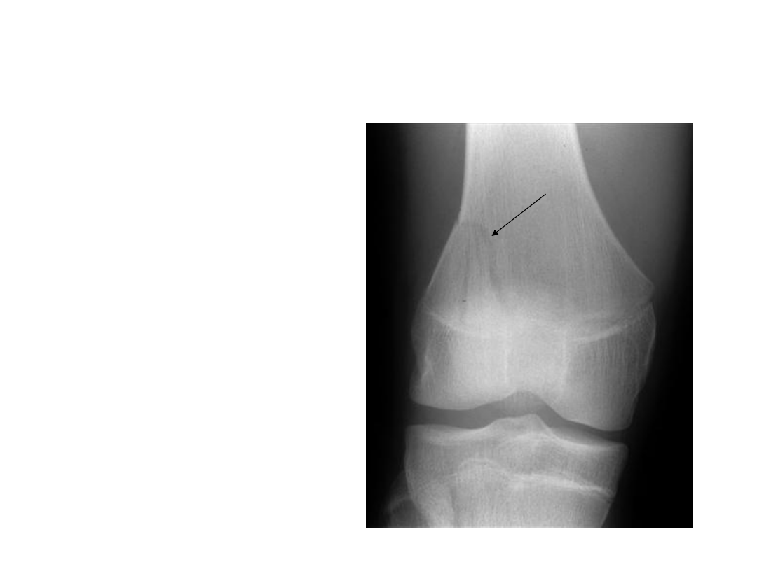
Salter Harris II fracture
• Salter Harris II fractures
are by far the most
common accounting for
approximately 75-80%
of physeal fractures. The
line traverses part of
the physis and a
corner of the
metaphysis. The
metaphyseal fragment
is sometimes referred to
as the Thurston Holland
fragment. Common sites
for Salter - Harris II
fractures include: the
distal radius, femur
and tibia
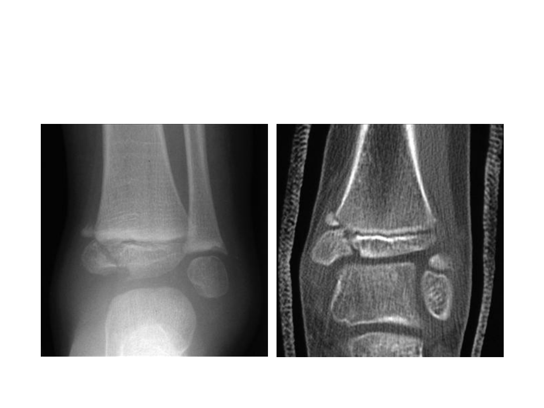
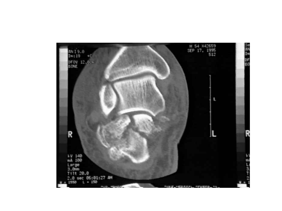
SalterHarris IV
fracture (a) radiograph and (b) coronal CT. There is
an oblique fracture passing through both the epiphysis and the
metaphysis with medial displacement
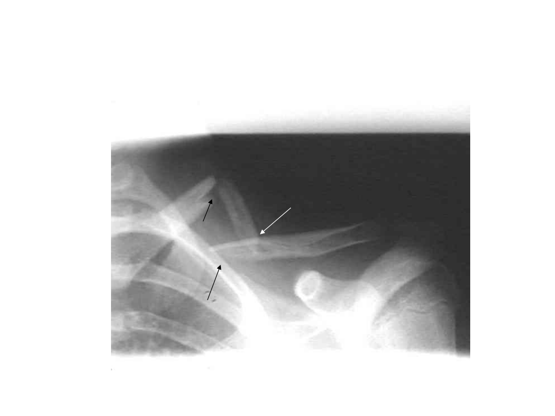
Clavicle - fracture
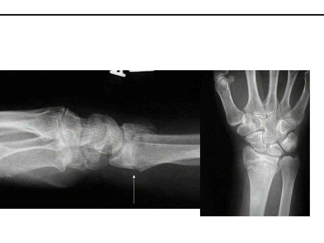
Dorsal Barton's Fracture / Dorsal Shearing Frx
- distal radius fracture w/ dislocation of radiocarpal joint;
- most common frx dislocation of the wrist joint
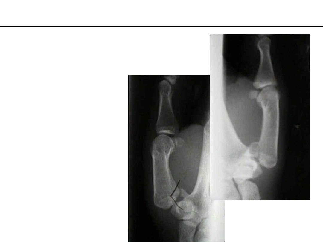
Bennett's Fracture Dislocation
- - most frequent of all thumb frx;
- described in 1882 by Dr. Edward Bennet;
- it is a frx dislocation, intra-articular frx at base of carpometacarpal joint of the thumb;
- mechanism of frx:
- results from axial blow directed against the partially flexed metacarpal; (ie. from fist fights)
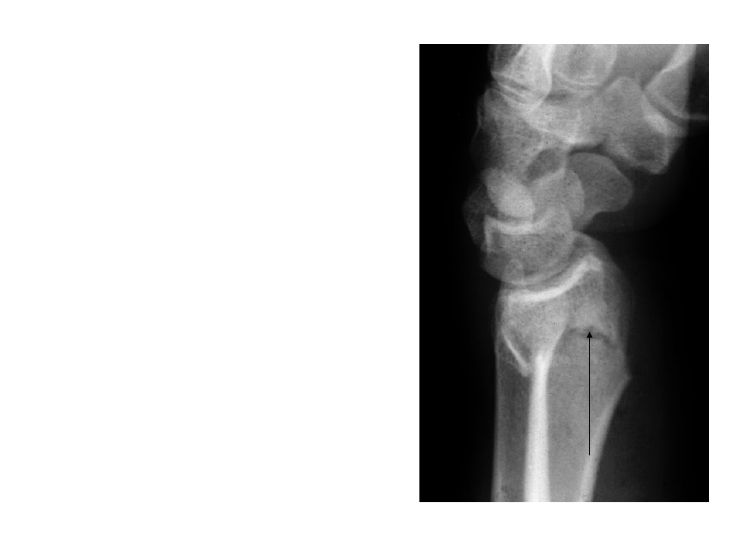
Colles’ fra
cture
•
A transverse that
extends from the
volar to the dorsal
surface of the distal
end of the radius.
This injury is
accompanied by
impaction and
displacement of the
dorsal radial surface
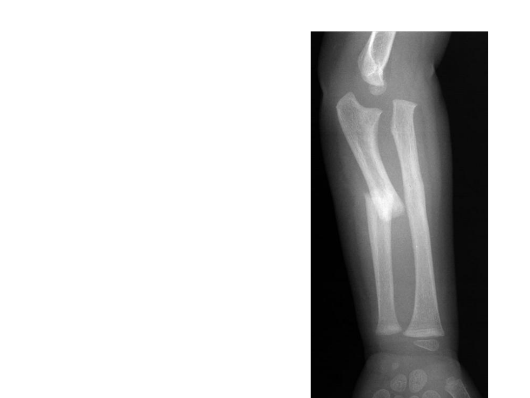
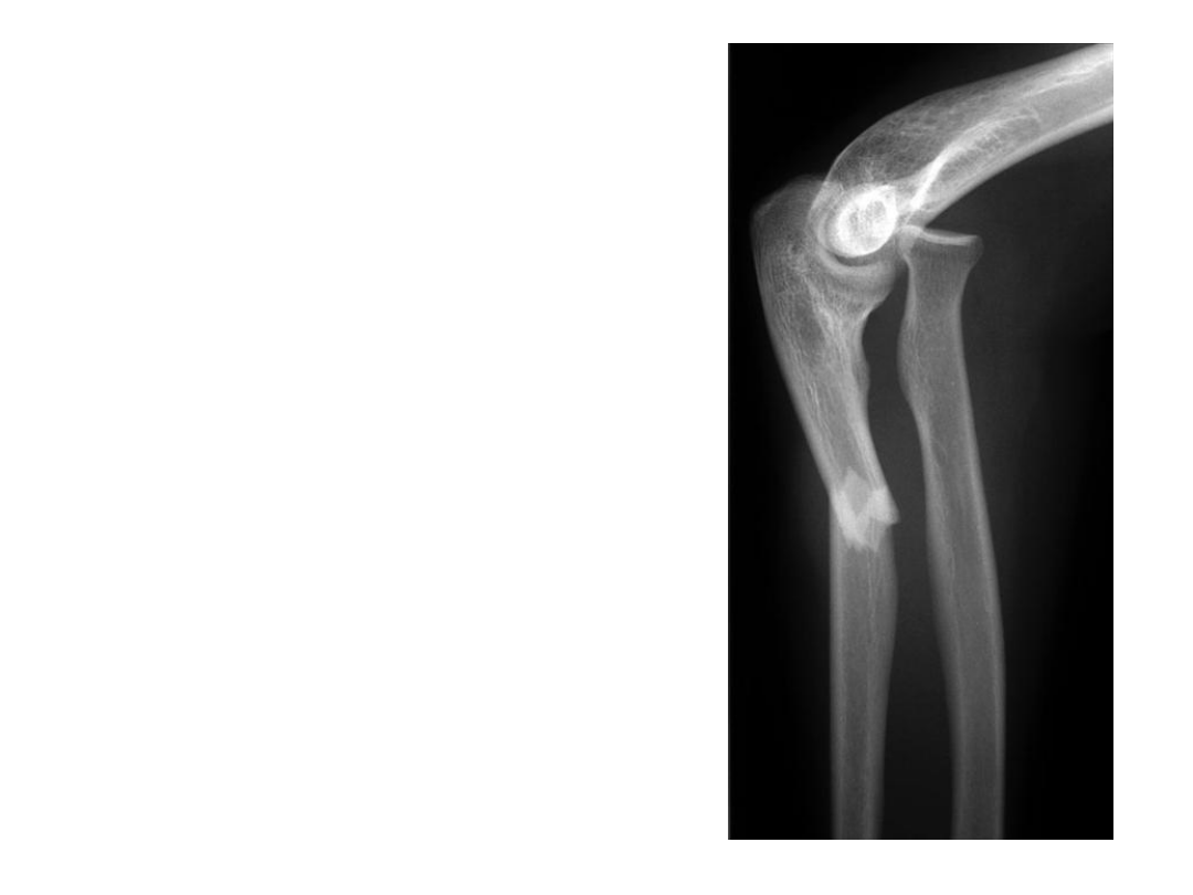
Monteggia fr
acture
• a fracture of the
proximal third of
the ulnar shaft in
conjunction with
radial head
dislocation and
tearing of the
annular ligament
Monteggia fracture -
dislocation
• A combined fracture of the ulna and
dislocation of the radial head.
• Type I: fracture of the middle or upper
third of the ulna with anterior dislocation
of the radial head and anterior angulation
of the ulna
• Type II: fracture of the middle or upper
third of the ulna with posterior dislocation
of the radial head and posterior angulation
of the ulna
• Type III: fracture of the ulna just distal to
the coronoid process with lateral
dislocation of the radial head
• Type IV: fracture of the upper or middle
third of the ulna with anterior dislocation
of the radial head and fracture of the upper
third of the radius below the bicipital
tuberosity.
• Type I injuries occur most frequently
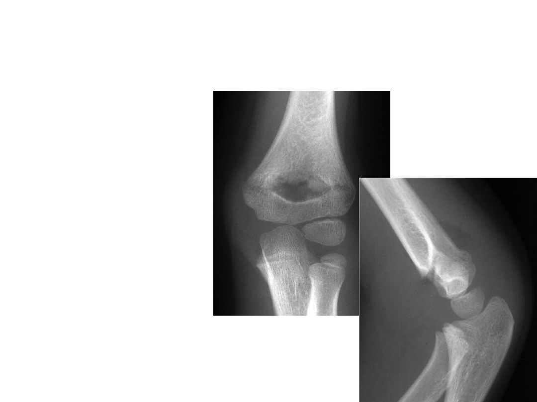
Supracondylar fracture
• a transverse
metaphyseal
fracture of the distal
humerus
• the commonest
elbow fracture in
childhood
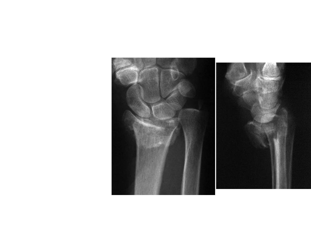
Smith’s fracture
• a fracture of the
distal portion of
the radius with
palmar
displacement.
• This type of injury
is also termed a
reverse Colles
fracture or reverse
Bartons fracture.
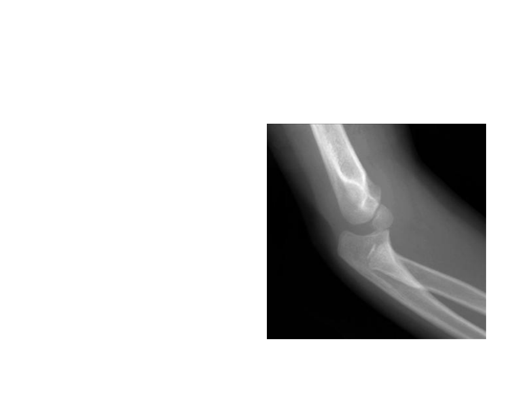
Elbow dislocation
• usually involves
displacement of
the proximal
radius and ulna
articulation from
the humerus
without disruption
of the radioulnar
articulation
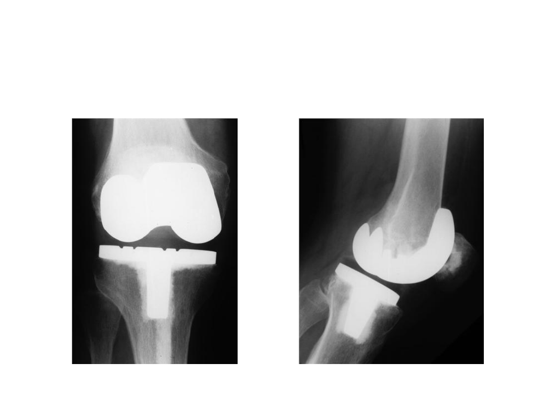
Total knee replacement
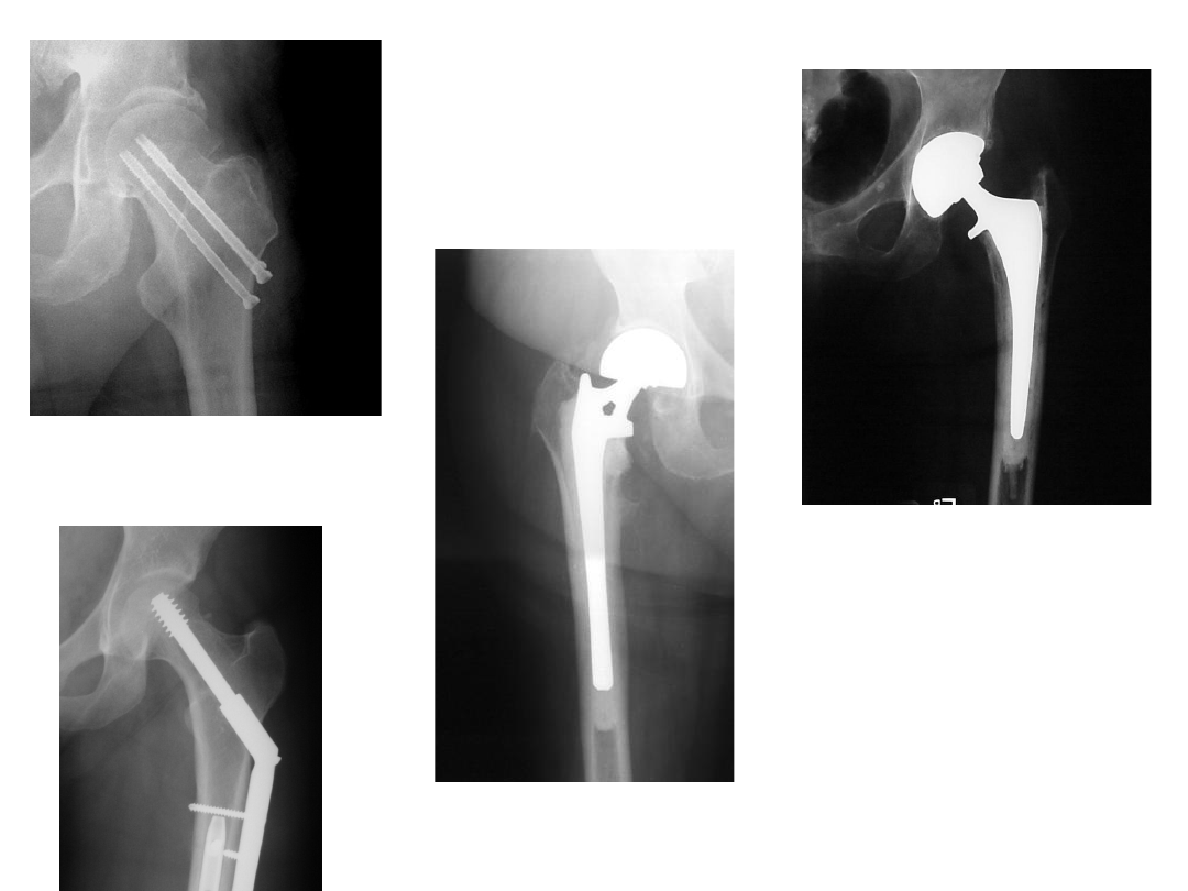
ORIF
Hemi
THR
-Total hip
replacement
FEMORAL NECK
FRACTURES
TREATMENT

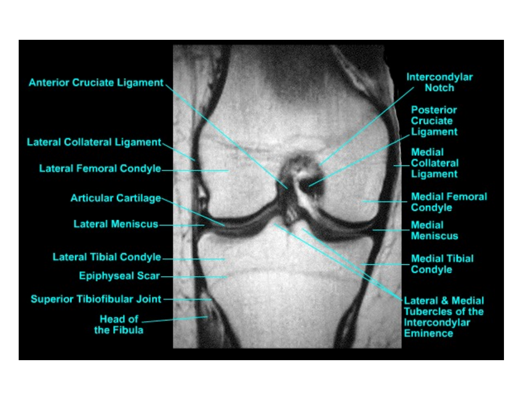
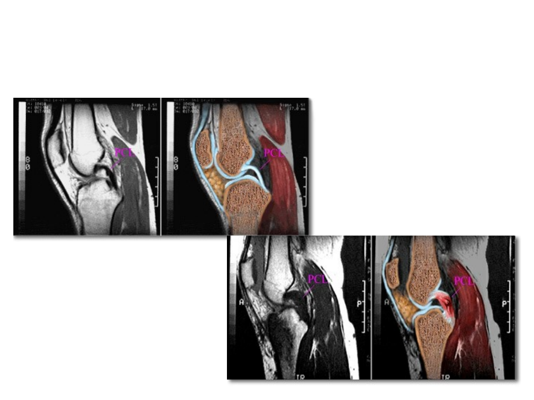
Cruciate ligaments
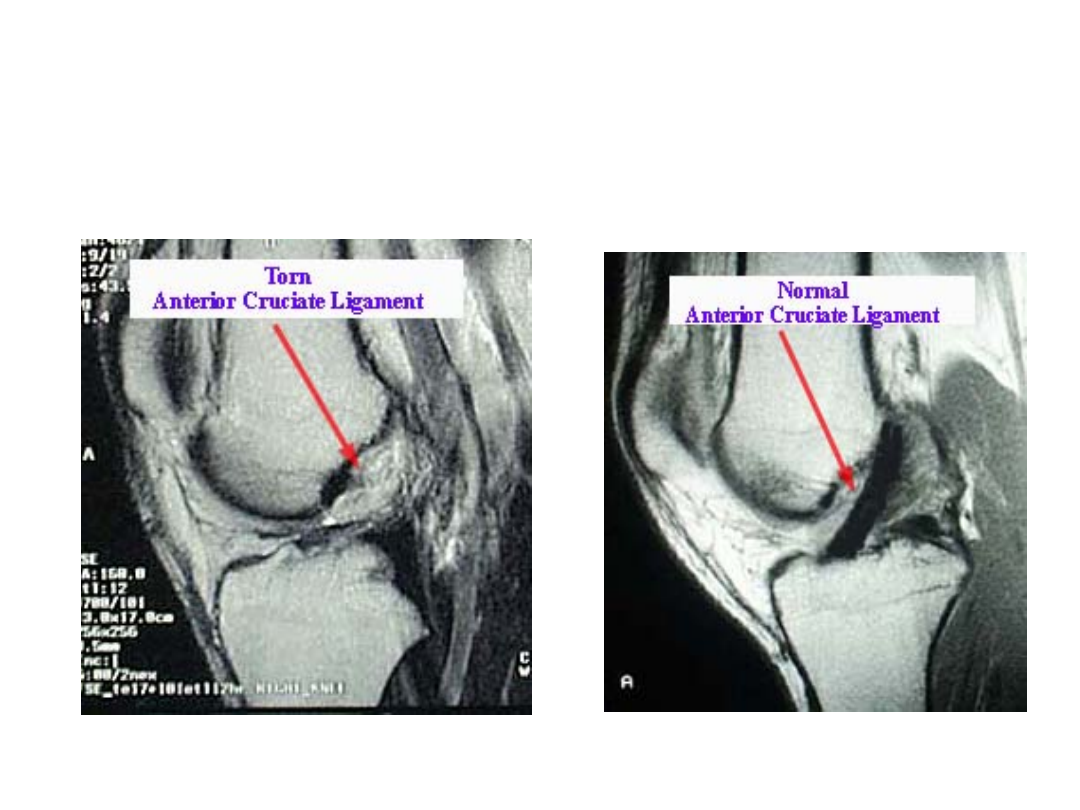
Diagnostic Imaging
Diagnostic Testing
Normal MRI
Torn PCL
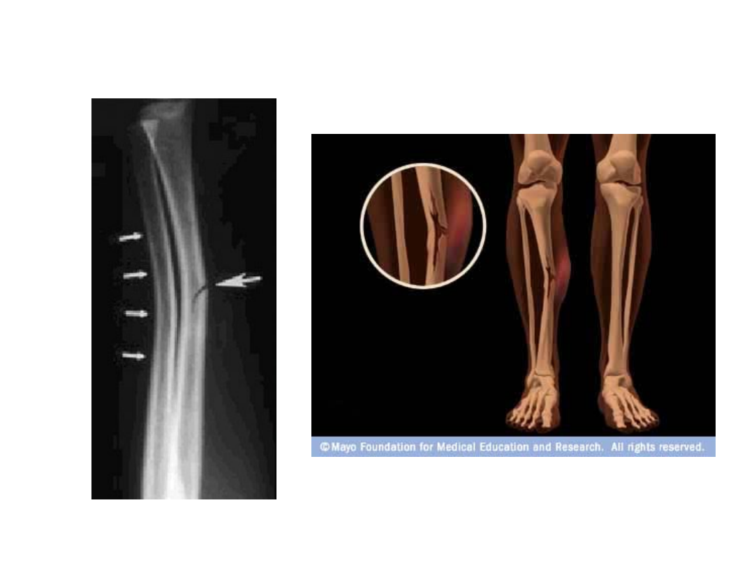
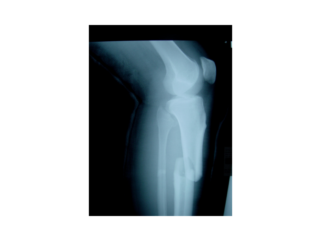
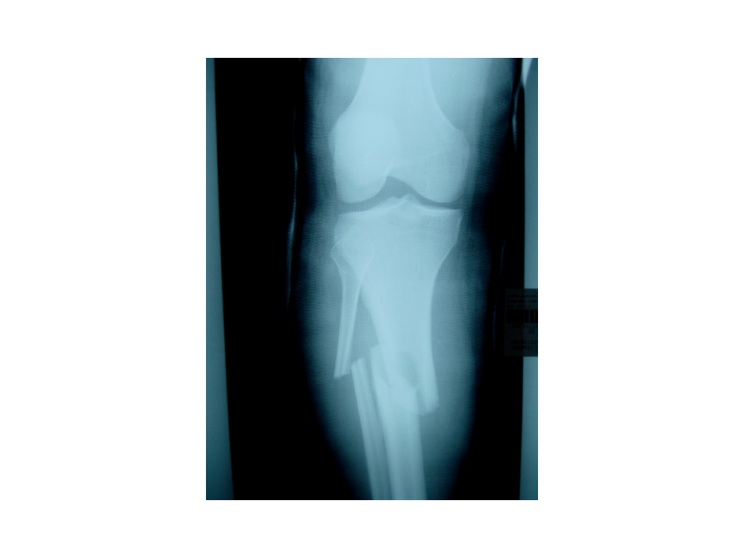
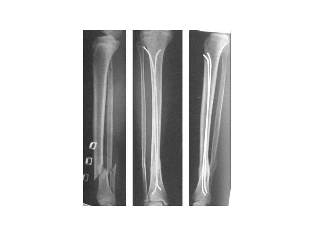
Greenstick fractures
Calcaneus fracture
Document Outline
- Slide 1
- Slide 2
- Slide 3
- Slide 4
- Slide 5
- Slide 6
- Slide 7
- Slide 8
- Slide 9
- Slide 10
- Slide 11
- Slide 12
- Slide 13
- Slide 14
- Slide 15
- Slide 16
- Slide 17
- Slide 18
- Slide 19
- Slide 20
- Slide 21
- Slide 22
- Slide 23
- Slide 24
- Slide 25
- Slide 26
- Slide 27
- Slide 28
- Slide 29
- Slide 30
Wyszukiwarka
Podobne podstrony:
Urazy kończyn
Urazy kończyn, biologia, Pierwsza pomoc
Urazy konczyn gornych
Urazy konczyn dolnych
Urazy kończyn(1)
Urazy kończyń krótko
1pomoc Urazy kończyn
Urazy glowy konczyn i tulowia
Urazy miednicy i konczyn dolnych
NAJCZĘSTSZE URAZY W OBRĘBIE KOŃCZYNY GÓRNEJ
L1 - Urazy kostno - stawowe kończyn, Ratownictwo Medyczne, Materiały ze studiów, Medycyna Ratunkowa
Urazy głowy, klatki piersiowej, kończyn
Urazy glowy konczyn i tulowia
Urazy miednicy i konczyn dolnych
Urazy głowy, tułowia i kończyn
Urazy narządu ruchu
więcej podobnych podstron