21
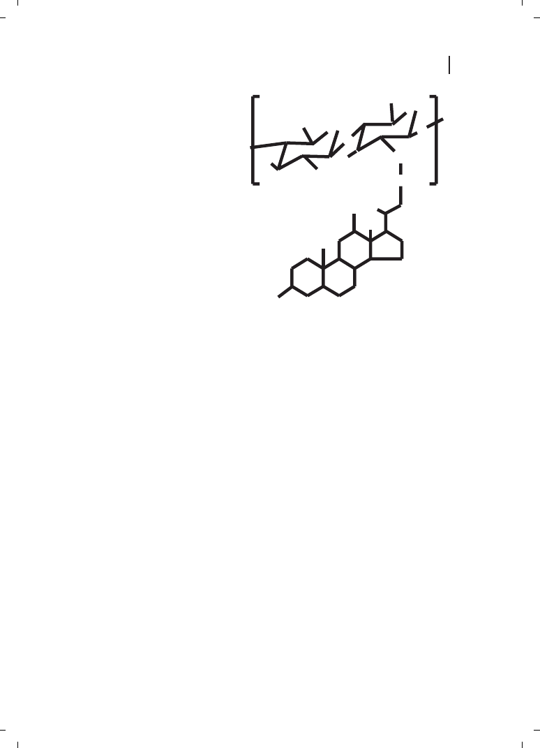
Nanoparticles as Non-Viral Transfection Agents
M. N. V. Ravi Kumar, Udo Bakowsky, and Claus-Michael Lehr
21.1
Introduction to Gene Delivery
Direct injection of naked DNA plasmids is possible, but relatively few cells take up the
DNA (1–3 %), leading to a small production of the encoded protein. The most important
use of naked DNA plasmids is in vaccine development, as the small amount of protein
produced can elicit a protective immune response. However, in most of the cases, it is
also well established that naked DNA is not suitable for in-vivo transport of genetic ma-
terial into selected cell body types due to its degradation by serum nucleases; hence, the
use of a carrier system is suggested. Unfortunately, there is no single system universally
applicable in vivo. An ideal system should be biocompatible, nonimmunogenic, and stable
in the bloodstream, protect DNA during transport, avoid entrapment by components of
the reticuloendothelial system (RES), be small enough to extravagate, and should be
cell- or tissue-specific to reach selected cells in the body. The delivery system is expected
319
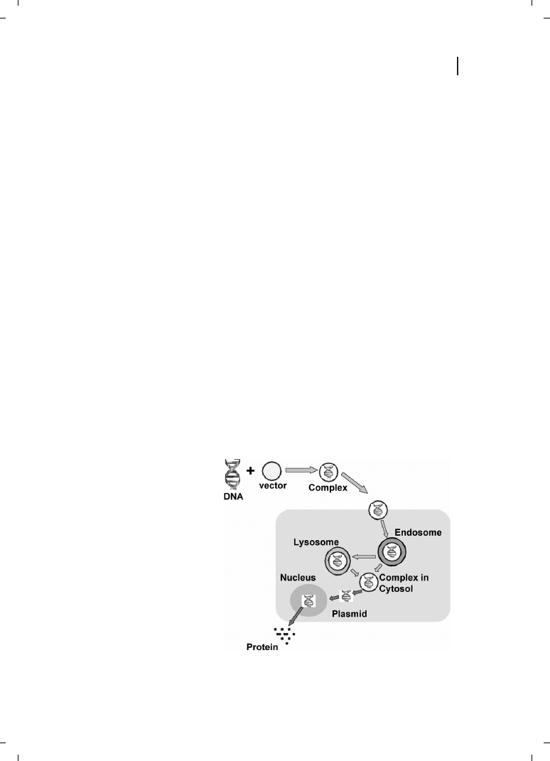
Figure 21.1
Schematic representation
of biological barriers that need to be
overcome by the gene transfer vector.
Nanobiotechnology. Edited by Christof Niemeyer, Chad Mirkin
Copyright
c 2004 WILEY-VCH Verlag GmbH & Co. K aA, Weinheim
ISBN 3-527-30658-7
G

to enter the cell via endocytosis, thereby avoiding its interaction with lysosomal enzymes,
and to facilitate endosomal escape, resulting in DNA delivery to the nucleus, as shown in
Figure 21.1. Thus, it is quite obvious that a successful gene delivery system must contain a
variety of structural elements responsible for the specific behavior.
In gene therapy, plasmid DNA is introduced into cells of patients to express the thera-
peutic proteins. On the other hand, an oligonucleotide is used to suppress the expression
of a disease-causing gene in antisense therapy. The clinical application of these new types
of gene-drugs is severely hindered by their instability in biological fluids and the low cel-
lular uptake efficiency due to the high molecular weight and polyanionic nature of the
nucleic acids. Thus, it is necessary to develop an efficient delivery system that can transfer
the gene/DNA in to the target site. So far, two major approaches have been tried and are
in use for gene delivery: these are viral vectors and nonviral vectors.
In spite of their relatively high efficacy, several major problems are associated with the
use of viral delivery systems in clinical treatment, particularly in relation to the risk of an
immune response against viral particles, and also to the risk of random integration
mediated by viruses or their recombination with wild-type viruses [1]. In an efficient
gene therapy, plasmid DNA is introduced into target cells, transcribed and the genetic in-
formation ultimately translated into the corresponding protein (see Figure 21.1).
Successful/efficient transfection is hampered by: (i) targeting the delivery systems to
the target cell; (ii) transport through the cell membrane; (iii) uptake and degradation in
endolysomes; and (iv) intracellular trafficking of plasmid DNA to the nucleus. Although,
viral vectors yield high transfection efficiency over a wide range of cell targets [2, 3], they
present major drawbacks, such as virally induced inflammatory responses and oncogenic
effects [4].
There is need for the development of safer and more effective gene delivery vehicles.
These should offer freedom to manipulate the complex stoichiometry, surface charge den-
sity, and hydrophobicity needed for interaction with the cellular lipid components. Catio-
nic phospholipids and cationic polymers are the two major types of nonviral gene delivery
vectors currently being investigated. Due to their permanent cationic charge, both types
interact electrostatically with negatively charged DNA and form complexes (lipo- or poly-
plexes). Despite the ease of fabrication of the lipoplexes, their low transfection efficiency
and toxicity has limited its success. However, polyplexes involving cationic polymers, on
the other hand are more stable than cationic lipids [5], although the transfection is rela-
tively low when compared to viral vectors. Cationic polymers have been used to condense
and deliver DNA both in vitro and in vivo. Several cationic polymers have been investi-
gated that lead to higher transfection efficiencies when compared to the other nonviral
vectors in use [5, 6]. They form polyelectrolyte complexes with plasmid DNA, in which
the DNA becomes better protected against nuclease degradation [7]. They also show struc-
tural variability and versatility, including the possibility of linking the targeting moieties
for gene expression mediated through specific receptors [5].
320
21 Nanoparticles as Non-Viral Transfection Agents
21.2
Nanoparticles for Drug and Gene Targeting
Pharmaceutical nanoparticles were first developed by Speiser and co-workers [8] during
the 1970s, and are defined as solid colloidal particles, less then 1 mm in size, that consist
of macromolecular compounds. Since then, a considerable amount of work has been car-
ried out on nanoparticles worldwide in the field of drug/gene delivery. Nanoparticles were
initially devised as carriers for vaccines and anticancer drugs [9], but their use for ophthal-
mic and oral delivery has also been investigated [10]. Drugs or other biologically active mo-
lecules are dissolved, entrapped or encapsulated in the nanoparticles, or are chemically
attached to the polymers or adsorbed to their surface. The selection of an appropriate
method for preparing drug-loaded nanoparticles depends on the physico-chemical proper-
ties of the polymer and the drug. On the other hand, the procedure and the formulation
conditions will determine the inner structure of these polymeric colloidal systems. Two
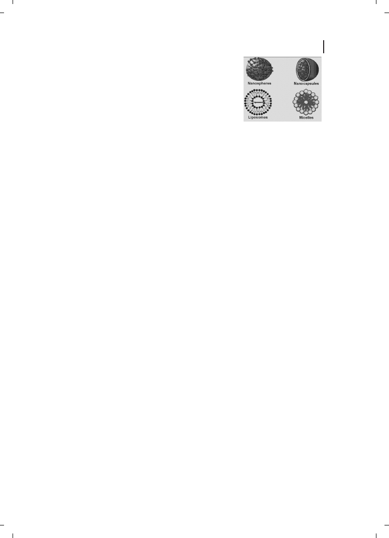
types of systems with different inner structures are possible :
x
A matrix-type system composed of an entanglement of oligomer or polymer units, de-
fined here as a nanoparticle or nanosphere.
x
A reservoir type system, consisting of an oily core surrounded by a polymer wall, defined
here as a nanocapsule.
Various colloidal nanoparticulate systems in use for drug/gene delivery are shown in Fig-
ure 21.2. In this chapter, we will discuss various nanoparticulate systems which are used
as gene carriers. Great care has been taken in compiling the literature; however, any omis-
sion in the references is purely inadvertent and is highly regretted.
21.3
Nonviral Nanomaterials in Development and Testing
21.3.1
Chitosan
Chitin is the most abundant natural aminopolysaccharide, and its annual production is
estimated to be almost as much as that of cellulose (Figure 21.3). Chitin has become of
major interest not only as an underutilized resource, but also as a new functional material
of high potential in various fields, and recent progress in chitin chemistry is quite note-
worthy. Chitosan is an aminopolysaccharide obtained by the alkaline deacetylation of
321
21.3 Nonviral Nanomaterials in Development and Testing
Figure 21.2
Structures of various nanoparticles in use for pharma-
ceutical applications.
chitin, a cellulose-like polymer which is present in fungal cell walls and the exoskeletons
of arthropods such as insects, crabs, shrimps, lobsters, and other vertebrates [11]. Chito-
san is a nontoxic, biocompatible polymer that has found a number of applications in drug
delivery [12], and recently has emerged as an alternative nonviral gene delivery system
[13]. Borchard [14] recently described chitosans as efficient gene delivery systems in his
review.
MacLaughlin et al. [15] studied chitosan and depolymerized chitosan oligomers as con-
densing carriers for in-vivo plasmid delivery. For forming the complexes with plasmid,
each chitosan oligomer or polymer (2 %, w/v) was dissolved in acetic acid by sonication
and the final concentration of the solution was brought to 0.4 % (w/v) by adding water;
the solution was sterile-filtered to remove particles above 200 nm. This chitosan solution
is then added to aqueous suspension containing 25–400 mg of plasmid in a total volume
of 1 mL by gentle pipetting to form nanocomplexes of a selected charge ratio [14]. The
solution was vortexed rapidly for 3–5 minutes and left for 30 minutes at room tempera-
ture to ensure complete complexation [15]. The in-vitro transfection was tested in Cos-1
322
21 Nanoparticles as Non-Viral Transfection Agents
Figure 21.3
Structural similarities between cellulose, chitin, and chitosan.

cells, and in-vivo expression in intestinal tissues. In vivo, higher levels of expression were
measured with plasmid/chitosan/GM225.1 formulation over naked plasmid in the upper
small intestine, the overall expression levels achieved using the DOTMA :DOPE based for-
mulation were lower than those achieved with plasmid/chitosan/GM225.1 complexes, and
there were a lower number of tissue extracts showing positive chloramphenicol acetyl-
transferase (CAT) gene expression (Table 21.1). It was interesting however to note that
the in-vitro results were the reverse of these findings. The parameters which influenced
323
21.3 Nonviral Nanomaterials in Development and Testing
Table 21.1
CAT gene expression in tissue extracts of rabbits dosed in the upper small intestine or colon
with plasmid/chitosan complexes
Formulation
Region
Rabbit number
CAT expression
*
#1
#2
#3
#4
[pg mg
–1
]
Dosed in upper small intestine
PP1
0
0
0
0
0
e 0
PP2
0
0
0
0
0
e 0
Plasmid/CT/GM225.1
PP3
6.12
5.01
0
5.83
4.24
e 2.87
(n = 4)
ENT1
1.45
0
8.10
6.62
4.04
e 3.92
ENT2
0
4.50
0
9.60
3.52
e 4.57
ENT3
6.16
0
11.41
4.32
5.47
e 4.72
Col
0
0
0
0
–
MLN
0
0
3.68
10.96
3.66
e 5.17
Plasmid in 10 % lactose
PP3
0
0
–
–
0
e 0
(n = 2)
ENT1
0
0
–
–
0
e 0
MLN
0
0
–
–
0
e 0
Plasmid/DOTMA :DOPE
PP1
0
0
0
6.75
1.69
e 3.37
(n = 4)
PP2
0
0
0
0
0
e 0
PP3
0
8.85
0
6.83
3.9
e 2.46
ENT1
0
0
0
10.13
2.53
e 5.07
ENT2
0
0
0
0
0
e 0
ENT3
0
0
0
0
0
e 0
Col
0
0
0
4.74
1.19
e 2.37
MLN
0
0
0
4.60
1.15
e 2.30
Dosed in the colon
Plasmid/CT/GM225.1
Col1
0
0
0
6.30
1.58
e 3.20
(n = 4)
Col2
0
0
7.47
6.30
3.57
e 4.13
Col3
0
0
0
0
0
e 0
MLN
5.97
10.11
12.14
0.00
7.06
e 5.36
Plasmid in 10 % lactose
PP3
0
0
–
–
0
e 0
(n = 2)
ENT1
0
0
–
–
0
e 0
MLN
0
0
–
–
0
e 0
Plasmid/DOTMA :DOPE
Col1
0
0
0
0
0
e 0
(n = 4)
Col2
0
0
0
0
0
e 0
Col3
0
0
0
0
0
e 0
MLN
0
0
0
0
0
e 0
*
Values ae mean
e SD.

particle size and stability included chitosan molecular weight, plasmid concentration, and
the charge ratio. Plasmid/chitosan complexes made of higher molecular-weight chitosan
were more stable to salt and serum challenge. Complexes of a 1 :2 (–/+) charge ratio were
shown to be most stable. In vitro, the highest level of expression in the absence of serum
was obtained using a 1 :2 (–/+) complex made with 102 kDa chitosan, and was approxi-
mately 250-fold lower than that observed with a positive control Lipofectamine
ä. Surpris-
ingly, particle size was found not to influence the expression, but inclusion of the pH-
sensitive endoosomolytic peptide GM227.3 in the formulation enhanced the levels of ex-
pression with the 1 :2 :0.25 (–/+/–) complex (200 mg mL
–1
), though expression levels were
very low (100-fold less) when compared to that of Lipofectamine.
Detailed investigations on chitosans as efficient gene transfection agents in vitro and in
vivo by Leong and co-workers [16–20] resulted in a series of papers describing various
modifications. They investigated the important parameters for the preparation of nanopar-
ticles and characterized the physico-chemical properties of the system. The protection for
encapsulated DNA by chitosan particles was confirmed [19]. These authors have also in-
vestigated the effects of co-encapsulating a lysoosomolytic agent chloroquine, on the trans-
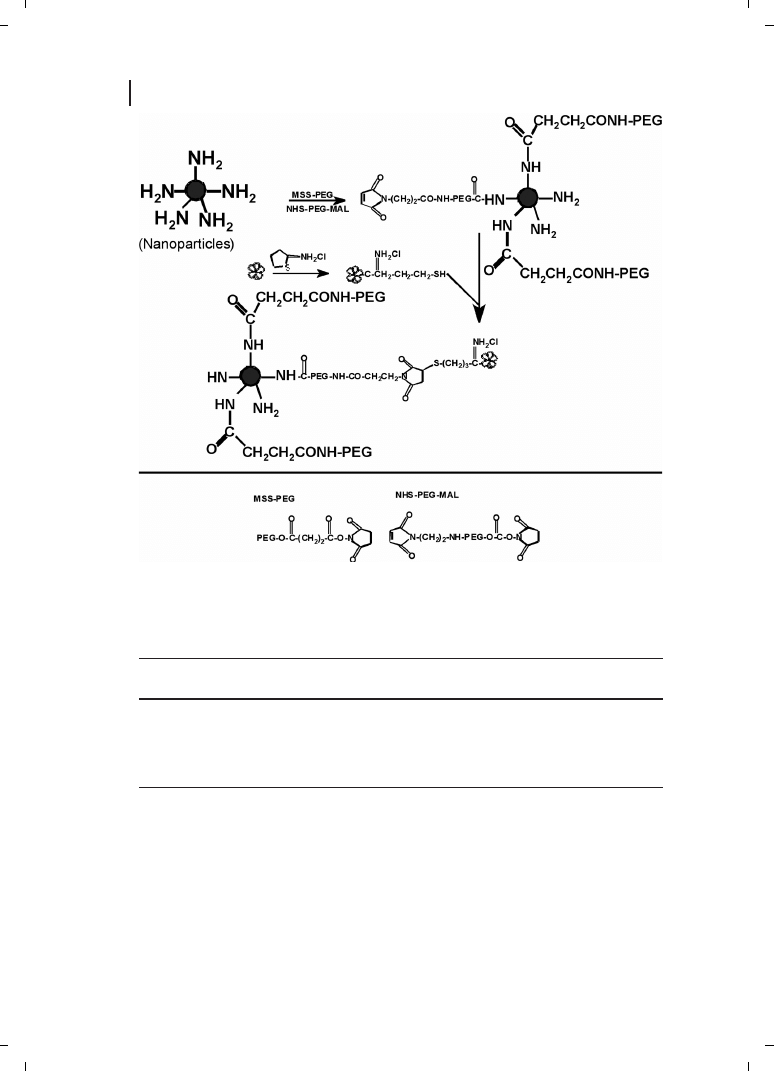
fection efficiency [19]. They have also proposed plausible schemes (Figure 21.4) for trans-
ferrin and KNOB protein conjugation in an attempt to improve the surface property re-
sulting in improved transfection efficiency [19]. Furthermore, the chitosan-DNA particles
were PEGylated (Figure 21.5) to improve their storage stability and to yield a formulation
that could be lyophilized without loss of the transfection ability [19]. Tissue distribution of
the chitosan-DNA nanoparticles and PEGylated nanoparticles following intravenous ad-
ministration was investigated [19], and clearance of the PEGylated nanoparticles was
found to be slower than that of the unmodified particles at 15 minutes, but with higher
deposition in the kidney and liver, though no difference was seen at the 1-hour time point
[19]. It was noted that the transfection efficiency was cell type-dependent, with three to
four orders of magnitude (in relative light units) higher than background level in
HEK293 cells, and two- to ten-fold lower than that achieved by Lipofectamine [19]. Moha-
patra and co-workers [20] used these chitosan nanoparticles [19] to demonstrate the pro-
tection factor of the particles towards acute respiratory syncytial virus infection in
BALB/c mice following intranasal gene transfer.
Hoggard et al. [21] used chitosan nanoparticles particles made from different molecular
weight chitosans (Table 21.2) for intratracheal administration in mice, and compared the
results obtained with those reported for polyethyleimine (PEI) in vitro. Following intratra-
cheal administration, both polyplexes were seen to be distributed to the mid-airways,
where transgene expression was observed in virtually every epithelial cell, using a sensi-
tive pLacZ reported containing a translational enhancer element [21]. However, a similar
kind of result as others was observed, where PEI polyplexes induced more rapid onset of
gene expression than that of chitosans [21].
Sato and co-workers [22], in their studies, optimized the transfection conditions for chit-
osan-mediated gene delivery. The transfection studies were carried out using a tumor cell
line (Human-lung Carcinoma A549 cells, HeLa cells and B16 melanoma cells; 1
q 10
5
cells/well). The transfection efficiency of the chitosan complexes was seen to depend
on the pH of the culture medium, the stoichiometry of pGL3 :chitosan, serum and the
molecular mass of chitosan [22]. Transfection efficiency at pH 6.9 was higher than that
324
21 Nanoparticles as Non-Viral Transfection Agents

at pH 7.6, and the optimum charge ratio of the pGL3 :chitosan was 1 :5. Chitosan poly-
mers of 15 and 52 kDa largely promoted luciferase activities. Transfection efficiency
mediated by chitosan of
i100 kDa was less than that by chitosan of 15 and 52 kDa [22].
Kim et al. [23] reported self-aggregates of deoxycholic acid-modified chitosan as being
DNA carriers. In order to control the size of the self-aggregates, chitosan was depolymer-
ized with various amounts of sodium nitrite, and hydrophobically modified with deoxy-
cholic acid to form self-aggregates in aqueous media (Figure 21.6). These authors observ-
ed that the size of the self-aggregates varied in the range of 130 to 300 nm in diameter,
where the chitosan molecular mass has influence on the sizes.
325
21.3 Nonviral Nanomaterials in Development and Testing
Figure 21.4
(a) Conjugation of
transferrin through periodate oxida-
tion; (b) conjugation of transferrin
through a reversible disulfide linkage.
326
21 Nanoparticles as Non-Viral Transfection Agents
Table 21.2
Transfection efficiency in 292 cells and particle size of different chitosan/pDNA complexes at
their optimal charge ratios
Chitosan
Charge ratio
Gene expression
Particle size
[–][
–1
][]
[+/–]
[pg mg
–1
protein]
[nm]
C(1;31)
3.6 :1
6.0
e 2.8
131
e 9
C(1;170)
3.6 :1
5.0
e 2.0
174
e 23
C(15;190)
3.0 :1
7.2
e 1.5
144
e 12
C(35;170)
3.0 :1
0.2
e 0.04
195
e 15
C(49;98)
3.6 :1
0.1
e 0.04
229
e 2
Charge ratios covering the range 0.6 :1–4.2 :1 (+/–) were investigated.
Cells were analyzed for CAT gene expression 48 hours after transfection. Data are expressed as mean values
e
SD from one representative experiment (n = 4) of three performed.
Chitosan represented by previously published nomenclature in which C is followed by two numbers : the first is
the degree of deacetylation (in %); the second is the molecular weight in kDa.
Figure 21.5
PEGylation of chitosan–DNA nanoparticles and conjugation of transferrin through a PEG
spacer.
Thanou et al. [24] reported quaternized chitosan oligomers as novel gene delivery vec-
tors in epithelial cell lines. They synthesized trimethylated chitosan (TMO) using oligo-
meric chitosan (
I20 monomer units), and observed spontaneous complex formation of
TMOs with RSV-a3 luciferase plasmid DNA with a size ranging from 200 to 500 nm
[24]. They also found the transfection efficiencies of chitoplexes to be lower than that of
DOTAP (N-[1-(2, 3-dioleoyloxy)propyl]-N,N,N-trimethylammonium sulfate)-DNA lipo-
plexes, when tested in Cos-1, whereas, both these complexes showed lower transfection
in Caco2 cells, with TMOs taking lead over DOTAP [24].
21.3.2
Liposomes and Solid Lipids
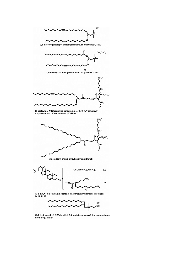
Felgner and co-workers [25] were the first to use cationic lipids dioleyltrimethylammo-
nium chloride (DOTMA) in a 1 :1 molar ratio with the neutral lipid dioleoylphosphati-
dyl-ethanolamine (DOPE) to condense and transfect DNA. Since then, a variety of cationic
lipids have been synthesized and evaluated for gene transfection. Because of their amphi-
philic nature, they easily form vesicular structures termed liposomes, when suspended in
water; given their cationic charge, these liposomes readily and efficiently interact with
DNA, thus forming so-called “lipoplexes”. Cationic liposomes are prepared by evaporation
of the organic solvent in which the cationic lipid (mixture) is dissolved, followed by hydra-
tion of the lipid film in aqueous buffer, and subsequent vortexing, which results in the
formation of multilamellar vesicles (MLVs). Small unilamellar vesicles are generated by
sonication or extrusion of the MLVs. Addition of DNA to performed cationic liposomes
triggers significant structural changes in both the liposomes and the DNA, whereby the
327
21.3 Nonviral Nanomaterials in Development and Testing
O
O
NH
CH
2
OH
NH
2
HO
O
O
CH
2
OH
HO
C=O
CH
3
C
H
3
HO
C
H
3
HO
Figure 21.6
Chemical structure of deoxycholic
acid-modified chitosan (DMAC).
328
21 Nanoparticles as Non-Viral Transfection Agents
Figure 21.7
Structures of some
cationic lipids commonly used in gene
therapy.

initial liposomal structure is lost and new structural organization, the lipoplex, is adopted
[26, 27]. The structures of some cationic lipids commonly used in gene therapy are shown
in Figure 21.7. Pedroso de Lima et al. [28] recently reviewed cationic lipid–DNA complexes
in gene delivery, where they discussed various aspects starting from biophysics to biologi-
cal applications.
Ochiya et al. [29] evaluated gene transfection in pregnant animals by testing several ca-
tionic liposomes conjugated with plasmid DNA carrying the b-galactosidase gene through
intravenous injection. These authors identified DMRIE-C reagent, which was a liposome
formulation of the cationic lipid DMRIE (1, 2-dimyristyloxypropyl-3-dimethyl-hydroxy
ethyl ammonium bromide) and cholesterol, as being suitable for their purpose. When
plasmid DNA/DMRIE-C complexes were administered intravenously to pregnant mice
at day 11.5 post coitus (p. c.), transferred genes were observed in several organs in
dams and were expressed (Table 21.3). Furthermore, gene expression was observed in
the progeny, although the copy numbers transferred into embryos were low [29].
Ishiwata et al. [30] developed a novel liposome formulation for gene transfection, con-
sisting of O,O
l-ditetradecanoyl-N-(a-trimethyl ammonioacetyl) diethanolamine chloride
(DC-6-14) as a cationic lipid (Figure 21.8), phospholipid and cholesterol in a molar
ratio of 4 :3 :3. The DC-6-14 liposome–DNA complexes were usually thought to have an
overall positive surface charge, but it was found to be DNA ratio-dependent, and the dia-
meter of the complex was also dependent on DNA concentration. The complexes had a
maximum diameter when the surface charge was neutral. These formulations showed ef-
fective gene transfection activity in cultured cells with serum and, when administered in-
traperitoneally to mice, the positively charged particles showed immediate accumulation
in lung tissues. By contrast, the negatively charged particles did not show any accumula-
tion [30]. The authors therefore concluded that surface modification of the liposome
329
21.3 Nonviral Nanomaterials in Development and Testing
Table 21.3
Distribution of the transfected gene in vivo
Organ
Number of
Plasmid form
copies/cell
Open circle
Linear
Supercoil
Brain
0
–
–
–
Heart
2–5
–
+
–
Lung
20–50
+
+
+
Liver
10–40
+
+
+
Kidney
2–10
–
+
–
Spleen
2–5
–
+
–
Pancreas
2–10
–
+
–
Stomach
1–2
–
+
–
Colon
0–2
–
+
–
Small intestine
0–2
–
+
–
Testis
0
–
–
–
Ovary
0
–
–
–
Note : Seven-week-old ICR male and female mice (n = 12) were injected i. v. with 100 mg pCMVb. SPORT bgal
plasmid DNA conjugated with 500 mg DMRIE-C reagent. Two days later, several organs were obtained, and the
genomic DNAs from them were subjected to Southern blot analysis for the presence of transferred gene.

would improve the biodistribution and hence the targetability of their DNA complexes
[30].
No single system thus far seems to transfect DNA efficiently into the nucleus, and at-
tempts are ongoing to achieve this. Oku et al. [31] reported a novel nonviral gene delivery
system which they named as the polycation liposome system (PCL). Basically, this ap-
proach was to combine the favorable properties of liposomes with the polycation, PEI.
The interesting point about PCL was that it did not require phosphatidylethanolamine
or cholesterol as a component, unlike most conventional liposomes [31]. Egg yolk phos-
phatidylcholine- and dipalmitoylphosphatidylcholine-based PCLs were found to be as ef-
fective as dioleoylphosphatidylethanolamine-based PCL for gene transfer [31]. These
authors examined the effect of molecular weight of the PEI on PCL-mediated gene trans-
fer and found that PEIs with molecular weights of 600 and 1800 Da to be quite effective,
whereas PEI of 25 000 Da was much less effective. Furthermore, they demonstrated an
increased transfection efficacy in the presence of serum, with effective gene transfer
being observed in all eight malignant and two normal cell lines tested, as well as in
Cos-1 cells [31]. Interestingly, the same authors also demonstrated gene transfer in
vivo : GFP and luciferase genes were expressed in mouse lung when the animals were
given tail vein injections, whereas gene expression occurred in the liver after portal
vein injection [31].
Huang and co-workers made very significant contributions to the liposomal approach in
gene delivery [32–40]. Li and Huang [41] have recently reviewed their developments in
nonviral delivery systems, with a major focus on cationic liposome entrapped, polyca-
tion-condensed DNA, which they termed LPD1. The original complex was composed of
poly-L-lysine (PLL), DNA, and cationic liposomes, which resulted in a high interaction be-
tween PLL and DNA, while liposomes led to the formation of a lipid shell on the surface
of the particles [33]. A recent review also highlighted the developments of LPD1, systemic
gene delivery, and mechanisms involved in cationic mediated gene delivery [41].
The other leading group in liposome research for nonviral gene delivery is that of Szoka
and co-workers [42–52]. These authors have contributed extensively to the fascinating area
of liposome research, and have applied cationic liposomes for targeting drugs to various
lesions as well as the delivery of genes. They suggested that intravenously administered
lipoplexes might serve as a depot for the extracellular release of naked DNA, and initially
330
21 Nanoparticles as Non-Viral Transfection Agents
C
13
H
27
-C-O-C
2
H
4
C
13
H
27
-C-O-C
2
H
4
N-C-CH
2
-N-CH
3
CH
3
CH
3
O
O
O
+
Cl
-
Figure 21.8
Chemical structure of
O,O
l-
ditetradecanoyl-
N-(a-trimethylammonioa-
cetyl) diethanolamine chloride (DC-6-
14).
considered that the naked DNA mediated gene delivery in the lung [49]. They introduced a
lung inflation-fixation protocol to examine the distribution and gene transfer efficiency of
fluorescently tagged lipoplexes using confocal microscopy within thick lung tissue sec-
tions [52]. Infusion of plasmid DNA at a rate of 80 mg min
–1
into the tail vein of a
mouse resulted in a DNA serum concentration of 800 mg mL
–1
. In spite of this high
level of transcriptionally active DNA, there was no significant gene expression in the
lung or any other organ tested [49]. In addition, when lipoplex containing a reporter
gene was injected, followed by an infusion of noncoding plasmid DNA as a potential com-
peting molecule for DNA released from the lipoplex, there was no effect on gene expres-
sion. Thus, it was concluded that the cationic lipid component of the lipoplex functions in
an active capacity beyond that of a simple passive matrix for plasmid DNA [49]. Baraldo et
al. [53] recently reported sphingosine-based liposomes as DNA vectors for intramuscular
gene delivery; these liposomes were formulated in a range of solutions with phosphatidyl-
choline, and then complexed with DNA. The physico-chemical characteristics of the
sphingosine/EPC liposomes and sphingosine/EPC/DNA lipoplexes were determined. It
was found that, by increasing the charge ratios, colloidally stable sphingosine/DNA parti-
cles with a 170 nm average diameter and a positive zeta potential were obtained [53].
These authors concluded that the cationic sphingosine/EPC/DNA complexes formed a
weakly compacted structure which potentially was labile in vivo and which might be use-
ful for in-vivo gene transfer [53].
Oberle et al. [54] showed that, under equilibrium conditions, lipoplex formation involves
a three-step mechanism, the interaction between plasmid and cationic liposomes being
investigated using atomic force microscopy (AFM) [54]. In the first step, the plasmids –
when interacting with the monolayer – display a strong tendency for orientational order-
ing. Subsequently, individual plasmids enwrap themselves with amphiphile molecules in
a multilamellar fashion. The size of the complex formed is determined by the supercoiled
331
21.3 Nonviral Nanomaterials in Development and Testing
Figure 21.9
Lipoplex (d = 300 nm)
composed of SAINT 2/DOPE 50/50
pCMVb plasmid.
size of the plasmid, and calculations reveal that the plasmid can be surrounded by three to
five bilayers of the amphiphile. The eventual size of the transfecting complex is finally
governed by fusion events between individually wrapped amphiphile/DNA complexes.
The AFM images of the liposome complexes are as shown in Figure 21.9.
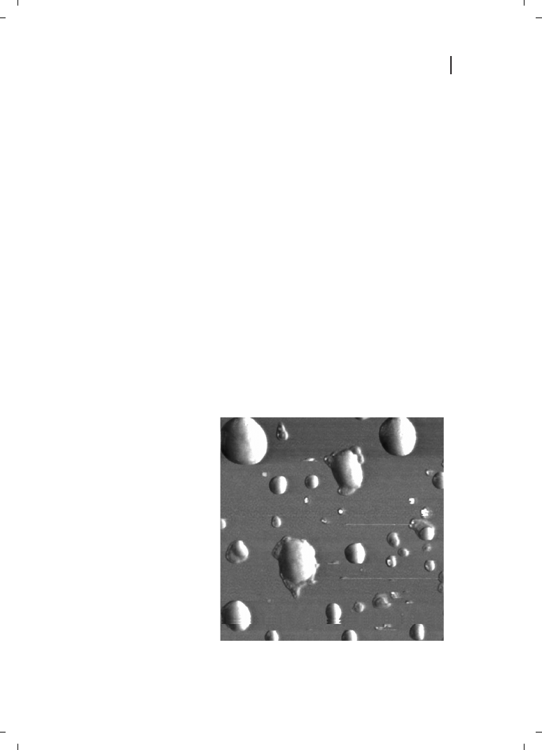
Müller and co-workers [55–58] carried out extensive research on solid lipid nanoparti-
cles for drug targeting as well as gene delivery. Müller et al. [59] have discussed the
state of art of solid lipid nanoparticles for pharmaceutical applications, and in recent in-
vestigations found that cationic solid-lipid nanoparticles (SLNs) could bind efficiently and
transfect plasmid DNA [60]. They produced highly cationic charged SLNs with a zeta po-
tential of up to +40 mV at pH 7.4 and 100 nm size, by using a hot homogenization tech-
nique. They also characterized the SLN–DNA complexes using AFM, and suggested fu-
sion of the SLNs after binding to the DNA, resulting in near-spherical lipid–DNA particles
with sizes between 300 and 800 nm (Figure 21.10). These investigations also demon-
strated that efficient transfection occurs in Cos-1 cells [60].
21.3.3
Poly-
L
-Lysine and Polyethylenimines
Poly-l-lysine (PLL) – one of the first polymers to be used in nonviral gene delivery [61, 62]
– is biodegradable but has a high toxicity which prevents its use in vivo. Initial reports
stated that, if prepared with PLL of suitable molecular weight and N/P ratio, 100 nm-
sized complexes are formed which are easily taken up by the cells, although the transfec-
tion remained lower [61]. The lack of amino groups was thought to prevent PLL com-
plexes from undergoing endoosomolysis, leading to low levels of transgene expression
[63]. Nevertheless, inclusion of targeting moieties or co-application of endoosomolytic
agents such as chloroquine [64] or fusogenic peptides [61] might improve reporter gene
expression.
Parker et al. [65] recently discussed methods for monitoring nanoparticle formation by
self-assembly of DNA with PLL. They compared three different methods viz., light scatter-
ing, inhibition of ethidium bromide fluorescence, and modified electrophoretic mobility
of DNA. Results obtained with the first two methods indicated that stable nanoparticles
form over the lysine/phosphate ratio range 0.6 to 1.0 [65], though no transfection studies
were reported.
332
21 Nanoparticles as Non-Viral Transfection Agents
Figure 21.10
Solid lipid nanoparticles (SLNs) (d
J100 nm) composed
of compritol ATO, Tween 80, Span 85, and distearoylethyl dimethyl
ammonium chloride form discrete, submicron particles with plasmid
DNA (upper half). Coalescence of the individual SLNs following inter-
action with DNA stabilize the complex. A single SLN can be seen in the
lower half of the image.

In an attempt to improve transfection efficiency and reduce cytotoxicity, Lee et al. [66]
utilized PEG grafted polylysine with fusogenic peptide. The method is initiated by com-
plexing the PEG-grafted PLL with DNA, and subsequently coating the positively charged
fusogenic peptide by ionic interaction onto the surface. This results in a net positively
charged complex [66]. The authors showed that the use of PEG-grafted PLL for peptide
coating significantly suppressed aggregation of the complexes via peptide mediated inter-
particulate crosslinking, and also enhanced transfection [66]. Hence, both PEG and pep-
tides play key roles in reducing cytotoxicity and improving transfection [74].
Gonzalez Ferreiro et al. [67] described the use of alginate/PLL particles for antisense oli-
gonucleotides wherein they utilized the gel-forming abilities of the alginate in the pres-
ence of calcium ions and further crosslinked it with PLL. Particle formation was found
to occur when the alginate concentration was 0.04–0.08 % (w/v) of the total formulation;
moreover, the particle size increased from nm to mm when the alginate concentration was
0.055 % (w/v) [67]. These authors found that with increasing PLL content, both the size
and encapsulation efficiency of the particle increased, but the release rate was decreased
[67].
Polyethylenimines (PEIs) were the first synthetic transfection agents to be discovered,
and have been widely studied, although the toxicity of PEI is a major drawback. Kircheis
et al. [68] reviewed the design and gene delivery of modified PEIs, and emphasized the
synthesis of modified PEIs, DNA condensation, particle size, cellular uptake of the parti-
cles, endosomal release, in-vivo gene delivery and many other features.
Sagara and Kim [69] more recently reported galactose-polyethylene glycol-PEI (Gal-PEG-
PEI) for gene delivery to hepatocytes. They found that transfection efficiency with 1 % Gal-
PEG-PEI in human hepatocyte-derived cell lines (HepG2) – a model of parenchymal cells
in the liver – was superior to PEI at corresponding optimal ratios of polymer to plasmid
DNA [69]. In HepGe cells, luciferase activity with 1 % Gal-PEG-PEI at an N/P ratio of 20
was 2.1-fold higher than that of PEI at an N/P ratio of 5. However, in mouse fibroblasts
(NIH-3T3) that had no asialoglycoprotein (ASGP) receptor, the transfection efficiency with
1 % Gal-PEG-PEI fell drastically to one-fortieth of that with PEI. These studies suggest
that Gal-PEG-PEI is more suited for targeting specific genes to the liver [69].
Rudolph et al. [70] reported modified PEIs for gene transfer to the lungs via either direct
intratracheal instillation or nebulization. In this study, PEG-coated PEI polyplexes were
investigated for their stability and interaction with human plasma and bronchoalveolar la-
vage fluid (BALF); their potential for gene delivery to the mouse lungs in vivo was also
examined. Gene transfer efficiency of the PEG-coated PEI polyplexes decreased as com-
pared with uncoated PEI polyplexes when administered intratracheally to the lung. The
higher the molecular weight of the copolymerized PEG, the stronger was the observed
gene transfer reduction. These authors speculated that the gene transfer was decreased
due to a reduced interaction of the coated gene vectors with the cell surface. To circumvent
this problem, transferrin was combined with PEI/DNA polyplexes for specific binding to
the cell surface, but this resulted in a decrease in gene transfer efficiency. Gene transfer of
the copolymer-protected and transferrin-modified gene vectors increased as compared
with the copolymer-protected gene vectors alone, but did not reach the level of uncoated
gene vectors. These data show that copolymers could be used effectively to shield poly-
plexes from interaction with components of the airway surface liquid. The increase in
333
21.3 Nonviral Nanomaterials in Development and Testing

gene expression upon transferrin modification of the coated PEI polyplexes still suggests a
targeting effect [70].
21.3.4
Poly(lactide-co-glycolide)
Various biodegradable polymers are being investigated for the formulations of nanoparti-
cles aimed at drug and gene delivery applications [71]. Poly(d,l lactide-co-glycolide)
(PLGA) and poly(lactic acid) (PLA) – both of which are FDA-approved biocompatible poly-
mers – have been the most extensively studied [72, 73]. Recently, Jain [72] reviewed the
PLGA nanoparticle preparation techniques, and reported an emulsion-solvent evaporation
technique to be the most widely used method to formulate PLA and PLGA nanoparticles,
while poly(vinyl alcohol) (PVA) is the most commonly used stabilizer.
Labhasetwar and co-workers [74–77] made several reports on aspects of PLGA nanopar-
ticles for gene delivery, viz., characterization of nanoparticle uptake by endothelial cells
[74], size dependency of nanoparticle-mediated gene transfection [75], effects on nanopar-
ticle properties and their cellular uptake associated with the residual PVA [76], and rapid
endolysosomal escape of the PLGA nanoparticles [77]. They followed the standard emul-
sion-solvent evaporation technique, which resulted in particles with a heterogeneous size
distribution. Moreover, they investigated the relative transfectivity of the smaller and the
larger-sized particles in cell cultures [75]. Those particles which passed through a mem-
brane of pore size 100 nm (mean diameter = 70
e 2 nm) were designated as smaller par-
ticles, whereas those retained on the membrane were deemed to be larger-sized nanopar-
ticles (202
e 9 nm). The smaller-sized nanoparticles showed a 27-fold higher transfection
than the larger-sized nanoparticles in a Cos-7 cell line, and a 4-fold higher transfection in
an HEK-293 cell line [75]. The surface charge, cellular uptake, and DNA release were si-
milar for the two fractions of nanoparticles, and suggests that other unknown factor(s)
might be responsible for the observed differences in transfection levels [75].
The same authors also studied the effects of residual PVA associated with PLGA nano-
particles on cellular uptake [76]. PVA removal during nanoparticle preparation is difficult
by water washing, and small amounts are inevitably associated with the particles. This oc-
curs despite repeated washings as PVA forms an interconnected network with the poly-
mer at the interface. The amount of residual PVA was found to depend on the initial
PVA concentration and type of organic solvent used in the emulsion, and affected particle
size, zeta potential, polydispersity index, and surface hydrophobicity of the nanoparticle
surface [76]. Particles with more residual PVA, despite their lower size, were seen to
have the lowest cellular uptake, but this may be due to the higher hydrophilicity of the
nanoparticle surface [76]. These authors also studied endolysosomal escape of the
PLGA nanoparticles [77], and showed the mechanism to be by selective reversal of the sur-
face charge of NPs (from anionic to cationic) in the acidic endolysosomal compartment,
which in turn causes the NPs to interact with the endolysosomal membrane and escape
into cytosol [77].
334
21 Nanoparticles as Non-Viral Transfection Agents
21.3.5
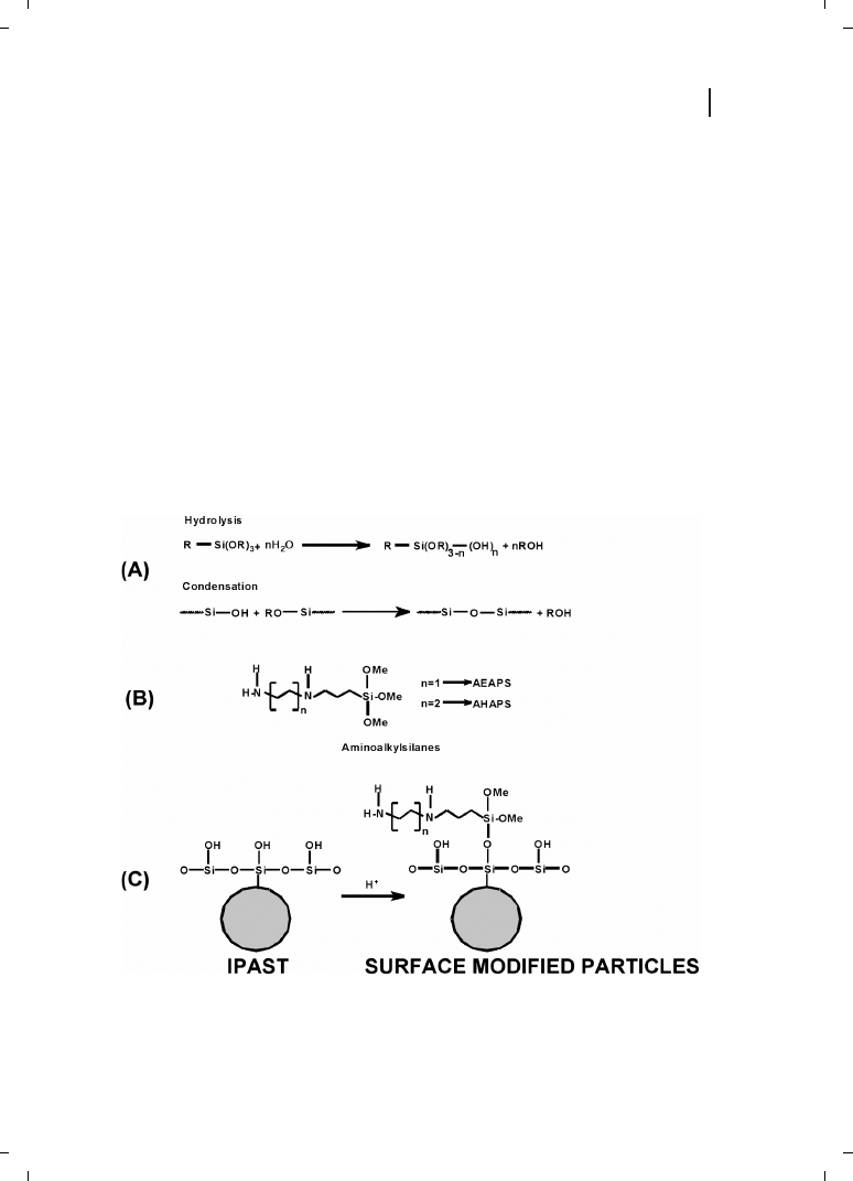
Silica
A major limitation of the polymers is the difficulty of including additional bioactive mo-
lecules (in order to enhance or modify the DNA activity) during the self-assembly process,
unless these are covalently linked to the polymer backbone. Recently, it was shown in the
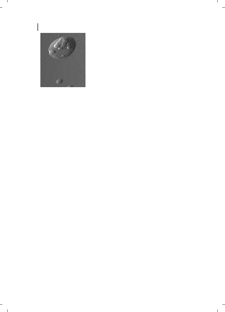
present author’s laboratory that the colloidal silica particles with cationic surface modifi-
cations interact with plasmid DNA and transfect in vitro [78, 79]. Surface-modified silica
nanoparticles were synthesized by modification of commercially available silica particles
(IPAST; Nissan Chemical Industries, Tokyo, Japan) with various aminoalkylsilanes, viz.,
N-(2-aminoethyl)-3-aminopropyltrimethoxysilane (AEAPS) and N-(6-aminohexyl)-amino-
propyltrimethoxysilane (AHAPS). The resulting particles have a hydrodynamic diameter
of 26 nm and a zeta potential up to +31 mV [78, 79]. The scheme for modification of
the silica particles is shown in Figure 21.11. In analogy to the terms lipoplex and polyplex,
we propose that be the nanoparticle–DNA complexes be described by the term “nanoplex”.
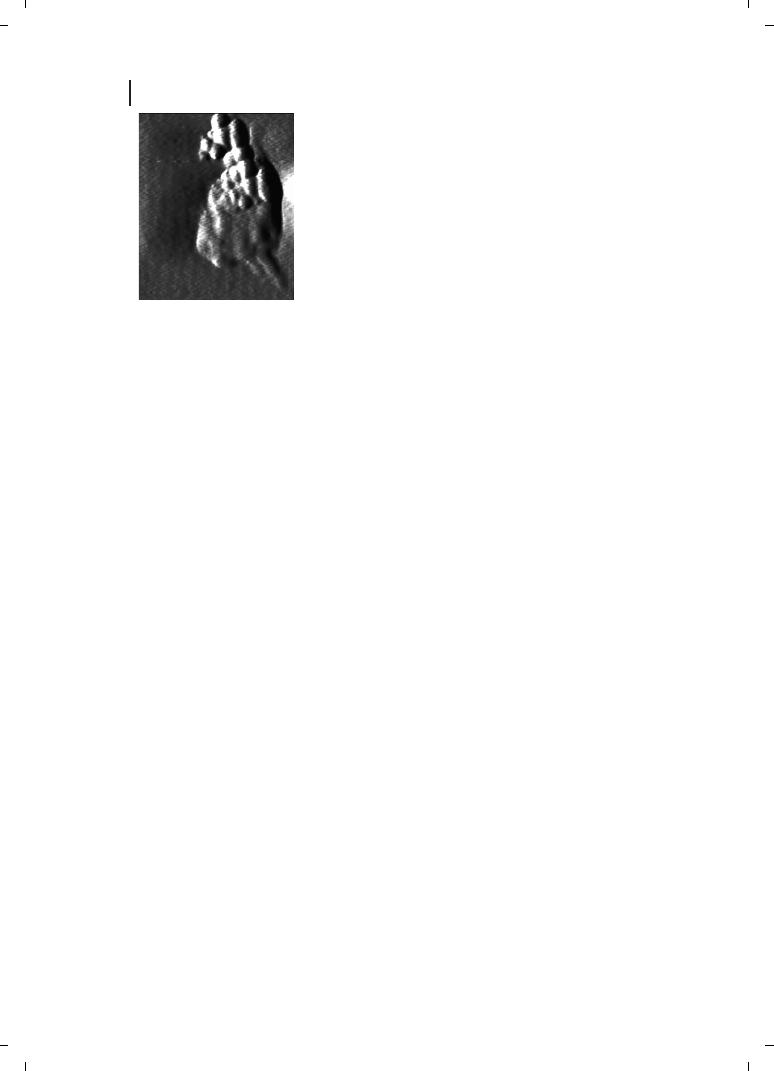
Transfection was strongly increased in the presence of 100 mM chloroquine in the incuba-
tion medium, and reached approximately 30 % of the efficiency of a 60-kDa PEI. In con-
trast to PEI, no toxicity was observed at the concentrations required. AFM of Si
2
6H–DNA
complexes revealed a spaghetti-meatball-like structure (Figure 21.12).
335
21.3 Nonviral Nanomaterials in Development and Testing
Figure 21.11
(A) Hydrolysis and condensation of unmodified particles. (B) Alkylaminoalkanes. (C)
Modification scheme.
We also investigated the stability of freeze-drying for the conservation of zwitterionic na-
noparticles and usefulness of different lyoprotective agents (LPAs) for the minimization of
particle aggregation [80]. The activity of the nanoparticles was measured as DNA-binding
capacity and transfection efficiency in Cos-1 cells before and after lyophilization. Massive
aggregation was found to occur in the absence of any LPA. Of the various LPAs screened
in the present investigations, trehalose and glycerol were found to provide exceptional
conservation of cationically modified silica nanoparticles with simultaneous preservation
of their DNA-binding and transfection activity in Cos-1 cells [80].
21.3.6
Block Copolymers
Block copolymers composed of the cationic segment and hydrophilic segments are ex-
pected spontaneously to associate with polyanionic DNA to form block copolymer mi-
celles. Kataoka and co-workers [81–85] pioneered this area of research, and recently pro-
vided an excellent review of block copolymer micelles for gene delivery and related com-
pounds [86]. They discussed various aspects of the synthesis of cationic block and graft
copolymers, the physico-chemical properties and the biological aspects including cellular
uptake and lysosomal escape [86]. Kataoka’s group is actively involved in block copolymers
of poly(ethylene glycol)-block-poly(l-lysine) (PEG-b-PLL) [82, 83], poly(ethylene glycol)-
block-poly(ethylenimine) (PEG-b-PEI) [87], and poly(ethylene glycol)-block-poly(dimethyla-
minoethyl methacrylate) (PEG-b-PPAMA) [84], all of which are synthesized from their re-
spective monomers using PEG with terminal functional groups as a macroinitiator (Fig-
ure 21.13).
The other active group involved in block copolymers is that of Kabanov et al. [88–92],
who carried out extensive investigations on Pluronic
r block copolymers as depots for
drugs as well as genes. In a recent review, these authors described Pluronic block copoly-
mers as novel functional molecules for gene therapy, with special focus on gene delivery
to skeletal muscle [93].
336
21 Nanoparticles as Non-Viral Transfection Agents
Figure 21.12
Cationic silica particles (d
J300 nm) self-associate with
plasmid DNA into submicron complexes that protect, release, and
transfect the DNA in vitro.

337
21.3 Nonviral Nanomaterials in Development and Testing
PEG-b-PLL
CH
3
(OCH
2
CH
2
)
m
NH
2
+ n
HN
O
R
1
O
O
CH
3
(OCH
2
CH
2
)
m
NH(COCHNH)
n
H R
1
= (C H
2
)
4
NHCOOCH
2
R
1
CH
3
(OCH
2
CH
2
)
m
NH(COCHNH)
n
H
R
2
HBr/AcOH
R
1
= (C H
2
)
4
NH
2
PEG-b-PAMA
m
+
CH
3
CH
2
O
CH
3
CH
2
O
CHCH
2
CH
2
O
-
K
+
O
CH
2
CH
2
+
CH
3
CH
C=O
(CH
2
)
2
NH(C H
3
)
2
CH
2
=
n
CH
2
C H
2
O
-
K
+
CH
3
C H
2
O
CH
3
C H
2
O
CHCH
2
CH
2
O(CH
2
CH
2
O)
m
CH
3
CH
2
O
CH
3
CH
2
O
CHCH
2
CH
2
O(C H
2
CH
2
O)
m
CH
3
C=O
(CH
2
)
2
NH(CH
3
)
2
(CH
2
CH)
n
H
PEG-b-PEI
CH
3
CH
2
O
CH
3
CH
2
O
CH
3
CH
2
O
CH
3
CH
2
O
CH
3
C=O
N
O
CH
3
+ n
CHCH
2
CH
2
O(CH
2
CH
2
O)
m
CH
2
CH
2
(NCH
2
CH
2
)
n
OH
CHCH
2
CH
2
O(CH
2
CH
2
O)
m
CH
2
CH
2
(NCH
2
CH
2
)
n
OH
CH
3
CH
2
O
CH
3
CH
2
O
CHCH
2
CH
2
O(CH
2
CH
2
O)
m
CH
2
CH
2
OSCH
3
O
O
Figure 21.13
Synthetic procedures for cationic copolymers.

21.4
Setbacks and Strategies to Improve Specific Cell Uptake of Nonviral Systems
Nonviral gene delivery has been used to express various genes in animal tissues, includ-
ing muscle, lung, skin, the central nervous system, and liver. For in-vivo gene delivery,
nonviral systems have the advantage of being more similar to pharmaceutical drugs in
terms of safety, uniformity, and administration. However, the transduction frequency of
the target cells is often barely adequate, and the persistence of cells expressing the trans-
duced gene in vivo is poor. Further development of nonviral gene delivery technology will
expand its application and provide a viable alternative to the use of viral vectors.
For in-vivo therapy it is necessary to address gene delivery vehicles to specific cell types
in order to avoid unwanted effects in nontarget cells. Targeting is achieved actively by in-
corporating structures, which facilitate exclusive uptake of the vector in certain tissues of
cell types. However, in some cases this can be achieved passively by taking advantage of
particular physiological conditions of the target tissue; for example, irregular endothelial
fenestration in tumors in conjugation with certain complex properties. To achieve an effi-
cient active targeting in vivo, the vector must fulfill two main requirements : (i) unspecific
interactions must be reduced by shielding net positive surface charges of the complexes by
using rather low nitrogen to phosphate content; and (ii) the targeting moiety which en-
ables uptake into a specific cell type needs to be incorporated. Various ligands are in
use depending upon the target structure, and these include folate, transferrin, lactose, ga-
lactose, mannose, low-density lipoprotein (LDL), fibroblast growth factor (FGF), endothe-
lial growth factor (EGF), and several antibodies. An early review of the strategies was pro-
vided by Nettelbeck et al. [94], and more recently by Merdan et al. [95].
21.5
Prospects for Nonviral Nanomaterials
Currently, gene therapy researchers are attempting to improve each vector, as well as to
match vector characteristics with diseases that they will target most successfully. It is likely
that, in the future, there will be many specialized vectors rather than one universal vector.
Some vectors may be required in situations where doctors require short term expression,
for example in the expression of a toxic gene product in cancer cells. However, in situa-
tions where long-term expression is required (e. g., for most genetic diseases), vectors
that deliver large and medium-sized sections of DNA would be required.
Gene insertion is not the only problem encountered, however, as the process is not auto-
matic and the vector must contain a mechanism to activate the therapeutic gene. Hence,
the vector must include a timing and regulatory “device” which allows the gene to be
turned on and off and change levels of the therapeutic protein over time. Such devices
are termed the gene’s “promoters”; they are complex and sometimes quite large, and pla-
cing them into a therapeutic vector is difficult.
It is clear from the foregoing sections that polymers display striking advantages as vec-
tors for gene delivery. They can be specifically tailored for a proposed application by choos-
ing an appropriate molecular weight, by coupling the cell- or tissue-specific targeting moi-
eties, and/or performing other modifications that confer certain physiological or physico-
338
21 Nanoparticles as Non-Viral Transfection Agents

chemical properties. After identifying a suitable polymer structure, a scale-up to the pro-
duction of large quantities is somewhat more straightforward. It is understood that all
these modifications/manipulations are possible only by using nonviral vectors.
The bottom line in any type of biomedical research is its relevance. Despite the excite-
ment that gene therapy might cause, the field is still in its infancy. Several clinical trials of
gene therapy have been completed or are under way, and all have provided information
that cannot be derived from tests conducted in animals. Although in theory this treatment
is good in principle, its efficacy has proved difficult to demonstrate in practice, and suc-
cess rates in clinical trials have been relatively low. Poor results may reduce the enthu-
siasm for genetic approaches, but the field is still new and the pace and surprises of
new discoveries continue to amaze.
Although in this chapter we have attempted to outline the most significant contribu-
tions on nonviral nanoparticulate gene delivery systems, it is very difficult to discuss
each and every reported system within the confines of the text. Nonetheless, an attempt
has been made to provide updated information on nonviral nanoparticulate gene delivery
systems, and in consideration of the reader’s interest we have included bibliographic in-
formation of those reports not discussed in the text [96–125].
Acknowledgments
M. N. V. R. K. is grateful to the Alexander Von Humboldt Foundation, Germany for provid-
ing personal fellowship. U. Bakowsky wishes to thank “Stiftung Deutscher Naturforscher
Leopoldina” (BMBF/LPD-9901/8-6).
339
References
References
[1]
V. Kabanov, V. A. Kabanov, Adv. Drug Del.
Rev. 1998, 30, 49–60.
[2]
Z. Q. Xiang, Y. Yang, J. M. Wilson,
H. C. Ertl, Virology 1996, 219, 220–227.
[3]
M. R. Knowles, K. W. Hohneker, Z. Zhou,
J. C. Olsen, T. L. Noah, P. C. Hu, M. W.
Leigh, et al. N. Engl. J. Med. 1995, 333,
823–831.
[4]
R. H. Simon, J. F. Engelhardt, Y. Yang,
M. Zepeda, S. Weber-Pendelton, M. Gross-
man, J. M. Wilson, Hum. Gene Ther. 1993,
4, 771–778.
[5]
S. C. De Smedt, J. Demeester, W. E. Hen-
nink, Pharm. Res. 2000, 17, 113–126.
[6]
M. C. Garnett, Crit. Rev. Ther. Drug Carrier
Syst. 1999, 16, 147–207.
[7]
K. Minagawa, Y. Matzusawa, K. Yoshikawa,
M. Matsumoto, M. Doi, FEBS Lett. 1991,
295, 67–69.
[8]
J. Kreuter, P. Speiser, J. Pharm. Sci. 1976,
65, 1624–1627.
[9]
P. Couvreur, L. Grislain, V. Lenaert,
F. Brasseur, P. Guiot, A. Biernacki, in :
P. Guiot, P. Couvreur (eds), Polymeric
Nanoparticles and Microparticles. CRC
Press, Boca Raton, 1986, pp. 27–93.
[10]
V. Labhasetwar, C. Song, R. J. Levy, Adv.
Drug Del. Rev. 1997, 24, 63–85.
[11]
R. A. A. Muzzarelli, Chitin, Pergamon
Press, Oxford, 1977.
[12]
M. N. V. Ravi Kumar, Reactive and Func-
tional Polymers 2000, 46, 1–27.
[13]
R. J. Mumper, J. J. Wang, J. M. Claspell,
A. P. Rolland, Proc. Int. Symp. Controll.
Rel. Bioactive Mater. 1995, 22,
178–179.
[14]
G. Borchard, Adv. Drug Del. Rev. 2001, 52,
145–150.

340
21 Nanoparticles as Non-Viral Transfection Agents
[15]
F. C. MacLaughlin, R. J. Mumper, J. Wang,
J. M. Tagliaferri, I. Gill, M. Hinchcliffe,
A. P. Rolland, J. Controll. Rel., 1998, 56,
259–272.
[16]
H.-Q. Mao, K. Roy, V. Truong-Le, et al.,
Proc. Int. Symp. Controll. Rel. Bioact. Mater.
Stockholm, Sweden, 1997, 24, 671–672.
[17]
K. W. Leong, H.-Q. Mao, V. L. Truong-Le, et
al., J. Controll. Rel., 1998, 53, 183–193.
[18]
K. Roy, H.-Q. Mao, S. K. Huang, K. W.
Leong, Nature Med. 1999, 5, 387–391.
[19]
H.-Q. Mao, K. Roy, V. L. Troung-Le, et al.,
2001, J. Controll. Rel. 2001, 70, 399–421.
[20]
M. Kumar, A. K. Behera, R. F. Lockey, J.
Zhang, et al., Hum. Gene Ther. 2002, 13,
1415–1425.
[21]
M. Koping-Hoggard, I. T. Guan, K. Ed-
wards, M. Nilsson, K. M. Varum, P. Ar-
tursson, Gene Ther. 2001, 8, 1108–1121.
[22]
T. Sato, T. Ishii, Y. Okahata, Biomaterials
2001, 22, 2075–2080.
[23]
Y. H. Kim, S. H. Gihm, C. R. Park, Biocon-
jug. Chem. 2001, 12, 932–938.
[24]
M. Thanou, B. I. Florea, M. Geldof,
H. E. Junginger, G. Borchard, Biomaterials
2002, 23, 153–159.
[25]
P. L. Felgner, T. R. Gadek, M. Holm,
R. Roman, et al., Proc. Natl. Acad. Sci. USA
1987, 84, 7413–7417.
[26]
E. K. Wasan, A. Fairchild, M. B. Balay,
J. Pharm. Sci. 1998, 87, 9–14.
[27]
A. A. P. Meekel, A. Wagenaar, J. Smister-
ova, J. E. Kroeze, Eur. J. Org. Chem. 2000,
2000, 665–673.
[28]
M. Pedroso de Lima, S. Simoes, P. Pires,
H. Faneca, N. Duzgunes, Adv. Drug Deliv.
Rev. 2001, 47, 277–294.
[29]
T. Ochiya, Y. Takahama, H. Baba-Toriyama,
M. Tsukamoto, Y. Yasuda, H. Kikuchi, M.
Terada, Biochem. Biophys. Res. Commun.
1999, 258, 358–365.
[30]
H. Ishiwata, N. Suzuki, S. Ando, H. Kiku-
chi, T. Kitagawa, J. Controll. Rel. 2000, 69,
139–148.
[31]
N. Oku, Y. Yamazaki, M. Matsuura,
M. Sugiyama, M. Hasegawa, M. Nango,
Adv. Drug Deliv. Rev. 2001, 52, 209–218.
[32]
C. Y. Wang, L. Huang, Proc. Natl. Acad. Sci.
USA 1987, 84, 7851–7855.
[33]
X. Gao, L. Huang, Biochemistry 1996, 53,
1027–1036.
[34]
F. L. Sorgi, S. Bhattacharya, L. Huang,
Gene Ther. 1997, 4, 961–968.
[35]
S. Li, M. A. Rizzo, S. Bhattacharya,
L. Huang, Gene Ther. 1998, 5, 930–937.
[36]
S. Li, L. Huang, Gene Ther. 1997, 4, 891–
900.
[37]
S. Li, W. C. Tseng, D. B. Stolz, S. P. Wu,
S. C. Watkins, L. Huang, Gene Ther. 1999,6,
585–594.
[38]
X. Zhou, L. Huang, Biochim. Biophys. Acta
1994, 1189, 195–203.
[39]
F. Liu, H. Qi, L. Huang, D. Liu, Gene Ther.
1997, 4, 517–523.
[40]
Y. Tan, F. Liu, Z. Li, L. Huang, Mol. Ther.
2001, 3, 673–682.
[41]
F. Liu, L. Huang, J. Controll. Rel. 2002, 78,
259–266.
[42]
L. G. Barron, K. B. Meyer, F. C. Szoka,
Jr., Hum. Gene Ther. 1998, 9, 315–323.
[43]
O. Zelphati, L. S. Uyechi, L. G. Barron,
F. C. Szoka, Jr., Biochim. Biophys. Acta
1998, 1390, 119–133.
[44]
S. C. Davis, F. C. Szoka, Jr., Bioconjug.
Chem. 1998, 9, 783–792.
[45]
J. Y. Legendre, S.-K. Huang, F. C. Szoka,
Jr., J. Liposome Res. 1998, 8, 347–366.
[46]
L. G. Barron, L. S. Uyechi, F. C. Szoka,
Jr., Gene Ther. 1999, 6, 1179–1183.
[47]
Y. Xu, S.-W. Hui, P. Frederik, F. C. Szoka,
Jr., Biophys. J. 1999, 77, 341–353.
[48]
S. Hirota, C. Tros de Llarduyu, L. Barron,
F. C. Szoka, Jr., Biotechniques 1999, 27,
286–289.
[49]
L. Barron, L. Gagne, F. C. Szoka, Jr., Hum.
Gene Ther. 1999, 10, 1683–1894.
[50]
F. Nicol, S. Nir, F. C. Szoka, Jr., Biophys.
J. 2000, 78, 818–829.
[51]
X. Guo, F. C. Szoka, Jr., Bioconjug. Chem.
2001, 12, 291–300.
[52]
L. S. Uyechi, G. Thurston, L. Gagne, F. C.
Szoka, Jr., 2001, Gene Ther., 8, 828–836.
[53]
K. Baraldo, N. Leforestier, M. Bureau, N.
Mignet, D. Scherman, Pharm. Res. 2002,
19, 1144–1149.
[54]
V. Oberle, U. Bakowsky, I. S. Zuhorn, D.
Hoekstra, Biophys. J. 2000, 79, 1447–1454.
[55]
R. H. Muller, S. Massen, H. Weyhers, F.
Specht, J. S. Lucks, Int. J. Pharm. 1996,
138, 85–94.
[56]
R. H. Muller, W. Mehnert, J. S. Lucks,
C. Schwarz, et al., Eur. J. Pharm. Biopharm.
1995, 41, 62–69.
[57]
C. Freitas, R. H. Muller, Eur. J. Pharm.
Biopharm. 1999, 47, 125–132.

341
References
[58]
A. Dingler, R. P. Blum, H. Niehus,
S. Gohla, R. H. Muller, J. Microencapsula-
tion 1999, 16, 751–767.
[59]
R. H. Muller, K. Mader, S. Gohla, Eur.
J. Pharm. Biopharm. 2000, 50, 161–177.
[60]
C. Olbrich, U. Bakowsky, C. M. Lehr, R. H.
Muller, C. Kneuer, J. Controll. Rel. 2001, 77,
345–355.
[61]
E. Wagner, C. Plank, K. Zatloukal, M.
Cotton, M. L. Birnstiel, Proc. Natl. Acad.
Sci. USA 1992, 89, 7934–7938.
[62]
M. A. Wolfert, P. R. Dash, O. Nazarova,
D. Oupicky, et al., Bioconjug. Chem. 1999,
10, 993–1004.
[63]
T. Merdan, K. Kunath, D. Fischer, J. Kope-
cek, T. Kissel, Pharm. Res. 2002, 19, 140–
146.
[64]
C. W. Pouton, P. Lucas, B. J. Thomas, A. N.
Uduehi, D. A. Milroy, S. H. Moss, J. Cont-
roll. Rel. 1998, 53, 289–299.
[65]
A. L. Parker, D. Oupicky, P. R. Dash, L. W.
Seymour, Anal. Biochem. 2002, 302, 75–80.
[66]
H. Lee, J. H. Jeong, T. G. Park, J. Controll.
Rel. 2002, 79, 283–291.
[67]
M. Gonzalez Ferreiro, L. Tillman, G.
Hardee, R. Bodmeier, Int. J. Pharm. 2002,
239, 47–59.
[68]
R. Kircheis, L. Wightman, E. Wagner, Adv.
Drug Deliv. Rev. 2001, 53, 341–358.
[69]
S. Kazuyoshi, S. W. Kim, J. Controll. Rel.
2002, 79, 271–281.
[70]
C. Rudolph, U. Schillinger, C. Plank, et al.,
Biochim. Biophys. Acta 2002, 1573, 75–83.
[71]
M. N. V. Ravi Kumar, N. Kumar, A. J.
Domb, M. Arora, Adv. Polym. Sci. 2002,
160, 1–73.
[72]
R. A. Jain, Biomaterials 2000, 21, 2475–
2490.
[73]
R. Gref, Y. Minamitake, M. T. Peracchia, et
al., Science 1994, 263, 1600–1603.
[74]
J. Davda, V. Labhasetwar, Int. J. Pharm.
2002, 233, 51–59.
[75]
S. Prabha, W.-Z. Zhou, J. Panyam, V. Lab-
hasetwar, Int. J. Pharm. 2002, 244, 105–
115.
[76]
S. K. Sahoo, J. Panyam, S. Prabha, V. Lab-
hasetwar, J. Controll. Rel. 2002, 82,
105–115.
[77]
J. Panyam, W.-Z. Zhou, S. Prabha, S. K.
Sahoo, V. Labhasetwar, FASEB J. 2002,
16,1217–1226.
[78]
C. Kneuer, M. Sameti, U. Bakowsky, T.
Schiestel, H. Schirra, H. Schmidt, C. M.
Lehr, Bioconjug. Chem. 2000, 11, 926–932.
[79]
C. Kneuer, M. Sameti, E. G. Haltner,
T. Schiestel, H. Schirra, H. Schmidt and
C. M. Lehr, Int. J. Pharm. 2000, 196,
257–261.
[80]
M. Sameti, G. Bohr, M. N. V. Ravi Kumar,
C. Kneuer, U. Bakowsky, M. Nacken,
H. Schmidt, C.-M. Lehr, Int. J. Pharm.
2003, 266, 51–60.
[81]
K. Kataoka, A. Harada, Y. Nagasaki, Adv.
Drug Deliv. Rev. 2001, 47, 113–131.
[82]
A. Harada, K. Kataoka, Macromolecules
1995, 28, 5294–5299.
[83]
K. Kataoka, H. Togawa, A. Harada, K. Ya-
sugi, T. Matsumoto, S. Katayose, Macro-
molecules 1996, 29, 8556–8557.
[84]
K. Kataoka, A. Harada, D. Wakebayashi,
Y. Nagasaki, Macromolecules 1999, 32,
6892–6894.
[85]
A. Harada, K. Kataoka, Science 1999, 283,
65–67.
[86]
Y. Kakizawa, K. Kataoka, Adv. Drug Deliv.
Rev. 2002, 54, 203–222.
[87]
Y. Akiyama, A. Harada, Y. Nagasaki,
K. Kataoka, Macromolecules 2000, 33,
5841–5845.
[88]
V. Y. Alakhov, E. Y. Moskaleva, E. V. Batra-
kova, A. V. Kabanov, Bioconjug. Chem. 1996,
7, 209–216.
[89]
E. V. Batrakova, S. Li, V. Y. Alakhov, A. V.
Kabanov, Polym. Prep. 2000, 41, 1639–1640.
[90]
P. Lemieux, N. Guerin, G. Paradis, R.
Proulx, et al., Gene Ther. 2000, 7, 986–991.
[91]
A. V. Kabanov, S. V. Vinogradov, Y. G. Suz-
daltseva, V. Y. Alakhov, Bioconjug. Chem.
1995, 6, 639–643.
[92]
S. V. Vinogradov, T. K. Bronich,
A. V. Kabanov, Bioconjug. Chem. 1998, 9,
805–812.
[93]
A. V. Kabanov, P. Lemieux, S. Vinogradov,
V. Alakhov, Adv. Drug Deliv. Rev. 2002, 54,
223–233.
[94]
D. M. Nettelbeck, V. Jerome, R. Muller,
Trends Genet. 2000, 16, 174–181.
[95]
T. Merden, J. Kopecek, T. Kissel, Adv. Drug
Deliv. Rev. 2002, 54, 715–758.
[96]
A. Prokop, E. Kozlov, W. Moore, J. M. Da-
vidson, J. Pharm. Sci. 2002, 91, 67–76.
[97]
I. Koltover, T. Salditt, J. O. Radler, C. R.
Safinya, Science 1998, 281, 78–81.
[98]
S. Simoes, V. Slepushkin, P. Pires, et al.,
Gene Ther. 1999, 6, 1798–1807.
[99]
M. O. Hotiger, T. N. Dam, B. J. Nickoloff,
T. M. Johnson, G. J. Nabel, Gene Ther. 1999,
6, 1929–1935.

342
21 Nanoparticles as Non-Viral Transfection Agents
[100]
Y. Yamazaki, M. Nango, M. Matsuura, Y.
Hasegawa, M. Hasegawa, N. Oku, Gene
Ther. 2000, 7, 1148–1155.
[101]
A. Chaudhuri, Pharmatechnology 2002, 1–4.
[102]
R. Chakraborty, D. Dasgupta, S. Adhya,
M. K. Basu, Biochem. J. 1999, 340, 393–396.
[103]
R. Weiskirchen, J. Kneifel, S. Weiskirchen,
E. Van de Leur, D. Kunz, A. M. Gressner,
BMC Cell Biol. 2000, 1, 4–12.
[104]
G. Gregoriadis, R. Saffie, J. Brian de Souza,
FEBS Lett. 1997, 402, 107–110.
[105]
J. Wang, P. C. Zhang, H. Q. Mao, K. W.
Leong, Gene Ther. 2002, 9, 1254–1261.
[106]
U. Z. Stammberger, A. N. Uduehi, et al.,
Ann. Thorac. Surg. 2002, 73, 432–436.
[107]
L. S. Siddall, L. C. Barcroft, A. J. Andrew,
Mol. Reprod. Dev. 2002, 63, 413–421.
[108]
K. Rittner, A. Benavente, S. Bompard, et
al., Mol. Ther. 2002, 5, 104–114.
[109]
H. Petersen, K. Holger, M. Klaus, et al.,
Biomacromolecules 2002, 3, 926–936.
[110]
F. M. Orson, L. Song, A. Gautam, et al.,
Gene Ther. 2002, 9, 463–471.
[111]
B. A. Lobo, S. A. Rogers, S. Choosakoonk-
roang, et al., J. Pharm. Sci. 2002, 91, 454–
466.
[112]
D. Y., Furgeson, R. N. Cohen, R. I. Mahato,
S. W. Kim, Pharm. Res. 2002, 19, 382–390.
[113]
M. Benns, R. I. Mahato, S. W. Kim,
J. Controll. Rel. 2002, 79, 255–269.
[114]
R. I. Mahato, S. W. Kim (eds), Pharmaceu-
tical Perspectives of Nucleic Acid-Based Ther-
apeutics, Taylor and Francis, London, 2002.
[115]
R. I. Mahato, O. D. Monera, L. C. Smith, A.
Rolland, Curr. Opin. Mol. Ther. 1999, 2,
226–243.
[116]
R. I. Mahato, L. C. Smith, A. Rolland, Adv.
Genet. 1999, 41, 95–156.
[117]
C. W. Pouton, L. W. Seymour, Adv. Drug
Deliv. Rev. 2001, 46, 187–203.
[118]
C. Perez, A. Samchez, D. Putnam, D. Ting,
R. Langer, M. J. Alonso, J. Controll. Rel.
2001, 75, 211–224.
[119]
J. Liaw, S.-F., F.-C. Hsiao, Gene Ther. 2001,
8, 999–1004.
[120]
M. Hashida, M. Nishikawa, F. Yamashita,
Y. Takakura, Adv. Drug Deliv. Rev. 2001, 52,
187–196.
[121]
P. K. Yadava, Molecular Biology Today, 2000,
1, 1–6.
[122]
M. Kurisawa, M. Yokoyama, T. Okano,
J. Controll. Rel. 2000, 69, 127–137.
[123]
V. L. Truong-Le, S. M. Walsh, E. Schweibert,
H.-Q. Mao, W. B. Guggino, J. T. August,
K. W. Leong, Arch. Biochem. Biophys. 1999,
361, 47–56.
[124]
H. Cohen, R. J. Levy, J. Gao, et al., Gene
Ther. 2000, 7, 1896–1905.
[125]
H. Cohen-Sacks, Y. Najajreh, V. Tchai-
kovski, et al., Gene Ther. 2002, 9, 1607–
1616.
Wyszukiwarka
Podobne podstrony:
Non viral Methods for siRNA Delivery
21 269 287 Effect of Niobium and Vanadium as an Alloying Elements in Tool Steels
21 H16 POST TRANSFUSION COMPLICATIONS 2nd part PL
non title as you want
opel vectra AS 21 2006
The challenge of developing green tea polyphenols as therapeutic agents
5ª A Trasmissioni non Mendel [21 10 08 h15 17 ] [modalità compatibilità]
Mierke, The Old Saxon Heliand Memoria as Cultural Transfer
Home Power 21 p17 Hydrogen As A Potential Fuel
Thiazolidinediones as anti cancer agents
Cadmium, chromium, lead, manganese and nickel concentrations in blood of women in non polluted areas
Prologue Archaeology, Animism and non human Agents
Software Diversity as a Defense Against Viral Propagation Models and Simulations
T7 Transformacja układu odniesienia
11 BIOCHEMIA horyzontalny transfer genów
W 21 Alkohole
więcej podobnych podstron