
Surg Today (2005) 35:185–195
DOI 10.1007/s00595-004-2924-0
Review Articles
Acute Mesenteric Ischemia: The Challenge of Gastroenterology
Hiroshi Yasuhara
Department of Surgery, Teikyo University Ichihara Hospital, 3426-3 Anesaki, Ichihara, Chiba 299-0111, Japan
Introduction
Acute intestinal ischemia is an abdominal emergency,
which should be distinguished from other critical condi-
tions such as panperitonitis caused by perforation of the
digestive tract. Above all, acute mesenteric ischemia
(AMI) is often lethal and in-hospital mortality rates
have remained high over the last 20 years, at 60%–
80%.
1–12
Although AMI accounts for only about 1%–
2% of gastrointestinal illnesses,
10
the incidence has been
increasing considerably.
13,14
It is paradoxical that cardiovascular surgeons
15
and
nephrologists
16
are more familiar with this emergency
abdominal condition than gastroenterologists, but they
are very likely to encounter AMI in patients undergoing
major vascular surgery and those on hemodialysis. Be-
cause of its relative infrequency, the incidence of AMI
has been underestimated and there are no established
guidelines for its diagnosis and treatment based on the
evidence of randomized controlled trials.
11
There is another drawback in establishing a general
consensus for the treatment of AMI, apart from its low
incidence. The pathophysiology of AMI, particularly
nonocclusive mesenteric ischemia (NOMI), is poorly
understood. Although recent studies have shed light on
the unique aspect of systemic influences secondary to
acute intestinal ischemia, such as bacterial translocation
and systemic inflammatory response syndrome (SIRS),
many predisposing factors for AMI, such as advanced
atherosclerosis or severely impaired cardiac function,
remain unclear. Thus, we review the literature on AMI
to present an overview on the current understanding of
this life-threatening condition.
Classification of Acute Intestinal Ischemia
There is still some confusion surrounding the terminol-
ogy related to intestinal ischemia. Some investigators
Abstract
Intestinal ischemia has been classified into three major
categories based on its clinical features, namely, acute
mesenteric ischemia (AMI), chronic mesenteric is-
chemia (intestinal angina), and colonic ischemia (is-
chemic colitis). Acute mesenteric ischemia is not an
isolated clinical entity, but a complex of diseases, in-
cluding acute mesenteric arterial embolus and throm-
bus, mesenteric venous thrombus, and nonocclusive
mesenteric ischemia (NOMI). These diseases have
common clinical features caused by impaired blood per-
fusion to the intestine, bacterial translocation, and sys-
temic inflammatory response syndrome. Reperfusion
injury, which exacerbates the ischemic damage of the
intestinal microcirculation, is another important feature
of AMI. There is substantial evidence that the mortality
associated with AMI varies according to its cause.
Nonocclusive mesenteric ischemia is the most lethal
form of AMI because of the poor understanding of its
pathophysiology and its mild and nonspecific symp-
toms, which often delay its diagnosis. Mesenteric
venous thrombosis is much less lethal than acute throm-
boembolism of the superior mesenteric artery and
NOMI. We present an overview of the current under-
standing of AMI based on reported evidence. Although
AMI is still lethal and in-hospital mortality rates have
remained high over the last few decades, accumulated
knowledge on this condition is expected to improve its
prognosis.
Key words Mesenteric ischemia · Nonocclusive mesen-
teric ischemia · Thrombus · Superior mesenteric artery
Reprint requests to: H. Yasuhara
Received: February 13, 2004 / Accepted: July 13, 2004

186
H. Yasuhara: Acute Mesenteric Ischemia
Table 1. Classification of intestinal ischemia according to the
obstructive mechanism
1. Occlusive intestinal ischemia
a. Arterial occlusion (thrombus and embolus)
1. Acute ischemia
2. Chronic ischemia
b. Ischemic colitis
c. Venous occlusion
2. Nonocclusive intestinal ischemia
Table 2. Classification of intestinal ischemia proposed by the
American Gastroenterological Association in 2000
1. Acute mesenteric ischemia
a. Major arterial occlusion
b. Minor arterial occlusion
c. Major embolus
d. Mesenteric venous thrombosis
e. Splanchnic vasoconstriction (nonocclusive mesenteric
ischemia)
2. Chronic mesenteric ischemia or intestinal angina
3. Ischemic colitis
classify intestinal ischemia according to the mechanisms
of vessel obstruction (Table 1), whereas others are more
concerned with its pathogenesis, and are indifferent
to whether the clinical onset of the disease is acute or
chronic. Venous mesenteric thrombosis is distinct from
arterial thromboembolism with respect to underlying
diseases such as coagulopathy or atherosclerosis. More
recently, the American Gastroenterological Associa-
tion (AGA) published an article classifying intestinal
ischemia into three major categories, focusing on the
clinical features: acute mesenteric ischemia (AMI),
chronic mesenteric ischemia (CMI), also known as in-
testinal angina, and colonic ischemia (CI), also known
as ischemic colitis (Table 2).
11
Acute mesenteric is-
chemia has several categories in the AGA classification,
including arterial thromboembolism, venous thrombo-
sis, and splanchnic vasoconstriction (see Nonocclusive
Mesenteric Ischemia). We discuss AMI as an abdominal
emergency.
Pathophysiology of Acute Intestinal Ischemia
The intestinal circulation is controlled by systemic
blood pressure as well as local autonomic mechanisms.
Circulating native and exogenous catecholamines
primarily induce vasoconstriction of the mesenteric
postcapillary venules and regulate the splanchnic vascu-
lar volume. Autonomic factors include the opposing
effects of
α- and β-adrenergic stimuli, producing vaso-
constriction and vasodilation, respectively. Intense per-
sistent vasoconstriction, induced by rennin, angiotensin,
vasopressin, thromboxanes, or leukotrienes, is con-
sidered to cause intestinal necrosis, as in NOMI (see
Nonocclusive Mesenteric Ischemia).
Normal intestinal circulation can be maintained even
with low blood flow and perfusion pressure, without
severe injury for several hours, because only 20%–25%
of the mesenteric capillaries remain patent for oxygen-
ation in the fasting condition.
17
The other capillaries are
recruited once intestinal ischemia develops.
In moderate ischemia, increased oxygen extraction
by the ischemic tissue compensates the impaired oxygen
supply; however, once the blood flow falls below 30 ml/
min per 100 g tissue
18
or the systemic blood pressure
falls below 40–70 mmHg,
19
the oxygen uptake becomes
flow dependent. Irreversible ischemia may develop
even in moderate ischemia of the intestine, because of
the reduction in blood flow caused by the stretched
intestinal well, as in “obstructive colitis.”
The relative imbalance in oxygen consumption and
supply also results in acute intestinal ischemia com-
bined with decreased oxygen supply. An experimental
study showed that the lipid component in enteral for-
mulas may cause intestinal ischemia by increasing meta-
bolic demands and mucosal oxygen uptake more than
other nutritional elements after ischemia reperfusion.
20
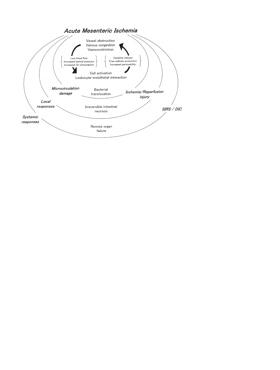
The clinical features of AMI originate from local and
systemic responses, which are induced by the impaired
microcirculation through the activation of a variety of
cells such as endothelium, monocytes, leukocytes, and
platelets (Fig. 1). Damage to the microcirculation leads
to irreversible intestinal necrosis locally, and dissemi-
nated intravascular coagulation (DIC) or systemic
inflammatory response syndrome (SIRS) in the whole
body or in remote organs systemically. Activated neu-
trophils, endothelium, monocytes, and platelets in the
ischemic intestine produce inflammatory cytokines,
such as tumor necrosis factor (TNF), interleukins,
platelet-activating factor (PAF), and leukotrienes,
through leukocyte–endothelial interactions. Endothe-
lial adhesion molecules such as E-selectin are upre-
gulated in the reperfused intestine.
21
The associated
DIC disturbs the intestinal microcirculation through
obstruction caused by leukocyte adhesion and platelet
aggregation.
22
Organ injury is also exacerbated by
vasoconstriction of the mesenteric vessels caused by
impaired production of nitric oxide by the damaged
endothelial cells.
23,24
Reperfusion injury is a feature of AMI. During is-
chemia, xanthine dehydrogenase, which exists predomi-
nantly in nonischemic tissue, is irreversibly converted to
xanthine oxidase. Reoxygenation of the ischemic intes-
tine converts a large amount of intracellular hypo-
xanthine to uric acid, which is catalyzed by xanthine
oxidase. Activated neutrophils in the reperfused intes-
tine release superoxide, catalyzed by nicotinamide
187
H. Yasuhara: Acute Mesenteric Ischemia
adenine dinucleotide phosphate (NADPH) oxidase
as well as elastase and collagenase. Furthermore,
myeloperoxidase from neutrophils produces chloride
ions from peroxide.
25
These reactions generate toxic
oxygen free radicals, such as superoxide (O
2
⫺
), peroxide
(H
2
O
2
), and hydroxyl radicals(
·
OH),
26
which damage
the cell membrane through lipid peroxidation. These
oxygen metabolites and enzymes of neutrophils also
cause serious damage to the surrounding tissue and dis-
tant organs by direct and indirect injury to the vascular
endothelium. It has been postulated that a high concen-
tration of xanthine dehydrogenase in the mesenteric
endothelium may be one of the reasons for the suscep-
tibility of the intestine to reperfusion injury.
Reperfusion induces interstitial edema and luminal
fluid accumulation through increased capillary perme-
ability.
27
The damaged intestinal microcirculation loses
its resistance to bacteria as well as to water, which leads
to endotoxemia
28
or bacteremia.
29,30
This bacterial trans-
location may play an important role in the development
of SIRS, adult respiratory distress syndrome (ARDS),
31
and cardiac dysfunction.
32
There is also substantial
evidence that the poor prognosis associated with acute
intestinal ischemia is strongly related to the multiple
organ failure caused by these sepsis-like systemic
responses.
33
Common Clinical Features and Diagnosis of AMI
Acute mesenteric ischemia caused by obstruction of the
superior mesenteric artery (SMA) is sometimes re-
ferred to as “acute mesenteric arterial syndrome,”
10
be-
cause of the common clinical features. The major symp-
tom of acute mesenteric ischemia is sudden and severe
abdominal pain out of proportion to the physical
findings. However, it should be noted that many elderly
patients have obscure symptoms and sometimes even
no early findings. Boley et al.
34
reported that patients
who present with abdominal pain and are at risk of AMI
are older than 50 years with congestive heart failure,
cardiac arrhythmias, recent myocardial infarction,
hypovolemia, hypotension, or sepsis. According to an-
other study, mesenteric arterial disease was found in
18% of patients aged over 65 years
35
and 70% of those
undergoing aortofemoral bypass.
36
Patients with AMI
may have a history of deep vein thromboses, arterial
embolism, collagen disease, or chronic postprandial
pain.
There is substantial evidence that the mortality varies
with the cause of AMI. Mesenteric venous thrombosis
(MVT) is much less lethal than acute thromboembolism
of the superior mesenteric artery and NOMI. In
general, survivors of AMI are younger, with less exten-
sive bowel infarction and a shorter clinical history.
37
Several retrospective studies have shown that prompt
diagnosis improves the survival rate significantly.
Another study found that the intestine was viable in
over 90% of patients if the duration of symptoms was
less than 12 h.
6
The plain X-ray findings suggestive of AMI include a
thickened bowel wall and a “ground-glass appearance”
in the abdomen.
38
However, as these findings are
nonspecific and the condition is associated with a high
Fig. 1. Local and systemic responses
in acute mesenteric ischemia. SIRS,
systemic inflammatory response syn-
drome; DIC, disseminated intravas-
cular coagulation

188
H. Yasuhara: Acute Mesenteric Ischemia
mortality rate, plain X-ray films are considered to be
useful only for excluding other possible causes of acute
abdominal pain, such as a perforated peptic ulcer.
The role of current radiographic and noninvasive
modalities is limited in the early diagnosis of AMI.
Computed tomography (CT) scans may reveal bowel
wall thickening, ascites, or occlusion of the mesenteric
arterial trunk. However, most of those abnormal signs
are nonspecific and found only in the late stage of acute
intestinal ischemia.
39,40
One study found that a correct
diagnosis was obtained by CT scans in only 26% of
suspected cases.
41
Another retrospective comparison of
plain X-ray films and CT scans in patients with proven
intestinal infarction demonstrated low specificity in
both examinations, of 30% and 39%, respectively.
42
Multislice spiral CT and magnetic resonance angiogra-
phy (MRA) are promising diagnostic modalities and
may be more useful than conventional CT scans be-
cause they provide high-resolution functional images
indicating low oxygen satruation.
43
However, clinical
evidence of the superiority of these diagnostic modali-
ties over CT is not yet available.
44
Duplex sonography is also a potentially useful diag-
nostic modality for acute intestinal ischemia, although it
is highly user-dependent and diminished blood flow can
only be confirmed in the mesenteric trunks.
45,46
More-
over, these recent imaging devices have only limited
value in the diagnosis of NOMI.
45,46
Selective mesenteric angiography and digital subtrac-
tion angiography (DSA) are still the gold standard for
the diagnosis of AMI
10,47–49
because they can identify
NOMI and provide important preoperative information
for mesenteric bypass surgery. Although routine an-
giography decreased the mortality rate without an
apparent increase in complications in many series,
10,50,51
the role of preoperative angiography is still contro-
versial in patients suspected of having AMI and those
with peritoneal signs. Moreover, it is impossible to per-
form selective mesenteric angiography for every patient
with suspected AMI in many hospitals. Some physicians
even claim that performing angiography will delay
surgical treatment for critically ill patients and, con-
sidering the low incidence of positive angiographic
findings, they recommend taking the patient directly to
surgery.
47
Serum markers, such as amylase, arterial pH, and
mucosal and seromuscular enzymes, frequently show
abnormalities in AMI. However, these enzymes usually
rise only after the development of a transmural intesti-
nal infarction.
52
A Swedish group recently found that a
D-dimer abnormality appears in the early stage of AMI
and reported promising results using this examination.
12
Other research groups investigated the
α subunit of
glutathione S-transferase (
α-GST), which is activated in
the intestine and liver to maintain cellular homeostasis
under oxidative stress, and found that
α-GST is a better
predictor of AMI than conventional biochemical tests.
53
However, experience with these serum markers is
limited.
It is generally agreed that exploratory laparotomy
and embolectomy with resection of the infarcted intes-
tine is mandatory if patients present with obvious peri-
toneal signs and symptoms. Segmental acute intestinal
ischemia can be treated by resection and primary anas-
tomosis (see Acute Mesenteric Arterial Embolus and
Thrombus). When blood perfusion of the anastomosis is
not confirmed, osteomy or the creation of a mucus fis-
tula should be done to monitor the viability of the intes-
tine postoperatively. In general, a routine second-look
procedure is done within 12–24 h postoperatively when
the viability of the site of anastomosis cannot be
confirmed at the initial operation.
54–56
Intestinal viability can be assessed intraoperatively
using a fluorescein method, Doppler ultrasound, or
laser Doppler.
57
With the fluorescein method, sodium
fluorescein is injected intravenously, and the dye leaks
out of the microvasculature and is deposited in the
interstitial tissue depending on the hypoxic damage.
This leakage pattern of fluorescein visualized under a
Wood’s lamp should assess the intestinal viability.
58,59
According to Bulkley et al., the fluorescein method
is more reliable for assessing intestinal viability than
either clinical criteria such as color, peristalsis, and pul-
sations in the mesentery, or Doppler ultrasound.
60,61
However, this method is highly subjective and requires
some experience to interpret the fluorescein pattern. A
fiberoptic fluorometer, which was recently introduced
to quantify the fluorescence concentration, has been
shown to better evaluate bowel viability.
58
Laser Dop-
pler and Doppler ultrasound may be more accurate for
assessing intestinal viability, but their major drawback
lies in the difficulty in achieving quick assessment of the
whole length of intestine.
Acute Mesenteric Arterial Embolus and Thrombus
The abdominal pain caused by an SMA embolus is usu-
ally of acute onset and out of proportion to the physical
findings. The embolus originates from the heart in most
patients, some of whom have a history of emboli in an
extremity or the brain. The abdominal pain associated
with SMA thrombosis is typically insidious and gradu-
ally progressive. Other patients may have symptoms
such as abdominal angina, consistent with chronic intes-
tinal ischemia, or signs of progressive sepsis, such as
dehydration, leukocytosis, bloody diarrhea, and eventu-
ally septic shock.
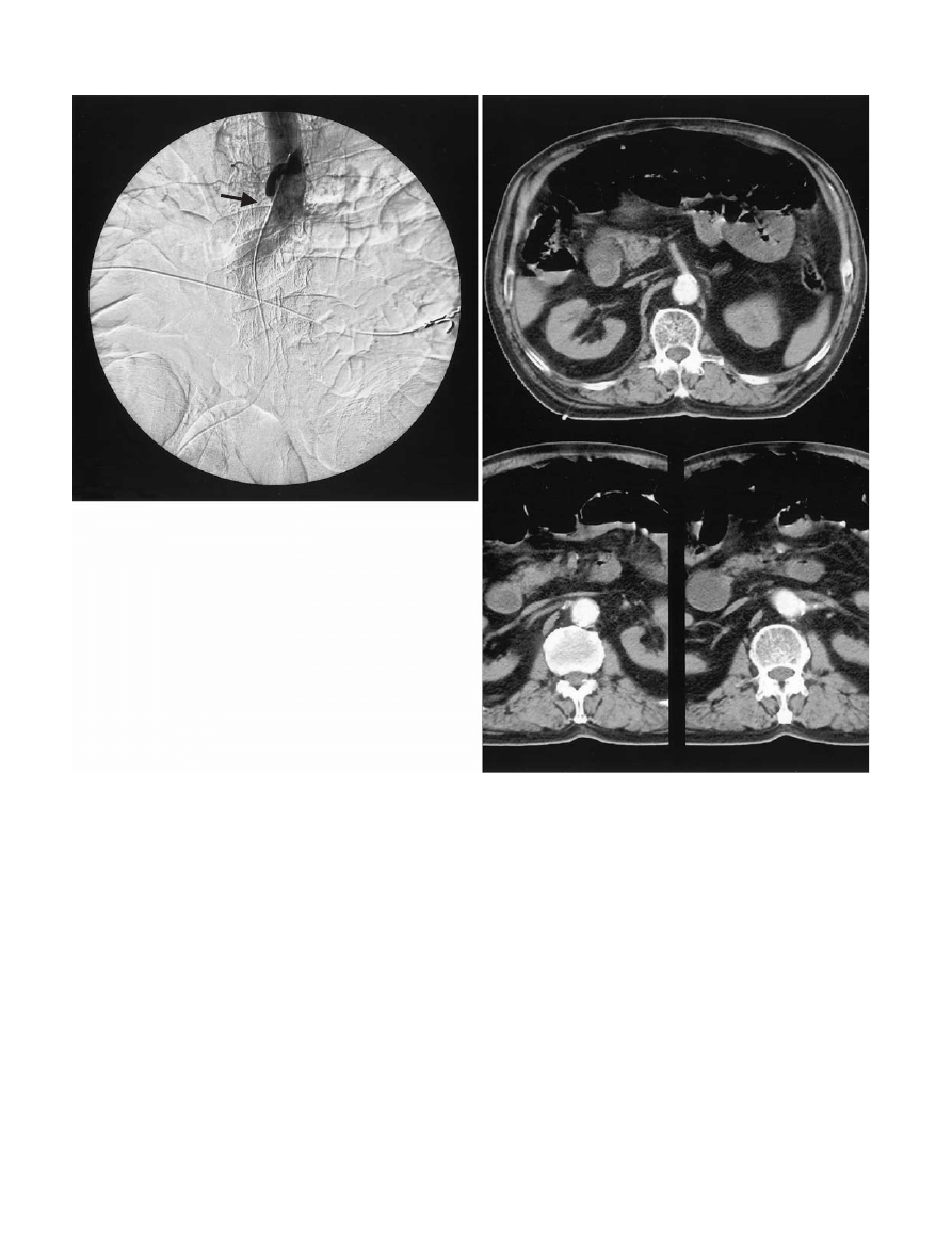
Angiography shows SMA thrombosis and embolus as
an abrupt cutoff of contrast in the vicinity of its origin
(Fig. 2). Although emboli to the SMA often appear as a
189
H. Yasuhara: Acute Mesenteric Ischemia
meniscus radiopaque sign within its lumen, the
angiographic findings differentiating between embolus
and thrombus are equivocal in many patients. It is
important to assess the development of collateral
vessels from the celiac axis or IMA connecting with
distal branches when total occlusion of the SMA is seen.
Enhanced collaterals may indicate chronic occlusion
of the SMA, such as that caused by mesenteric
thrombosis.
Once the diagnosis of embolus has been established,
emergency embolectomy must be performed through
an arteriotomy in the SMA, using a Fogarty catheter. It
is important to establish if the ischemic lesion extends
beyond the oral part of the ligament of Treitz in the
SMA thrombus, although the proximal jejunum is usu-
ally unaffected by an SMA embolus.
10
When mesenteric
thrombosis is diagnosed intraoperatively, an aortome-
senteric bypass should be performed, using the saphen-
ous vein as the conduit because of the risk of infection
associated with a prosthetic graft in the setting of bowel
ischemia or infarction. However, some surgeons prefer
a prosthesis over the saphenous vein, to avoid kinking
of the graft in the abdominal cavity.
Various other therapeutic approaches have been
established for SMA thromboembolism, including
intra-arterial perfusion with a thrombolytic agent and
vasodilators with interventional radiology procedures.
The indications for these treatments depend on whether
Fig. 2. a Typical abrupt cutoff sign of the superior mesenteric
artery in a 72-year-old man with a thrombus caused by atrial
fibrillation. b Abdominal computed tomography scan of the
same patient. It is difficult to see the obstruction of the artery
in this image
a
b
190
H. Yasuhara: Acute Mesenteric Ischemia
there are any signs of peritonitis, the extension of me-
senteric occlusion, and if the mesenteric occlusion is
in the distal or proximal area. Thrombolytic therapy is
most likely to be successful if treatment is started within
12 h of the onset of symptoms and if the thrombus par-
tially occludes the SMA trunk or occludes a single SMA
branch distal to the ileocolic artery.
62–69
It was reported
that a routine transcatheter infusion of papaverine im-
proved survival in highly selected patients with major
emboli of the SMA, although the experience is
limited.
50,70
Mesenteric Venous Thrombosis
Mesenteric venous thrombosis (MVT) is relatively rare
and characterized by the insidious onset of abdominal
discomfort. Some patients complain of diffuse intermit-
tent abdominal pain lasting several days or weeks. The
symptoms are usually less severe than those caused by
an SMA embolus. Mesenteric venous thrombosis can be
classified into acute and chronic types, depending on the
duration of symptoms.
71
The acute type of MVT, in
which symptoms last less than 4 weeks, accounts for
only 5%–15% of patients with AMI.
72,73
Rhee et al.
reported that only 9% of patients with MVT presented
with symptoms of less than 24 h duration.
54
Mesenteric venous thrombosis is also classified
according to its etiology, into primary and secondary
MVT. Primary MVT may be spontaneous or idiopathic,
not associated with any other etiologic factor,
74
whereas
patients with the following underlying conditions are
classified as having secondary MVT: hypercoagulability,
cirrhosis, splenomegaly, cancer, infection, trauma, pan-
creatitis, hematologic disease, inflammatory bowel
disease, or diverticular disease.
54
According to some
researchers, about 20% of cases are idiopathic and 80%
are secondary.
75,76
The number of patients with second-
ary MVT has increased considerably over the last two
decades because of the recognition of previously un-
known factors, such as hematological disorders includ-
ing protein C and S deficiency, antithrombin III
deficiency, dysfibrinogenemia, abnormal plasminogen,
polycythemia vera, thrombocytosis, sickle cell disease,
and factor V Leiden mutation.
77–80
Thus, the MVT could
be considered to be an analogy of deep venous throm-
bosis of the lower limb. In fact, both disease have many
predisposing factors in common, including abnormal
coagulopathy. Localized MVT can also develop second-
ary to volvulus, intussusception, or strangulation of the
bowel.
Contrast-enhanced CT has been found to be more
valuable for the diagnosis of MVT in contrast to its
limited role in the diagnosis of AMI or NOMI.
81
There-
fore, CT is done as the initial diagnostic examination in
patients with severe abdominal pain and a history of
deep vein thrombosis or a familial history of hyperco-
agulability.
82
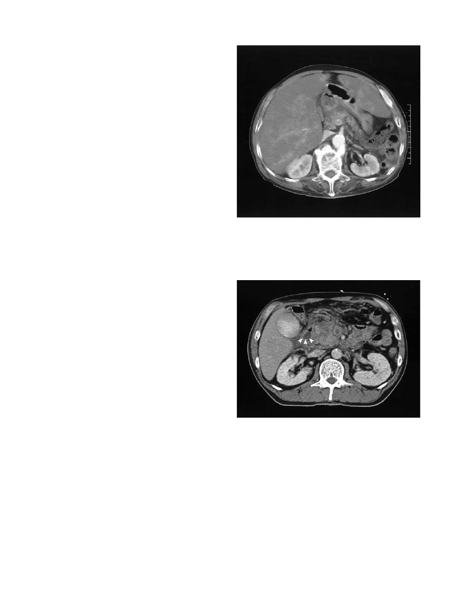
Computed tomography can show throm-
bosis in the superior mesenteric vein, portal vein, and
splenic vein, with or without bowel wall thickening or
pneumatosis, in many asymptomatic patients (Figs. 3
and 4) Miller and Berland advocated duplex Doppler
examination as the first diagnostic choice because it is as
durable as CT scans.
83
Magnetic resonance imaging has
Fig. 3. Abdominal computed tomography scan showing por-
tal vein thrombosis of 24 h duration associated with massive
liver necrosis in a 67-year-old patient who died of multiple
organ failure after massive intestinal necrosis
Fig. 4. Mesenteric venous thrombosis associated with acute
pancreatitis. Computed tomography scan showing radiopaque
image in the superior mesenteric vein and a swollen pancre-
atic head

191
H. Yasuhara: Acute Mesenteric Ischemia
also been reported to be sensitive for diagnosing
MVT,
84
although it costs more than almost any other
diagnostic modality.
Serum laboratory test findings, such as leukocytosis,
and elevated serum levels of lactate and amylase, are
not useful for the diagnosis of acute MVT. The diagnos-
tic value of D-dimer, which is used extensively for the
diagnosis of venous thromboembolism of the extremi-
ties, remains controversial.
85
In symptomatic patients with acute MVT, the choice
of treatment is determined by the severity of peritoneal
signs. In the absence of peritoneal signs, anticoagulant
therapy should be started immediately. Patients are first
treated with heparin for 7–10 days, then an oral regimen
of Coumadin or warfarin sodium for 3–6 months.
86
Thrombolysis is also a treatment of choice. The throm-
bolytic agents can be infused using several approaches,
such as via the SMA,
86,87
via the internal jugular vein,
88
or transhepatically via the portal vein.
89,90
Exploratory laparotomy should be performed imme-
diately for all patients with peritoneal signs. Normal
laboratory test results should not preclude exploration
and the patient should receive continuous heparin infu-
sion regardless of the risk of bleeding. Perioperative
anticoagulation therapy decreases the risk of recurrence
of thrombosis and ultimately improves survival.
71
Venous thrombectomy is not usually recommended for
acute MVT because thrombosis often recurs and results
in distal diffuse extention.
71
For asymptomatic patients with an incidental diagno-
sis based on CT scan findings, either no therapy or 3–6
months’ systemic anticoagulant administration is rec-
ommended, although there is no available evidence sup-
porting this therapeutic decision.
The rate of recurrence for acute MVT, which gener-
ally occurs within 30 days, is high.
10
The long-term sur-
vival of patients with chronic MVT depends on their
underlying diseases and appears to be better than that
of patients with acute MVT.
77
All patients who have
suffered recurrent MVT should be kept on warfarin
sodium for the rest of their life.
Nonocclusive Mesenteric Ischemia
Nonocclusive mesenteric ischemia is defined as “intesti-
nal necrosis with a patent arterial tree” and has also
been termed “hemorrhagic enteropathy,” “hemor-
rhagic necrosis of gastrointestinal tract,” “intestinal inf-
arction without mesenteric vascular occlusion,” and
“hemorrhagic necrotizing enteropathy.”
91
Nonocclusive
mesenteric ischemia is the most lethal form of AMI,
with mortality rates of up to 70%–100%. Nonocclusive
mesenteric ischemia was previously thought to be rare,
but its frequency is increasing,
13
and its overall incidence
is estimated at about 1 in every 5000 hospital admis-
sions,
48
which accounts for 25%–60% of all bowel
infarctions.
The frequency of NOMI among patients undergoing
cardiac surgery
15
and those on hemodialysis
16,92–94
has
dramatically increased over the last few decades. In fact,
9%–20% of deaths among patients on hemodialysis are
attributable to NOMI. Newman and colleagues also
noted that 22% of patients with bowel infarction had
renal failure as comorbidity.
95
It was also reported that
hemodialysis-induced hypotension triggers NOMI in
patients with signs of atherosclerosis.
92
Colonic ischemia (CI) is another important
comorbidity involved in the pathogenesis of NOMI.
Colonic ischemia frequently occurs after cardiac func-
tion has been optimized and the presumed cause of
mesenteric vasoconstriction has been corrected,
96
for
example, after major cardiovascular surgery or a hy-
potensive episode caused by rupture of an abdominal
aortic aneurysm. Colonic ischemia is often associated
with systemic conditions, such as vasculitis; the use of
various medications that can induce intense vasospasm,
such as oral contraceptives,
β-blockers, diuretics, and
digitalis; and colonic obstructive lesions, such as carci-
noma. Therefore, some investigators suggest that
reperfusion after NOMI could induce CI and it is rea-
sonable to assume that reperfusion injury may explain
the high incidence of CI.
The high mortality rates associated with NOMI are
frequently attributed to a delay in diagnosis because of
its mild and nonspecific symptoms compared with other
types of AMI. An abrupt onset of severe abdominal
pain may be less common. The most common symptom
is a gradual onset of crampy, periumbilical abdominal
pain, which progresses to constant pain. Some patients
may not complain of any apparent symptoms because of
their severe underlying illness and many patients have
experienced a recent episode of hemorrhagic shock or
sepsis. Abdominal distension and feeding intolerance
may be the early manifestations of NOMI, and in its late
stage, fever metabolic acidosis or hypovolemic shock
can develop in patients receiving enteral feeding.
Reinus et al.
97
reviewed the symptomatology of
NOMI and concluded that it most often develops in
patients older than 60 years with associated underlying
cardiovascular disease, and frequently abdominal pain,
distension, and leukocytosis. The patients at highest risk
include those with disorders predisposing to atheroscle-
rosis, such as diabetes mellitus, advanced age, hyperten-
sion, dyslipidemia, a history of smoking, and arterial
occlusive disease.
98
Several hypotheses have been proposed to explain
the pathogenesis of NOMI, among which persistent and
irreversible vasoconstriction is thought to be the most
important. Previous experimental and clinical studies
have demonstrated that long-standing vasoconstriction

192
H. Yasuhara: Acute Mesenteric Ischemia
can become persistent and irreversible.
96
Vasoconstric-
tion of the splanchnic resistance vessels occurs during
cardiogenic/hemorrhagic shock or sepsis. Although the
intestine can tolerate a short period of hypoxia (see
Pathophysiology of Acute Intestinal Ischemia), persis-
tent vasoconstriction may induce critical intestinal is-
chemia.
99
This persistent vasoconstriction is also likely
to induce NOMI after the restoration of blood flow
following embolectomy of the SMA.
Experimental studies suggest that persistent vasocon-
striction is primarily mediated by angiotensin II and
vasopressin derived from the kidney and pituitary
gland.
100,101
(see Pathophysiology of Acute Intestinal
Ischemia.) The histological findings of NOMI resemble
those induced by angiotensin II in animals.
102,103
Angio-
tensin II is generated via the renin-angiotensin system,
which is stimulated by the hypoperfused kidney. A dis-
proportionate distribution of angiotensin II receptors
on splanchnic vascular smooth muscle cells may induce
mesenteric vasoconstriction.
104
Intestinal mucosal damage in NOMI begins at the
villous tip,
105
which is characterized by the features of
mucosal circulation. In vasoconstriction severe enough
to reduce intestinal blood flow by 30%–50% in experi-
mental animals, the volume of blood supplying the in-
testinal villi remains unchanged, but blood flow velocity
to the villus tip is reduced. The reduced velocity of
blood flow in the villus increases oxygen shunting from
artery to vein via a “countercurrent exchange” mecha-
nism. This is why the villous tip is susceptible to
vasoconstriction.
Angiography must be performed to diagnose NOMI
before intestinal infarction occurs.
70,98
The classic
angiographic finding of NOMI is spasming and narrow-
ing of multiple branches of the mesenteric arteries.
Irregularities in the branches, spasm of the arcades,
or impaired filling of intramural vessels may also be
seen.
106
Although some physicians are skeptical about
the usefulness of angiography to improve the short-
term prognosis,
94
it may prevent unnecessary dissection
around the SMA, which could exacerbate vasoconstric-
tion. One of the advantages of preoperative angio-
graphy is that pharmacoangiographic treatment (see
below) can be initiated simultaneously when the diag-
nosis of NOMI is made. Early diagnosis by angiography
followed by intra-arterial papaverine infusion may be
the best option for improving survival and maintaining
intestinal integrity.
The therapeutic options for NOMI vary according to
the interval between the onset of symptoms and the
start of treatment. Initial treatment should include cor-
rection of any underlying causes, such as congestive
heart failure, arrhythmia, or hypovolemia. When the
diagnosis of NOMI has been made, pharmacoangio-
graphic procedures are utilized. Papaverine hydrochlo-
ride (30–60 mg/h) is infused through a catheter in the
SMA to relieve the vasoconstriction and prevent it be-
ing persistent.
70,93,98,107
The presence of peritoneal signs
or an ischemic time of longer than 12 h may indicate the
need for urgent laparotomy. Papaverine infusion should
be continued during and after surgery. When the diag-
nosis of peritonitis is made intraoperatively, large seg-
ments of intestine must be resected, which often leads to
short bowel syndrome. However, intestinal resection
must be done in areas of questionable viability to pre-
serve a longer segment of intestine. A second-look
exploration is necessary to confirm the viability of the
remaining intestine. Sheridan et al. reported that the
accuracy of prediction of intestinal viability using clini-
cal criteria, such as intestinal color, arterial pulsation,
and peristalsis, was only 58%.
108
Conclusions
Acute mesenteric ischemia is not a single clinical entity
but rather a complex of diseases with many clinical
features caused by impaired blood perfusion to the in-
testine. Despite advanced diagnostic modalities, AMI is
still a life-threatening condition, and although many
predisposing factors, such as aging and atherosclerosis,
are beyond the physician’s control, accumulated knowl-
edge on this condition is expected to improve its prog-
nosis through a multidisciplinary approach.
References
1. Batellier J, Keny R. Superior mesenteric artery embolism:
eighty-two cases. Ann Vasc Surg 1990;4:112–6.
2. Boley SJ, Feinstein FR, Sammartano R, Brandt LJ. New concept
in the management of emboli of the superior mesenteric artery.
Surg Gynecol Obstet 1981;153:561–9.
3. Inderbitzi R, Wagner HE, Seiler C, Stirnemann P, Gertsch P.
Acute mesenteric ischemia. Eur J Surg 1992;158:123–6.
4. Lazaro T, Sierra L, Gesto R, Villafana W, Fonseca J, Porto J,
et al. Embolization of the mesenteric arteries: surgical treatment
in twenty three consecutive cases. Ann Vasc Surg 1986;1:311–5.
5. Levy PJ, Krausz MM, Manny J. Acute mesenteric ischemia:
improved results — a retrospective analysis of ninety-two pa-
tients. Surgery 1990;107:372–80.
6. Vellar ID, Doyle JC. Acute mesenteric ischemia. Aust N Z J
Surg 1977;47:54–61.
7. Clavien PA, Muller C, Harder F. Treatment of mesenteric inf-
arction. Br J Surg 1987;74:500–3.
8. Finucani PM, Arunachalam T, O’Dowd J, Pathy J. Acute mesen-
teric infarction in elderly patients. J Am Geriatr Soc 1989;37:
355–8.
9. Mishima Y. Acute mesenteric ischemia. Jpn J Surg 1988;18:615–
9.
10. Schneider TA, Longo WE, Ure T, Vernava AM III. Mesenteric
ischemia: acute arterial syndromes. Dis Colon Rectum
1994;37:1163–74.
11. AGA technical review on intestinal ischemia. Gastroenterology
2000;118:954–68.

193
H. Yasuhara: Acute Mesenteric Ischemia
12. Acosta S, Nilsson TK, Bjorck M. Preliminary study of D-dimer
as a possible marker of acute bowel ischaemia. Br J Surg 2001;
88:385–8.
13. Corder AP, Taylor I. Acute mesenteric ischaemia. Postgrad Med
J 1993;69:1–3.
14. Bjorck M, Troeng T, Bergqvist D. Risk factors for intestinal
ischemia after aortoiliac surgery: a combined cohort and case
control study of 2824 operations. Eur J Vasc Surg 1997;13:531–
9.
15. Gennaro M, Ascer E, Matano R, Jacobowitz IJ, Cunningham
JN, Uceda P. Acute mesenteric ischemia after cardiopulmonary
bypass. Am J Surg 1993;166:2321–36.
16. Diamond S, Emmett M, Henrich WL. Bowel infarction as a
cause of death in dialysis patients. JAMA 1986;256:2545–7.
17. Boley SJ, Brandt LJ, Veith FJ. Ischemic disease of the intestine.
Curr Probl Surg 1978;15:1–85.
18. Wilcox MG, Howard TJ, Plaskon LA, Unthank JL, Madura JA.
Current theories of pathogenesis and treatment of non-occlusive
mesenteric ischemia. Dig Dis Sci 1995;50:709–15.
19. Bulkley GB, Kvietys PR, Perry MA, Granger DN. Effects of
cardiac tamponade on colonic hemodynamics and oxygen up-
take. Am J Physiol 1983;244:G604–12.
20. Crissinger KD, Tso P. The role of lipids in ischemia/reperfusion-
induced changes in mucosal permeability in developing piglets.
Gastroenterology 1992;102:1693–9.
21. Russell J, Epstein CJ, Grisham MB, Alexander JS, Yeh KY,
Granger DN. Regulation of E-selectin expression in postis-
chemic intestinal microvasculature. Am J Physiol Gastrointest
Liver Physiol 2000;278:G878–85.
22. Harward TR, Brooks DL, Flynn TC, Seeger JM. Multiple organ
dysfunction after mesenteric artery revascularization. J Vasc
Surg 1993;18:459–67.
23. Ma XL, Johnson G III, Lefer AM. Mechanisms of inhibition of
nitric oxide production in a murine model of splanchnic artery
occlusion shock. Arch Int Pharmacodyn Ther 1991;311:89–103.
24. Carey C, Siegfried MR, Ma XL, Weyrich AS, Lefer AM.
Antishock and endothelial protective actions of a NO donor in
mesenteric ischemia and reperfusion. Circ Shock 1992;38:209–
16.
25. Simpson R, Alon R, Kobzik L, Valeri CR, Shepro D, Hechtman
HB. Neutrophils and non-neutrophil-mediated injury intestinal
ischemia-reperfusion. Ann Surg 1993;218:444–53.
26. McCord JM, Roy RS. The pathophysiology of superoxide: role
in inflammation and ischemia. Can J Physiol Pharmacol 1982;60:
1346–52.
27. Granger DN, McCord JM, Parks DA, Hollwarth ME. Xanthine
oxidase inhibitors attenuate ischemia induced vascular perme-
ability changes in the cat intestine. Gastroenterology 1986;90:80–
4.
28. Turnage RH, Guice KS, Oldham KT. Endotoxemia and remote
organ injury following intestinal reperfusion. J Surg Res 1994;56:
571–8.
29. Grissinger KD, Granger DN. Mucosal injury induced by is-
chemia and reperfusion in the pelvic intestine: influence of the
age and feeding. Gastroenterology 1989;98:920–6.
30. Koike K, Moore EE, Moore FA, Read RA, Carl VS, Banerjee
A. Gut ischemia/reperfusion produces lung injury independent
of endotoxin. Crit Care Med 1994;22:1438–44.
31. Fullerton DA, Hahn AR, Koike K, Banerjee A, Harken AH.
Intracellular mechanisms of pulmonary vasomotor dysfunction
in acute lung injury caused by mesenteric ischemia-reperfusion.
Surgery 1993;114:360–6.
32. Jamieson WG, DeRose G, Harris KA, Pliagus G, Stafford L.
Myocardial and circulatory performance during the ischemic
phase of superior mesenteric artery occlusion. Can J Surg
1993;36:435–9.
33. Grotz MR, Deitch EA, Ding J, Xu D, Huang Q, Regel G. Intes-
tinal cytokine response after gut ischemia: role of gut barrier
failure. Ann Surg 1999;229:478–86.
34. Boley SJ, Sprayregan S, Veith FJ, Siegeiman SS. An aggressive
roentgenolgic and surgical approach to acute mesenteric is-
chemia. Surg Ann 1973;355–78.
35. Roobottom CA, Dubbins PA. Significant disease of the celiac
and superior mesenteric arteries in asymptomatic patients: pre-
dictive value of Doppler sonography. Am J Roentgenol 1993;
161:985–8.
36. Pokrovskii AV, Kazanchian PO, Iudin VI, Varava BN, Khabriev
TA, Shilenok DV. Indications for and the methods of reva-
scularization of visceral branches in aorto-femoral reconstruc-
tion. Vestn Khir 1990;144:3–10.
37. Wilson C, Gupta R, Gilmour DG, Imrie CW. Acute superior
mesenteric ischaemia. Br J Surg 1987;74:279–81.
38. Wolf EL, Spraregen S, Bakal CW. Radiology in intestinal is-
chemia: plain films, contrast and other imaging studies. Surg Clin
North Am 1992;72:104–24.
39. Smerud MJ, Johnson CD, Stephens DH. Diagnosis of bowel
infarction: a comparison of plain films and CT scans in 23 cases.
Am J Radiol 1990;154:99–103.
40. Taourel PG, Deneuville M, Pradel JA, Regent D, Bruel JB.
Acute mesenteric ischemia: diagnosis with contrast-enhanced
CT. Radiology 1996;199:632–6.
41. Alpern MB, Glazer GM, Francis IR. Ischemic or infarcted
bowel: CT findings. Radiology 1988;166:149–52.
42. Smerud MJ, Johnson CD, Stephens DH. Diagnosis of bowel
infarction: a comparison of plain films and CT scans in 23 cases.
Am J Radiol 1990;154:99–103.
43. Klein HM, Klosterhalfen B, Kinzel S, Jansen A, Seggewiss C,
Weghaus P, et al. CT and MRI of experimentally induced me-
senteric ischemia in porcine model. J Comput Assist Tomogr
1996;20:254–61.
44. Chan FP, Li KC, Heiss SG, Razavi MK. A comprehensive ap-
proach using MR imaging to diagnose acute segmental mesen-
teric ischemia in a porcine model. Am J Roentgenol 1999;
173:523–9.
45. Bowersox JC, Zwolak RM, Walsh DB, Schneider JR, Musson A,
Labombard FE, et al. Duplex ultrasonography in the diagnosis
of celiac and mesenteric artery occlusive disease. J Vasc Surg
1991;14:780–6.
46. Danse EM, Van Beers BE, Goffette P, Dardenne AN, Laterre
PF, Pringot J. Acute intestinal ischemia due to occlusion of the
superior mesenteric artery: detection with Doppler sonography.
J Ultrasound Med 1996;15:323–6.
47. Bradbury AW, Brittenden J, McBride K, Ruckley CV. Mesen-
teric ischemia: a multidisciplinary approach. Br J Surg 1995;82:
1446–59.
48. Stoney RJ, Cunningham CG. Acute mesenteric ischemia.
Surgery 1993;114:489–90.
49. Clavien PA. Diagnosis and management of mesenteric infarc-
tion. Br J Surg 1990;77:601–3.
50. Clark RA, Gallant TE. Acute mesenteric ischemia: angiographic
spectrum. Am J Radiol 1984;142:555–62.
51. Aakhus T, Evensen A. Angiography in acute mesenteric
insufficiency. Acta Radiol Diag 1978;19:945–54.
52. Kurland B, Brandt LJ, Delaney HM. Diagnostic tests for intesti-
nal ischemia. Surg Clin North Am 1992;72:88–105.
53. Gearhart SL, Delaney CP, Senagore AJ, Banbury MK, Remzi
FH, Kiran RP, et al. Prospective assessment of the predictive
value of alpha-glutathione S-transferase for intestinal ischemia.
Am Surg 2003:324–9.
54. Rhee RY, Gloviczki P, Mendonca CT, Patterson TM, Serry RD,
Sarr MG, et al. Mesenteric venous thrombosis: still a lethal dis-
ease in the 1990s. J Vasc Surg 1994;20:688–97.
55. Levy P, Krausz MM, Manny J. The role of second-look
procedure in improving survival time for patients with mesen-
teric venous thrombosis. Surg Gynecol Obstet 1990;170:287–
91.
56. Zuidema GD, Reed D, Turcotte JC, Fry WJ. Superior mesen-
teric artery embolectomy. Ann Surg 1964;159:549–53.

194
H. Yasuhara: Acute Mesenteric Ischemia
57. Ahn H, Lindhagen J, Nilsson GE, Salerud EG, Jodal M, Lundgren
O. Evaluation of laser Doppler flow in the assessment of intestinal
blood flow in the cat. Gastroenterology 1985;88:951–7.
58. Mann A, Fazo VW, Lucas FV. A comparative study of the use of
fluorescein and the Doppler device in the determination of intes-
tinal viability. Surg Gynecol Obstet 1982;154:53–5.
59. Killewich LA, Peterson GJ. Arterial embolic and occlusive dis-
eases. Semin Colon Rectal Surg 1993;4:205–11.
60. Bulkley GB, Zuidema GD, Hamilton SR, O’Mara CS,
Klacsmann PG, Horn SD. Intraoperative determination of small
intestinal viability following ischemic injury. Ann Surg 1981;194:
628–35.
61. Wright CB, Hobson RW. Prediction of intestinal viability using
Doppler ultrasound techniques. Am J Surg 1975;129:642–5.
62. Badiola CM, Scoppetta DJ. Rapid revascularization of an
embolic superior mesenteric artery occlusion using pulse-spray
pharmacomechanical thrombolysis with urokinase. Am J
Roentgenol 1997;169:55–7.
63. Boyer L, Delorme JM, Alexandre M, Boissier A, Gimbergues P,
Glanddier G, et al. Local fibrinolysis for superior mesenteric
artery thromboembolism. Cardiovasc Intervent Radiol 1994;17:
214–6.
64. Flickinger EG, Johnsrude IS, Ogburn NL, Weaver MD, Pories
WJ. Local streptokinase infusion for superior mesenteric artery
thromboembolism. Am J Roentgenol 1983;140:771–2.
65. McBride KD, Gaines PA. Thrombolysis of a partially occluding
superior mesenteric artery thromboembolus by infusion of strep-
tokinase. Cardiovasc Intervent Radiol 1994;17:164–6.
66. Pillari G, Doscher W, Fierstein J, Ross W, Loh G, Berkowitz BJ.
Low-dose streptokinase in the treatment of celiac and superior
mesenteric artery occlusion. Arch Surg 1983;118:1340–2.
67. Regan F, Karistad RR, Magnusan TH. Minimally invasive man-
agement of acute superior mesenteric artery occlusion: com-
bined urokinase and laparoscopic therapy. Am J Gastroenterol
1996;91:1019–21.
68. Gallego AM, Ramirez P, Rodriguez JM, Buenos FS, Robles R,
Capel A, et al. Role of urokinase in the superior mesenteric
artery embolism. Surgery 1996;120:111–3.
69. Simo G, Echenagusia AJ, Camunez F, Turegano F, Cabrera A,
Urbano J. Superior mesenteric arterial embolism: local
fibrinolytic treatment with urokinase. Radiology 1997;20:775–9.
70. Boley SJ, Sprayregan S, Siegelman SS, Veith FJ. Initial results
from an aggressive approach to acute mesenteric ischemia.
Surgery 1977;82:848–55.
71. Rhee RY, Gloviczki P. Mesenteric venous thrombosis. Surg Clin
North Am 1997;77:327–38.
72. Kairaluoma MI, Karkola P, Heikkinen E, Huttunen R, Mokka
REM, Larmi TKI. Mesenteric infarction. Am J Surg
1977;133:188–93.
73. Ottinger LW, Austen WG. A study of 136 patients with mesen-
teric infarction. Surg Gynecol Obstet 1967;251–61.
74. Kitchens CS. Evolution of our understanding of the pathophysi-
ology of primary mesenteric venous thrombosis. Am J Surg
1992;163:346–8.
75. Grendell JH, Ockner RK. Mesenteric venous thrombosis.
Gastroenterology 1982;82:358–72.
76. Abdu R, Zakhour BJ, Dallis DJ. Mesenteric venous thrombosis
— 1911 to 1984. Surgery 1987;101:383–8.
77. Inagaki H, Sakakibara O, Miyake H, Eimoto T, Yura J. Mesen-
teric venous thrombosis in familial free protein S deficiency. Am
J Gastroenterol 1993;88:134–8.
78. Ostermiller W Jr, Carter R. Mesenteric venous thrombosis sec-
ondary to polycythemia vera. Am J Surg 1969;35:407–9.
79. Tollefson DFJ, Friedman KD, Marlar RA, Bandyk DF, Towne
JB. Protein C deficiency; a cause of unusual or unexplained
thrombosis. Arch Surg 1988;123:881–4.
80. Wilson C, Walker ID, Davidson JF, Imrie CW. Mesenteric
venous thombosis and antithrombin III deficiency. J Clin Pathol
1987;40:906–8.
81. Harward TRS, Green D, Bergan JJ, Rizzo RJ, Yao JST. Mesen-
teric venous thrombosis. J Vasc Surg 1989;9:328–33.
82. Boley SJ, Kaleya RN, Brandt LI. Mesenteric venous thrombosis.
Surg Clin North Am 1992;72:183–202.
83. Miller VE, Berland LL. Pulsed Doppler duplex sonography and
CT of portal vein thrombosis. Am J Roentgenol 1985;145:73–6.
84. Gehl HB, Bohndorf K, Klose KC, Gunther RW. Two-
dimensional MR angiography in the evaluation of abdominal
vein with gradients refocused sequences. J Comput Assist
Tomogr 1990;14:619–24.
85. Brill-Edwards P, Lee A. D-dimer testing in the diagnosis of
acute venous thromboembolism. Thromb Haemost 1999;82:688–
94.
86. Poplausky MR, Kaufman JR, Geller SC, Waltmna AC. Mesen-
teric venous thrombosis treated with urokinase via the superior
mesenteric artery. Gastroenterology 1996;110:1633–5.
87. Train JS, Ross H, Weiss JD, Feingold ML, Khoury-Yacoub A,
Khoury PT. Mesenteric venous throbosis: successful treatment
by intraarterial lytic therapy. J Vasc Interv Radiol 1998;9:461–
4.
88. Rivitz SM, Geller SC, Hahn C, Waltman AC. Treatment of acute
mesenteric venous thrombosis with transjugular intramesenteric
urokinase infusion. J Vasc Interv Radiol 1995;6:219–23.
89. Yankes JR, Uglietta JP, Grant J, Braun SD. Percutaneous
transhepatic recanalization and thrombolysis of the superior
mesenteric vein. Am J Roentgenol 1988;151:289–90.
90. Bilbao JI, Rodriguez-Cabello J, Longo J, Zornoza G, Paramo J,
Lecumberri FJ. Portal thrombosis: percutaneous transhepatic
treatment with urokinase — a case report. Gastrointest Radiol
1989;14:326–8.
91. Haglund U, Lundgren O. Non-occlusive acute intestinal vascular
failure. Br J Surg 1979;66:155–8.
92. Diamond S, Emmett M, Henrich W. Bowel infarction as a cause
of death in dialysis patients. JAMA 1986;256;2545–7.
93. John AS, Tuerff SD, Kerstein MD. Nonocclusive mesenteric
infarction in hemodialysis patients. J Am Coll Surg 2000;190:84–
8.
94. Bender JS, Ratner LE, Hagnuson TH, Zenilman ME. Acute
abdomen in the hemodialysis patient population. Surgery
1995;117:494–7.
95. Newman TS, Mgnuson TH, Ahrendt SA, Smith-Meek MA,
Bender JS. The changing face of mesenteric infarction. Am Surg
1998;64:611–6.
96. Boley SJ, Regan JA, Tunick PA, Everhard ME, Winslow PR,
Veith FJ. Persistent vasoconstriction — a major factor in
nonocclusive mesenteric ischemia. Curr Top Surg Res 1971;3:
425–33.
97. Reinus JF, Brandt LJ, Boley SJ. Ischemic diseases of the bowel.
Gastroenterol Clin North Am 1990;19:319–43.
98. Zeier M, Wiesel M, Rambusek M, Ritz E. Non-occlusive mesen-
teric infarction in dialysis patients: the importance of prevention
and early intervention. Nephrol Dial Transplant 1995;10:771–
3.
99. Reda JA, Rush BF, Lysz TW, Machiedo GW. Organ distribution
of gut-derived bacteria caused by bowel manipulation or is-
chemia. Am J Surg 1990;159:85–90.
100. Bailey RW, Bulkley GB, Hamilton SR, Morris JB, Haglund UH.
Protection of the small intestine from nonocclusive mesenteric
ischemic injury due to cardiogenic shock. Am J Surg 1987;153:
108–16.
101. McNeill JR, Stark RD, Greenway CV. Intestinal vasoconstric-
tion after hemorrhage. Roles of vasopressin and angiotensin.
Am J Physiol 1970;219:1342–7.
102. Banks RO, Gallavan RH, Zinner MJ, Bulkley GB, Harper SL,
Granger DN, et al. Vasoactive agents in control of the mesen-
teric circulation. Fed Proc 1985;44:2743–9.
103. Bulkley GB, Womack WA, Downey JM, Kvietys PR, Granger
DN. Collateral blood flow in segmental intestinal ischemia:
effects of vasoactive agents. Surgery 1986;100:157–65.

195
H. Yasuhara: Acute Mesenteric Ischemia
104. Gunther S, Gibrone MA Jr, Alexander RW. Identification and
characterization of the high affinity vascular angiotensin II
receptor in rat mesenteric artery. Circ Res 1980;47:278.
105. Williams LF. Vascular insufficiency of the intestines. Gastroen-
terology 1971;61:757–77.
106. Clark RA, Gallant TE. Acute mesenteric ischemia: angiographic
spectrum. Am J Roentgenol 1984;142:555–62.
107. Ward D, Vernava AM, Kaminski DL, Ure T, Peterson G,
Garvin P, et al. Improved outcome by identification of high-risk
nonocclusive mesenteric ischemia, aggressive reexploration, and
delayed anastomosis. Am J Surg 1995;170:5777–81.
108. Sheridan WG, Lowdes RH, Williams GT, Young HL. Determi-
nation of a critical level of tissue oxygenation in acute intestinal
ischemia. Gut 1991;33:762–6.
Wyszukiwarka
Podobne podstrony:
Acute mesenteric ischemia Text 03
Acute extremities ischemia Text
Visceral Ischemia Text 01
02 csb text
Wyk 02 Pneumatyczne elementy
02 OperowanieDanymiid 3913 ppt
02 Boża radość Ne MSZA ŚWIĘTAid 3583 ppt
OC 02
PD W1 Wprowadzenie do PD(2010 10 02) 1 1
02 Pojęcie i podziały prawaid 3482 ppt
WYKŁAD 02 SterowCyfrowe
02 filtracja
02 poniedziałek
21 02 2014 Wykład 1 Sala
Genetyka 2[1] 02
więcej podobnych podstron